Development and characterization of patient derived xenografts from non small cell lung cancer brain metastases - Nature
←
→
Page content transcription
If your browser does not render page correctly, please read the page content below

www.nature.com/scientificreports
OPEN Development and characterization
of patient‑derived xenografts
from non‑small cell lung cancer
brain metastases
Andrew M. Baschnagel1,2*, Saakshi Kaushik1, Arda Durmaz3, Steve Goldstein2,4,
Irene M. Ong2,4,5, Lindsey Abel1, Paul A. Clark1, Zafer Gurel1, Ticiana Leal2,6, Darya Buehler2,7,
Gopal Iyer1,2, Jacob G. Scott8 & Randall J. Kimple1,2,9*
Non-small cell lung cancer (NSCLC) brain metastasis cell lines and in vivo models are not widely
accessible. Herein we report on a direct-from patient-derived xenograft (PDX) model system of
NSCLC brain metastases with genomic annotation useful for translational and mechanistic studies.
Both heterotopic and orthotopic intracranial xenografts were established and RNA and DNA
sequencing was performed on patient and matching tumors. Morphologically, strong retention of
cytoarchitectural features was observed between original patient tumors and PDXs. Transcriptome
and mutation analysis revealed high correlation between matched patient and PDX samples
with more than more than 95% of variants detected being retained in the matched PDXs. PDXs
demonstrated response to radiation, response to selumetinib in tumors harboring KRAS G12C
mutations and response to savolitinib in a tumor with MET exon 14 skipping mutation. Savolitinib also
demonstrated in vivo radiation enhancement in our MET exon 14 mutated PDX. Early passage cell
strains showed high consistency between patient and PDX tumors. Together, these data describe a
robust human xenograft model system for investigating NSCLC brain metastases. These PDXs and cell
lines show strong phenotypic and molecular correlation with the original patient tumors and provide a
valuable resource for testing preclinical therapeutics.
Brain metastases from non-small cell lung cancer (NSCLC) are unfortunately a common event that occur in
more than a quarter of patients with stage IV disease at d iagnosis1, and up to 30% of patients developing central
nervous system (CNS) progression during the course of their d isease1–3. Once diagnosed, the prognosis remains
poor, with a median survival of 9–15 months4. Treatment options are evolving as patients treated with systemic
therapies have better control of their extracranial disease and live longer, which allows time for brain metastases
to develop.
Standard treatments for brain metastases are surgical resection and stereotactic radiosurgery for limited
lesions or whole brain irradiation for multiple metastases5. Whole brain irradiation has significant toxicity5 and
cytotoxic chemotherapy has a limited role due to its inability to cross the blood–brain b arrier5,6. A proportion
of patients with brain metastases harboring alterations in EGFR or ALK can achieve intracranial control with
small molecule tyrosine kinase i nhibitors7,8; however most patients with NSCLC do not harbor these genomic
1
Department of Human Oncology, School of Medicine and Public Health, University of Wisconsin, 600 Highland
Avenue, K4/B100‑0600, Madison, WI 53792, USA. 2University of Wisconsin Carbone Cancer Center, School of
Medicine and Public Health, University of Wisconsin, Madison, WI, USA. 3Case Western Reserve University,
Cleveland, OH 44195, USA. 4Department of Biostatistics and Medical Informatics, University of Wisconsin School
of Medicine and Public Health, University of Wisconsin, Madison, WI, USA. 5Department of Obstetrics and
Gynecology, University of Wisconsin School of Medicine and Public Health, University of Wisconsin, Madison,
WI, USA. 6Division of Hematology/Oncology, Department of Medicine, School of Medicine and Public Health,
University of Wisconsin, Madison, WI, USA. 7Department of Pathology and Laboratory Medicine, University
of Wisconsin School of Medicine and Public Health, University of Wisconsin, Madison, WI, USA. 8Department
of Radiation Oncology, Taussig Cancer Institute, Cleveland Clinic, 10201 Carnegie Ave, Cleveland, OH 44195,
USA. 93107 WIMR, 1111 Highland Ave., Madison, WI 53705, USA. *email: baschnagel@humonc.wisc.edu;
rkimple@humonc.wisc.edu
Scientific Reports | (2021) 11:2520 | https://doi.org/10.1038/s41598-021-81832-1 1
Vol.:(0123456789)
www.nature.com/scientificreports/
Figure 1. Patient Characteristics and summary of clinically relevant gene mutations from the patient non-
small cell lung cancer (NSCLC) brain metastasis samples in which patient-derived xenografts (PDXs) were
established. Age at diagnosis, gender, smoking pack-years, synchronous brain metastasis, yes or no, prior
radiotherapy yes or no, programmed death-ligand 1 (PD-L1) immunohistochemistry (IHC) percent positive.
alterations. In a phase II study, patients with NSCLC and brain metastases with PD-L1 expression of at least
1%9 benefited from PD-1 blockade, with CNS responses (30%) similar to extracranial responses, indicating that
immunotherapy can result in prolonged survival in a subset of patients. However, these response rates are far
below those seen following standard of care radiotherapy. Overall, there is a need to develop therapies that have
better intracranial efficacy with less toxicity.
One barrier to improving the therapeutic approach to brain metastases is the lack of robust preclinical brain
metastasis models. To our knowledge, there are currently no commercial NSCLC brain metastasis cell lines
available and in vivo brain metastasis models are not widely accessible. Patient-derived xenografts (PDXs) are
a well-established model to study cancers in the preclinical s etting10. PDXs are established by obtaining tumor
samples directly from patients and subsequently implanting and passaging these tumors in immunodeficient
mice10. PDXs can be grown in the flank or hind leg of mice or as orthotopic brain implants. For brain tumors,
orthotopic intracranial implants allow cancer cells to more closely resemble their original patient tumors both
phenotypically and genotypically given the influence of the brain m icroenvironment11.
Here, we report on the development and characterization of a cohort of NSCLC brain metastasis PDX models,
including both flank and orthotopic intracranial xenografts as well as two NSCLC brain metastasis-derived cell
lines. We demonstrate the utility of these PDXs by assessing their response to radiation and selumetinib in KRAS
mutated xenografts and savolitinib in a MET exon 14 mutated xenograft.
Results
Patient characteristics. Fourteen patients with NSCLC brain metastases were consented for tumor collec-
tion and establishment of xenografts. Overall, nine brain metastasis samples have given rise to viable xenograft
tumors with a take rate of 64%. Clinical characteristic of the patient and their tumors that successfully formed
xenografts are shown in Fig. 1. The average age of the donors was 61 (range 38–84). All tumors were adenocar-
cinomas except one, which was a sarcomatoid carcinoma with an adenocarcinoma (TTF-1 positive) component.
The majority (71%) were diagnosed as synchronous brain metastases. Two patients underwent surgical resection
for salvage treatment after progression following whole brain irradiation.
All nine patient brain metastasis samples underwent amplicon DNA sequencing (Fig. 1). The most common
genomic alterations were TP53 mutations in six tumors and KRAS mutations in five tumors. Other relevant
genomic alterations include MYC amplification and loss of CDKN2A. One tumor also harbored a MET exon
14 splice site mutation. Programmed death-ligand 1 (PD-L1) expression of the patient brain metastasis tumors
ranged from < 1% to 100% (Fig. 1 and Supplemental Table 1).
Scientific Reports | (2021) 11:2520 | https://doi.org/10.1038/s41598-021-81832-1 2
Vol:.(1234567890)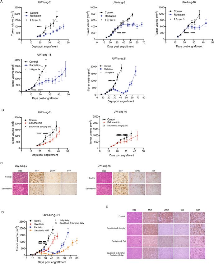
www.nature.com/scientificreports/
Establishment and characterization of xenografts and cell lines. All nine PDX tumors were pas-
saged at least 5 times as subcutaneous xenografts with a mean time between passages of 46.4 days (22–114 days).
There were no differences between time of passage from initial tumor implantation to first passage versus fourth
to fifth passage (p = 0.52). Morphologically, strong retention of cytoarchitectural features was observed between
patient tumor and subcutaneous PDX. Figure 2 shows representative H&E images of the patient sample and
PDX passage 1 and passage 5. TTF-1 expression status was also highly consistent between patient tumor and
PDX (Fig. 2). Genetic profiles of the PDXs were confirmed with STR analysis (Supplemental Table 2). All PDXs
showed 89–100% allelic match to the original patient samples. PDXs continued to retain their STR profiles from
passage one to passage five (Supplemental Table 2). Comprehensive tumor growth rate assessment was carried
out for five PDXs (Fig. 3A). Orthotopic intracranial models were successfully generated from two tumor sam-
ples; University of Wisconsin (UW)-lung-2 and UW-lung-16. Tumors were directly implanted into the brain
after initial subcutaneous passage. They were not passaged in culture. MRI images shows the formation of brain
metastasis and adjacent H&E confirmed tumor, which was morphologically similar to the patient and flank
tumors (Fig. 3B). STR testing confirmed 100% and 92.8% matching of alleles for UW-lung-2 and UW-lung-16
orthotopic tumors, respectively. Cell lines were successfully established from two PDXs, UW-lung-2 and UW-
lung-16, following at least 15 in vitro passages (Fig. 3C). STR testing of subsequent passages confirmed that
these lines retained their genetic profile (Supplemental Table 2). UW-lung-2 and UW-lung-16 cells were also
transfected with luciferase and successfully formed tumors when implanted into the brain of mice (Fig. 3D). In
addition, intracardiac injection of UW-lung-2 cells resulted in hematogenous metastases in the brains of 9 out
of 10 mice.
Transcriptome analysis of patient and PDX tumors. RNA-Seq analysis was performed on four
patient and corresponding flank PDX samples. One PDX (UW-lung-16) was sequenced twice and was used as a
control replicate. Mouse-specific reads were filtered in silico from the PDX RNA samples before aligning reads to
the human genome. Overall, patient and PDX samples highly correlated with each other (Fig. 4A). The average
Pearson correlation coefficient for case-matched tumors was 0.88 and ranged from 0.86 to 0.90. Using a super-
vised analysis comparing matched patient tumors and PDXs, there were 21 Kyoto Encyclopedia of Genes and
Genomes (KEGG) pathways that were significantly over represented in patient tumor samples (q < 0.001) and 12
processes that were significantly over represented in the PDX tumors (q < 0.001) (Supplemental Table 3). There
were 231 Gene Ontology (GO) annotations overrepresented in patients (q < 0.001) and 185 GO annotations sig-
nificantly overrepresented in PDX tumors (Supplemental Table 4). The top 10 most significantly overrepresented
GO annotations in the patient tumor samples consisted of cellular processes associated with immune regulation
while the top pathways overrepresented in the PDXs included those related to cell cycle and DNA replication
(Fig. 4B).
Principal component analysis (PCA) of matched patient and PDX tumor gene expression revealed distinct
clustering of PDXs compared to patient tumor samples when viewed through PC1 and PC2 (Fig. 4C–E). PC2
appeared to be the main contributor of the differences between patient and PDX samples (Fig. 4C). By contrast,
viewing the PCA through PC1/PC3 and PC1/PC4 revealed close clustering of matched PDX samples to their
corresponding patient sample. (Fig. 4D and 4E). On Gene Ontology enrichment analysis, PC2 is highly repre-
sented by immune cell processes (Fig. 4F). PC1, PC3 and PC4 have little representation of immune processes
and appear to represent differences in intrinsic tumor biology (Supplemental Table 4).
Given the differences seen in PC2, we hypothesized that PDXs would associate most similarly to their matched
patient tumors compared with unmatched tumors after controlling for the differences in immune cell popula-
tions. We performed an analysis removing the top 1% of genes contributing to PC2. This removed 219 genes that
were the most prevalent genes associated with immune cell populations (Fig. 4F), and a stronger transcriptional
resemblance between patient and PDX tumor pairs was seen. The average Pearson correlation coefficient for
case-matched tumors and PDXs increased from 0.88 to 0.90 (Fig. 4G).
Patient and PDX variant allele fraction analysis (VAF). Variants detected by RNA-Seq for UW-
lung-2, UW-lung-4, UW-lung-5 and UW-lung-16 were compared between patient tumors and PDXs grown in
the brain and in the flank. High correlations were observed between samples with Pearson correlation coeffi-
cients ranging between 0.794 and 0.839 (Fig. 5A,B). For UW-lung-2 a comparison between patient brain metas-
tasis and PDX grown in the flank and PDX grown in the brain was also performed. Overall, there were 1,979
alterations detected in the UW-lung-2 patient brain metastasis with 96.5% of these alterations identified in the
PDX samples (Fig. 5A). There was a higher correlation in VAF distribution (R2 = 0.848 and 0.794, respectively) of
shared variants, between the patient and the PDX brain metastasis compared to patient and PDX flank tumors.
Overall, these findings indicate that the mutation landscape is conserved between patient and PDX samples.
In addition, our orthotopic intracranial PDX more closely resembles the originating patient brain metastasis
compared to the heterotopic PDX.
PDX response to radiotherapy. Given that radiation is the predominate therapeutic modality for
the treatment of brain metastases, five PDXs were tested for response to standard fractionated radiotherapy
(Fig. 6A). All tumors responded to radiation with tumor growth inhibition values at the end of the experiments
of 51% for UW-lung-2, 42% for UW-lung-5, 53% for UW-lung-16, 58% for UW-lung-18 and 92% for UW-
lung-21 (p < 0.01 for all).
Scientific Reports | (2021) 11:2520 | https://doi.org/10.1038/s41598-021-81832-1 3
Vol.:(0123456789)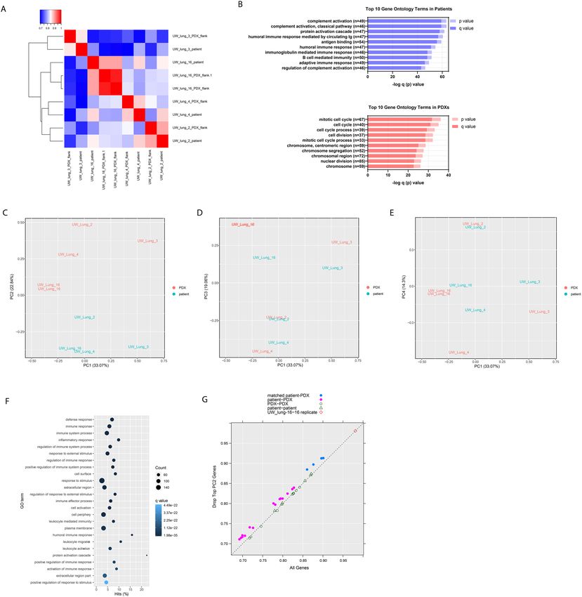
www.nature.com/scientificreports/

                                            Figure 2.  Histopathologic features of nine patient non-small cell lung cancer (NSCLC) brain metastases and
                                            corresponding NSCLC brain metastasis patient-derived xenografts (PDXs). Shown are photomicrographs
                                            hematoxylin and eosin (H&E) of patient tumor, PDX passage 1 and passage 5 and thyroid transcription factor-1
                                            (TTF-1) of patient tumor and passage 1. Images at 20X.

          Scientific Reports |   (2021) 11:2520 |               https://doi.org/10.1038/s41598-021-81832-1                                             4

Vol:.(1234567890)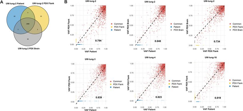
www.nature.com/scientificreports/

                                  Figure 3.  Establishment of subcutaneous flank and orthotopic intracranial patient non-small cell lung cancer
                                  (NSCLC) brain metastasis xenografts. (A) Subcutaneous flank xenograft growth characteristics of University of
                                  Wisconsin (UW)-lung-2, UW-lung-5, UW-lung-16 and UW-lung-18 and UW-lung-21. Tumors were derived
                                  directly from patients’ tumors. (B) Representative MR images and histology of UW-lung-2 and UW-lung-16
                                  orthotopic intracranial xenografts. Tumors were first passaged subcutaneously and then directly implanted
                                  into the brain. They were not passaged in culture. (C) Pictures of established cell lines UW-lung-2 (passage 20)
                                  and UW-lung-16 (passage 20). (D) Representative images of UW-lung-16 luciferase-expressing cells grown as
                                  orthotopic intracranial tumors.

Scientific Reports |   (2021) 11:2520 |               https://doi.org/10.1038/s41598-021-81832-1                                                     5

                                                                                                                                             Vol.:(0123456789)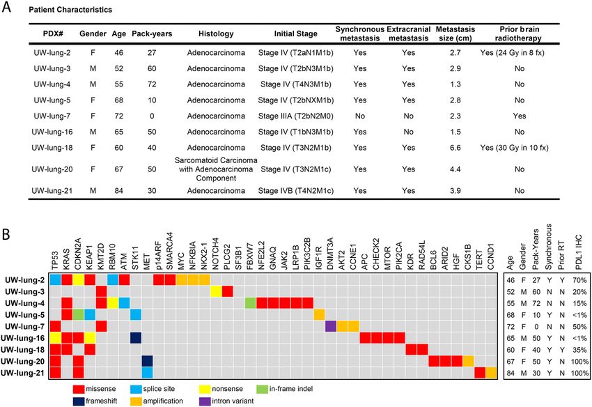
www.nature.com/scientificreports/
Figure 4. RNAseq shows high correlation between patient and patient-derived xenografts (PDXs) tumor
samples. (A) Correlation heatmap and hierarchical clustering of patient and PDX tumor samples. (B) Top 10
Gene Ontology (GO) terms as summarized by bar charts showing the p value (lighter hue) and false discovery
rate (FDR) q value (darker hue). Pathways are labeled along the y-axis; the number of genes annotated within
each pathway is indicated in parentheses. (C–E) Principal component analysis (PCA) clustering of patient and
PDX tumor RNA samples. (F) Top GO terms overrepresented in PC2; showing the number of genes (count), the
q-value and the percent hits. (G) Pearson correlation coefficient of all genes on the x-axis and after removing the
top 1% (219 genes) contributing to PC2.
PDX response to MEK inhibitor selumetinib. KRAS is one of the most commonly mutated genes in
lung adenocarcinoma12, and in our cohort five PDX tumors harbored KRAS mutations. Mutations in KRAS leads
to activation of the mitogen-activated protein kinase (MAPK) pathway involving MAPK kinase (MEK). As an
initial effort to assess the utility of these PDXs for therapeutic response to molecular targeted therapy, we selected
two tumors harboring KRAS G12C mutations and tested their response to the MEK inhibitor selumetinib. Both
tumors responded to selumetinib; however, tumor growth delay was only significant in UW-lung-2 (p = 0.005)
and not significant in the UW-lung 16 (p = 0.34) (Fig. 6B). IHC analysis revealed a decrease in phospho-MAPK,
Scientific Reports | (2021) 11:2520 | https://doi.org/10.1038/s41598-021-81832-1 6
Vol:.(1234567890)www.nature.com/scientificreports/
Figure 5. Conservation of patient-derived xenografts (PDXs) mutational landscape. (A) Venn diagram of all
variants detected in UW-lung-2 patient brain metastasis, brain PDX and flank PDX. (B) Scatterplots displaying
the correlation between patient brain metastasis variant allele frequency (VAF) and flank PDX, patient brain
metastasis VAF and brain PDX and flank PDX and brain PDX. The R2 value is represented by the black value in
the lower right-hand corner of each plot.
pS6 and Ki67 with treatment of selumetinib at 24 h in UW-lung-2. In UW-lung-16, there was minimal change in
phospho-MAPK and Ki-67, but a decrease in pS6 (Fig. 6C) with selumetinib treatment at 24 h.
PDX response to MET inhibitor savolitinib. MET exon 14 skipping mutations occur in 3–4% of lung
adenocarcinomas and results in the deletion of the intracellular juxtamembrane domain of the receptor, leading
to enhanced signaling and tumor proliferation12,13. Small molecule inhibitors have shown therapeutic efficacy
in NSCLC harboring MET exon 14 mutations13. We therefore tested the therapeutic response of UW-lung-21,
which harbors a MET exon 14 mutation, to the selective MET inhibitor savolitinib. Savolitinib alone signifi-
cantly inhibited tumor growth in UW-lung-21 with a tumor growth inhibition value of 46% (p < 0.01, Fig. 6D).
We next tested the capacity of savolitinib to augment the response of radiation. Previous studies have shown
mixed results when combining radiation with MET inhibitors14–16. The combination of savolitinib and radiation
significantly delayed the growth compared to vehicle control, savolitinib or radiation alone (p < 0.01; Fig. 6D).
The mean time from start of treatment for tumors to reach a size of 1000 mm3 was 12.3 days for vehicle-treated
mice; 21.6 days for savolitinib-treated mice, 40.5 days for irradiated mice and 54.7 days in mice that received
savolitinib plus irradiation (p < 0.001). The absolute growth delay was 9.3 days for savolitinib alone, 28.2 days
for irradiation alone and 42.4 days for savolitinib plus irradiation. The radiation dose enhancement factor for
savolitinib was 1.2, indicating that savolitinib enhances radiation-induced tumor growth delay. IHC performed
on UW-lung-21-treated tumors demonstrated inhibition of phospho-MET and pS6 and decrease in Ki67 with
savolitinib treatment (Fig. 6E).
Discussion
Brain metastases from NSCLC frequently occur, remain difficult to treat and if not controlled can lead to sig-
nificant neurologic compromise and ultimately d eath2. Current standard approaches involving surgery and/or
whole brain radiotherapy, while effective, can significantly impair quality of life. Stereotactic radiosurgery can
provide excellent control but continued development of new brain metastases remains a c hallenge2,5. Despite
molecular-targeted agents that penetrate the blood–brain barrier, disease progression in the brain remains a
problem. The lack of preclinical NSCLC brain metastasis models is one barrier that may be preventing the
development of effective therapeutic strategies.
Here we describe the development of a panel of NSCLC brain metastasis PDXs and demonstrate their preclini-
cal utility in evaluating therapies. We accomplished a high engraftment efficiency, and showed that these PDXs
closely resemble the patient’s original metastatic tumor by retaining their morphologic and molecular features.
In addition, we demonstrated the capability of these tumors to grow as cell lines and as intracranial implants
making them a valuable resource to study brain metastases. To expand upon their translational potential, we
characterized the PDXs response to radiotherapy, demonstrated tumor growth inhibition with MEK inhibition in
KRAS mutated PDXs and showed significant growth delay with the MET inhibitor savolitinib in a PDX harbor-
ing a MET exon 14 mutation. Furthermore, combining savolitinib with clinically meaningful doses of radiation
resulted in radiosensitization in our MET exon 14 mutated NSCLC PDX. This combination provides a potential
treatment strategy for patients with brain metastases from exon 14 mutated NSCLC.
Scientific Reports | (2021) 11:2520 | https://doi.org/10.1038/s41598-021-81832-1 7
Vol.:(0123456789)www.nature.com/scientificreports/
Figure 6. Therapeutic effects of radiation, selumitinib and savolitinib on xenograft growth delay. (A) Radiation-induced tumor growth
delay curves of UW-lung-2, UW-lung-5, UW-lung-16, UW-lung-18 and UW-lung-21. Radiation was given as 2 Gy per fraction for
either 5 days or 10 days as indicated. (B) KRAS G12C mutated UW-lung -2 and UW-lung-16 subcutaneous flank xenografts treated with
vehicle or selumitinib 25 mg/kg BID for 10 days. (C) Immunohistochemistry of UW-lung-2 and UW-lung-16 tumor sections showing
modulation of KRAS-downstream signaling (p-Erk and p-S6) following a one-time dose of 25 mg/kg BID of selumitnib, collected at 24 h.
(D) MET exon 14 mutated UW-lung-21 subcutaneous flank xenografts treated with vehicle, savolitinib 2.5 mg/kg, radiation or radiation
plus savolitinib 2.5 mg/kg for 10 days. (E) Immunohistochemistry of UW-lung-21 tumor sections showing modulation of MET signaling
(MET, p-MET, p-Erk and p-S6) and decrease in Ki-67 following a single dose of 2.5 mg/kg of savolitinib, collected at 24 h. Points, mean
tumor volume in mice after treatment; bars, SEM. The scale bar represents 50 mm and all images are to the same scale.
Scientific Reports | (2021) 11:2520 | https://doi.org/10.1038/s41598-021-81832-1 8
Vol:.(1234567890)www.nature.com/scientificreports/
PDXs have both advantages and disadvantages compared to other NSCLC brain metastasis models. PDXs
allow for quick testing of molecular-targeted therapies, recapitulate human tumors and are cost effective17. Other
available models include human and murine cell line xenografts, spontaneous murine models, and humanized
mouse xenografts.
Human cell line models generally show a homogeneous histology and lack human stroma, which is important
for modeling the metastatic process. These cell lines are derived by serially passaging cells in culture, inoculating
them in immunodeficient mice and selecting cells that seed the brain. There are two known available NSCLC
cell lines that have tropism for the brain, H2030-BrM3 and PC9-BrM3, both established from lymph nodes of
patients with lung adenocarcinoma and not from brain metastasis t umors18. Syngeneic murine cell line models
allow for assessment of immunotherapies but are dependent on mouse tumors cell lines, which are typically
driven by specific driver mutations and may not reliably recapitulate human disease. The only reported syngeneic
murine cell line model used Lewis lung carcinoma cells either directly implanted into the brain or forming brain
tumors through hematogenous spread by intracardiac i njections19.
Spontaneous brain metastases can be modeled in immunocompetent, genetically engineered mice (GEM) by
manipulating oncogenes or tumor suppressor g enes20,21. Currently there are no available NSCLC brain metas-
tasis GEM models; however, two models of melanoma22,23 and one model of small cell lung cancer24 have been
reported. In these models, the formation of metastases is limited because primary tumors grow quickly and
animals usually have to be euthanized before brain metastases d evelop25.
Compared to cell line and GEM models, PDXs are directly established from patients and thus closely repro-
duce the molecular, genetic and histological heterogeneity of the original tumor. The disadvantage of PDXs is the
lack of an immune system—however, this can be overcome by using humanized mouse PDX models. Humanized
mouse models engraft components of the human immune system into otherwise immunocompromised mice26.
Ideally, humanized mice would have the same immune system from which the PDX was derived. However, it is
difficult to obtain CD34+ cells from cancer patients, and therefore, an allogeneic immune approach is typically
used27. These are complex models and currently no humanized mouse models for NSCLC brain metastasis exist.
In conclusion, we have developed a panel of human derived NSCLC brain metastasis xenografts and cell
lines, which have the capability of forming intracranial tumors. These PDXs display a spectrum of differentiation
typical of clinical pulmonary adenocarcinoma samples, and a high degree of phenotypic and genetic consistency
between original patient tumor and xenograft. The use of early passage xenografts and cell strains provides a
powerful system for investigating novel therapeutics and combination therapies, and for testing hypotheses of
mechanisms of therapeutic response and resistance in human NSCLC brain metastases.
Materials and methods
Patient selection. Patients with newly diagnosed or recurrent NSCLC brain metastases, undergoing surgi-
cal resection, were consented to participate in the University of Wisconsin Health Sciences Translational Sci-
ence Bio Core Biobank Institutional Review Board approved protocol (IRB # 2016–0934). Written consent was
obtained from all subjects and all methods were performed in accordance with relevant guidelines and regula-
tions of the University of Wisconsin Health Sciences Institutional Review Board.
Mice. Immunodeficient NOD SCID gamma (NSG) (Envigo, Indianapolis) and Hsd:athymic Nude-Foxn1nu
(Envigo, Indianapolis) mice were used for PDX development. All mice were kept in the Association for Assess-
ment and Accreditation of Laboratory Animal Care approved Wisconsin Institute for Medical Research Animal
Care Facility, and studies were carried out in accordance with an approved animal protocol. All animal studies
were in accordance with the Animal Research Reporting of In Vivo (ARRIVE) guidelines.
DNA sequencing. After expert pathologist assessment, solid tumor genomic DNA was isolated from for-
malin-fixed, paraffin-embedded tumor samples. DNA libraries were prepared using the KAPA Hyper Prep Kit,
hybridized to the xT probe set, and amplified with the KAPA HiFi HotStart ReadyMix. The amplified target-
captured DNA tumor libraries were sequenced to an average target depth of 500 × on an Illumina HiSeq 4000
using the Tempus xT (Tempus labs Chicago, IL, USA) gene panel. The panel analyzes single nucleotide variants
(SNVs), indels, and copy number variants in 596 genes and genomic rearrangements in 21 genes with an aver-
age coverage of 5 00x28. Variants were called using Freebayes (version 1.0.2). Clinically relevant mutations were
identified using the Cancer Genome Interpretor29 and ClinVar30. Data was graphed using Microsoft Excel 2016.
Establishment of xenografts. Patient tissue collected during surgery was added to a 1.5 ml tube with
media (Dulbecco’s Modified Eagle Medium with 10% fetal bovine serum, 1% penicillin/streptomycin, and
2.5 μg/mL amphotericin B) and minced with sharp, sterile scissors into a slurry. Matrigel (#354,230, BD Bio-
sciences, Inc) was added to the media in a 1:1 ratio and injected subcutaneously into NSG mice with an 18 gauge
needle. Subsequent passages were made in a similar fashion into either NSG or athymic nude mice. Cryopreser-
vation of xenografts was accomplished by mixing minced tumor pieces with complete transport media supple-
mented with 10% dimethylsulfoxide (DMSO) as previously described31–34. Tumors were frozen in controlled rate
freezers to − 80 °C overnight and transferred to liquid nitrogen for long-term storage. To thaw tumors, aliquots
were warmed to 37 °C in a heated water bath and tumor tissue was washed twice in transport complete media
(without DMSO) and immediately implanted into mice.
Establishment of cell lines. Tumors taken from early PDX passages were dissociated into single cell sus-
pensions and seeded in T-25 flasks in media (Dulbecco’s Modified Eagle Medium with 10% fetal bovine serum,
1% penicillin/streptomycin, and 2.5 μg/mL amphotericin B) and incubated at 37 °C in a humidified atmosphere
Scientific Reports | (2021) 11:2520 | https://doi.org/10.1038/s41598-021-81832-1 9
Vol.:(0123456789)www.nature.com/scientificreports/
of 5% CO2. Media was changed regularly, and cells were passaged once they reached 70% confluence. Myco-
plasma testing and short tandem repeat profiling was performed during passaging.
Short tandem repeat (STR) profiling. DNA typing by multiplex polymerase chain reaction (PCR) of short
repetitive elements of highly polymorphic markers was performed to assess genetic stability of xenografts and cell
lines. STR profiling and analysis was performed using the PowerPlex 16 HS System (Promega, Madison WI).
Luciferase transfection. Luciferase-expressing cells were generated using a pLV-TurboGFP_Luciferase
vector from VectorBuilder via lentiviral transduction. Stably transduced PDX + Luciferase cell lines were selected
with puromycin (2 μg/ml, Sigma-Aldrich) and single-cell cloned for strong luciferase activity.
Orthotopic intracranial implants. Tissue was enzymatically dissociated to single cells and 2 × 10⁵ cells
were suspended in 5 µl of phosphate-buffered saline. Using a Hamilton syringe, the cells were stereotactically
injected under sterile conditions into the right striatum of anesthetized NOD SCID mice at 1 µL/min at the
following coordinates referenced from bregma: 0 mm antero-posterior, + 2.5 mm medio-lateral, and − 3.5 mm
dorso-ventral. The scalp was closed with 6-0 polypropylene suture. Mice were placed on a heating pad in ster-
ile cages and allowed to awaken from anesthesia. One week after injection animals were placed onto a small-
animal MRI scanner (4.7-T horizontal bore imaging/spectroscopy system; Varian, Palo Alto, CA), and T1- and
T2-weighted images were obtained. As per animal protocol, once neurological symptoms were observed, mice
were immediately euthanized by perfusion fixation with 10% paraformaldehyde and moved to 70% ethanol
solution after 48 h. Brains were then excised, formalin fixed, embedded in paraffin, and processed for histology.
In vivo bioluminescence imaging. Tumor growth in the brain was quantified using IVIS Spectrum bio-
luminescence imaging system (Perkin Elmer) starting on day 3. Prior to the in vivo imaging, the mice were anes-
thetized with isoflurane. D-Luciferen firefly/potassium salt solution (150 mg/kg) was injected 18 min before each
scan. The IVIS system detects the emitted light with a CCD camera, yielding a quantitative readout expressed as
radiance (photon/sec/cm2/sr).
Immunohistochemistry. At each passage, tumor was fixed in 10% neutral-buffered formalin and embed-
ded in paraffin blocks. Five-μm sections of PDX and matching patient tumors were stained with hematoxylin
and eosin (H&E) and imaged on an Olympus BX51 microscope (Olympus America, Inc). TTF-1 (clone SPT24,
BioCare, 1:100) staining was performed with a BenchMark Ultra IHC/ISH System (Roche Diagnostics, Rot-
kreuz, Switzerland); according to manufactures established protocol. Comparisons between patient tumor, first
passage xenograft, and subsequent xenograft passages were made by a pathologist on the basis of overall tumor
cytoarchitectural features. In addition, each patient’s tumor and the first PDX passage was stained for TTF-1.
For in vivo immunohistochemistry, 5 μm sections from formalin fixed paraffin embedded samples were
deparaffinized with Xylene and hydrated through graded solutions of ethanol. Antigen retrieval was conducted
in sodium citrate retrieval buffer (pH 6.0) followed by washing in running water. Slides were washed in PBS
and then incubated with 0.3% hydrogen peroxide solution. Blocking was carried out using 10% goat serum in
PBS and then incubated with the primary antibody (Supplemental Methods) diluted in 1% goat serum in PBS
containing 0.1% Triton X-100 overnight at 4 °C. Slides were washed with PBS next day; secondary antibody
was used (SignalStain Boost IHC Detection Reagent (HRP, Rabbit) CST #8114). Staining was detected using
diaminobenzidine (Vector Laboratories, Inc. #SK-4100). The slides were counterstained with 1:10 hematoxylin
(Thermo Scientific #TA-125-MH) solution for 2 min, then dehydrated in ethanol and xylene solutions and
sections were covered with coverslip with Cytoseal (Thermo Scientific #8312-4). Details of antibodies used are
listed in the Supplemental Methods.
RNA sequencing. Total RNA was isolated by using the QIAGEN AllPrep Kit. RNA sequencing libraries
were prepared using the KAPA Stranded RNA-Seq Kit with RiboErase (HMR; KAPA Biosystems). RNA was
fragmented to a size of 100–200 nucleotides and amplified for 11 cycles. Final libraries were quantified on a high-
sensitivity bioanalyzer chip and sequenced on the NextSeq 550 (Illumina).
Gene expression and pathway analysis. RNA-Seq data were aligned to human reference sequence
GRCh38 using RSEM35, and expression quantification per gene was computed. Raw read counts were normal-
ized by the trimmed mean of M-values method36. Differential gene expression (DE) analysis was performed
using a likelihood ratio test with the R package edgeR37,38. The p-values were adjusted to control for false dis-
coveries using the Benjamini–Hochberg method39. Principal component analysis (PCA) was performed on log2
transformed count per million values. The gene lists were sorted as increasing or decreasing. For the increasing
rankings, the genes with positive log fold change (logFC) were sorted by q-value in increasing order, followed
by genes with negative logFC sorted by q-value in decreasing order. For the decreasing rankings, the increasing
list was reversed.
Gene ontology (GO) and gene set enrichment analyses (GSEA) were performed using goseq40 and
GSEAPreranked41, and p-values were adjusted with Benjamini–Hochberg method. The GOseq analysis was
performed using the annotation package Bioconductor TxDb.Hsapiens.UCSC.hg38.knownGene: Annotation
package for TxDb object(s). (Team BC, Maintainer BP (2019), R package version 3.4.6.). GSEAPreranked was
performed using the KEGG subset of the canonical pathways gene sets from MSigDB version 742,43. For identify-
ing annotations overrepresented in the supervised approach, the top 1% (219) of the genes were extracted from
Scientific Reports | (2021) 11:2520 | https://doi.org/10.1038/s41598-021-81832-1 10
Vol:.(1234567890)www.nature.com/scientificreports/
the differential expressing (DE) increasing rankings; in the unsupervised approach, the top 1% (219 genes) were
extracted from the PC2 increasing rankings. For identifying annotations overrepresented in the patient tumors
increasing rankings were used and for PDX samples decreasing rankings were used. To mitigate the high false
discovery rate with GSEAPreranked44, a filtering step as described by Yoon et al.44 was implemented. Pearson
correlation coefficient of the log2 transformed gene expression values was computed, first using all of the genes
for each pair of samples and then excluding the top 1% of genes from the PC2 increasing rankings.
Mutation calling from RNA‑Seq. Bulk RNA-Seq files are pre-processed for quality control using fastp
arameters45. STAR aligner in two-pass mode is used to align the reads to GRCh38 and GRCm38
with default p
46
genomes . Aligned bam files for PDX data are further filtered to remove reads aligning to the host genome with
high confidence using R package XenofilteR47. Following read splitting and duplicate removal, we have utilized
Strelka2 in RNA mode to call variants with default p arameters48. Variants passing the default filters and read
depth 20 are annotated using Ensembl VEP and filtered to include only high impact variants for downstream
analysis.
Tumor growth delay experiments. Tumor cells (5 × 10⁶) suspended in saline were injected subcu-
taneously into the hind leg. When tumors grew to a mean volume of 150 mm3, mice were randomized into
control group, radiation alone group (2 Gy per fractions in 1–2 weeks), selumitinib group (25 mg/kg BID
for 2 weeks), savolitinib group (2.5 mg/kg for 2 weeks) or radiation plus savolitinib group (2.5 mg/kg for
2 weeks). Selumitinib and savolitinib were administered by PO gavage. Each experimental and control group
contained
10 mice. Tumor
volume was measured twice weekly with Vernier calipers and calculated using:
V = π6 x largediameter x(smalldiamter)2. Tumors were followed until the group reached a mean size of
1,500–2,000 mm3. Specific tumor growth delay was calculated as the number of days from start of treatment
for the mean of the treated tumors to grow to 1000 m m3 minus the number of days for the mean of the control
group to reach the same size. The percentage of tumor growth inhibition (%TGI = 1 – [changes of tumor volume
in treatment group/changes of tumor volume in control group] × 100) was used for the evaluation of anti‐tumor
efficacy. The radiation dose enhancement factor was calculated by dividing the normalized tumor growth delay
in mice treated with both savolitinib and radiation by the absolute growth delay in mice treated with radiation
alone. Normalized tumor growth was defined as the time in days for tumors to reach 1000 m m3 in mice treated
with the combination of savolitinib and radiation minus the time in days in mice treated with savolitinib alone.
Statistical analysis. Differences between tumor mean values was determined using a Student’s t-test. Data
are presented as the mean ± standard error of the mean (SEM). A probability level of a p value of < 0.05 was
considered significant. All graphs and analyses were performed and graphed using GraphPad Prism version 8.0
(GraphPad Software, San Diego, CA).
Received: 12 November 2020; Accepted: 12 January 2021
References
1. Waqar, S. N. et al. Non-small-cell Lung cancer with brain metastasis at presentation. Clin. Lung Cancer 19(4), e373–e379 (2018).
2. Langer, C. J. & Mehta, M. P. Current management of brain metastases, with a focus on systemic options. J. Clin. Oncol. Off. J. Am.
Soc. Clin. Oncol. 23(25), 6207–6219 (2005).
3. Sun, A. et al. Prophylactic cranial irradiation vs observation in patients with locally advanced non-small cell lung cancer: a long-
term update of the NRG oncology/RTOG 0214 phase 3 randomized clinical trial. JAMA Oncol. 5(6), 847–855 (2019).
4. Sperduto, P. W. et al. Estimating survival in patients with lung cancer and brain metastases: an update of the graded prognostic
assessment for lung cancer using molecular markers (lung-molGPA). JAMA Oncol. 3(6), 827–831 (2017).
5. Baschnagel, A., Wolters, P. L. & Camphausen, K. Neuropsychological testing and biomarkers in the management of brain metas-
tases. Radiat. Oncol. 3, 26 (2008).
6. Deeken, J. F. & Loscher, W. The blood-brain barrier and cancer: transporters, treatment, and Trojan horses. Clin. Cancer Res. 13(6),
1663–1674 (2007).
7. Soria, J. C. et al. Osimertinib in untreated EGFR-mutated advanced non-small-cell lung cancer. N. Engl. J. Med. 378(2), 113–125
(2018).
8. Peters, S. et al. Alectinib versus crizotinib in untreated ALK-positive non-small-cell lung cancer. N. Engl. J. Med. 377(9), 829–838
(2017).
9. Goldberg SB, Schalper KA, Gettinger SN, et al. Pembrolizumab for management of patients with NSCLC and brain metastases:
long-term results and biomarker analysis from a non-randomised, open-label, phase 2 trial. Lancet Oncol. (2020).
10. Tentler, J. J. et al. Patient-derived tumour xenografts as models for oncology drug development. Nat. Rev. Clin. Oncol. 9(6), 338–350
(2012).
11. Camphausen, K. et al. Influence of in vivo growth on human glioma cell line gene expression: convergent profiles under orthotopic
conditions. Proc. Natl. Acad. Sci. U S A. 102(23), 8287–8292 (2005).
12. Cancer Genome Atlas Research N. Comprehensive molecular profiling of lung adenocarcinoma. Nature. 2014;511(7511):543–550.
13. Awad, M. M. et al. MET Exon 14 mutations in non-small-cell lung cancer are associated with advanced age and stage-dependent
MET genomic amplification and c-Met overexpression. J. Clin. Oncol. Off. J. Am. Soc. Clin. Oncol. 34(7), 721–730 (2016).
14. Baschnagel, A. M. et al. Crizotinib fails to enhance the effect of radiation in head and neck squamous cell carcinoma xenografts.
Anticancer Res. 35(11), 5973–5982 (2015).
15. Bhardwaj, V. et al. C-Met inhibitor MK-8003 radiosensitizes c-Met-expressing non-small-cell lung cancer cells with radiation-
induced c-Met-expression. J. Thorac. Oncol. 7(8), 1211–1217 (2012).
16. Tumati, V. et al. Effect of PF-02341066 and radiation on non-small cell lung cancer cells. Oncol. Rep. 29(3), 1094–1100 (2013).
Scientific Reports | (2021) 11:2520 | https://doi.org/10.1038/s41598-021-81832-1 11
Vol.:(0123456789)www.nature.com/scientificreports/
17. Lee, H. W. et al. Patient-derived xenografts from non-small cell lung cancer brain metastases are valuable translational platforms
for the development of personalized targeted therapy. Clin. Cancer Res. 21(5), 1172–1182 (2015).
18. Nguyen, D. X. et al. WNT/TCF signaling through LEF1 and HOXB9 mediates lung adenocarcinoma metastasis. Cell 138(1), 51–62
(2009).
19. Zhang, Z., Hatori, T. & Nonaka, H. An experimental model of brain metastasis of lung carcinoma. Neuropathology 28(1), 24–28
(2008).
20. Heyer, J., Kwong, L. N., Lowe, S. W. & Chin, L. Non-germline genetically engineered mouse models for translational cancer research.
Nat. Rev. Cancer 10(7), 470–480 (2010).
21. Sharpless, N. E. & Depinho, R. A. The mighty mouse: genetically engineered mouse models in cancer drug development. Nat. Rev.
Drug. Discov. 5(9), 741–754 (2006).
22. Cho, J. H. et al. AKT1 activation promotes development of melanoma metastases. Cell Rep. 13(5), 898–905 (2015).
23. Kato, M. et al. Transgenic mouse model for skin malignant melanoma. Oncogene 17(14), 1885–1888 (1998).
24. Meuwissen, R. et al. Induction of small cell lung cancer by somatic inactivation of both Trp53 and Rb1 in a conditional mouse
model. Cancer Cell 4(3), 181–189 (2003).
25. Gomez-Cuadrado, L., Tracey, N., Ma, R., Qian, B. & Brunton, V. G. Mouse models of metastasis: progress and prospects. Dis.
Model. Mech. 10(9), 1061–1074 (2017).
26. Morton, J. J., Bird, G., Refaeli, Y. & Jimeno, A. Humanized mouse xenograft models: narrowing the tumor-microenvironment gap.
Cancer Res. 76(21), 6153–6158 (2016).
27. Choi, Y. et al. Studying cancer immunotherapy using patient-derived xenografts (PDXs) in humanized mice. Exp. Mol. Med. 50(8),
99 (2018).
28. Beaubier, N. et al. Clinical validation of the tempus xT next-generation targeted oncology sequencing assay. Oncotarget 10(24),
2384–2396 (2019).
29. Tamborero, D. et al. Cancer genome interpreter annotates the biological and clinical relevance of tumor alterations. Genome Med.
10(1), 25 (2018).
30. Landrum, M. J. et al. ClinVar: improving access to variant interpretations and supporting evidence. Nucleic Acids Res. 46(D1),
D1062–D1067 (2018).
31. Kimple, R. J. et al. Development and characterization of HPV-positive and HPV-negative head and neck squamous cell carcinoma
tumorgrafts. Clin. Cancer Res. 19(4), 855–864 (2013).
32. Stein, A. P. et al. Influence of handling conditions on the establishment and propagation of head and neck cancer patient derived
xenografts. PLoS ONE 9(6), e100995 (2014).
33. Swick, A. D. et al. Defining the boundaries and expanding the utility of head and neck cancer patient derived xenografts. Oral
Oncol. 64, 65–72 (2017).
34. Stein, A. P. et al. Xenograft assessment of predictive biomarkers for standard head and neck cancer therapies. Cancer Med. 4(5),
699–712 (2015).
35. Li, B. & Dewey, C. N. RSEM: accurate transcript quantification from RNA-Seq data with or without a reference genome. BMC
Bioinform. 12, 323 (2011).
36. Robinson, M. D. & Oshlack, A. A scaling normalization method for differential expression analysis of RNA-seq data. Genome Biol.
11(3), R25 (2010).
37. McCarthy, D. J., Chen, Y. & Smyth, G. K. Differential expression analysis of multifactor RNA-Seq experiments with respect to
biological variation. Nucleic Acids Res. 40(10), 4288–4297 (2012).
38. Robinson, M. D., McCarthy, D. J. & Smyth, G. K. edgeR: a bioconductor package for differential expression analysis of digital gene
expression data. Bioinformatics 26(1), 139–140 (2010).
39. Benjamini, Y. & Hochberg, Y. Controlling the false discovery rate: a practical and powerful approach to multiple testing. J. Roy.
Stat. Soc. Ser. B (Methodol.) 57(1), 289–300 (1995).
40. Young, M. D., Wakefield, M. J., Smyth, G. K. & Oshlack, A. Gene ontology analysis for RNA-seq: accounting for selection bias.
Genome Biol. 11(2), R14 (2010).
41. Subramanian, A. et al. Gene set enrichment analysis: a knowledge-based approach for interpreting genome-wide expression
profiles. Proc. Natl. Acad. Sci. U S A. 102(43), 15545–15550 (2005).
42. Kanehisa, M. & Goto, S. KEGG: kyoto encyclopedia of genes and genomes. Nucleic Acids Res. 28(1), 27–30 (2000).
43. Liberzon, A. et al. Molecular signatures database (MSigDB) 30. Bioinformatics 27(12), 1739–1740 (2011).
44. Yoon, S., Kim, S. Y. & Nam, D. Improving gene-set enrichment analysis of RNA-Seq data with small replicates. PLoS ONE 11(11),
e0165919 (2016).
45. Chen, S., Zhou, Y., Chen, Y. & Gu, J. fastp: an ultra-fast all-in-one FASTQ preprocessor. Bioinformatics 34(17), i884–i890 (2018).
46. Dobin, A. et al. STAR: ultrafast universal RNA-seq aligner. Bioinformatics 29(1), 15–21 (2013).
47. Kluin, R. J. C. et al. XenofilteR: computational deconvolution of mouse and human reads in tumor xenograft sequence data. BMC
Bioinform. 19(1), 366 (2018).
48. Kim, S. et al. Strelka2: fast and accurate calling of germline and somatic variants. Nat Methods 15(8), 591–594 (2018).
Acknowledgements
We would like to thank Dr. M. Shahriar Salamat and University of Wisconsin Department of Pathology and
Laboratory Medicine for initial tissue processing of patient samples. We also would like to acknowledge the
University of Wisconsin Carbone Cancer Center Translational Science BioCore Biobank for providing tumor
samples for patient derived xenograft development.
Author contributions
A.M.B. and R.J.K. conceived and supervised the project. S.K., A.D., L.A. and S.G. performed the majority of
experiments and data analysis with the assistance of A.M.B., I.O., J.G.S. and R.J.K. A.M.B., S.K, A.D., D.B. and
R.J.K. prepared the figures. A.M.B., S.K. and R.J.K. wrote the manuscript. All authors discussed the results,
provided input and reviewed the manuscript.
Funding
This project was supported in part by grants from the American Cancer Society (RSG-16-091-01-TBG), the UW
Paul P. Carbone Young Investigator Award, the University of Wisconsin Lung Disease Oriented Team, and the
University of Wisconsin Carbone Cancer Center Support Grant (P30 CA014520).
Competing interests
The authors declare no competing interests.
Scientific Reports | (2021) 11:2520 | https://doi.org/10.1038/s41598-021-81832-1 12
Vol:.(1234567890)www.nature.com/scientificreports/
Additional information
Supplementary Information The online version contains supplementary material available at https://doi.
org/10.1038/s41598-021-81832-1.
Correspondence and requests for materials should be addressed to A.M.B. or R.J.K.
Reprints and permissions information is available at www.nature.com/reprints.
Publisher’s note Springer Nature remains neutral with regard to jurisdictional claims in published maps and
institutional affiliations.
Open Access This article is licensed under a Creative Commons Attribution 4.0 International
License, which permits use, sharing, adaptation, distribution and reproduction in any medium or
format, as long as you give appropriate credit to the original author(s) and the source, provide a link to the
Creative Commons licence, and indicate if changes were made. The images or other third party material in this
article are included in the article’s Creative Commons licence, unless indicated otherwise in a credit line to the
material. If material is not included in the article’s Creative Commons licence and your intended use is not
permitted by statutory regulation or exceeds the permitted use, you will need to obtain permission directly from
the copyright holder. To view a copy of this licence, visit http://creativecommons.org/licenses/by/4.0/.
© The Author(s) 2021
Scientific Reports | (2021) 11:2520 | https://doi.org/10.1038/s41598-021-81832-1 13
Vol.:(0123456789)You can also read