Slap the Cap The Role of Capnography in EMS - Bob Page, BAS, NREMT-P, CCEMT-P, NCEE
←
→
Page content transcription
If your browser does not render page correctly, please read the page content below
Slap the Cap
       The Role of Capnography in EMS


        50
        40
        30
        20
        10
         0
                           Time


        50
        40
        30
        20
        10
         0
                           Time

       Bob Page, BAS, NREMT-P, CCEMT-P, NCEE
  Slap
  the
  Cap
      2


                                                    Slap the Cap! The Role of Capnography in EMS
                                                                                                                                                                                     Table of Contents

                       Objectives and Introduction                                                                                                                                                                                                                                                                                                                                                                                                                                                                                      2
Section 1
                       Relevant Anatomy and Physiology Review                                                                                                                                                                                                                                                                                                                                                                                                                                                           3-11
Section 2
                       Technology of Capnography                                                                                                                                                                                                                                                                                                                                                                                                                                                                                    12
                                                                      How EtCO2 is Measured
                                                                      Definitions: Capnography/Capnometry                                                                                                                                                                                                                                                                                                                                                                                                       12-14
50                     Waveform Analysis                                                                                                                                                                                                                                                                                                                                                                                                                                                                        15-19
40
30
                                                                      Normal Capnogram
20
10
 0
                                                                      Normal Abnormal
                                                                                                                                                                                                                                        Time
                       Abnormal Waveforms
                                                                      Rebreathing
                                                                      Bronchospasm
Section 3: Clinical Applications
                       Intubated Patient                                                                                                                                                                                                                                                                                                                                                                                                                                                                        19-22
                                                                      Tube confirmation
                                                                      Ventilation management
                                                                      Curare Cleft
                                                                      Trending
                       Non Intubated Triage and Management                                                                                                                                                                                                                                                                                                                                                                                                                                                      22-29
                                                                      Respiratory Triage
                                                                      Sedation
                                                                      Perfusion and Seizures

                                                                                          ©1998,
  2012
  Bob
  Page,
  Edutainment
  Consulting
  and
  Seminars,
  LLC
Slap
the
Cap
3
Introduction:
You are participating in one of the first nationally
presented courses on capnography in emergency medicine.
This comprehensive course is designed to supply you with the
knowledge background necessary to understand the full
spectrum use of capnography as a diagnostic tool. Just as the
12 Lead ECG is a diagnostic tool for acute coronary syndromes,
capnography is a diagnostic tool for ventilation and perfusion.
It is an objective, fast, and accurate way to triage, assess and
monitor the ABC’s in almost all aspects of the emergency
medicine. This handout, while informative alone, is designed as
a supplement to Bob Page’s Course: Slap the Cap, which offers
far more extensive practice, case presentations and
explanations.
OBJECTIVES
By the end of this session, you will be able to:
Ø Describe the structure and function of the upper and
lower airways.
Ø Describe the mechanics and science of ventilation and
respiration.
Ø Describe the basic physiology of perfusion.
Ø Describe the relationship between ventilation and
perfusion.
Ø Describe the principles behind CO2 measurement.
Ø Describe the various methods of EtCO2 measurement
including quantitative and qualitative capnometry and
capnography.
Ø Describe the technology of EtCO2 measurement including
mainstream, sidestream and microstream sampling.
Ø Identify the components of a normal capnogram waveform.
Ø Identify abnormal capnogram waveforms as related to
various airway, breathing and circulation problems.
Ø Discuss the various clinical applications of capnography.
Ø Given various cases, discuss the role of capnography in
identifying the problem and in the management of the
patient.
©1998,
2012
Bob
Page,
Edutainment
Consulting
and
Seminars,
LLC
  Slap
  the
  Cap
     4


Anatomy and Physiology Review
This course is based on the performance of individuals that
take capnography courses. While it would be easy to show
what to look for on a capnogram, not knowing how or what it is
telling you can be a double-edged sword.
                                                                                                                                                                                        The Circle of Life
In the popular Children’s movie and play, “The Lion King” the
hit song and theme is about the great circle of life. In a way, our
body also has a circle of life. We are put here on earth and
consume the oxygen that is given to us, in return we must give
back CO2. The human circle of life can be visualized with this
graphic.

                                                                                                                                                                                                                                         Lungs


                                          CO2

                                   transported
                                                                                                                                                                                                                                                                                                                                                   O2
  to
  cells

                                     to
  Lungs


                                                                                                  CO2
  in
  Blood
                                                                                                                                                                                                   Metabolism


                                                                                          ©1998,
  2012
  Bob
  Page,
  Edutainment
  Consulting
  and
  Seminars,
  LLC
  Slap
  the
  Cap
     5


      As we breathe in oxygen, it enters in our lungs and through the
alveoli it diffuses into the blood stream. Oxygen is then carried, bound to
hemoglobin molecule (on-loads) on the Red Blood Cells to the body cells
that can use them for metabolism. The oxygen dissociates (offloads) from
the hemoglobin molecule and diffuses into the cells.
                       Once the cells use oxygen for metabolism, the net product is energy
(ATP) and waste product CO2. CO2 is then diffused into the blood stream
and carried back to the lungs as bicarbonate Ion. Once in the lungs, the
CO2 is released out of the body as you exhale. In effect, air goes in and out
and blood must go round and round to put it quite simply.
                       With normal physiology, and circulation is adequate, a prescribed
amount of CO2 should be exhaled. This is a fundamental life process.
Capnography is the only non-invasive tool that can measure this
fundamental life process. This is the main reason why capnography is
such a valuable assessment tool.

Upper Airway

The physiology of the upper
airway structures is to warm,
filter and humidifies the air that
you breathe in.
The various structures also
cause a resistance to airflow on
exhalation that offers a positive
end expiratory pressure or
“PEEP”. PEEP is one of the
three mechanisms you have to
prevent collapse of the alveoli
or atelectasis. The alveoli are
the only place in the body you
can exchange gas with the
environment.
People with pathologies such as
emphysema often need to
“purse” their lips to provide
PEEP because the disease
destroys two of the three
mechanisms. What would
happen to PEEP if this patient
were to be intubated?
                                                                                          ©1998,
  2012
  Bob
  Page,
  Edutainment
  Consulting
  and
  Seminars,
  LLC
  Slap
  the
  Cap
     6


Lower Airway

Larynx
Trachea
Carina


Left and Right Main stem
Bronchi
25 divisions of the
bronchial tree

Bronchioles
This area from the nose to
the bronchioles is known as
DEAD SPACE AIR. By
definition this is air not
available for gas exchange.
This particular type is called
anatomical dead space
because there are not alveoli
until you get to the end of the
airway.
When the dead space is alveolar , this occurs when you have pathology
such as air but no blood, blood but no air, or air and blood but the
exchange surface is compromised. Understanding this pathology is the
key to uncovering the cause and to reverse the situation.

DEFINITION REVIEW:
Dead Space Air: Air that is not available for gas exchange.
Anatomical Dead Space: Air that occupies the space between the nose
and bronchioles that never exchanges gas. (about 150ml in the average
adult)
Alveolar Dead Space: When gas exchange doesn’t occur because air is
present but no blood is available to exchange gas. Or there is blood but no
air. It could also be because the exchange surface is compromised by
pulmonary edema, pulmonary effusion, or swollen membranes.


                                                                                          ©1998,
  2012
  Bob
  Page,
  Edutainment
  Consulting
  and
  Seminars,
  LLC
Slap
the
Cap
7
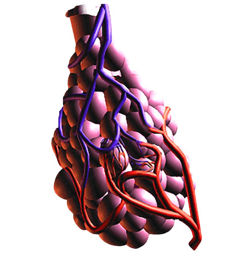
Bronchioles and Capillaries
This is where the gas exchange occurs in the lungs. It is the only place in
the body where you exchange gas with the environment. This juncture is
where most of the pathology we encounter occurs. The alveoli have
elastin fibers around them to allow them to stretch and reform their shape.
The inside of the alveolar membrane has surfactant to allow re-inflation
and to prevent atelectasis.
Between the capillary and alveolar
membrane there is a thin layer of
fluid that O2 and CO2 travel across
to exchange gas. In some
circumstances, pathology and
leaking capillaries into the
interstitial space increase the fluid.
This makes it harder to exchange
gas and can results in atelectasis.
CPAP is a useful tool for this
pathology as it splints open the
alveoli and allows higher pressure
for gas exchange and can even
prevent fluid intrusion.
©1998,
2012
Bob
Page,
Edutainment
Consulting
and
Seminars,
LLC
  Slap
  the
  Cap
     8


RULE of LIFE # 1: Air Must Go In and Out
The Physiology of Breathing
       Why do you breathe? Although you need oxygen to live, you breathe
to get rid of CO2. CO2 is produced as a by-product of cellular metabolism.
The chemoreceptors in you brain sense the levels of CO2 and then report
to the medulla. The medulla triggers the ventilator effort. So, as a result of
eliminating the CO2, you get oxygen in return. This is the circle of life as
the plants take in the CO2 and give us back oxygen.
      Some people with pathology may breathe on a hypoxic drive; that is
they breathe when oxygen levels are low. We were all told to watch out
giving oxygen to a hypoxic breather because we could shut them down.
       What we are talking about is depressing their stimulus to breathe. If
a person loses their stimulus to live, we say they have clinical depression.
To diagnose that, a trained screener that asks a series of questions will
interview the patient. If one loses their stimulus to breathe that is called
respiratory depression. So we screen for that by asking, “Are you too sad
to breathe?” Well not really, but I think you can see something here.
                                                                                                                           Breathing is a Chemical Thing!
So if we want to know if breathing is adequate, then we ask the question:
WWMD? What Would Medulla Do?
       Since the medulla triggers the effort of breathing, just what
happens? The diaphragm contracts and moves down. The intercostal
muscles contract and pull up. This makes the chest wall expand and
creates a negative pressure inside of your lungs, so air comes in. So we
are all negative pressure breathers. In order for us to breathe, we have to
create a negative pressure on the environment. This requires an intact
thoracic bellows, and a lot of energy to make our muscles do the work of
breathing.
       That being said, this leads to one of the signs of adequate breathing,
(or so we are taught) to be chest rise and fall. Well not so fast. In a flail
chest or a open chest wound, the bellows is not intact so when the chest
expands, the flail segment or hole in the chest will negate the pressure
change, so we have chest rise and fall without air movement. That is not at
all adequate. That is why you are taught to seal open chest wounds and
secure a flail chest.
       One more interesting note: To take a normal tidal volume breath at
rest the chest need only create a 2mm/hg pressure difference to make air

                                                                                          ©1998,
  2012
  Bob
  Page,
  Edutainment
  Consulting
  and
  Seminars,
  LLC
  Slap
  the
  Cap
     9


move into the chest. In most people this will only require a 2cm excursion
of the chest wall. Hardly noticeable to count.
                                                                                                                                                                    A Question of Volume

500ml is normal Tidal
Volume for adult at rest
150ml of that is
anatomical dead space
350 alveolar volume for
gas exchange

Total Lung Capacity is
about 6000ml
Vital capacity is the
maximum amount you
can exhale in a forceful
exhalation, about 4800ml
This leaves about
1200ml residual volume.
The larger the volume
the more alveoli that are
recruited and the more
gas exchange occurs.
Definitions:
Hyperventilation:
Blowing off more CO2
that you are making. This creates a deficit and in unchecked, can lead to
cerebral vasoconstriction and a left shift of the oxyhemoglobin
dissociation curve, not allowing O2 to be released at the cellular level. The
patient then becomes hypoxic, with a pulse ox reading of 100%. This is
similar to the effects of CO poisoning.
Hypoventilation. Making more CO2 than you can exhale. As CO2 rises in
hypoventilation, this can lead to acidosis and a right shift of the curve not
allowing O2 to bind easily to hemoglobin, so it cannot be carried to the
cells to be used. As a result the cells become hypoxic. Pulse oximetry in
acidosis will drop off after a few minutes.

                                                                                          ©1998,
  2012
  Bob
  Page,
  Edutainment
  Consulting
  and
  Seminars,
  LLC
  Slap
  the
  Cap
   10


So the true measure of adequate oxygenation should include the
measurement of expired CO2. To oxygenate adequately, one must have
normal CO2 output. This is because oxygenation is dependent upon the
RBC’s ability to on load and offload oxygen. Hyper or hypoventilation will
hinder this ability.
The FICK principle of oxygen transport shed further light on this when it
mentioned that RBC’s must be able to on load and offload O2 as a
condition necessary for perfusion to occur.
Other conditions mentioned by Fick including having enough oxygen,
having enough RBC’s and having the pressure to transport them around
the body. That is commonly referred to as rule #2 of life. “Blood must go
round and round.”
                                                                                                                                                                                                           The Balance
Optimal gas exchange occurs when there are equal ratios of blood and
gas available. This is called the V/Q ratio; that is ventilation to perfusion
ratio. There is a physiologic balance inside of your body. It is based on
normal anatomy and physiology.

                                                                                       ©1998,
  2012
  Bob
  Page,
  Edutainment
  Consulting
  and
  Seminars,
  LLC
  Slap
  the
  Cap
   11


Earlier we discussed anatomical dead space. That referred to airway
structures that carry air down to the alveoli. Once there it can be
exchanged as ventilation is accomplished and respiration. The
physiological balance exists as follows.
In the upper lobes of the lungs, V>Q so there is limited amount of gas that
can be exposed because there is not sufficient blood supply there
available for gas exchange.
In the middle lobes, V=Q so this is where most gas exchange occurs on
normal tidal volume breaths.
In the lower lobes of the lungs, V
  Slap
  the
  Cap
   12


SECTION 2: The Technology of Capnography
It goes by many names but I have to clear the air with the correct names
so we all can have a conversation about capnography;
Qualitative Capnometry: Measure the CO2 by the quality of the color
change between purple and yellow. Make no mistake, this is simply
detection of CO2, it is not a measurement therefore it has very limited use
in emergency medicine. Even its accuracy in confirming ET tube
placement is questionable, so much in fact that it is recommended to have
another device BESIDES that one.
Quantitative Capnometry: Measure the CO2 and give us a value. Values
are useful for triage, assessing the severity of a condition and trending
therapy and response to therapy.
Quantitive Capnography: Measure the CO2 and express it with a waveform
and a value. This gives you an numeric value and a waveform.
Measuring ETCO2
The technology by which CO2 is measured is called infrared spectroscopy.
That’s a fancy way of saying take a sample of air and send IR light through
it and measure the results. All devices measure CO2 this way, some use
broad spectrum beams, while other use more specific beams only for CO2,
Machine that use broad spectrum beams must be compensated for when
giving nitrous oxide or O2 concentration>40%

                                                                                       ©1998,
  2012
  Bob
  Page,
  Edutainment
  Consulting
  and
  Seminars,
  LLC
  Slap
  the
  Cap
   13


Definitions of Measure: ETCO2 values.
Two terms that are synonymous are PetCo2 and ETCO2. “Pet” is “Peak End
Tidal.” ETCO2 is a slightly shortened version. The Peak End Tidal is the
highest level the CO2 obtains.
The normal range for CO2 in the body is 35 – 45mm/hg. The ETCO2 in
cases of normal perfusion have a very close correspondence with Blood
Gas CO2 (PaCO2). The mean difference is 2 and the average difference is
up to 5mm/hg difference. In some studied it even closer than that. So, In
cases or normal perfusion, capnography can be used as a triage tool to
determine the CO2 levels in the body via exhaled CO2 readings.
35-45 mm/hg                                                                                                                                                                                                                                              Normal
Less than 35mm/hg                                                                                                                                                                                                                                        HYPOCAPNIA
More than 45mm/hg                                                                                                                                                                                                                                        HYPERCAPNIA
Hypocapnia: ETCO 2 less than 35mm/hg
Clinical reasons for HYPOCAPNIA:
       1.    Hyperventilation syndrome. The patient, for whatever reason
is blowing off more CO2 than they are making. Or what is worse, we could
the one doing the hyperventilation. This can be due to many reasons
including compensation for a metabolic condition, i.e, DKA or involuntary
reflex such as in Cushing’s response to a closed injury and ICP.
REMEMBER capnography does not tell you why or how they are
hyperventilating, it simply measures the CO2. You can take the reading in
the context of the physical exam and that could be very helpful.
      2.    Hypoperfusion: Shock. If blood doesn’t get back to the lungs
to exchange gas, the amount of CO2 coming out will be diminished. The
following conditions will cause hypocapnia: Shock, hypotension,
pulmonary embolism, doing CPR are just a few of the hypoperfusion
reasons for hypocapnia.
       3.     Hypothermia: Since CO2 is produced as a normal byproduct
of metabolism, in a hypothermic state, the body’s metabolism is slowed
down, so less CO2 is produced soles is blown off. By the same mechanism,
fever (increased metabolism) will produces spikes in CO2. For the most
part, healthy people will increase their breathing to compensate for this.
This is called effortless tachypnea, which is increased respiratory rate,
without dyspnea. Most are not even aware they are breathing fast.

                                                                                       ©1998,
  2012
  Bob
  Page,
  Edutainment
  Consulting
  and
  Seminars,
  LLC
  Slap
  the
  Cap
   14


Hypercapnia: ETCO2 more than 45mm/hg
Clinical reasons for HYPERCAPNIA:

                      1.                                            Ventilatory failure. The purpose of ventilation is to normalize
                                                                    the CO2 in the body. In a hypercapnic state the CO2 being
                                                                    exhaled is the same as the CO2 in the body indicating
                                                                    hypoventilation and failure of the ventilatory system to auto-
                                                                    correct the CO2 levels. This condition, if allowed to exist, can
                                                                    lead to acidosis, which can lead to hypoxic cells. Another
                                                                    important note to make about hypercapnia is that if there is
                                                                    higher than normal to high CO2 exhaled then perfusion to the
                                                                    lungs has to be adequate. In other words, a patient cannot be
                                                                    hypercapnic and hypotensive.

                      2.                                            CO2 retention. It is well known that some end-stage COPD
                                                                    patients retain CO2. Over time, CO2 levels increase slowly
                                                                    and the increase is usually overlooked by the medulla. In end-
                                                                    stage COPD, the stimulus to breathe can become the hypoxic
                                                                    drive (low O2) because the medulla is numb to the CO2’s
                                                                    signals. For this reason, the patient with COPD can retain
                                                                    CO2, but still maintain normal acid-based balance by
                                                                    compensating with the kidneys. When the stimulus to breathe
                                                                    is the hypoxic drive, increased amounts of oxygen given
                                                                    supplementary could cause respiratory depression by
                                                                    suppressing the low O2 stimulus. This is why we are told to be
                                                                    careful administering oxygen to late COPD. The way you can
                                                                    tell that the CO2 retainer is a hypoxic breather is after oxygen
                                                                    administration the CO2 levels will increase above their normal
                                                                    baseline. This is why it is so important to measure ETCO2
                                                                    before oxygen administration.

                      3.                                            Acidosis: Acidosis occurs when the blood Ph drops below
                                                                    7.35. Respiratory acidosis is present when there is high
                                                                    ETCO2 and low Ph. Acidosis causes a right shift of the
                                                                    oxyhemoglobin dissociation curve causing oxygen to have
                                                                    difficulty binding to hemoglobin where it can be carried to the
                                                                    cells. This causes reduced oxygenation to the cells.

                                                                                       ©1998,
  2012
  Bob
  Page,
  Edutainment
  Consulting
  and
  Seminars,
  LLC
  Slap
  the
  Cap
   15


Reading the Wave forms (Riding the Waves)

“An ETCO2 value without a wave form is like a heart rate without an EKG.”
(B. Page, 1998)

       Imagine caring for a patient with extreme tachycardia without any
way of determining the rhythm you’re about to treat. Although we will
treat tachycardia, the selection of medications can be lethal if the wrong
one is chosen. That is why emergency care providers spend hours
mastering EKG interpretation skills. Although the rate is why we treat, the
ECG is what we treat. The good news is, there are very few capnograms
to understand. Let’s look at a waveform analysis.

In reading a waveform, it’s important to realize that capnography
measures CO2 flow, not airflow.

Phase I is called respiratory baseline (A-B). This is at the start of
exhalation and is dead space air without CO2. As the alveolar CO2 makes
its way out of the body, you’ll see a vertical upstroke called Phase II.

Phase II is called respiratory upstroke (B-C). During respiratory upstroke
the CO2 from the alveoli rapidly rises and is measured upon exhalation.
For most patients this should be nearly vertical. It then makes an abrupt
90-degree turn to Phase II, the expiratory plateau.

Phase III is called the expiratory plateau (C-D). The expiratory plateau
should be flat, like a plateau. During this phase, smaller alveoli gradually
increase the CO2 until it reaches peak level. At the peak level is where the
ETCO2 is actually measured and a value is given.

Phase IV is inspiratory downslope. During this phase, inhalation occurs
and the CO2 is rapidly purged from the airways and the alveoli as pressure
is brought in. This concludes the capnogram and makes the square
waveform that you see pictured.

                                                                                       ©1998,
  2012
  Bob
  Page,
  Edutainment
  Consulting
  and
  Seminars,
  LLC
Slap
the
Cap
16
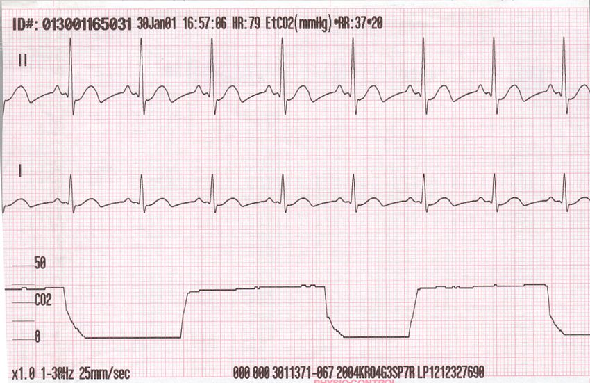
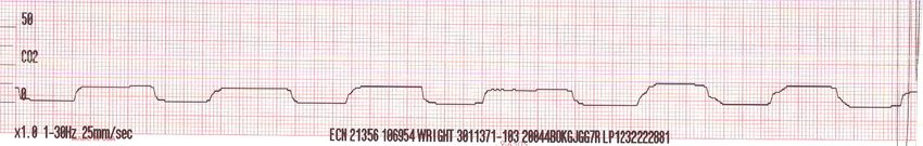
So the normal capnogram will be square with a flat plateau and an ETCO2
between 35 and 45. What can we say about this? A square waveform
means there is no obstruction to CO2 flow. A flat plateau means they’re
exhaling their CO2 to peak level, and a flat baseline means there’s no re-
breathing.
The normal capnogram.
When you first discover capnography you’ll notice a difference between
the on-screen wave form and the printed waveform. This is a result of a
technology trade-off so that a waveform can be seen on the screen.
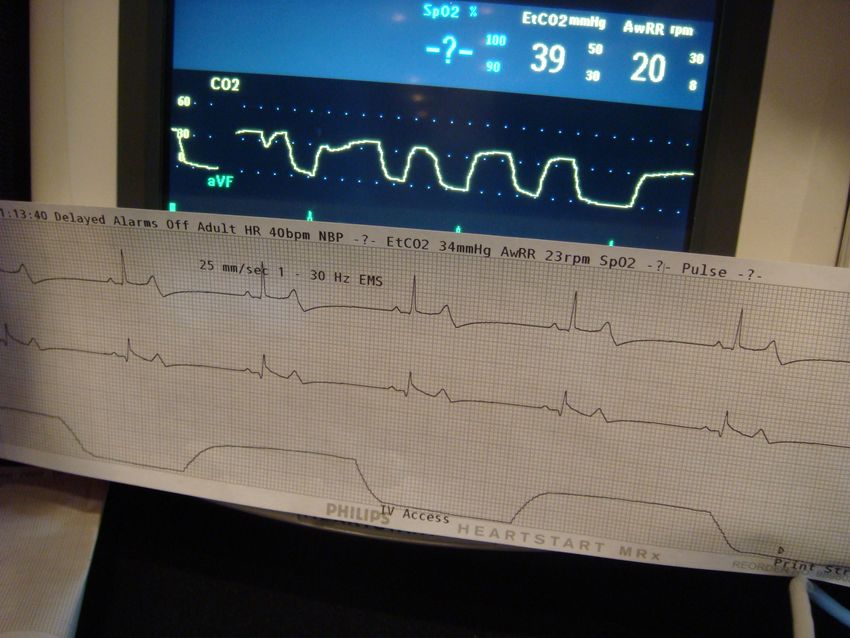
Remember that the normal ECG speed is 25 mm per second. That means
that the normal monitor screen can display just three seconds at a time. If
a patient is breathing once every five seconds, then it would be impossible
to see a wave form on the screen. Therefore, the manufacturers, through
software, have increased the speed of the CO2 channel (user definable) to
ten times its normal rate. This allows the user to see waveforms across
the screen in real time. These waveforms, however, are not diagnostic
except for the presence of a square waveform or a flat-line. In other
words, to accurately diagnose a waveform, the user should print the
waveform real time on the paper. It is best practice to have a dedicated
channel on your multi-parameter monitor, preset to capnography. Never
try to interpret a waveform, other than a square or a flat line, on a monitor
scale. Always print one out.
Direct comparison between printed versus onscreen capnogram.
©1998,
2012
Bob
Page,
Edutainment
Consulting
and
Seminars,
LLC
Slap
the
Cap
17
Steps to Capnogram Interpretation.
Step I. Is there CO 2 present? (Do you have a waveform? Any wave
form?)
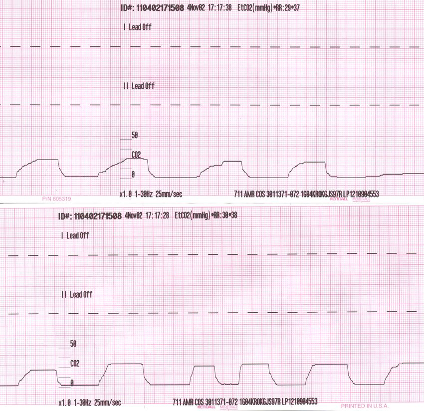
Step 2. Look for re-breathing. With re-breathing comes a breath-to-
breath increase in the respiratory baseline. Rebreathing patterns such as
below have been recorded in Morbid obesity, late term pregnancy, and on
“low end, preset ventilators” that the exhalation valves did not open.
Step 3. Respiratory upstroke. It should be a square. However,
observe for sloping or prolongation.
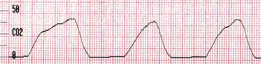
Step 4. Look at the alveolar expiratory plateau. It should be flat.
If the expiratory upstroke is curved over that affects the plateau, this is
known as alveolar emptying. An uneven alveolar emptying occurs
secondary to a bronchospasm. This is different from standard
inflammatory response. During inflammation, most small airways are
narrowed, however they are narrowed evenly throughout. Therefore CO2
is eliminated at the same rate, causing a square waveform. These
patients may wheeze chronically because of this narrowing. However,
this wheeze is characterized by a single tone, also known as a
monophonic wheeze. The waveform will be square in most inflammatory
processes. However, when a bronchospasm occurs, the lower airways
are narrowed at different diameters causing an uneven alveolar emptying.
This causes the capnogram to slope towards the plateau instead of
forming a square waveform. This pattern, sometimes called a shark’s fin,
is truly diagnostic of a bronchospasm.
©1998,
2012
Bob
Page,
Edutainment
Consulting
and
Seminars,
LLC
Slap
the
Cap
18
Uneven Alveolar
Emptying
Bronchospasm
(Not a Shark fin)
Most commonly
seen in asthma
Not all bronchospasms will have a shark’s fin. A true shark’s fin exists
when you lose the alveolar plateau altogether. This is a serious condition,
which means that the CO2 is higher than it shows and the patient is
probably in autopeep. What can be said is that a square wave form all but
rules out a bronchospasm because the alveoli are emptying equally. This
is an objective finding. Remember, it must be assessed on the actual print
out and not on the monitor scope.
True “shark’s
Fin” waveform
Loss of
Plateau
Step 5. Finally, read the CO 2 . The end title CO2 value is a valuable
triage and trending tool. The normal mean CO2 is 40, with the range being
35-45 mm mercury. Having a normal CO2 and a square waveform
indicates the following:
1. There is no obstruction to CO2 emptying.
2. Their breathing is adequate. (A CO2 of 40 means the
perfusion is normal, therefore the CO2 reading should equal
the blood gas CO2, so ventilation is normal.)
3. Their perfusion is adequate because you cannot get a normal
or high CO2 reading while hypoperfusing. In other words, the
ABCs are intact.
ETCO2 = 40
The Normal
Capnogram
This tool can
give you an
objective
©1998,
2012
Bob
Page,
Edutainment
Consulting
and
Seminars,
LLC
  Slap
  the
  Cap
   19


measurement of the patient’s airway patency, breathing adequacy, and
circulatory proficiency with every breath they take. It is the ultimate
measure of a fundamental life process called diffusion.

So the benefits of capnography are, you get a value and a waveform. It
gives you rapid, objective and reliable assessment data of the ABCs. This
data is measurable and trendable. In the old days, we used to triage
patients with a “quick look”. This allowed us to assess the dead for
shockable rhythms so that time to therapy can be reduced. Today, we
have capnography for the living (or the dead), and so we say “slap the cap”
as our quick look for the living. With objective evidence of the ABCs it can
help us rapidly identify immediate life threats.

Section 3: Clinical Applications

The intubated patient.

Part I. To confirm tube placement.
This is probably the most common use of capnography, yet limiting
oneself to this use only is a huge waste. In the beginning, color change
devices would detect CO2 levels. This is widely believed to be able to
accurately predict when the endotracheal tube is misplaced in the
esophagus. Theoretically, there should be no CO2 exhaled from the
esophagus, on the trachea. However, in low perfusion states, this is not a
very accurate reading and the manufacturer even suggests using another
confirming device besides this one.

Since modern capnography can measure CO2 all the way down to one
(NOTE: This is an objective measurement, not a color change), the
numbers however, cannot confirm correct tube placement because low
CO2 readings can also come out of the esophagus or will also show when
the tube is in the hypopharynx. For this reason, there is “no value of CO2
that would confirm endotracheal placement”.

This leaves the waveform that is the gold standard for endotracheal tube
confirmation. Tube confirmation is confirmed with a SQUARE waveform.
With a square waveform, the tube cannot be in the esophagus, or the
hypopharynx. It must be in the trachea, regardless of the value of the
return of CO2.

                                                                                       ©1998,
  2012
  Bob
  Page,
  Edutainment
  Consulting
  and
  Seminars,
  LLC
Slap
the
Cap
20
Time
and
date
stamped
indicating
time
care
turned
over
to
hospital
Square
waveform
indicating
correct
tracheal
placement
Right mainstem intubation. A square waveform can occur with a right
mainstem intubation because the tube is still in the main airway.
Therefore, auscultation in the fifth intercostal space midaxillary,
bilaterally, is necessary to rule out right mainstem intubation.
Not only can capnography square waveform confirm correct endotracheal
tube placement, but it can also confirm most BIADs (blind insertion airway
devices).
Q&A: Can capnography confirm ET tube placement?
YES with SQUARES
Can capnography detect esophageal intubation?
YES no Squares!
Can capnography detect hypopharyngeal intubation?
Yes, no Squares
Can capnography detect right mainstem intubation?
NO, Still must listen to both sides, even with squares
TUBE and CAPNOGRAPHY PEARLS
v Measured ETCO2 alone cannot confirm ET Tube placement, the
square waveform must be printed to confirm.
v Low perfusion states will produce a smaller “square” but you can
still confirm the tube is in the trachea.
v In cardiac arrest, CPR must be performed, and ventilation must
occur in order for capnography to even show anything on the
capnogram.
©1998,
2012
Bob
Page,
Edutainment
Consulting
and
Seminars,
LLC
Slap
the
Cap
21
v In some cases of pediatric or infant intubation, it may be necessary
for a cuffed tube be used with a small amount of air in the cuff to get
a waveform as leaking air around the tube will prevent from coming
up the tube to be measured.
Setup for confirmation. Now this a brand specific task, so check with your
own monitor for setup instructions. There ARE some basic premises you
need to follow that all devices will need to address.
1. Make sure the device has capnography waveform set up default on
one of the channels. Turn on the machine as you check equipment.
2. Make sure the capnography filter line is already plugged in and
sampling, while you are preparing the equipment. This will eliminate
the delay that can happen with sensor warm-up times and auto zero.
3. Place the airway CO2 sensor on the ET Tube or have it ready for use
ON THE FIRST BREATH to check correct placement!
4. PRINT the waveform immediately as you confirm. Leave
capnography unit attached to patient for tube vigilance, trending,
and ventilation check.
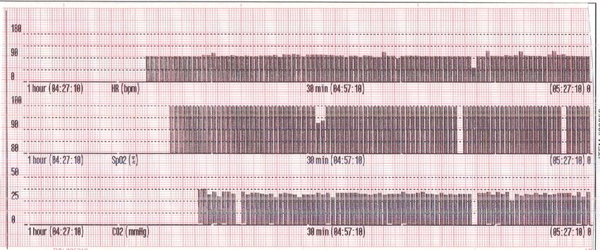
TRENDING after intubation, or the entire call.
One of the huge advantages of these monitors is that most manufacturers
offer some sort of trending options for user defined parameters. Below is
a trend of ETCO2 Heart Rate and SPO2 before during and after an RSI
procedure. This type of documentation is a great defense tool.
Note that the objective proof shows that the patient did not desaturate
during the procedure and that the heart did not increase or decrease (no
catecholamine response) and the CO2 remained stable (adequate
ventilation and tube did not come out)
©1998,
2012
Bob
Page,
Edutainment
Consulting
and
Seminars,
LLC
Slap
the
Cap
22
Part II. Adequate Ventilation after intubation
While it is imperative that we conform endotracheal tube placement in the
trachea, more important is how the patient is ventilated after intubation. A
tube in the trachea/airway eliminates the dead space cause by a facemask
and resistance of upper airway structures, but it also make it incredible
easy to over ventilate (hyperventilate) the patient. The following
capnogram illustrates a patient intubated and confirmed but is being too
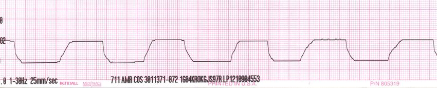
aggressively ventilated, resulting in an ETCO2 of less than 20.
This is unacceptable and causes harm to the patient. However, how would
someone even know how much to squeeze the bag anyway? Subjective
“criteria” such as breaths per minute and a certain “quality of chest rise”
is NOT reliable. The body measures CO2. Why don’t we all? With an
objective, reliable tool such as capnography, it is easy to accomplish this
safely and effectively.
Part III. Early warning time for intubated patient waking up.
While RSI may be necessary in some cases to facilitate intubation, it can
also create problems for airway management when the paralytic wears off.
As a neuromuscular blockage begins to wear off, the diaphragm will be
the first to try to move. This cannot be seen by observation but is easily
detected by a capnogram. This is called a curare cleft.
Curare cleft
This little indention tells you that you have maybe 3 minutes to sedate the
patient before they begin to waken or start to fight the tube.
©1998,
2012
Bob
Page,
Edutainment
Consulting
and
Seminars,
LLC
  Slap
  the
  Cap
   23


PART 3: CLINICAL APPLICATIONS
The Non Intubated Patient
Although many systems and protocols mandate the use of Capnography
for intubated patient, another area that is commonly overlooked in in the
non intubated patient.
This has the potential to be used on nearly every patient encounter as a
triage tool. Refer to my website, www.multileadmedics.com to download
the actual cases from the capnography page. This also has extensive
bibliography of many of the resources used in the preparation of this
project. Bob will also show clear cut examples in the Slap the Cap course.

Below however, is a synopsis of clinical uses for capnography in the non
intubated patient.

Part 1 How do you do this?
All manufacturers of capnography products make adapters for the
intubated and non-intubated patient. The non intubated patients has
several options:
Nasal cannula
Oral/Nasal Cannula: The can also be use to administer O2 saving extra
money spent on separate cannula.

                                                                                       ©1998,
  2012
  Bob
  Page,
  Edutainment
  Consulting
  and
  Seminars,
  LLC
  Slap
  the
  Cap
   24


“Puffer” mouth piece over an intubation adapter
This device works well if you
do not have a continuous
nasal cannula. You can do a
quick triage by having them
puff into the mouthpiece and
get a spot check, then
perform the therapy and
check again after therapy.
No manufacturer
recommends giving updraft
treatments directly through
capnography filter lines
although it can be done.

It has also been demonstrated that the nasal cannula devices can be used
under CPAP masks. (see the March 2012 supplement on Capnography for
Oridion/JEMS magazine on my website) The readings are reliable, just
about 10mm/hg (CO2 washout) lower than baseline without CPAP but the
waveform is not altered.
Oral/Nasal cannulas have also been used successfully under a non
rebreather mask as well.
Depending on the manufacturer, the device plugs into the main machine
and the reading is set up there. Again, check which one you have to find
out how to do it. PRACTICE OFTEN. Learn how the device works.

                                                                                                                                                                  Patient Talking Artifact!

                                                                                       ©1998,
  2012
  Bob
  Page,
  Edutainment
  Consulting
  and
  Seminars,
  LLC
Slap
the
Cap
25
Part 2: Some Specific Uses
RESPIRATORY
Asthma: Capnography can be used a triage tool for asthma attacks. It can
assess the severity and can give an accurate measure of Arterial blood
gas thus helping the caregiver to triage the severity of the attack. It can
also be used to trend and document therapy success or not.
Same patient before albuterol (left) and after albuterol (right) This is
objective proof this was a bronchospasm because the drug corrected the
bronchospasm. This response came within three minutes of therapy time.
A patient with a square waveform does not have a bronchospasm, so the
benefit of a bronchodilator must be carefully examined and weighed
against the risk of tachycardia.
COPD: capnography can be used to determine if bronchodilator therapy is
necessary, and also can assess and trend possible CO2 retainers.
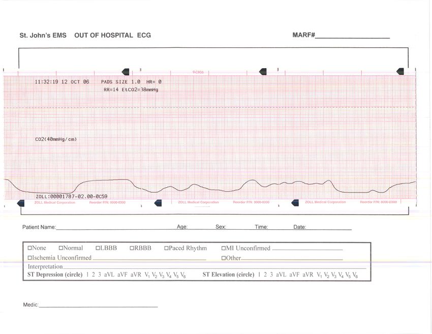
CHF with Pulmonary Edema: This is perhaps the most difficult diagnosis to
make anywhere (CHF versus COPD) It has been reported that as much as
50% of patients with CHF were given unnecessary bronchodilator therapy.
The capnography can triage the severity an also prevent this mistake:
This capnogram shows a high ETCO2 (ventilator failure), but there is no
objective evidence of bronchospasm, avoiding unnecessary and possibly
harmful therapy.
©1998,
2012
Bob
Page,
Edutainment
Consulting
and
Seminars,
LLC
  Slap
  the
  Cap
   26


Can we really use this device to triage a respiratory patient? Does the
blood gas CO2 correspond with the ETCO2? Below is an abstract from a
2005 article that was published in the Annuals of Emergency Medicine.
There are others, see my website and the articles, or Google this article
and it will lead you to others.

So as you can see, for the respiratory patient, the evidence to date
supports the value of capnography as a triage tool. Furthermore, the
waveform is the only objective reliable tool for assessing bronchospasm.
Not only can capnography be used to triage, it can also be used to trend
therapy effectiveness and monitor for relapses.

                                                                                       ©1998,
  2012
  Bob
  Page,
  Edutainment
  Consulting
  and
  Seminars,
  LLC
  Slap
  the
  Cap
   27


• Sedation: Anybody sedated for any reason must be monitored for
     respiratory depression. Download the standards (from my website)
     from all organizations and see where your profession falls in place
     on the use of capnography Drugs such as recreational ones, legal
     ones, narcotics, benzo’s, depressants can all cause respiratory
     depression. Recent and well-done studies have proven the benefits
     of capnography in enhanced patient safety: How does capnography
     show respiratory depression? The ETCO2 will trend up with a normal
     baseline indicating hypoventilation, and hypercapnia.

What is this? A PCA Pump with
Capnography override

• Supports current ASA and Joint
     Commission standards
     mandating CO2 monitoring
     for all anesthesized patients
     (intubated and non-
     intubated)

• Provides an additional safety
      net at the bedside to
      continuously monitor
      patient respiratory response
      to infusion therapy

                                                                                       ©1998,
  2012
  Bob
  Page,
  Edutainment
  Consulting
  and
  Seminars,
  LLC
Slap
the
Cap
28
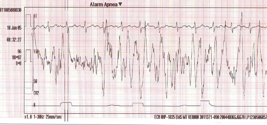
Seizures: When someone has a Generalized Seizure, such as a tonic/
clonic, it affects both hemispheres of the brain and the medulla. When the
medulla is involved, the patient does not breath during seizure activity.
This is easily determined by capnography during the seizure, (see below
capnogram or refer to in class cases I present.
The capnogram shows small ineffective breaths (small waveforms) this is
noisemaking (involuntary) as thoracic muscle contract during the seizure.
This is proof that the seizure is on going. When the patient goes post-ictal,
the breathing will resume. If it does not, the patient needs to be ventilated
to normal ETCO2 and anti-seizure meds need to be given.
Any generalized seizure will cause apnea during the seizure, though most
are short in duration (2-10 seconds), not breathing is usually not noticed.
Metabolic Uses: DKA
Since CO2 is carried in the blood stream and bicarbonate Ion, it has a
direct clinical relationship to serum bicarb levels. Therefore, if the patient
has a high Blood glucose, measure their ETCO2. If it is less than 29, then
the patient has DKA. The blood gas bicarb will show a very low level as
well, indication metabolic acidosis.
Pulmonary Embolism:
This is easy. The combination of ETCO2 and an ABG CO2 can easily call a
V/Q mismatch. All you need then is a CT scan to figure out where it is and
they are on their way. A high blood gas CO2 and a low ETCO2 tells us the
CO2 is not getting to the lungs to be exhaled.
©1998,
2012
Bob
Page,
Edutainment
Consulting
and
Seminars,
LLC
  Slap
  the
  Cap
   29


Perfusion?
The 2010 AHA criteria recommend capnography for monitoring the
effectiveness of CPR, and for detecting the presence of ROSC (return of
spontaneous circulation. Here is a trend that shows ROSC after 4 min of
CPR and a defibrillation.

ROSC will show a sharp increase in CO2 levels. When this happens, stop
pushing and start squeezing the BVM to return the CO2 to normal. Most
cases that have ROSC has ETCO2 that go into the 70-90’s!
Trauma?
In Tension Pneumothorax, pressure in the chest collapses a lung and then
presses on the right side of the heart making it hard to fill with blood. It
only takes about 7mm/hg pressure to stop the blood flow into the right
atria. The first and must reliable sign of a TENSION pneumothorax is the
sudden drop in perfusion that is picked up immediately on a capnogram.
By the same token, when the chest is successfully decompressed, it is not
a rush of air but a sudden increase in ETCO2 that confirms decompression
success. Furthermore, the capnogram can be used to keep watch in case
it develops again.
The same is true for Pericardial Tamponade and cardiocentesis. In each
of these obstructive forms of hypoperfusion, the capnogram will remain
square because it is a perfusion problem, not an airway problem, but you
knew that, right?
Closed Head Injury. ITLS and the Brain Trauma Foundation have taken the
lead in recommending capnography as the way titrate CO2 ventilations in
the patient with a closed head injury. If the patient has a GCS of less than
9 and they are posturing, have unequal pupils, or dropped two in front of
you, then they should be selectively ventilated to an ETCO2 between 30-
35mm/hg. If the patient does not the signs (above) of deterioration, then
ventilate the patient to levels, 35-45. Never ever bag them to lower than
25mm/hg. It causes cerebral vasoconstriction and creates an alkalosis not
allowing O2 to dissociate from hemoglobin, make the brain injury worse.

                                                                                       ©1998,
  2012
  Bob
  Page,
  Edutainment
  Consulting
  and
  Seminars,
  LLC
  Slap
  the
  Cap
   30


In Conclusion:
There are many more cases to be seen, through my seminars and
publications. I hope you enjoyed the seminar! I really get into it because I
believe this will help you do your job better and since we are health care,
it is our patients that will benefit from this in the end.

It is easy to say no to a new idea, Throughout 2011 I surveyed 3800 of
students in my capnography class. About 30% had had a formal
capnography course before, and of those, the average time in class was 1
hour. There is no way to gain adequate knowledge and experience doing
this. Since most systems require capnography for one purpose, tube
confirmation, so that is all they ever pursue knowledge on. Ignorance (the
lack of knowledge thereof) is the biggest factor in the slow adoption of this
tool. And this lack of understanding and knowledge goes to all levels of
Emergency services.
It is my hope and prayer that this class has touched you personally and
sparked a fire that will help carry this idea to all levels of medicine. It takes
great courage to take a new idea and run with it. Please let me know if
there is anything I can do to help you.

For more information on your instructor or for information on these or
other classes I present, or to get to your area for a class, visit the web site
at

www.multileadmedics.com
or e-mail me at lead2noclue@mac.com
Bob Page, BAS, NREMT-P, CCEMT-P, NCEE
Edutainment Consulting and Seminars, LLC

                                                                                       ©1998,
  2012
  Bob
  Page,
  Edutainment
  Consulting
  and
  Seminars,
  LLC
You can also read