Congenital Brain abnormalities and Zika Virus: What the Radiologist Can Expect to See Prenatally and Postnatally1 - Neurotalk
←
→
Page content transcription
If your browser does not render page correctly, please read the page content below
This copy is for personal use only. To order printed copies, contact reprints@rsna.org
Original Research
Congenital Brain Abnormalities
and Zika Virus: What the
n Special Report
Radiologist Can Expect to See
Prenatally and Postnatally1
Patricia Soares de Oliveira-Szejnfeld, MD
Purpose: To document the imaging findings associated with con-
Deborah Levine, MD
genital Zika virus infection as found in the Instituto de
Adriana Suely de Oliveira Melo, MD, PhD
Pesquisa in Campina Grande State Paraiba (IPESQ) in
Melania Maria Ramos Amorim, MD, PhD northeastern Brazil, where the congenital infection has
Alba Gean M. Batista, MD been particularly severe.
Leila Chimelli, MD, PhD
Amilcar Tanuri, MD, PhD Materials and From June 2015 to May 2016, 438 patients were referred
Renato Santana Aguiar, PhD Methods: to the IPESQ for rash occurring during pregnancy or for
Gustavo Malinger, MD, PhD suspected fetal central nervous system abnormality. Pa-
Renato Ximenes, MD tients who underwent imaging at IPESQ were included, as
Richard Robertson, MD well as those with documented Zika virus infection in fluid
Jacob Szejnfeld, MD, PhD or tissue (n = 17, confirmed infection cohort) or those
with brain findings suspicious for Zika virus infection,
Fernanda Tovar-Moll, MD, PhD
with intracranial calcifications (n = 28, presumed infection
cohort). Imaging examinations included 12 fetal magnetic
resonance (MR) examinations, 42 postnatal brain com-
puted tomographic examinations, and 11 postnatal brain
MR examinations. Images were reviewed by four radiolo-
1
gists, with final opinion achieved by means of consensus.
From the Dept of Diagnostic Imaging, Federal Univ of São
Paulo, São Paulo, Brazil (P.S.d.O.S., R.X., J.S.); Foundation
Inst for Education and Research in Diagnostic Imaging,
Results: Brain abnormalities seen in confirmed (n = 17) and pre-
Dept of Diagnostic Imaging, Federal Univ of São Paulo, São sumed (n = 28) congenital Zika virus infections were simi-
Paulo, Brazil (P.S.d.O.S., J.S.); Dept of Radiology, Beth Israel lar, with ventriculomegaly in 16 of 17 (94%) and 27 of 28
Deaconess Medical Ctr, Harvard Medical School, Boston, (96%) infections, respectively; abnormalities of the corpus
Mass (D.L.); Instituto de Pesquisa Professor Amorim callosum in 16 of 17 (94%) and 22 of 28 (78%) infections,
Neto, Campina Grande, PB, Brazil (A.S.d.O.M., M.M.R.A.);
respectively; and cortical migrational abnormalities in 16
Instituto de Saúde Elpidio de Almeida, Campina Grande,
PB, Brazil (A.S.d.O.M., M.M.R.A.); Faculdade de Ciências
of 17 (94%) and 28 of 28 (100%) infections, respectively.
Médicas de Campina Grande, Campina Grande, PB, Brazil Although most fetuses underwent at least one examination
(A.S.d.O.M.); Hosp Municipal Pedro I, Serviço Municipal that showed head circumference below the 5th percentile,
de Atendimento Transdisciplinar a Gestantes e Bebês head circumference could be normal in the presence of se-
com Infecção Congênita por Zika Virus, Campina Grande, vere ventriculomegaly (seen in three fetuses). Intracranial
PB, Brazil (A.S.d.O.M., A.G.M.B.); Universidade Federal calcifications were most commonly seen at the gray mat-
de Campina Grande, PB, Brazil (M.M.R.A.); Laboratory of
ter–white matter junction, in 15 of 17 (88%) and 28 of 28
Neuropathology, State Inst of Brain, Rio de Janeiro, Brazil
(L.C.); Departamento de Genética, Instituto de Biologia, Uni- (100%) confirmed and presumed infections, respectively.
versidade Federal do Rio de Janeiro, Rio de Janeiro, Brazil The basal ganglia and/or thalamus were also commonly in-
(A.T., R.S.A.); Div of Ultrasound in Obstetrics & Gynecology, volved with calcifications in 11 of 17 (65%) and 18 of 28
Lis Maternity Hosp, Tel Aviv Sourasky Medical Ctr, Sackler (64%) infections, respectively. The skull frequently had a
Faculty of Medicine, Tel Aviv Univ, Tel Aviv, Israel (G.M.); collapsed appearance with overlapping sutures and redun-
Fetal Medicine Foundation Latinamerica–FMFLA, Centrus–
dant skin folds and, occasionally, intracranial herniation
Fetal Medicine, Campinas, Brazil (R.X.); Boston Children’s
Hosp, Boston, Mass (R.R.); Inst of Biomedical Sciences and
of orbital fat and clot in the confluence of sinuses.
National Ctr for Structural Biology and Bioimaging, Federal
Univ of Rio de Janeiro, Rio de Janeiro, Brazil (F.T.M.); and Conclusion: The spectrum of findings associated with congenital Zika
D’Or Inst for Research and Education, Rua Diniz Cordeiro virus infection in the IPESQ in northeastern Brazil is il-
30, Botafogo, Rio de Janeiro, RJ, Brazil 22881-100 (F.T.M.). lustrated to aid the radiologist in identifying Zika virus
Received July 7, 2016; revision requested July 14; revision
infection at imaging.
received July 22; accepted July 27; final version accepted
August 3. Address correspondence to F.T.M. (e-mail:
Fernanda.tovarmoll@idor.org).
q
RSNA, 2016
q
RSNA, 2016 Online supplemental material is available for this article.
Radiology: Volume 281: Number 2— 2016 n radiology.rsna.org 1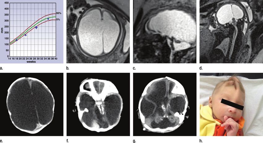
SPECIAL REPORT: Congenital Brain Abnormalities and Zika Virus: What the Radiologist Can Expect to See Soares de Oliveira-Szejnfeld et al
M
uch has been written recently at prenatal ultrasonography (US), and/ infection, viral infection was confirmed
regarding Zika virus in preg- or (c) postnatal microcephaly or other by means of serologic findings or reverse
nancy and the increased risk CNS malformation that was believed transcription polymerase chain reaction
of microcephaly in fetuses exposed to to be characteristic of congenital infec- (RT-PCR). An assay (Rapid Test DPP
the virus. The outbreak of infection tion. During this period, 384 pregnant Zika IgM/IgG Assay; Chembio, Medford,
in Brazil, especially in the northeast women with a rash or a history of CNS NY) was used to detect immunoglobulin
part of the country (1), has been of abnormality at US (group 1) and 47 ne- G and immunoglobulin M against Zika
particular concern. The virus has onates with postnatal physical examina- virus in the fluids. Virus genome was
been found in the fluids of pregnant tion findings suggestive of microcephaly identified in fluids and/or tissues with
mothers and during autopsy in the (head circumference , 33 cm) (group 2) RT-PCR by using specific primers and
brains of neonates with microceph- were enrolled in this protocol. Group 1 probes to identify the region of Zika
aly (2–5). Much of the concern in the exclusion criteria included not returning virus (13). Dengue virus and chikun-
media regarding the teratogenicity of for imaging at IPESQ, no CNS abnor- gunya virus infection were excluded by
Zika virus infection has focused on mality identified at US examination at using enzyme-linked immunosorbent
brain findings of microcephaly. How- IPESQ, classic findings of isolated CNS assay and RT-PCR. The patients tested
ever, as documented in many case abnormality not characteristic of infec- negative for toxoplasmosis, syphilis,
series, there are a variety of brain tion, diagnosis of a genetic syndrome or varicella-zoster virus, Parvovirus B19,
abnormalities that can be found in aneuploidy, and lack of postnatal images rubella, Cytomegalovirus, and herpes
fetuses exposed to intrauterine Zika for review. Group 2 exclusion criteria virus (TORCH) infection, as well as hu-
virus infection (2–11). These include included not returning for computed to- man immunodeficiency virus.
abnormalities in ventricular size, gray mography (CT) or magnetic resonance As described previously (4,5), pre-
and white matter volume loss, brain- (MR) imaging examination at IPESQ natal US was performed by fetal med-
stem abnormalities, and calcifications. and no calcification on postnatal images. icine specialists using either a Voluson
Although the current outbreak has Initially, we used a head circumference E8 unit (General Electric, Milwaukee,
centered in Brazil, according to the U.S. criterion of 32.5 cm for microcephaly as Wis) with transvaginal probes or a
Centers for Disease Control and Preven- an exclusion criterion for the postnatal Samsung XG or WS80 unit (Sam-
tion, there are currently 51 countries diagnosis, but after review of the imaging sung, Seoul, South Korea) with 2–9-
or territories in which active transmis- findings, there was a newborn with nor- MHz probes. MR imaging of the fetus
sion of Zika virus has been reported mal head circumference but severe ven- was performed with a 3-T Skyra unit
(12). It is important for radiologists to triculomegaly and calcifications similar
understand the type of abnormalities to those seen in the other confirmed and
associated with congenital Zika virus in- presumed Zika virus infections; there- Published online before print
fection to aid in recognition of disease fore, this neonate was included in our 10.1148/radiol.2016161584 Content codes:
and appropriate counseling of patients. population with presumed Zika virus in- Radiology 2016; 281:1–16
The purpose of this special report is to fection. After exclusions, there were im-
document the imaging findings associ- aging studies of 31 fetuses in 30 pregnant Abbreviations:
CNS = central nervous system
ated with congenital Zika virus infection women (which included one set of twins)
IPESQ = Instituto de Pesquisa in Campina Grande State
as found in patients seen at the Instituto and 45 neonates (which included the 31 Paraiba
de Pesquisa in Campina Grande State fetuses that underwent prenatal imaging, RT-PCR = reverse transcription polymerase chain reaction
Paraiba (IPESQ) in northeastern Brazil. as well as an additional 14 neonates that TORCH = toxoplasmosis, syphilis, varicella-zoster virus, par-
were enrolled postnatally; Fig 1). vovirus B19, rubella, cytomegalovirus, and herpes virus
Results in 10 of these patients have
Materials and Methods Author contributions:
been reported previously (4,5); however, Guarantors of integrity of entire study, P.S.d.O.S., D.L.,
This retrospective review includes im- these prior publications were not of suf- A.S.d.O.M., A.G.M.B., A.T., R.X., F.T.M.; study concepts/
aging and autopsy data from an insti- ficiently large sample size to provide esti- study design or data acquisition or data analysis/interpreta-
tutional review board–approved study mates of the types of abnormalities seen tion, all authors; manuscript drafting or manuscript revision
that allowed for imaging and follow-up in infected neonates, and the images for important intellectual content, all authors; approval of
final version of submitted manuscript, all authors; agrees to
of presumed Zika virus infection in preg- were not formally reviewed by multiple
ensure any questions related to the work are appropri-
nant women and their neonates. Writ- fetal and neonatal imaging experts. ately resolved, all authors; literature research, P.S.d.O.S.,
ten informed consent was obtained from Prenatal and/or postnatal Zika virus D.L., A.S.d.O.M., M.M.R.A., L.C., R.S.A., R.X., R.R., F.T.M.;
the pregnant women and/or the parents infection investigations were performed clinical studies, P.S.d.O.S., A.S.d.O.M., A.G.M.B., L.C.,
of newborns. From June 2015 to May on the patients’ fluids (blood, urine, am- G.M., R.X., R.R., F.T.M.; experimental studies, A.T., R.S.A.,
2016, 438 patients were referred to the niotic fluid, and/or cord blood) or tis- F.T.M.; statistical analysis, P.S.d.O.S., D.L., J.S., F.T.M.; and
manuscript editing, P.S.d.O.S., D.L., A.S.d.O.M., L.C., R.S.A.,
IPESQ for one or more of the following: sues (placenta or brain and other organ
G.M., R.X., R.R., J.S., F.T.M.
(a) pregnancy with rash, (b) fetal cen- tissue at autopsy), as described previ-
tral nervous system (CNS) abnormalities ously (4). In the group with confirmed Conflicts of interest are listed at the end of this article.
2 radiology.rsna.org n Radiology: Volume 281: Number 2— 2016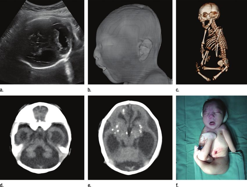
SPECIAL REPORT: Congenital Brain Abnormalities and Zika Virus: What the Radiologist Can Expect to See Soares de Oliveira-Szejnfeld et al Figure 1 Figure 1: Flowchart for subject inclusion and exclusion in the confirmed and presumed Zika virus (ZikV ) infection cohorts. ∗ = Gestational ages for fetal MR exam- inations were 26 weeks (with a second MR examination at 30 weeks), 29 weeks, 29 weeks (with a second MR examination at 37 weeks), 32 weeks, 34 weeks, and four examinations at 36 weeks (one in a set of twins). (Siemens Healthcare, Erlangen, Ger- Neurological findings on prenatal yielding 17 fetuses and/or neonates), many) or a 1.5-T Espree unit (Siemens MR images and postnatal CT and MR testing for Zika virus by means of RT- Healthcare) with an eight-channel body images were subjectively described by PCR and/or serologic analysis gener- coil and standard acquisition protocols four radiologists with experience in fetal ated positive results in amniotic fluid (Table E1 [online]). Postnatal head CT and/or neonatal neuroradiology (D.L., in nine women (with 10 gestational was performed with a 16-section CT an obstetric and fetal MR imager with sacs), cord blood in seven women, scanner (Siemens Healthcare). Post- 20 years of experience; R.R. and F.T.M., neonatal brain during autopsy in natal MR imaging was performed with pediatric neuroradiologists with 20 and three neonates, and placenta in one a 1.5-T Espree brain MR imaging unit 16 years of experience, respectively; neonate, with multiple positive sites (Siemens Healthcare). Postmortem and P.S.d.O.S., a neuroradiologist with in two women (one of whom had brain examination was performed in 12 years of experience). Findings were twins; Table E2 [online]). A rash stillborns or neonates who died within agreed on in consensus. In a minority of characteristic of Zika virus infection 48 hours of birth after obtaining paren- cases, select images from obstetric US was present in the first trimester in tal consent to perform autopsy. Brain were also available for review. 13 of 16 women (81%) with confirmed tissue images were acquired with a infection (one with twins) and in 22 64-channel multisection CT scanner of 28 women (78%) with presumed (GE Healthcare) and a 3-T MR imag- Results infection. ing unit (Achieva; Philips, Best, the In the 16 women with confirmed in- The imaging findings are described Netherlands). fection (including one set of twins, in Table 1. Figures depicting fetal Radiology: Volume 281: Number 2— 2016 n radiology.rsna.org 3
SPECIAL REPORT: Congenital Brain Abnormalities and Zika Virus: What the Radiologist Can Expect to See Soares de Oliveira-Szejnfeld et al
Table 1
Imaging Findings in 17 Confirmed and 28 Presumed Zika Virus Infections
No. of Confirmed No. of Presumed Infections
Infections at with Microcephaly and
Prenatal and/or Calcifications on CT Images
Abnormality Postnatal Imaging or Postnatal MR Images Comments
Parenchymal volume loss 17 (100) 28 (100) …
Mild to moderate 6 (35) 17 (61) …
Severe 12 (70) 11 (39) …
Cortical abnormalities 16 (94) 28 (100) Fifteen subjects were categorized as having both polymicrogyria and
irregular areas of sulci and/or gyri not otherwise specified.
Lissencephaly 2 (12) 6 (21) …
Polymicrogyria or pachygyria 11 (65) 14 (50) …
Irregular areas of sulci and/or gyri 5 (29) 21 (75) …
not otherwise specified
Corpus callosum abnormalities 16 (94) 22 (78) CT findings of agenesis and/or dysgenesis of the corpus callosum
were frequently difficult owing to relatively large fornices and some
anterior corpus callosum being present, but pronounced colpocephaly
without visualization of the body of the corpus callosum was used
for this diagnosis at CT. At MR imaging and US, direct visualization
of the corpus callosum was used for diagnosis.
Ventriculomegaly 16 (94) 27 (96) It can be difficult to characterize ventriculomegaly as mild or moderate,
since the occipital horns were often dilated out of proportion to the
frontal horns owing to parieto-occipital gray and white matter loss.
Asymmetrical venticulomegaly was observed in six of 17 and five
of 28 of the confirmed and presumed infections, respectively.
Mild 4 (24) 5 (18) …
Moderate 7 (41) 9 (32) …
Severe 5 (29) 13 (46) …
Septations in occipital horns 5 (29) 3 (11) …
Cerebellum abnormalities 14 (82) 21 (75) Twelve subjects had abnormalities of cerebellar hemispheres and vermis.
Hemisphere hypoplasia or 14 (82) 11 (39) …
maldevelopment
Vermis hypoplasia 10 (59) 19 (68) …
Brainstem hypoplasia and/or atrophy 12 (70) 6 (21) The high percentage in the confirmed infections reflects that these are
predominantly prenatally diagnosed infections compared with the
presumed infections, which has many neonates with only postnatal
images. Prenatal imaging includes sagittal fetal MR imaging, which
allows for better visualization of the brainstem compared with axial
postnatal CT.
Calcifications 17 (100) 28 (100) This was an inclusion criterion for the presumed infection cohort.
Periventricular 11 (65) 4 (14) All instances with periventricular calcifications had the periventricular
calcifications in areas of parenchymal thinning.
Cortical 4 (24) 4 (14) At times, a layered appearance can be seen with cortical, gray, and
white matter.
Gray matter–white matter junction 15 (88) 28 (100) …
Basal ganglia and/or thalamus 11 (65) 18 (64) …
Brainstem 3 (18) 4 (14) …
Cerebellum 1 (6) 1 (4) …
Soft tissues of the neck 0 1 (4) …
Heterogeneous material, some 9 (53) 8 (28) At unenhanced CT, it is helpful to compare material in the confluence
of which could be thrombus of the sinuses region to the basilar artery to determine if the
in the region of the attenuating and/or heterogeneous material is due to thrombus
confluence of sinuses rather than dehydration and hemoconcentration effect.
Note.—Numbers in parentheses are percentages.
4 radiology.rsna.org n Radiology: Volume 281: Number 2— 2016SPECIAL REPORT: Congenital Brain Abnormalities and Zika Virus: What the Radiologist Can Expect to See Soares de Oliveira-Szejnfeld et al
Figure 2
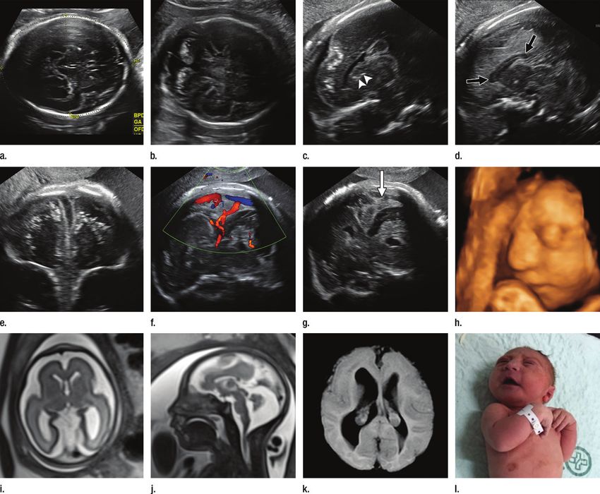
Figure 2: (a) Axial, (b) sagittal, and (c) coronal fetal T2-weighted MR images in a 29-year-old woman with confirmed Zika virus infection, initially seen for charac-
teristic rash at 12 weeks of gestational age. (d) Axial postnatal CT image and (e–g) axial and (h) coronal T2-weighted MR images obtained in her male neonate. The
fetal MR images obtained at 34 weeks (a–c) show asymmetrical ventriculomegaly with a septation in the right occipital horn (arrowhead on a), small frontal lobes,
thinning of the occipital parenchyma (left worse than right), underdeveloped sylvian fissures, and regions of thickened cortex, as in the right frontal lobe, which is
suggestive of polymicrogyria (arrow on a). There is abnormal, increased signal intensity in the white matter. The postnatal CT image (d) obtained in the 22-day-old
neonate shows punctate calcifications at the gray matter–white matter junction and asymmetrical ventriculomegaly. The T2-weighted MR images obtained at 26 days
(e–h) show septation in the ventricle (arrowhead on e). Note how the right ventricle has relatively decreased in size compared with the prenatal image, whereas the
left ventricle has increased in size. Under-rotation of the hippocampus (arrowheads on f ) is demonstrated. There is clear asymmetry of the gyral pattern on g, which
is relatively smooth in the left occipital region, with abnormal folds in the right occipital and frontoparietal regions (arrows on g). Subependymal cysts are visualized on
h, which are not seen on fetal MR images.
anatomy (Figs 2–8), neonatal micro- ventriculomegaly and normal head the head circumference was 38 cm at
cephaly (Figs 9–15; Figs E1–E3 [on- circumference at a scan conducted at birth. The CT images showed calcifica-
line]), and one neonate with normal 17 weeks of gestational age, and the tions in the subcortical region, thala-
head circumference but with charac- circumference remained within the mus, basal ganglia, and brainstem. No
teristic calcifications in association with normal range later in pregnancy. In sulci were seen; however, the paren-
severe ventriculomegaly (Fig 16 ) are 23 of 26 fetuses that underwent serial chyma was extremely thin. There was
illustrated. Movie clips of sequences of prenatal US, head circumference re- pontocerebellar hypoplasia and Dan-
images are also available in Movies 1– mained under the 5th percentile until dy-Walker spectrum anomaly, and the
E5 (online). birth, which led to a diagnosis of mi- corpus callosum was not visualized.
In all but one fetus with confirmed crocephaly at birth. However, it is no- The most remarkable change in the
Zika virus infection at prenatal imag- table that the three fetuses with head brain parenchyma, present on all neona-
ing, the head circumference percen- circumference in the normal range at tal images, was the reduction in paren-
tile was at or below the 5th percentile birth showed severe ventriculomegaly, chymal volume. Abnormalities of corti-
in at least one US examination per- which we presume was due to the en- cal development associated with volume
formed during the second trimester larged, obstructed ventricles. For this changes were observed in 16 of 17 con-
of pregnancy (Table 2). However, one reason, we included Figure 16, which firmed infections (94%) and 28 of 28 pre-
fetus with severe parenchymal and depicts a neonate referred to IPESQ sumed infections (100%). Abnormalities
brainstem malformation had severe for potential Zika virus infection, but of the corpus callosum were present in
Radiology: Volume 281: Number 2— 2016 n radiology.rsna.org 5SPECIAL REPORT: Congenital Brain Abnormalities and Zika Virus: What the Radiologist Can Expect to See Soares de Oliveira-Szejnfeld et al
Figure 3
Figure 3: Images in the case of a 34-year-old woman with confirmed Zika virus infection, initially seen for a rash at 8 weeks of gestation. Fetal head cir-
cumference was in the normal range at 12 and 16 weeks (5.8 cm and 11.9 cm, respectively) but then decreased to the 10th percentile at 22 weeks and was
below the 3rd percentile in subsequent imaging examinations. (a–h) US images obtained at 30 weeks. Head circumference on the axial image (a) measured 24
cm (,2.3 percentile, corresponding to a gestational age of 26 weeks 3 days). Note the open sylvian fissures and relatively smooth cortex, which are abnormal
findings at this gestational age. Oblique axial image (b) shows cerebellar calcifications (seen as the echogenic outer contour of the cerebellar hemispheres) and
inferior vermian hypoplasia with associated enlarged cisterna magna. A sagittal transvaginal image (c) shows calcifications in the basal ganglia (arrowheads)
and more bulky calcification at the gray matter–white matter junction. Another sagittal transvaginal image (d) shows a relatively small corpus callosum (arrows).
A coronal image (e) shows cortical and subcortical white matter calcifications in a linear pattern. The gyral pattern is abnormal, which is suggestive of polymi-
crogyria. A sagittal color Doppler image (f) shows a stretched appearance of vessels coursing into the posterior fossa. An oblique axial gray-scale transvaginal
image in the posterior fossa (g) shows heterogeneous material in the confluence of sinuses due to blood clot (arrow). A three-dimensional US image of the face
(h) shows a sloping forehead, compatible with frontal lobe hypoplasia. (i) Axial and (j) sagittal fetal MR images obtained at 29 weeks show atrophic frontal lobes,
wide sylvian fissures, enlarged posterior fossa, abnormal gyral pattern, prominent cerebrospinal fluid spaces, and inferior vermian hypoplasia. On i, note the
diffuse hypointense and undersulcated cortex, which is suggestive of mineralization and polymicrogyria. The hypoplastic corpus callosum can be seen on the
sagittal view (j), as well as the inferior vermian hypoplasia, enlarged cisterna magna, and heterogeneous signal intensity in the confluence of sinuses. There is a
subjectively thin spinal cord. (k) Postnatal axial MR image obtained at 81 days shows small frontal lobes and cortical thickening. The choroid plexi are enlarged.
(l) Photograph of the neonate after birth.
6 radiology.rsna.org n Radiology: Volume 281: Number 2— 2016SPECIAL REPORT: Congenital Brain Abnormalities and Zika Virus: What the Radiologist Can Expect to See Soares de Oliveira-Szejnfeld et al
Figure 4
Figure 4: Images in the case of an 18-year-old woman, first seen for rash at 10 weeks of pregnancy, with confirmed Zika virus infection. US findings obtained
at 20 weeks of gestational age were reportedly normal, with normal head circumference of 17.5 cm. At 37 weeks of gestational age, (a) sagittal transvaginal and
(b) coronal transabdominal US images obtained with the head upside down show a small head circumference (26.4 cm, corresponding to 28 weeks 5 days, below
the 3rd percentile), moderate ventriculomegaly with dense intracranial calcifications (arrowheads on a), and abnormal head shape with flattened appearance and
thickened skin (arrow on a). On the sonogram, it is difficult to precisely localize the calcifications, given the thin parenchyma. (c, d) Axial bone window CT images,
(e) sagittal localizer CT image, and (f–h) axial CT images show microcephaly with cerebral atrophy, and, despite ventriculomegaly, the extra-axial cerebrospinal
fluid spaces are still prominent. The dense calcifications are predominantly located in the subcortical white matter at the gray matter–white matter interface. There
is markedly abnormal skull shape with some eversion of the bones at the suture sites (particularly frontoparietal sites), with redundant skin folds (particularly in
the parieto-occipital region). (i) Sagittal T1-weighted, ( j, k) coronal T2-weighted, and (l) axial susceptibility-weighted MR images obtained at 1 month of age show
an undersegmented midbrain, severe microcephaly, open sylvian fissures, and polymicrogyria. The dense calcifications are evident on the susceptibility-weighted
image. On the sagittal images (a, e, i), note the small supratentorial compartment and associated skull deformity.
16 of 17 confirmed infections (94%) and (96%). This was asymmetrical in six of cortical underdevelopment or atrophy.
22 of 28 presumed infections (78%). 17 confirmed infections and five of 28 Other findings associated with the ven-
Lateral ventricles were enlarged in presumed infections. Despite ventricu- tricles were septations in the ventricle
16 of 17 confirmed infections (94%) lomegaly, the extra-axial spaces are (typically in the occipital horns), which
and 27 of 28 presumed infections frequently still prominent because of were frequently difficult to distinguish
Radiology: Volume 281: Number 2— 2016 n radiology.rsna.org 7SPECIAL REPORT: Congenital Brain Abnormalities and Zika Virus: What the Radiologist Can Expect to See Soares de Oliveira-Szejnfeld et al
Figure 5
Figure 5: Images in the case of a 33-year-old woman who had a rash at 10 weeks of pregnancy, with confirmed Zika virus infec-
tion. US performed at 19 weeks of gestational age showed a head circumference in the normal range (16.6 cm). (a, b) Sagittal and
(c) coronal transvaginal US images obtained at the next US examination at 27 weeks 2 days of gestational age, however, showed
the fetal head circumference to be 21.6 cm, which corresponded to 23 weeks 3 days (,2.3 percentile, not shown). There was
mild ventriculomegaly with septations in the occipital horns. Calcifications could be seen at the gray matter–white matter junction.
The cerebrum was atrophic. There was blood clot in the region of the confluence of sinuses (arrow). (d) Coronal, (e) axial, and (f)
sagittal T2-weighted fetal MR images obtained at 32 weeks show septations in the ventricles and an abnormal-appearing cortex,
with a thickened and undersulcated cortex most marked on the left, compatible with polymicrogyria. The sagittal midline view shows
microcephaly, blood clot in the region of the confluence of sinuses, and prominent skin folds. (g, h) Axial T2-weighted and (i) sagittal
T1-weighted postnatal MR images obtained in the 4-week-old neonate show diffuse gyral abnormality and abnormal myelination.
The septations in the ventricles are again seen. The sagittal image shows thinning of the spinal cord at the craniocervical junction.
8 radiology.rsna.org n Radiology: Volume 281: Number 2— 2016SPECIAL REPORT: Congenital Brain Abnormalities and Zika Virus: What the Radiologist Can Expect to See Soares de Oliveira-Szejnfeld et al Figure 6 Figure 6: Images in the case of a 24-year-old woman pregnant with twins, with characteristic rash at 9 weeks of pregnancy and confirmed Zika virus infection. (For each pair of images, the first image is of twin A, and the second image is of twin B.) At 14 weeks of gestational age, the fetal head size of both twins was normal. The head size never went below the 3rd percentile for either fetus in examinations at 19–28 weeks. (a, b) Sagittal and (c, d) axial fetal MR images were obtained at 36 weeks. (e, f ) Axial and (g, h) surface reconstruction postnatal CT images and (i, j) axial T2-weighted and (k, l) coronal MR images were obtained 1 week after delivery at 38 weeks of gestational age. There is severe microcephaly with profound frontal lobe hypoplasia. Calcifications in the subcortical white matter at the gray matter–white matter junction are visualized. Both twins have a flattened appearance of the pons. The spinal cord is atrophic (best seen on a). Redundant skin is seen in the occipital region. There is polymicrogyria involving the frontal and parietal regions and atrophic cortex and white matter in the occipital regions. Each twin has hypoplasia of the corpus callosum, with prominent fornices. There is abnormal myelination in the occipital region that, in twin A (i), has the appearance of a cyst or septation within the ventricle. The cerebellum is somewhat small and nodular. There is lack of rotation of the hippocampi. from subventricular cysts. Subependy- The most common finding was irreg- other three, two did not have a fetal mal cysts were occasionally visualized. ular areas of sulci and/or gyri not sonogram for review, and one only un- Abnormalities of cortical develop- otherwise specified, but focal cortical derwent a scan at 10 weeks of gesta- ment were present in all patients but malformation was also observed. In tion, which is too early to assign this with a substantial variation regarding addition, six patients had the appear- diagnosis. The cortical development the type of abnormality, hemispheric ance of lissencephaly. In three fetuses, abnormalities were usually asymmet- symmetry, and severity (Table 1). this was diagnosed prenatally. Of the rical. In general, the sulci were less Radiology: Volume 281: Number 2— 2016 n radiology.rsna.org 9
SPECIAL REPORT: Congenital Brain Abnormalities and Zika Virus: What the Radiologist Can Expect to See Soares de Oliveira-Szejnfeld et al
Figure 7
Figure 7: Images in the case of a pregnant 20-year-old woman, without history of rash, who was referred for fetal brain abnormality at US with confirmed
Zika virus infection. (a) Graph of head circumference during pregnancy shows small head size in the second trimester, which increased in the third trimester. US
(not shown) showed moderate ventriculomegaly starting at 23 weeks, which progressed to severe ventriculomegaly by the third trimester, associated with the
increase in head circumference. (b) Axial, (c) sagittal, and (d) coronal fetal MR images obtained at 36 weeks show severe asymmetric ventriculomegaly, marked
parenchymal thinning and/or atrophy, sloping forehead, and elevation of the hypoplastic cerebellar vermis with hypoplastic cerebellar hemispheres. The brainstem
is thin, and the midbrain is foreshortened. The spinal cord is irregular, thin, and nonvisualized in parts, and then thicker and possibly mineralized. (e, f) Axial CT
images obtained 1 day postnatally show severe asymmetrical ventriculomegaly with dense calcifications in the brainstem, cerebellum, and gray matter–white
matter interface (temporal lobes on f) and marked parenchymal atrophy. (g) Axial CT image obtained in the 6-week-old infant, after shunting, showed persistent
ventriculomegaly and periventricular, midbrain, thalamic, and cerebellar calcifications. (h) Photograph of the neonatal face shows redundant skin folds and skull
asymmetry.
prominent, and wide sylvian and in- identified. However, these were usually the vermis, associated with an enlarged
terhemispheric fissures were identi- present in more severe manifestations cisterna magna.
fied in most neonates, as well as ab- of infection, being associated with dys- Abnormalities of the corpus cal-
normal myelination. morphic brainstem, stenosis of the aq- losum—usually thin, dysgenetic, and
Calcification regions were predomi- ueduct, and secondary supratentorial hypoplastic or even absent—were fre-
nantly located in the gray matter–white hydrocephaly. Calcification in the brain- quently observed. Other changes in-
matter junction in our series (88% in stem was a common finding at autopsy cluded under-rotation of the hippocam-
the confirmed infection cohort and (in three of three of the neonates with pus and thickened fornices.
100% in the presumed infection co- confirmed infection that underwent In some imaging studies, an en-
hort). Calcifications were also iden- autopsy). larged confluence of the dural venous
tified in the thalamus, basal ganglia, Abnormalities of the brainstem sinuses had heterogeneous material. In
cortex, and periventricular regions. It were identified. The pons was often a few fetal sonograms that were avail-
is important to mention that the latter thin and atrophic. There was frequently able for review, this was demonstrated
were only present in neonates where a kink seen at the pontomedullary to be blood clot (Fig 3g). In many
there was substantial thinning of the junction. The spinal cord was thinned postnatal CT studies, there was hy-
brain parenchyma; thus, the precise and at times irregular in its appearance. perattenuating material in this region,
location of calcifications was difficult Other posterior fossa abnormalities in- which could be either thrombus or
to determine. Although less common, cluded cerebellar hemisphere hypopla- hematocrit effect (due to dehydration
infratentorial calcifications were also sia, vermis hypoplasia, and elevation of with hemoconcentration). In many
10 radiology.rsna.org n Radiology: Volume 281: Number 2— 2016SPECIAL REPORT: Congenital Brain Abnormalities and Zika Virus: What the Radiologist Can Expect to See Soares de Oliveira-Szejnfeld et al
Figure 8
Figure 8: Fetal and postnatal images in the case of an 18-year-old pregnant woman initially seen for a rash at 12 weeks of
gestation, with confirmed Zika virus infection. Signs of joint contractures were identified at US at 17 weeks, but head circum-
ference was in the normal range, then decreased to the 15th percentile at 21 weeks and was below the 3rd percentile at
subsequent scans at 24 and 36 weeks (19.0 cm and 25.0 cm, respectively). (a) Axial transabdominal US performed at 36 weeks
shows ventriculomegaly, calcifications in the thalamus and basal ganglia, and enlarged cisterna magna. (b) Soft-tissue, (c) body
bone reconstruction, and (d, e) axial postmortem CT images show an abnormal profile, moderate ventriculomegaly, parenchymal
atrophy, splaying of the cerebellar hemispheres, inferior vermian hypoplasia, and calcifications in the subcortical white matter,
thalamus, and basal ganglia. The corpus callosum is not visualized. The gyral pattern is abnormally smooth for a term neonate.
The postnatal bone reconstruction image showed the severe joint contractures, similar to those observed on (f) a photograph of
the neonate.
fetal and neonatal MR studies, there orbital fat into the cranial vault. Body conjunctivitis, and, in rare instances,
was fluid posterior to the confluence abnormalities included arthrogryposis. Guillain-Barré syndrome (15).
of the sinuses that was either similar It is well recognized that Zika virus
to cerebrospinal fluid in attenuation or crosses the fetal-placental barrier. Zika
signal intensity or contained fluid with Discussion virus has been isolated from the brain
a higher protein content than that of Zika virus is a single-strand RNA Flavi- and cerebrospinal fluid of neonates born
cerebrospinal fluid. The location of the virus (1). It is transmitted by infected with congenital microcephaly and iden-
fluid collection is likely related to the female mosquito vectors, such as the tified in the amniotic fluid and placental
unusual head shape and overlapping Aedes aegypti mosquito. Diagnosis of tissue of mothers who had experienced
sutures. This abnormal head shape Zika virus infection is complicated by clinical symptoms consistent with Zika
was frequently associated with redun- the fact that it is asymptomatic in up virus infection during their pregnancies
dant skin folds. to 80% of infections (14). The com- (2–5,8,15,16). Zika virus has also been
Other findings include orbital abnor- mon symptoms tend to be mild and shown to lead to neurotoxiticity and to
malities, such as asymmetrical microp- nonspecific, including headache, fever, impair human neurosphere growth in
thalmia, cataracts, and herniation of the and rash. Other symptoms include experimental models (17).
Radiology: Volume 281: Number 2— 2016 n radiology.rsna.org 11SPECIAL REPORT: Congenital Brain Abnormalities and Zika Virus: What the Radiologist Can Expect to See Soares de Oliveira-Szejnfeld et al
Figure 9 Figure 10 Figure 11
Figure 10: Oblique axial CT image in a 3-month- Figure 11: Axial CT image in a 1-month-old
Figure 9: Axial CT image in a 1-week-old female old male infant with a head circumference of 27.5 male neonate with a head circumference of 27 cm
neonate with a head circumference of 31 cm at cm at birth, with presumed Zika virus infection. at birth, with presumed Zika virus infection. Note
birth and presumed Zika virus infection. The frontal Note the misshapen skull, ventriculomegaly, absent ventriculomegaly with septation (arrow) in the right
lobes are slightly hypoplastic, with mild underoper- corpus callosum, diffuse parenchymal volume occipital horn and striking subcortical calcifications.
cularization of the hypoplastic sylvian fissures. There loss, diffuse cortical migrational abnormality, and There are also cortical and periventricular calcifica-
are scattered subcortical calcifications (arrowheads), calcifications, most likely subcortical but difficult to tions. The gyral pattern is diffusely abnormal, and
and there is high-attenuating material (arrow) in the classify secondary to parenchymal thinning. the skull is deformed.
region of the confluence of sinuses, likely represent-
ing blood products.
Figure 13 Figure 14
Figure 12
Figure 13: Axial CT image in a 1-month-old
female neonate with a head circumference of 31 Figure 14: Axial CT image in a 2-month-old
Figure 12: Oblique coronal CT image in a cm at birth, with presumed Zika virus infection. Note female infant with a head circumference of 32.5 cm
1-month-old male neonate with a head circumfer- severe ventriculomegaly, subcortical calcifications, at birth, with presumed Zika virus infection. Note the
ence of 29.5 cm at birth, with presumed Zika virus and diffuse gyral abnormality. asymmetrical volume loss, more marked on the right
infection. Note the misshapen skull; ventriculomeg- than the left. Also note the diffusely irregular sulci
aly; absent corpus callosum; cerebellar hypoplasia and gyri bilaterally.
(arrows); thin parenchyma with diffuse gyral There are many causes of microceph-
abnormality; calcifications in the thalamus, basal aly, the most common being infections
ganglia, and subcortical white matter; and cerebellar
(such as TORCH infections and human virus, it is clear that there are devel-
hemispheres.
immunodeficiency virus), teratogens opmental insults that lead to micren-
(including maternal exposure to heavy cephaly (small brain) and associated
Microcephaly is a nonspecific term metals, alcohol, and radiation), genetic microcephaly (small head) (6,18–21).
that refers to a head circumference abnormalities and syndromes, and It is important to recognize that almost
smaller than normal for gestational age. growth restriction. In the case of Zika all of the infections at our institution
12 radiology.rsna.org n Radiology: Volume 281: Number 2— 2016SPECIAL REPORT: Congenital Brain Abnormalities and Zika Virus: What the Radiologist Can Expect to See Soares de Oliveira-Szejnfeld et al
occurred in women who had a charac- is 1%–13% (22). However, as the flow applying our exclusion criteria. In the
teristic rash in the late first trimester. of women referred to the IPESQ for report by França et al, one in five def-
This correlates well with the finding of assessment demonstrates, it is clear inite or probable Zika virus infections
severe cerebral dysmorphisms asso- that many pregnant women with a yielded head circumferences in the
ciated with infection during a time of rash in Brazil are never shown to have normal range (more than 22 standard
rapid brain development. According to congenitally infected fetuses, although deviations below the median of the In-
the U.S. Centers for Disease Control some certainly could have less severe ternational Fetal and Newborn Growth
and Prevention, the risk of microceph- infection that has thus far been undiag- Consortium for the 21st Century, or
aly after maternal infection with Zika nosed. For women who have neonates INTERGROWTH-21st, standard), and
virus in the first trimester of pregnancy with findings suggestive of severe mi- for one-third of definite and probable
crocephaly but who have a history of infections, there was no history of a
Figure 15 rash or for those who had a rash in the rash during pregnancy (24). This sug-
third trimester, we can hypothesize that gests that our series is biased to the
there was an unrecognized or asymp- more severe infections. However, in the
tomatic first-trimester exposure and/or series by França et al (24), the more
infection. severe infections were localized in the
As of July 2, 2016, the Brazilian northeast region of Brazil, since 97%
Ministry of Health had been notified of definite or probable infections were
of 8301 cases of microcephaly and from the northeast region, where 28%
confirmed 1656 infections (23). In a of all births in Brazil occur. This sug-
recent study, França et al reported on gests that there could be additional un-
the follow-up of 1501 cases in which known factors that exacerbate the fetal
Zika virus infection was suspected, of infection in this region. Coinfections, in
which 602 were deemed to be defi- addition to those already excluded, as
nitely or probably due to Zika virus (76 well other environmental factors, will
definite infections, 54 highly probable need to be explored further.
infections, 181 moderately probable There are many nomograms for head
Figure 15: Axial CT image in a 3-month-old infections, and 291 somewhat proba- circumference size. Current guidance in
female infant with cord blood positive for Zika virus ble infections) (24). The incidence of Brazil is to use the standards from the
(confirmed Zika virus infection) and a head circum- confirmed infection in our study with INTERGROWTH-21st study for fetuses
ference of 28.5 cm at birth. The mother had a rash respect to referral population is similar (25), the INTERGROWTH-21st study
at 12 weeks. Bilateral cataracts are seen, as well to what has been seen in the larger Bra- for infants (25), and World Health Or-
as abnormal cerebellar vermis and fat herniating zilian population. In our study, of 432 ganization criteria for full-term neonates
posteriorly from the orbits through the superior women initially screened, only findings (26). These charts show that a head
orbital fissure into the cranial vault. in 44 patients are reported here after circumference of 32 cm is at about 22
Figure 16
Figure 16: Axial CT images in a 5-month-old male infant with a head circumference of 38 cm at birth. The mother had a rash at 11
weeks. The size of the head is likely secondary to hydrocephalus. Unlike most of our cohort, the head was still round in shape. However,
calcifications are present in the subcortical region, thalamus, basal ganglia, and brainstem. No sulci were seen; however, the paren-
chyma is extremely thin. There is pontocerebellar hypoplasia, nonvisualization of the corpus callosum, and Dandy-Walker spectrum
anomaly. This is an example of how Zika virus infection can be missed if only newborns with microcephaly are assessed.
Radiology: Volume 281: Number 2— 2016 n radiology.rsna.org 13SPECIAL REPORT: Congenital Brain Abnormalities and Zika Virus: What the Radiologist Can Expect to See Soares de Oliveira-Szejnfeld et al
Table 2
Head Circumference Percentiles with Respect to Gestational Age in Fetuses with Prenatal US Images
Gestational Age
Infection Group and Head Circumference Gestational Age
Case No. 14–16 Weeks 17–20 Weeks 21–24 Weeks 25–30 Weeks 31–35 Weeks 36 Weeks at Birth (cm) at Birth (wk)
Confirmed infection
C1 … 5 … ,3 5 40 36.5 41
C2 5 5 5 3–5 ,3 3 30.5 40
C3 … 50 90 25 … … 35 36
C4 … … 25 … 3 ,3 31.5 41
C5 50 25 … 5 5 ,5 33 39
C6 … 10 5 5 … 25 35 39
C7 … 50 … ,3 ,3 ,3 28 39
C8 … 50 … 5 ,3 … 28.5 39
C9 25 25 … 3–5 … … 28 38
C10 5 5 … 3 … … 26 38
C11 … … 50 5 3 ,3 29 40
C12 25 5 10 … … ,3 29.5 40
C13 … 50 … … … ,3 28 40
C14 … … … … 25 3 30 41
C15 50 … 50 … 5 … 32.5 41
C16 … … 10 … … ,3 27.5 39
Presumed infection
P1 … … 10 5 … … 31 38
P2 10 50 … 5 ,3 … 27 38
P3 … … … … ,3 … 28.5 40
P4 … 10 … … … … 29 40
P5 … 40 50 ,3 3 3 31 39
P6 … … … … … ,3 27.5 39
P7 … 25 5 … ,3 … 31 39
P8 … … … 5 … ,1 30 39
P9 … … 5 … ,3 … 28 39
P10 … 75 25 25 3 3 31.5 40
P11 … … … … ,3 … 27 36
P12 50 25 … … … … 27 38
P13 … … 25 … … ,1 30 37
P14 … … … … … ,3 29 39
P15 … … … ,3 … … 29.5 40
Note.—Data are percentages, unless indicated otherwise. If more than one examination was conducted in a given time period, measurements from the first examination were used.
standard deviations below the mean for a combination of the small brain as it instances, a normal (or even increased)
both boys and girls at term. However, develops and a result of what, at some head circumference may be present.
this threshold will naturally include some point, was likely a larger head size (due Another unusual finding that sug-
normally developing neonates and also to ventriculomegaly) that then decom- gests skull collapse is that neonates
be inaccurate for neonates born prema- presses. Cerebral atrophy may also con- have orbital fat herniation into the cra-
turely. However, it should be recognized tribute, giving the skull the collapsed nial vault. Thus, some of the ocular find-
that to cast a broad net to find neonates shape with everted and/or cupped su- ings could be secondary to the process
with congenital Zika virus infection, we tures and overriding bones in the occipi- of skull deformation itself rather than
must bear in mind that not all neonates tal region, causing redundant and folded direct infection of the eye. For example,
will have microcephaly at birth. skin. In part, this is also likely due to nerve and blood flow interruption could
The striking imaging features of the the head and skin continuing to grow, be due to herniated tissue.
severe micrencephaly associated with while the size of the brain regresses. We used brain calcifications as in-
Zika virus include a markedly abnormal However, in some fetuses and/or neo- clusion criteria for the postnatal as-
head shape. The unusual appearance nates, the ventricle and/or brain atro- sessment to exclude microcephaly from
of the skull, we hypothesize, is due to phy has not yet occurred, and in these causes other than infection, such as
14 radiology.rsna.org n Radiology: Volume 281: Number 2— 2016SPECIAL REPORT: Congenital Brain Abnormalities and Zika Virus: What the Radiologist Can Expect to See Soares de Oliveira-Szejnfeld et al
unrecognized prematurity or congenital There have been many reports of with normal head size or brain abnor-
syndromes. While this could have led small series of imaging findings in fe- malities without calcifications could
to exclusion of some infections with- tuses and neonates with congenital Zika have been missed. In addition, it could
out intracranial calcifications, it led virus infection. In 2016, Mlakar et al de- be that some of the cohort had disease
to a homogeneous group of neonates scribed one pregnancy at 29 weeks with origin for microcephaly other than Zika
with strikingly similar parenchymal microcephaly and intracranial calcifica- virus. For example, we excluded a neo-
abnormalities. In our series, the most tions, with an earlier, second-trimester nate with microcephaly and confirmed
common location for calcifications was sonogram that showed no abnormality Zika virus infection due to aneuploidy
the gray matter–white matter junction (6). Also in 2016, Calvet et al described with trisomy 13 syndrome. However,
(88% in the confirmed infection cohort two pregnant women who underwent other infections or syndromes could be
and 100% in the presumed infection co- US at 22 weeks, which showed micro- present but not yet identified in either
hort), which is an area not classically cephaly (2). Sarno et al described a our confirmed infection cohort or our
or commonly targeted in other con- stillbirth at 32 weeks, with microceph- presumed infection cohort. Findings on
genital infections. The location of the aly, intracranial calcifications, and fetal MR images can lead to underestimation
calcifications at the gray matter–white hydrops (7). Driggers et al in 2016 de- of the incidence of calcifications, and
matter interface could suggest a vascu- scribed decreased fetal head circumfer- evaluation of CT images makes charac-
lar component to the infection, as other ence between 16 and 21 weeks, with terization of subtle parenchymal abnor-
processes that preferentially affect the brain abnormalities (8). In a study by malities and corpus callosum abnormal-
gray matter–white matter junction have Brasil et al (3), of 88 pregnant women ities difficult. Further imaging studies
been posited to be due to changes in with rash, 72 tested positive for Zika on these neonates as they grow will be
arterial configuration from straight ves- virus. Fetal US was performed in 42 helpful in further assessment of areas
sels in the cortex to coiled vessels in the fetuses, and abnormalities were seen involved with the infection. Finally, we
subcortical white matter (27). in 12, including intrauterine growth re- focused on brain findings in this review.
In the classic TORCH infections, striction with or without microcephaly Additional sites of infection and associ-
the brain calcifications are periventric- and ventricular calcifications (3). ated pathologic abnormalities will likely
ular and cortical, although rare cases Schuler-Faccini et al described 35 be identified in the future.
of basal ganglia and thalamus calcifi- neonates with microcephaly, including It is well recognized that transpla-
cations have been reported (28,29). brain calcifications, ventriculomgaly, cental transmission of viruses, even in
Other findings include intraventricular and cortical and/or subcortical atrophy subclinical maternal infection, can lead
adhesions, callosal abnormalities, peri- (9). In 2016, Hazin et al (10) and de to severe congenital abnormalities. As
ventricular pseudocysts, sulcation, and Fatima Vasco Aragao et al (11) each de- in other infections, serial imaging can
gyral abnormalities (29–31), similar to scribed 23 neonates with microcephaly demonstrate evolution of findings. Pre-
what we describe in this report. who had CT findings that included intra- natal sonograms may show normal or
However, unlike most patients with cranial calcifications, ventriculomegaly, decreased head circumference and,
congenital Cytomegalovirus, the pa- abnormal gryal pattern, and abnormal rarely, increased head circumference.
tients with documented or presumed white matter attenuation. Guillemette- Almost all neonates will show intrapa-
Zika virus infection described in this re- Artur et al described three neonates renchymal calcifications more severe
port had severe microcephaly. This may with congenital Zika virus infection, than what are typically seen in TORCH
be due to the first-trimester nature of with micrencephaly in all three, small infections and frequently occur at the
most of the infections reported herein. cerebellum in two, occipital subependy- gray matter–white matter junction,
It could also be due to the viral load in mal pseudocysts in two, polymicrogyria which is an unusual location for the cal-
the Zika virus infections, which we as- in three, corpus callosum abnormalities cifications of other congenital infections.
sume are severe infections. It could be in two, and hypoplastic brainstem in We hope the illustrations of these many
that congenital Cytomegalovirus is di- one (32). These findings are all similar fetuses and neonates will aid others in
agnosed in a range of infections from to what we report. the event that the unfortunate epidemic
mild to severe, whereas we may be fo- Our study had limitations. What we of congenital Zika virus continues.
cusing our results on the severe Zika present here is a convenience sample
Acknowledgments: We gratefully acknowledge
virus infections. In our cohort, almost of imaging findings for illustrative pur- the members of the Brazilian Network BRAZIKA
all fetuses and neonates had dramati- poses. Our cohort was obtained from a (Rede Internacional de Estudos Sobre Zika no
cally abnormal cerebral volume, abnor- referral center for high-risk pregnancy. Brasil).
mal cortical folding pattern, and/or re- Thus, we have no information on inci- Disclosures of Conflicts of Interest: P.S.d.O.S.
gions of lissencephaly, pachygyria, and/ dence of the Zika virus in the general disclosed no relevant relationships. D.L. Activ-
or polymicrogyria. We hypothesize that population or risk estimates for trans- ities related to the present article: disclosed
no relevant relationships. Activities not related
the cortical abnormalities visualized are mission to the fetus. Because of the
to the present article: author received roy-
due at least in part to arrested cortical manner in which we accrued subjects, alties from Elsevier and UpToDate. Other re-
development at various stages. neonates with congenital infection but lationships: disclosed no relevant relationships.
Radiology: Volume 281: Number 2— 2016 n radiology.rsna.org 15SPECIAL REPORT: Congenital Brain Abnormalities and Zika Virus: What the Radiologist Can Expect to See Soares de Oliveira-Szejnfeld et al
A.S.d.O.M. disclosed no relevant relationships. in presumed Zika virus related congenital php/cidadao/principal/agencia-saude/24437-
M.M.R.A. disclosed no relevant relationships. infection and microcephaly: retrospective ministerio-da-saude-confirma-1-656-ca-
A.G.M.B. disclosed no relevant relationships. case series study. BMJ 2016;353:i1901. sos-de-microcefalia. Updated July 7, 2016.
L.C. disclosed no relevant relationships. A.T. Accessed July 7, 2016.
disclosed no relevant relationships. R.S.A. dis- 12. Centers for Disease Control and Prevention.
closed no relevant relationships. G.M. disclosed All Countries & Territories with Active Zika 24. França GV, Schuler-Faccini L, Oliveira WK,
no relevant relationships. R.X. disclosed no rel- Virus Transmission. http://www.cdc.gov/ et al. Congenital Zika virus syndrome in
evant relationships. R.R. disclosed no relevant zika/geo/active-countries.html. Updated July Brazil: a case series of the first 1501 live-
relationships. J.S. disclosed no relevant relation- 26, 2016. Accessed July 29, 2016. births with complete investigation. Lancet
ships. F.T.M. disclosed no relevant relationships. 2016 Jun 29. [Epub ahead of print]
13. Lanciotti RS, Kosoy OL, Laven JJ, et al. Ge-
netic and serologic properties of Zika virus 25. Papageorghiou AT, Ohuma EO, Altman
associated with an epidemic, Yap State, DG, et al; International Fetal and Newborn
References Micronesia, 2007. Emerg Infect Dis 2008; Growth Consortium for the 21st Century (IN-
14(8):1232–1239. TERGROWTH-21st). International standards
1. Campos GS, Bandeira AC, Sardi SI. Zika
virus outbreak, Bahia, Brazil. Emerg Infect for fetal growth based on serial ultrasound
14. Duffy MR, Chen TH, Hancock WT, et al.
Dis 2015;21(10):1885–1886. measurements: the Fetal Growth Longitudi-
Zika virus outbreak on Yap Island, Federated
nal Study of the INTERGROWTH-21st Pro-
States of Micronesia. N Engl J Med 2009;
2. Calvet G, Aguiar RS, Melo AS, et al. Detection ject. Lancet 2014;384(9946):869–879.
360(24):2536–2543.
and sequencing of Zika virus from amniotic
fluid of fetuses with microcephaly in Brazil: 15. Faria NR, Azevedo RdoS, Kraemer MU, et 26. World Health Organization. Head Cir-
a case study. Lancet Infect Dis 2016;16(6): al. Zika virus in the Americas: early epidemi- cumference-for-Age. http://www.who.int/
653–660. ological and genetic findings. Science 2016; childgrowth/standards/hc_for_age/en/. Ac-
352(6283):345–349. cessed June 12, 2016.
3. Brasil P, Pereira JP Jr, Raja Gabaglia C, et
al. Zika virus infection in pregnant women in 16. Martines RB, Bhatnagar J, Keating MK, 27. Nonaka H, Akima M, Hatori T, Nagayama
Rio de Janeiro—preliminary report. N Engl J et al. Notes from the field: evidence of Zika T, Zhang Z, Ihara F. The microvasculature
Med 2016 Mar 4. [Epub ahead of print] virus infection in brain and placental tissues of the cerebral white matter: arteries of the
from two congenitally infected newborns and subcortical white matter. J Neuropathol Exp
4. Melo A, Aguiar R, Amorim M, et al. Con- Neurol 2003;62(2):154–161.
two fetal losses—Brazil, 2015. MMWR Morb
genital Zika virus infection: beyond neonatal
Mortal Wkly Rep 2016;65(6):159–160. 28. Estroff JA, Parad RB, Teele RL, Benacerraf
microcephaly. JAMA Neurol (in press).
17. Garcez PP, Loiola EC, Madeiro da Costa R, BR. Echogenic vessels in the fetal thalami and
5. Oliveira Melo AS, Malinger G, Ximenes R, et al. Zika virus impairs growth in human basal ganglia associated with cytomegalovi-
Szejnfeld PO, Alves Sampaio S, Bispo de neurospheres and brain organoids. Science rus infection. J Ultrasound Med 1992;11(12):
Filippis AM. Zika virus intrauterine infec- 2016;352(6287):816–818. 686–688.
tion causes fetal brain abnormality and mi-
18. Rasmussen SA, Jamieson DJ, Honein MA, 29. Picone O, Simon I, Benachi A, Brunelle F,
crocephaly: tip of the iceberg? Ultrasound
Petersen LR. Zika virus and birth defects— Sonigo P. Comparison between ultrasound
Obstet Gynecol 2016;47(1):6–7.
reviewing the evidence for causality. N Engl and magnetic resonance imaging in assess-
6. Mlakar J, Korva M, Tul N, et al. Zika virus J Med 2016;374(20):1981–1987. ment of fetal cytomegalovirus infection. Pre-
associated with microcephaly. N Engl J Med nat Diagn 2008;28(8):753–758.
2016;374(10):951–958. 19. Besnard M, Eyrolle-Guignot D, Guillemette-
Artur P, et al. Congenital cerebral malfor- 30. Moinuddin A, McKinstry RC, Martin KA,
7. Sarno M, Sacramento GA, Khouri R, mations and dysfunction in fetuses and new- Neil JJ. Intracranial hemorrhage progress-
et al. Zika virus infection and stillbirths: a borns following the 2013 to 2014 Zika virus ing to porencephaly as a result of congeni-
case of hydrops fetalis, hydranencephaly and epidemic in French Polynesia. Euro Surveill tally acquired cytomegalovirus infection—an
fetal demise. PLoS Negl Trop Dis 2016;10(2): 2016;21(13). illustrative report. Prenat Diagn 2003;23
e0004517. (10):797–800.
20. de Paula Freitas B, de Oliveira Dias JR,
8. Driggers RW, Ho CY, Korhonen EM, et al. Prazeres J, et al. Ocular findings in infants 31. Teissier N, Fallet-Bianco C, Delezoide AL,
Zika virus infection with prolonged mater- with microcephaly associated with pre- et al. Cytomegalovirus-induced brain mal-
nal viremia and fetal brain abnormalities. N sumed Zika virus congenital infection in Sal- formations in fetuses. J Neuropathol Exp
Engl J Med 2016;374(22):2142–2151. vador, Brazil. JAMA Ophthalmol 2016 Feb Neurol 2014;73(2):143–158.
9. [Epub ahead of print]
9. Schuler-Faccini L, Ribeiro EM, Feitosa IM, 32. Guillemette-Artur P, Besnard M, Eyrolle-
et al. Possible association between Zika 21. Miranda-Filho DdeB, Martelli CM, Ximenes Guignot D, Jouannic JM, Garel C. Prenatal
virus infection and microcephaly—Bra- RA, et al. Initial description of the pre- brain MRI of fetuses with Zika virus infec-
zil, 2015. MMWR Morb Mortal Wkly Rep sumed congenital Zika syndrome. Am J tion. Pediatr Radiol 2016;46(7):1032–1039.
2016;65(3):59–62. Public Health 2016;106(4):598–600.
10. Hazin AN, Poretti A, Turchi Martelli CM, 22. Johansson MA, Mier-y-Teran-Romero L,
et al. Computed tomographic findings in Reefhuis J, Gilboa SM, Hills SL. Zika and
microcephaly associated with Zika virus. N the risk of microcephaly. N Engl J Med 2016;
Engl J Med 2016;374(22):2193–2195. 375(1):1–4.
11. de Fatima Vasco Aragao M, van der Linden 23. Brazilian Ministry of Health. Ministério da
V, Brainer-Lima AM, et al. Clinical features Saúde Confirma 1.656 Casos de Microce-
and neuroimaging (CT and MRI) findings falia. http://portalsaude.saude.gov.br/index.
16 radiology.rsna.org n Radiology: Volume 281: Number 2— 2016You can also read