Infant and Adult Brains Are Coupled to the Dynamics of Natural Communication
←
→
Page content transcription
If your browser does not render page correctly, please read the page content below
878698
research-article2019
PSSXXX10.1177/0956797619878698Piazza et al.Infant–Adult Coupling During Natural Communication
ASSOCIATION FOR
Research Article PSYCHOLOGICAL SCIENCE
Psychological Science
Infant and Adult Brains Are Coupled to 2020, Vol. 31(1) 6–17
© The Author(s) 2019
Article reuse guidelines:
the Dynamics of Natural Communication sagepub.com/journals-permissions
DOI: 10.1177/0956797619878698
https://doi.org/10.1177/0956797619878698
www.psychologicalscience.org/PS
Elise A. Piazza1,2 , Liat Hasenfratz1,2, Uri Hasson1,2, and
Casey Lew-Williams2
1
Princeton Neuroscience Institute, Princeton University, and 2Department of Psychology, Princeton University
Abstract
Infancy is the foundational period for learning from adults, and the dynamics of the social environment have long
been considered central to children’s development. Here, we reveal a novel, naturalistic approach for studying live
interactions between infants and adults. Using functional near-infrared spectroscopy (fNIRS), we simultaneously and
continuously measured the brains of infants (N = 18; 9–15 months of age) and an adult while they communicated
and played with each other. We found that time-locked neural coupling within dyads was significantly greater when
dyad members interacted with each other than with control individuals. In addition, we characterized the dynamic
relationship between neural activation and the moment-to-moment fluctuations of mutual gaze, joint attention to
objects, infant emotion, and adult speech prosody. This investigation advances what is currently known about how the
brains and behaviors of infants both shape and reflect those of adults during real-life communication.
Keywords
communication, development, infancy, naturalistic, language, functional near-infrared spectroscopy, neural coupling,
open data, open materials
Received 8/11/18; Revision accepted 8/16/19
The ability to communicate during the first years of life temporal lag) to the responses in a speaker’s brain, and
requires the development of common ground with stronger speaker–listener coupling is associated with
other people. This involves learning to engage with better comprehension (Stephens, Silbert, & Hasson,
social environments using a range of behaviors, includ- 2010). Importantly, this coupling in higher-order areas
ing eye gaze, facial expressions, and speech. By adult- is not simply driven by shared exposure to the same
hood, we interact with others following a set of norms perceptual input; areas of the default-mode network
shared by our community of speakers. (including medial prefrontal cortex) are coupled only
Recent work with adults indicates that shared under- when participants have a shared understanding of a
standing is associated with shared neural responses story, not simply when identical stimuli are presented
across people in a set of higher-order brain regions (Lerner, Honey, Silbert, & Hasson, 2011; Simony et al.,
(Hasson, Ghazanfar, Galantucci, Garrod, & Keysers, 2016). Speaker–listener neural coupling has been found
2012). Responses in these areas correlate strongly using both functional MRI (fMRI; Stephens et al., 2010)
across people when input is interpreted in a similar and functional near-infrared spectroscopy (fNIRS; Jiang
way, irrespective of its form (Honey, Thompson, Lerner, et al., 2012; Liu et al., 2017), showing strong consistency
& Hasson, 2012). For example, written and spoken ver- between the two methodologies.
sions of the same story evoke comparable neural
responses across readers and listeners (Regev et al.,
2019). Furthermore, effective storytelling reflects the
Corresponding Author:
successful transfer of information between brains: Elise A. Piazza, Princeton University, Princeton Neuroscience Institute
Responses in a listener’s linguistic and higher-order and Department of Psychology, Princeton, NJ 08540
brain areas are coupled (i.e., correlated with a short E-mail: elise.piazza@gmail.com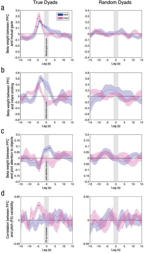
Infant–Adult Coupling During Natural Communication                                                                      7

    How does this shared neural code emerge in our            member of the dyad interacted with another person in
brains as we begin learning during the first years of         the room. Furthermore, we predicted that activation in
life? The ability to understand language input does not       areas of the brain involved in mutual understanding (in
emerge automatically. On the contrary, children’s             particular, prefrontal and parietal areas; Liu et al., 2017)
knowledge of the conventional ways of using words to          would be related to natural communicative behaviors,
communicate ideas, desires, and intentions must emerge        such as mutual gaze, joint attention to objects, infant
over time from interactions with other members of a           smiling, and adult speech prosody.
community of speakers (Brazelton, Koslowski, & Main,
1974; Tomasello, 1992; Vygotsky, 1978). Two-way inter-
                                                              Method
actions provide continuous information about these
communicative conventions, in the form of feedback            Participants
from (and to) caregivers, and transactional models of
development emphasize the roles of both infants and           Eighteen infants (age: M = 11.3 months, range = 9.8–
caregivers in shaping communication and learning (see         14.9 months; 9 female) with no history of hearing prob-
Sameroff, 2009). For instance, mothers modify the pitch,      lems and no known developmental delays participated
rhythm, and overall spectral quality of their voices          in the experiment. One experimenter with extensive
when talking to their infants (Fernald & Kuhl, 1987;          parenting experience performed the adult role in all
Piazza, Iordan, & Lew-Williams, 2017), even changing          experimental sessions. This experimenter was generally
their prosody in real time on the basis of their children’s   aware of the basic hypothesis (i.e., stronger coupling
emotional feedback (N. A. Smith & Trainor, 2008). On          when she interacted with the infant than when she
the other side of the interaction, infants preferentially     ignored the infant), but she was naive with respect to
attend to and learn from communicatively relevant infor-      details, such as the behavioral cues or brain areas that
mation (Ferguson & Lew-Williams, 2016; Vouloumanos            might be relevant. One of the included 18 infants was
& Werker, 2004), and infants’ gaze following and point-       excluded from behavioral analyses because of video
ing predict later language outcomes (Brooks & Meltzoff,       malfunction; 3 additional infants were unable to par-
2008). This generates an interesting prediction: that         ticipate in the experiment because they refused to wear
dynamic coupling between infant and adult brains may          the fNIRS cap, and 21 additional infants were excluded
support the successful exchange of information during         from statistical analyses because of excessive signal noise
everyday interactions.                                        or artifacts (15 of these infants grabbed the cap or
    Here, we aimed to understand the ways in which the        squirmed excessively, resulting in significant head
brains of infants and adults are coupled in real time,        motion). The sample size was based on sizes used in
both to each other and to natural social behaviors. To        previous studies that measured neural coupling in com-
do so, we developed a new dual-brain fNIRS paradigm           munication paradigms with fMRI (e.g., Stephens et al.,
for simultaneously measuring the brains of an adult           2010) and fNIRS ( Jiang et al., 2012; Liu et al., 2017).
caregiver and 9- to 15-month-old infants while they
engaged in continuous, everyday, two-way interactions,
                                                              Procedure
including playing, singing, and reading. fNIRS provides
a noninvasive measure of changes in blood oxygen-             We used a dual-brain LABNIRS system (Shimadzu Sci-
ation resulting from neural activity while being mini-        entific Instruments, Columbia, MD) to simultaneously
mally sensitive to motion artifacts, thus allowing            record brain activity from the adult and infant in each
multiple participants to move and interact freely, face       dyad. We recorded from 57 channels (3 cm in the adult,
to face, while wearing comfortable caps (Boas, Elwell,        2.5 cm in the infants) across the cortex of each partici-
Ferrari, & Taga, 2014). Our fNIRS hyperscanning (two-         pant. These channels covered prefrontal cortex (PFC),
brain) paradigm opens new experimental possibilities          temporoparietal junction, and parietal cortex (i.e., areas
for studying the reciprocal dynamics of natural com-          involved in prediction, language processing, and under-
munication among multiple people, such as one per-            standing other people’s perspectives; Lerner et al.,
son’s brain predicting another person’s upcoming              2011). The locations of these channels were homolo-
behavior. This approach contrasts with that of previous       gous across the infant and the adult (see Fig. 1a). Caps
studies, which used a single fMRI scanner to measure          were positioned on the basis of known anatomical
neural coupling between adult speakers and listeners          landmarks according to the international 10-20 system
during one-way communication. We predicted that               (e.g., the center point of the cap was approximately at
infant-to-caregiver brain-to-brain coupling would be          Cz). Our primary intersubject correlation (ISC) analyses,
higher when the infant and adult interacted with each         which aimed to determine the spatial extent of cou-
other, compared with a control condition in which each        pling, focused on deoxyhemoglobin because it is less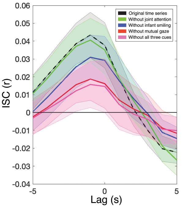
8                                                                                                                                Piazza et al.

    Fig. 1. Example of an interaction between an adult and infant during the together condition (a) and the corresponding intersubject cor-
    relation (ISC) in one channel pair (b). The graph shows the concentration of deoxyhemoglobin across the length of an interaction during
    the together condition, separately for the adult and infant. The ISC between the adult and infant, computed from a single right prefrontal
    cortex (PFC) channel, is shown at the bottom right.

likely than oxyhemoglobin to include systemic effects                      Muehlemann, & Wolf, 2010); we also low-pass-filtered
and therefore measures more spatially precise cortical                     (0.5 Hz) and high-pass-filtered (0.02 Hz) the signal to
activation (Boas et al., 2014; Hirsch, Zhang, Noah, &                      remove physiological noise and drift, respectively. We
Ono, 2017).                                                                conducted additional control analyses that demonstrated
   All adult–infant dyads participated in two 5-min con-                   that our ISC results could not be driven by motion arti-
ditions. In the together condition, the adult experi-                      facts (see Fig. S2 in the Supplemental Material available
menter engaged directly with the child by playing with                     online). To calculate ISC, we computed a Pearson correla-
a consistent set of toys, singing nursery rhymes, and                      tion between one channel in the adult and one channel
reading Goodnight Moon (Fig. 1a). The child sat on his                     in the infant (Fig. 1b).
or her parent’s lap, and the parent was told to keep the                      Because of the presence of long-range temporal
child comfortable but not to communicate with the                          autocorrelation in fNIRS time series, we estimated the
child in any way. In the control (apart) condition,                        statistical likelihood of each observed correlation using
the experimenter turned 90° away from the child and                        a bootstrapped permutation procedure. This procedure
told a story to another adult experimenter using adult-                    is based on surrogate data generated using phase ran-
directed speech, while the child interacted quietly with                   domization (see Simony et al., 2016), which preserves
his or her parent. The order of the two conditions was                     the mean and autocorrelation of the original signal but
counterbalanced across participants. This comparison                       randomizes the phases after applying a fast Fourier
allowed us to test whether coupling was stronger when                      transform. For each of 3,249 channel combinations (57
the adult and child directly communicated with each                        infant × 57 adult), we computed a group-average Pear-
other than when they were engaged in a similarly com-                      son correlation coefficient based on phase-scrambled
municative task but not communicating with each other.                     time series in the adult and infant of each dyad. We
Importantly, in both conditions, the members of the                        performed this procedure 20,000 times to yield a null
dyad shared common perceptual input: They could                            distribution of ISCs for each channel combination and
hear the same speech, see the same toys, and look at                       computed a p value by calculating the proportion of
faces in both conditions. The critical difference was that                 null values that exceeded the true ISC value for that
in the together condition, the members of a dyad inter-                    channel combination; p values of 0 (in which the entire
acted directly with each other, and in the apart condi-                    null distribution fell below the true ISC) were set to 1
tion, they each interacted with someone else.                              divided by the number of bootstrapped samples. To
                                                                           correct for multiple comparisons, we applied the false-
                                                                           discovery-rate procedure (Benjamini & Hochberg, 1995;
Preprocessing and analysis                                                 q < .05).
We removed motion artifacts using moving standard                             During the together condition, we continuously
deviation and spline interpolation (Scholkmann, Spichtig,                  recorded video and audio of the dyadic interaction.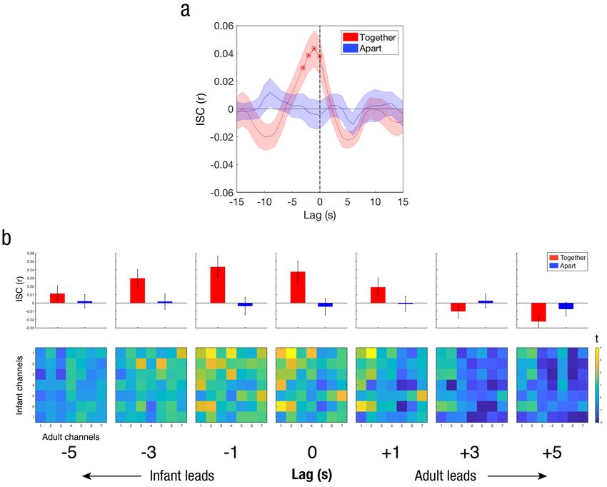
Infant–Adult Coupling During Natural Communication                                                                      9

Later, a research assistant coded every 500 ms whether         and socially salient, dynamic behaviors measured
or not there was mutual gaze (joint eye contact)               throughout the together condition (mutual gaze, infant
between the adult and infant, whether or not the infant        smiling, joint attention, and adult speech prosody).
was smiling, and whether or not the infant and adult
were engaged in joint attention to an external object,         Infant–adult neural coupling is
such as a toy or book. We did not code adult smiling
because the adult experimenter smiled more than 95%
                                                               present only during joint interaction
of the time; the nearly continuous adult smiling left          In the together (but not the apart) condition, we found
minimal opportunities for the infant to react to indi-         significant coupling between many PFC channels and
vidual instances of smiling. Two additional research           some parietal channels of infants and the adult (see
assistants each coded these behaviors in subsets of the        Fig. 2b). ISC between the infant and adult brains was
data (two thirds of the data sets each), and the scores        measured during the together and apart conditions, and
were highly reliable across the three raters (Cronbach’s       these actual correlation values were compared with null
α = .70, averaged across the behavioral measures;              distributions of correlation values. We included all
Cronbach, 1951). After filtering out background noise          homologous (i.e., same channel across brains) and non-
from the audio recordings in Adobe Audition, we ana-           homologous (i.e., different channels across brains)
lyzed continuous fundamental frequency (F0) using              channel pairings in this analysis. Statistical significance
Praat software (Version; 6.0.41; Boersma & Weenink,            was determined using a permutation procedure based
2009). Only six infants displayed any smiling through-         on phase-randomized surrogate data, and multiple-
out the videos, and this subset was used in the smiling-       comparisons correction was performed across channel
related analyses. During the 5-min duration of the             pairs (see the Method section). These results revealed
together condition, mutual gaze was present 27% of the         that communication between an infant and a caregiver
time (on average across dyads); the corresponding rates        was reflected in significant intersubject coupling
of infant smiling, joint attention, and adult speech were      throughout cortical areas involved in social and narra-
5.5%, 40%, and 49%, respectively.                              tive processing. Specifically, as shown in Figure 2b, we
   To perform brain-behavior comparisons, we first             found eleven significant channel pairs (primarily in the
downsampled the fNIRS time series in each channel to           PFC) in the together condition and no significant chan-
match the behavioral data (2 Hz for mutual gaze, smil-         nel pairs in the apart condition. The spatial selectivity
ing, and joint attention, 5 Hz for F0 variability) before      of this pattern of coupling indicated that the effects
computing time-lagged correlations. Here, we report            were not due to widespread, global arousal throughout
oxyhemoglobin for clarity of interpretation, as this signal    the brain.
reflects increases in local cortical activation in relation       To further assess infant–adult coupling, we com-
to the onset of behavioral events and is commonly              pared the strength of ISC between the infant and adult
reported in similar event-related analyses (e.g., Emberson,    brains during the together and apart phases in the PFC
Boldin, Robertson, Cannon, & Aslin, 2018).                     channels, which exhibited the most robust coupling in
                                                               the whole-brain analysis. Each participant had seven
                                                               channels covering the PFC. For each dyad, we com-
Results                                                        puted the average ISC across all 49 pairwise combina-
The brain responses of the infants and the adult experi-       tions of PFC channels (Infant Channel 1 vs. Adult
menter were recorded simultaneously using fNIRS (Fig.          Channel 1, Infant Channel 1 vs. Adult Channel 2, etc.).
1) in two conditions: together and apart. In the together      To assess the temporal dynamics of the infant–adult
condition, each adult-infant dyad participated in face-        coupling, we shifted the two signals relative to each
to-face sets of playful interactions with each other, and      other in time and performed the above statistical com-
in the apart condition, they interacted with other             parison (together vs. apart) at shifts (lags) of –15 s
individuals.                                                   (infant’s PFC activity leading) to +15 s (adult’s PFC
   In the first set of analyses (Figs. 2 and 3), we assessed   activity leading) in 1-s increments (Fig. 3a).
the significance of neural coupling between infants and           Significant infant–adult neural coupling was found
the adult experimenter in both conditions using a              at a lag of 0 and at negative lags of –1, –2, and –3 s
robust bootstrapped phase-scrambling analysis across           (infant leading), but only for the together condition
all available cortical channels and a form of cross-           (Fig. 3a; see also Fig. S1 in the Supplemental Material).
correlation that validated the temporal specificity of         Infant–adult neural coupling in the apart condition did
coupling in one cortical region of interest—the PFC. In        not exceed chance at any lag. Similarly, infant–adult
the second and third sets of analyses (Figs. 4 and 5),         neural coupling was significantly greater in the together
we measured the relationship between the neural acti-          condition than in the apart condition only at the same
vation of each participant in each dyad (adult, infant)        set of lags (0, –1, –2, and –3 s; see Fig. S1). The pattern
10 Piazza et al.
a
Together r Apart r
Together Apart
5 .1 5 .1
10 10
15 15
.05 .05
20 20
Infant Channel
Infant Channel
25 25
30 .0 30 .0
35 35
40 –.05 40 –.05
45 45
50 50
–.1 –.1
55 55
10 20 30 40 50 10 20 30 40 50
Adult Channel Adult Channel
b
Infant Adult Infant Adult
Fig. 2. Spatial extent of infant–adult neural coupling. The intersubject correlation matrices (a) show the relation between all infant
and adult channels, separately for the together and apart conditions. The brain diagrams (b) show significantly coupled channel pairs
(in red), determined by phase-scrambling analysis and corrected for multiple comparisons using the false-discovery-rate procedure
(q < .05; Benjamini & Hochberg, 1995). The solid line indicates a homologous channel pair; dotted lines indicate nonhomologous
channel pairs. N = 18 dyads.
of channel combinations that drove these channel- results were not driven by coupling in sensorimotor
averaged results can be seen in Figure 3b (bottom row), areas as a result of joint motion.
which shows t statistics (together – apart) for all 49
pairwise channel correlations at each lag. These find- The PFC plays a dynamic role in joint
ings suggest that the infant PFC may have slightly led eye contact, infant emotion, and joint
the adult PFC. For these time-shifted analyses, p values
were corrected using false-discovery rate across lags
attention to objects
(q < .05; Benjamini & Hochberg, 1995). This lag in Infant–adult coupling emerges from continuous feed-
neural responses between the child and the experi- back between interlocutors through eye contact, facial
menter helped to ensure that the neural coupling was expressions, vocal prosody, and other cues. Which
not a mere reflection of shared perceptual input. If behavioral cues are most directly involved in the cou-
responses were locked to shared audiovisual input, pling between the infant and adult brains during the
then neural coupling should have peaked at Lag 0. interaction? To start probing this question, we asked an
Additionally, the relative strength of PFC (compared independent rater (whose scores were validated by two
with parietal) coupling reinforces the idea that these additional raters; see the Method section) to view andInfant–Adult Coupling During Natural Communication 11 Fig. 3. Temporal dynamics of infant–adult coupling in the prefrontal cortex (PFC). The graph in (a) shows the intersubject correlation (ISC) between the adult and infant PFC signals across relative shifts (lags) of the two signals, separately for the together and apart conditions. Asterisks indicate significant lags, corrected for multiple comparisons. The top row in (b) shows mean ISC values across all 49 pairwise (infant–adult) combinations of PFC channels, averaged across dyads and shown separately for each condition at lags between –5 s and +5 s. The bottom row in (b) shows t maps comparing infant–adult ISCs (together vs. apart) for all pairwise PFC channel combinations at lags between –5 s and +5 s. Error bands (a) and error bars (b) represent standard errors of the mean. N = 18 dyads. code the video recording of each infant–adult interac- Next, we performed linear regression using the tion on a frame-by-frame basis along three behavioral behavioral time series to predict the infants’ and the dimensions: (a) the presence of mutual gaze (i.e., adult’s brain responses. We did so to examine whether whether the adult and infant were looking directly at responses in PFC increased during moments of mutual each other’s faces), (b) the signaling of a smile by the gaze, of infant smiling, and of joint attention. For each infant, and (c) the presence of joint attention (i.e., dyad, we performed this regression in each channel of whether the adult and infant were looking at the same the adult and infant. We averaged the resulting beta object, such as a toy or book). We then shifted all fNIRS weights within our specific region of interest (PFC) and time series relative to the behavioral time series, taking computed group-level statistics across dyads (see Fig. into account a canonical hemodynamic lag of 4 s to 5 S1). To assess the temporal alignment between the neu- s, before performing regression between the brain and ral and behavioral time series, we shifted the two sig- behavioral signals. When we instead convolved the nals relative to each other in time and performed the behavioral time series with a canonical hemodynamic above steps at lags from –15 s (brain leading) to +15 s response function (Arichi et al., 2012), the results (behavior leading) in 1-s increments (see the Method (described in Fig. 4) were highly similar. section).
12 Piazza et al.
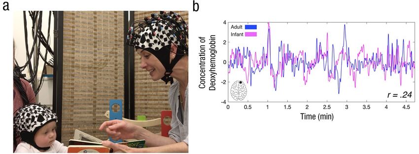
Fig. 4. Time-lagged relationship between neural responses in the prefrontal cortex (PFC) and
continuous measures of (a) mutual gaze, (b) infant smiling, (c) joint attention to objects, and
(d) pitch (F0) variability of the adult’s speech. Results are shown separately for the adult and
for infants (n = 17 for all analyses except smiling, n = 6). The left column shows results for true
dyads, and the right column depicts control results using random brain-behavior assignments.
Asterisks indicate time lags at which coefficients significantly exceeded 0 after correction for
multiple comparisons across lags. Error bands represent standard errors of the mean.Infant–Adult Coupling During Natural Communication 13
(e.g., singing) might have reliably increased both
behavioral and neural activity at a coarse level across
all dyads because of similarities in overall task
structure.
Finally, we assessed to what extent the direct infant–
adult brain-to-brain coupling (reported in Fig. 3) was
modulated by mutual gaze, infant smiling, and joint
attention to objects. We did this by regressing out these
three behavioral time courses from the fNIRS (PFC)
signals of each dyad (both infant and adult) before
recalculating infant–adult ISC. Specifically, we first
regressed out each behavioral time series at all possible
lags from –15 s to +15 s relative to each infant and adult
fNIRS time series (after accounting for a 4-s hemody-
namic lag). This analysis completely removed the influ-
ence of each behavior, regardless of the temporal offset
at which it was most correlated with brain activation.
After regressing out each behavior, we recalculated ISC
from lags of –5 s (infant leading) to +5 s (adult leading;
see Fig. 5). At the peak of the curve (lag = –1 s), brain-
to-brain coupling significantly decreased after the
removal of infant smiling, t(16) = 2.09, p < .05, Cohen’s
d = 0.52, 95% confidence interval (CI) for d = [–0.16,
1.21]; mutual gaze, t(16) = 3.12, p < .01, Cohen’s d =
Fig. 5. Time-lagged intersubject correlation (ISC). Results are shown
0.78, 95% CI for d = [0.08, 1.48]; and all three behaviors
for five analyses: one using the original prefrontal cortex (PFC) time
series (dashed line, which represents the curve for the together con- together (infant smiling, mutual gaze, and joint atten-
dition shown in Fig. 3a) and the other four after regressing out the tion), t(16) = 3.01, p < .01, Cohen’s d = 0.75, 95% CI for
time course of joint attention to objects from the PFC time series, the d = [0.06, 1.45]. However, removing joint attention alone
time course of infant smiling, the time course of mutual gaze, and all
three cues. Error bands represent standard errors of the mean. N = 17.
did not significantly decrease coupling, t(16) = –0.79,
p = .44, Cohen’s d = –0.20, 95% CI for d = [–0.87, 0.48].
This suggests that mutual gaze and infant smiling may
In both the infant and adult brains, PFC activation have contributed more to neural coupling than did joint
slightly preceded moments of mutual gaze (i.e., joint attention to objects, but differences in sample size and
eye contact between the infant and adult; Fig. 4a, left). in the frequency of each behavior suggest the need for
Specifically, the relationship between brain and behav- further research on this finding.
ior peaked at about 5 s before the initiation of the
mutual gaze (see Fig. S1). This suggests that the PFC The infant PFC is dynamically linked to
of both individuals anticipated—or even drove—an
the pitch variability of adult speech
increase in joint social behavior. PFC activity also mod-
ulated the presence of infants’ smiles. Here, we observed Another factor known to facilitate communication with
a more distinct pattern of results between infants and infants is the intonation of infant-directed speech, also
the adult (Fig. 4b, left). Specifically, both infant and known as “motherese.” We assessed the relationship
adult PFC responses increased before the initiation of between infant and adult neural responses and the
an infant smile, with the infant PFC responses preced- dynamics of the adult’s infant-directed speech, focusing
ing the adult’s; this confirmed our general finding that on F0 variability because of its influence on infant atten-
the infants’ neural dynamics led the adult’s. Finally, the tion (Fernald & Kuhl, 1987) and language processing
adult PFC showed a time-locked relationship to the (Trainor & Desjardins, 2002). To do this, we measured
time course of joint attention to objects (Fig. 4c, left). the relationship between F0 variability and the fNIRS
Control analyses randomly reassigning each neural time signal across time bins in the together condition. We
series to the behavioral data from a different dyad (Figs. chose a bin size of 200 ms because the syllabic rate of
4a–4c, right) showed no significant brain–behavior rela- child-directed speech is typically close to 4 Hz to
tionship for any of the three measures at any lag. This 5 Hz (Ryan, 2000). In each time bin in which there were
result ensured that the effects we observed were due recorded F0 values, we extracted the standard deviation
to the unique, dyad-specific dynamics of a given inter- of F0 of the adult’s voice as well as the average fNIRS
action and not simply the fact that a particular task activation in that bin. After once again accounting for14 Piazza et al.
the hemodynamic lag, we computed the correlation observed in the PFC was not significantly mediated by
between these two measures, averaged across the two variability in the adult’s speech prosody over time. This
most frontal PFC channels (Bonferroni-corrected for all finding is consistent with our finding of lack of modula-
possible pairs of PFC channels; α = .002). tion of the adult PFC responses by the F0 variability of
An increase in infant PFC response amplitude was her own voice (Fig. 4d, left).
reliably followed by an increase in pitch variability of
the caregiver’s speech. Figure 4d (left) shows that PFC
Discussion
responses in the infant brain were significantly corre-
lated with the F0 variability of the adult’s speech at lags From the beginning of life, infants’ survival depends
of –4 s to –5 s (see Fig. S1) after false-discovery-rate on successful communication with adult caregivers.
correction across lags (from –15 s to +15 s, as in the Researchers have uncovered important details about
analyses above). The adult PFC was not significantly how the behaviors of infants and adults are coupled
correlated with this measure at any lag, suggesting that during natural communication (Cohn & Tronick, 1988;
the adult’s own pitch variability was not directly related Marsh, Richardson, & Schmidt, 2009), but very little is
to the responses in her PFC. This influence of the known about how their brains interact during this pro-
infants’ neural dynamics on the adult’s speech is con- cess. Our research, using an infant-friendly imaging
sistent with the direction of coupling (infant preceding technique, provides the first demonstration of the
adult) found in the main ISC analyses and indicates the dynamic role played by both the developing and mature
experimenter’s sensitivity to the state of the infant. For brain during live social interaction. Moreover, we began
example, she may have increased her vocal excitement uncovering the relationships between various commu-
in response to a range of possible infant behaviors that nicative behaviors and infant–adult neural alignment.
could be linked to an infant PFC response. Once again, During coupled interactions, infant and adult brains
a control analysis randomly reassigning each neural are dynamically linked to important social cues (gaze,
time series to the behavioral data from a different dyad smiling, joint attention, and speech prosody), although
(Fig. 4d, right) showed no significant brain–behavior to differing extents. Our findings represent a crucial
relationship at any lag, suggesting that the infant PFC’s step toward understanding how infants’ brain responses
relationship to the variability of adult speech was due both anticipate and process the most important struc-
to dyad-specific dynamics. ture from adults’ input and how adults’ brains represent
In addition, for each dyad, we found time windows infants’ emotional feedback as adults strive to engage
containing the highest 10% of F0 variability for that infants in everyday communication. This opens new
session and compared infant PFC activation (again, in possibilities for understanding the independence versus
the first two channels) with activation during all other interdependence of two brains, the role of coupling in
moments (lower 90% in F0 variability). Moments of early learning, individual differences in early processing
particularly high pitch variability were preceded by and learning, and the many dimensions of coupling in
significantly higher infant PFC activation than other infant–caregiver interactions over time.
moments (see Fig. S3a in the Supplemental Material)— The findings support transactional development
two-tailed t test, t(16) = 4.35, p < .001, Cohen’s d = 1.09, models, which emphasize not only the role of adults’
95% CI for d = [0.37, 1.81]. input but children’s potential role in shaping their own
Finally, we assessed the extent to which direct input (Sameroff, 2009; for related ideas, see L. B. Smith,
infant–adult brain-to-brain coupling was modulated by Jayaraman, Clerkin, & Yu, 2018). Our results expand on
the adult speaker’s pitch variability. We did so by these frameworks and demonstrate a new application
regressing its time course at all lags from –15 s to +15 s of dynamic-systems theory (Thelen & Smith, 1996) by
relative to each infant and adult fNIRS time series (after showing how adults’ and infants’ brains reflect each
accounting for a 4-s hemodynamic lag), as in the partial other during natural, social interaction—mediated,
regression analysis above. After regressing out F0, we importantly, by sensitivity to each other’s behaviors. Over-
recalculated ISC at lags of –5 s to +5 s (see Fig. S3b in all, we found that prefrontal activity in infants’ brains
the Supplemental Material). The inclusion in this analy- slightly preceded similar activity in the adult’s brain,
sis of fNIRS data only at time points containing speech despite the relative lack of overt behavioral influence
resulted in a slightly noisier baseline ISC curve here exhibited by infants (e.g., minimal vocalization relative
(Fig. S3b, black line) than in Figure 3a (red line), but to the adult). This is consistent with fMRI evidence
the shape remained highly consistent. At the peak of showing that the activity in the medial and dorsolateral
the curve (Lag 0), we found no significant change in PFC of an adult listener often precedes a speaker’s PFC
ISC after removing F0 variability, t(16) = –0.84, p = .41, activity; this anticipatory signal is likely to relate to
Cohen’s d = –0.21, 95% CI for d = [–0.88, 0.46], suggest- prediction of upcoming events in a story (Dikker, Sil-
ing that the infant–adult brain-to-brain coupling bert, Hasson, & Zevin, 2014; Stephens et al., 2010). OurInfant–Adult Coupling During Natural Communication 15
findings suggest that the adult was sensitive to subtle enhanced coupling between infants and adults during
behavioral cues from the infants (likely via a combina- direct (vs. indirect) gaze (Leong et al., 2017) and showing
tion of explicit and implicit processes), which in turn parents’ neural responsiveness to infants’ attention (Wass
modified the adult’s brain responses and behaviors in et al., 2019). Importantly, our design enabled us not only
real time in order to improve alignment with, and maxi- to simultaneously measure neural responsiveness of both
mize information transfer to, the infants. Recent devel- infants and adults to a variety of natural communicative
opmental work has emphasized a feedback loop cues but also to compare the relative contributions of
between developmental changes to the body, the sta- these different cues to coupling. For example, mutual
tistics of input, and brain networks (Byrge, Sporns, & gaze appears to play a stronger role in driving ISC than
Smith, 2014), and our investigation recasts this proposal joint attention to objects does (Fig. 5). Future work will
within a multibrain framework. benefit from analyses of a broader range of behavioral
Importantly, we measured alignment between brain cues known to be important for successful communica-
and behavior from both sides of the dyadic interaction, tion and learning, such as hand actions and other ges-
in support of this dynamic view. In most cases, neural tures (Brand, Baldwin, & Ashburn, 2002; Yu & Smith,
responses preceded behavior, but the two brains were 2013), contingency of adult feedback on infant vocaliza-
differentially engaged with each cue depending on its tions (Goldstein, King, & West, 2003), and facial expres-
social relevance. For instance, both adult and infant sions. Whether in lab manipulations or unconstrained
PFCs were significantly coupled to the time course of interactions, systematic comparisons of neural coupling
mutual gaze—a social cue common to both individuals— and a range of behaviors will unravel how brains syn-
at lags that skewed slightly negatively (Fig. 4a, left), chronize during naturalistic communication.
which implies that neural alignment preceded and Because of the constraints of fNIRS research (and
anticipated joint eye contact. The adult PFC tracked developmental neuroscience in general), our current
joint attention to objects in a time-locked manner and sample was limited to infants who could sit relatively
anticipated infant smiling (Figs. 4b and 4c, left), prob- still during an interaction with a stranger. To enhance
ably because the adult was motivated to use the toys our understanding of natural variability in how caregiv-
to engage the infants and elicit positive emotional ers and children align their brains and behaviors,
responses; the infant PFC, on the basis of our conser researchers will need new approaches to study how
vative corrections, showed a nonsignificant trend infants with different temperaments and communicative
toward anticipating the infants’ own smiles (Fig. 4b, abilities couple with adults. In particular, our findings
left). Finally, the infant PFC anticipated changes in the prompt comparisons of the reciprocal dynamics of
adult’s infant-directed speech (Fig. 4d, left), likely brain-to-brain coupling in typically developing popula-
because the adult used extreme pitch contours in tions and the breakdown of neural responses to social
response to a range of infant behaviors, possibly to cues in disorders such as autism spectrum disorder
highlight a certain word. This finding differs from those (Elsabbagh et al., 2009).
of previous studies showing that variation in speech To conclude, our investigation represents an innovative
prosody drives infants’ attention (Fernald & Kuhl, 1987) approach to the study of social interaction—namely, by
and that musical features drive the adult medial PFC tracking the back-and-forth relationships between brains
( Janata et al., 2002). and behaviors during live communication. Studying this
Why did PFC activation often precede behavioral cues? process in 1-year-old infants has the potential to advance
It could be that the PFC was predicting behaviors, con- models of socially embedded cognition and learning in
tributing to the generation of those behaviors, or respond- development. This research highlights the value of exam-
ing to earlier, mediating social events. Future investigations ining the perspectives of both the developing and mature
of the causal link between brain and behavior (via mea- members of dyads, an approach that will enable a rich
sures of additional brain regions, real-time feedback, or understanding of how infants and adults work together
transcranial magnetic stimulation) could help to distin- to facilitate playful, shared communication.
guish these interpretations and further clarify how the
PFC and other areas organize the statistics of social and Action Editor
nonsocial input throughout development. Rebecca Treiman served as action editor for this article.
Our investigation of the complementary neural repre-
sentations of second-to-second social dynamics advances Author Contributions
what is currently known about infant–adult interactions, E. A. Piazza, C. Lew-Williams, and U. Hasson designed the
building on previous fNIRS research showing heightened experiment, E. A. Piazza and L. Hasenfratz collected the data,
medial PFC activation in infants in response to direct gaze E. A. Piazza analyzed the data with feedback from U. Hasson
(Urakawa, Takamoto, Ishikawa, Ono, & Nishijo, 2015). It and C. Lew-Williams. E. A. Piazza drafted the manuscript, and
also expands on electroencephalogram research showing all authors edited the manuscript.16 Piazza et al.
ORCID iD Brand, R. J., Baldwin, D. A., & Ashburn, L. A. (2002). Evidence
for ‘motionese’: Modifications in mothers’ infant-directed
Elise A. Piazza https://orcid.org/0000-0001-6729-8559
action. Developmental Science, 5, 72–83.
Brazelton, T. B., Koslowski, B., & Main, M. (1974). The ori-
Acknowledgments gins of reciprocity: The early mother-infant interaction.
We thank Ariella Cohen, Alice Wang, Mia Sullivan, Sagi Jaffe- In M. Lewis & L. A. Rosenblum (Eds.), The effect of the
Dax, Eva Fourakis, and Carolyn Mazzei for assistance with infant on its caregiver (pp. 49–76). Oxford, England:
data collection, behavioral coding, analysis feedback, and Wiley-Interscience.
participant recruitment. Brooks, R., & Meltzoff, A. N. (2008). Infant gaze following and
pointing predict accelerated vocabulary growth through
Declaration of Conflicting Interests two years of age: A longitudinal, growth curve modeling
study. Journal of Child Language, 35, 207–220.
The author(s) declared that there were no conflicts of interest
Byrge, L., Sporns, O., & Smith, L. B. (2014). Developmental
with respect to the authorship or the publication of this article.
process emerges from extended brain-body-behavior net-
works. Trends in Cognitive Sciences, 18, 395–403.
Funding Cohn, J. F., & Tronick, E. Z. (1988). Mother-infant face-to-
This work was supported by the Princeton University C. V. face interaction: Influence is bidirectional and unre-
Starr Fellowship (to E. A. Piazza), the Eric and Wendy Schmidt lated to periodic cycles in either partner’s behavior.
Transformative Technology Award (to E. A. Piazza, U. Hasson, Developmental Psychology, 24, 386–392.
and C. Lew-Williams), National Institutes of Health (NIH) Cronbach, L. J. (1951). Coefficient alpha and the internal
Grant 5DP1HD091948 (to U. Hasson), and NIH Grants structure of tests. Psychometrika, 16, 297–334.
R01HD095912 and R03HD079779 (to C. Lew-Williams). Dikker, S., Silbert, L. J., Hasson, U., & Zevin, J. D. (2014).
On the same wavelength: Predictable language enhances
Supplemental Material speaker-listener brain-to-brain synchrony in posterior
superior temporal gyrus. The Journal of Neuroscience,
Additional supporting information can be found at http://
34, 6267–6272.
journals.sagepub.com/doi/suppl/10.1177/0956797619878698
Elsabbagh, M., Volein, A., Csibra, G., Holmboe, K., Garwood,
H., Tucker, L., . . . Johnson, M. H. (2009). Neural corre-
Open Practices
lates of eye gaze processing in the infant broader autism
phenotype. Biological Psychiatry, 65, 31–38.
Emberson, L. L., Boldin, A. M., Robertson, C. E., Cannon,
G., & Aslin, R. N. (2018). Expectation affects neu-
All data, analysis scripts, and materials needed to repro-
ral repetition suppression in infancy. Developmental
duce this study have been made publicly available via the
Cognitive Neuroscience, 37, Article 100597. doi:10.1016/j
Open Science Framework and can be accessed at https://
.dcn.2018.11.001
osf.io/udxqp/. The design and analysis plans for the experi-
Ferguson, B., & Lew-Williams, C. (2016). Communicative sig-
ment were not preregistered. The complete Open Practices
nals support abstract rule learning by 7-month-old infants.
Disclosure for this article can be found at http://journals
Scientific Reports, 6, Article 25434. doi:10.1038/srep25434
.sagepub.com/doi/suppl/10.1177/0956797619878698. This article
Fernald, A., & Kuhl, P. (1987). Acoustic determinants of infant
has received the badges for Open Data and Open Materials.
preference for motherese speech. Infant Behavior and
More information about the Open Practices badges can be found
Development, 10, 279–293.
at http://www.psychologicalscience.org/publications/badges.
Goldstein, M. H., King, A. P., & West, M. J. (2003). Social inter-
action shapes babbling: Testing parallels between bird-
References song and speech. Proceedings of the National Academy
Arichi, T., Fagiolo, G., Varela, M., Melendez-Calderon, A., of Sciences, USA, 100, 8030–8035.
Allievi, A., Merchant, N., . . . Edwards, A. D. (2012). Hasson, U., Ghazanfar, A. A., Galantucci, B., Garrod, S., &
Development of BOLD signal hemodynamic responses Keysers, C. (2012). Brain-to-brain coupling: A mecha-
in the human brain. NeuroImage, 63, 663–673. nism for creating and sharing a social world. Trends in
Benjamini, Y., & Hochberg, Y. (1995). Controlling the false Cognitive Sciences, 16, 114–121.
discovery rate: A practical and powerful approach to Hirsch, J., Zhang, X., Noah, J. A., & Ono, Y. (2017). Frontal
multiple testing. Journal of the Royal Statistical Society B: temporal and parietal systems synchronize within and
Methodological, 57, 289–300. across brains during live eye-to-eye contact. NeuroImage,
Boas, D. A., Elwell, C. E., Ferrari, M., & Taga, G. (2014). 157, 314–330.
Twenty years of functional near-infrared spectroscopy: Honey, C. J., Thompson, C. R., Lerner, Y., & Hasson, U. (2012).
Introduction for the special issue. NeuroImage, 85, 1–5. Not lost in translation: Neural responses shared across
Boersma, P. & Weenink, D. (2009). Praat: Doing phonetics by languages. The Journal of Neuroscience, 32, 15277–15283.
computer (Version 6.0.41) [Computer software]. Retrieved Janata, P., Birk, J. L., Van Horn, J. D., Leman, M., Tillmann,
from http://www.praat.org/ B., & Bharucha, J. (2002). The cortical topography ofInfant–Adult Coupling During Natural Communication 17
tonal structures underlying Western music. Science, 298, of the default mode network during narrative comprehen-
2167–2170. sion. Nature Communications, 7, Article 12141. doi:10
Jiang, J., Dai, B., Peng, D., Zhu, C., Liu, L., & Lu, C. (2012). .1038/ncomms12141
Neural synchronization during face-to-face communica- Smith, L. B., Jayaraman, S., Clerkin, E., & Yu, C. (2018). The
tion. The Journal of Neuroscience, 45, 16064–16069. developing infant creates a curriculum for statistical learn-
Leong, V., Byrne, E., Clackson, K., Georgieva, S., Lam, S., ing. Trends in Cognitive Sciences, 22, 325–336.
& Wass, S. (2017). Speaker gaze increases information Smith, N. A., & Trainor, L. J. (2008). Infant-directed speech
coupling between infant and adult brains. Proceedings of is modulated by infant feedback. Infancy, 4, 410–420.
the National Academy of Sciences, USA, 114, 13290–13295. Stephens, G. J., Silbert, L. J., & Hasson, U. (2010). Speaker–
Lerner, Y., Honey, C. J., Silbert, L. J., & Hasson, U. (2011). listener neural coupling underlies successful communi-
Topographic mapping of a hierarchy of temporal recep- cation. Proceedings of the National Academy of Sciences,
tive windows using a narrated story. The Journal of USA, 107, 14425–14430.
Neuroscience, 31, 2906–2915. Thelen, E., & Smith, L. B. (1996). A dynamic systems approach
Liu, Y., Piazza, E. A., Simony, E., Shewokis, P. A., Onaral, B., to the development of cognition and action. Cambridge,
Hasson, U., & Ayaz, H. (2017). Measuring speaker–listener MA: MIT Press.
neural coupling with functional near infrared spectroscopy. Tomasello, M. (1992). The social bases of language acquisi-
Scientific Reports, 7, Article 43293. doi:10.1038/srep43293 tion. Social Development, 1, 67–87.
Marsh, K. L., Richardson, M. J., & Schmidt, R. C. (2009). Social Trainor, L. J., & Desjardins, R. J. (2002). Pitch characteristics
connection through joint action and interpersonal coordi- of infant-directed speech affect infants’ ability to discrimi-
nation. Topics in Cognitive Science, 1, 320–339. nate vowels. Psychonomic Bulletin & Review, 9, 335–340.
Piazza, E. A., Iordan, M. C., & Lew-Williams, C. (2017). Mothers Urakawa, S., Takamoto, K., Ishikawa, A., Ono, T., & Nishijo, H.
consistently alter their unique vocal fingerprints when com- (2015). Selective medial prefrontal cortex responses dur-
municating with infants. Current Biology, 27, 3162–3167. ing live mutual gaze interactions in human infants: An
Regev, M., Simony, E., Lee, K., Tan, K. M., Chen, J., & Hasson, U. fNIRS study. Brain Topography, 28, 691–701.
(2019). Propagation of information along the cortical Vouloumanos, A., & Werker, J. F. (2004). Tuned to the sig-
hierarchy as a function of attention while reading and nal: The privileged status of speech for young infants.
listening to stories. Cerebral Cortex, 29, 4017–4034. doi:10 Developmental Science, 7, 270–276.
.1093/cercor/bhy282 Vygotsky, L. (1978). Interaction between learning and devel-
Ryan, B. P. (2000). Speaking rate, conversational speech acts, opment. In M. Gauvain & M. Cole (Eds.), Readings on the
interruption, and linguistic complexity of 20 pre-school development of children (Vol. 23, pp. 34–41). New York,
stuttering and non-stuttering children and their mothers. NY: Scientific American Books.
Clinical Linguistics & Phonetics, 14, 25–51. Wass, S. V., Noreika, V., Georgieva, S., Clackson, K.,
Sameroff, A. (2009). The transactional model. In A. Sameroff Brightman, L., Nutbrown, R., . . . Leong, V. (2019).
(Ed.), The transactional model of development: How Parental neural responsivity to infants’ visual attention:
children and contexts shape each other (pp. 3–21). How mature brains influence immature brains during
Washington, DC: American Psychological Association. social interaction. PLOS Biology, 16(12), Article e2006328.
Scholkmann, F., Spichtig, S., Muehlemann, T., & Wolf, M. (2010). doi:10.1371/journal.pbio.2006328
How to detect and reduce movement artifacts in near-infra- Yu, C., & Smith, L. B. (2013). Joint attention without gaze
red imaging using moving standard deviation and spline following: Human infants and their parents coordinate
interpolation. Physiological Measurement, 31, 649–662. visual attention to objects through eye-hand coordina-
Simony, E., Honey, C. J., Chen, J., Lositsky, O., Yeshurun, Y., tion. PLOS ONE, 8(11), Article e79659. doi:10.1371/jour
Wiesel, A., & Hasson, U. (2016). Dynamic reconfiguration nal.pone.0079659You can also read