PRIOR SARS-COV-2 INFECTION RESCUES B AND T CELL RESPONSES TO VARIANTS AFTER FIRST VACCINE DOSE - SCIENCE
←
→
Page content transcription
If your browser does not render page correctly, please read the page content below
REPORTS

                                                                                                                           Cite as: C. J. Reynolds et al., Science
                                                                                                                               10.1126/science.abh1282 (2021).

Prior SARS-CoV-2 infection rescues B and T cell responses
to variants after first vaccine dose
Catherine J. Reynolds1†, Corinna Pade2†, Joseph M. Gibbons2†, David K. Butler1, Ashley D. Otter3, Katia
Menacho4, Marianna Fontana5,6, Angelique Smit5, Jane E. Sackville-West7, Teresa Cutino-Moguel4, Mala K.
Maini8, Benjamin Chain8, Mahdad Noursadeghi8, UK COVIDsortium Immune Correlates Network‡, Tim
Brooks3, Amanda Semper3, Charlotte Manisty4,9, Thomas A. Treibel4,9, James C. Moon4,9, UK COVIDsortium
Investigators‡, Ana M. Valdes10,11, Áine McKnight2§, Daniel M. Altmann12§, Rosemary Boyton1,13§*
1
 Department of Infectious Disease, Imperial College London, London, UK. 2Blizard Institute, Barts and the London School of Medicine and Dentistry, Queen Mary University
of London, London, UK. 3National Infection Service, Public Health England, Porton Down, UK. 4St Bartholomew’s Hospital, Barts Health NHS Trust, London, UK. 5Royal Free
London NHS Foundation Trust, London, UK. 6Division of Medicine, University College London, London, UK. 7James Wigg Practice, Kentish Town, London, UK. 8Division of
Infection and Immunity, University College London, London, UK. 9Institute of Cardiovascular Science, University College London, London, UK. 10Academic Rheumatology,
Clinical Sciences, Nottingham City Hospital, Nottingham, UK. 11NIHR Nottingham Biomedical Research Centre, Nottingham University Hospitals NHS Trust and University of

                                                                                                                                                                           Downloaded from http://science.sciencemag.org/ on May 29, 2021
Nottingham, Nottingham, UK. 12Department of Immunology and Inflammation, Imperial College London, London, UK. 13Lung Division, Royal Brompton and Harefield
Hospitals, London, UK.
*Corresponding author: Email: r.boyton@imperial.ac.uk
†These authors contributed equally to this work.
§These authors contributed equally to this work.
‡UK COVIDsortium Investigators and UK COVIDsortium Immune Correlates Network collaborators and affiliations are listed in the supplementary materials.

SARS-CoV-2 vaccine rollout has coincided with the spread of variants of concern. We investigated if single
dose vaccination, with or without prior infection, confers cross protective immunity to variants. We
analyzed T and B cell responses after first dose vaccination with the Pfizer/BioNTech mRNA vaccine
BNT162b2 in healthcare workers (HCW) followed longitudinally, with or without prior Wuhan-Hu-1 SARS-
CoV-2 infection. After one dose, individuals with prior infection showed enhanced T cell immunity, antibody
secreting memory B cell response to spike and neutralizing antibodies effective against B.1.1.7 and B.1.351.
By comparison, HCW receiving one vaccine dose without prior infection showed reduced immunity against
variants. B.1.1.7 and B.1.351 spike mutations resulted in increased, abrogated or unchanged T cell responses
depending on human leukocyte antigen (HLA) polymorphisms. Single dose vaccination with BNT162b2 in
the context of prior infection with a heterologous variant substantially enhances neutralizing antibody
responses against variants.

During worldwide rollout of SARS-CoV-2 vaccines it is vital                            mutation, in addition to E484K, the latter implicated in es-
to understand how vaccination influences immune responses                              cape from neutralizing antibody (nAb) (5, 6).
and protection among those who have had prior natural                                      The Pfizer/BioNTech mRNA vaccine BNT162b2 encodes a
SARS-CoV-2 infection. This is a knowledge-gap since a history                          prefusion stabilized, membrane-anchored SARS-CoV-2 full-
of previous infection was an exclusion criterion in Phase III                          length spike protein modified by two proline substitutions (1,
vaccine trials (1). Countries have adopted diverse approaches,                         7, 8). A two-dose regimen of 30 μg BNT162b2, 21d apart, con-
including the UK policy to maximize deployment of first                                fers 95% protection against Wuhan-Hu-1 SARS-CoV-2 (1),
doses to the largest possible numbers by extending the time                            eliciting high nAb titers, as well as CD4 cell and CD8 re-
interval to second dose. At the end of 2020, it became appar-                          sponses (8). When given as a single 60 μg dose, BNT162b1
ent several virus variants had emerged (2, 3) and that these                           induced virus Ab neutralization, but T cell responses were re-
might impact vaccine rollout. The B.1.1.7 variant, possessing                          duced compared with the standard prime-boost regime (8).
the spike N501Y mutation, first emerged in the UK in Decem-                            Single 30 μg dose, BNT162b1 was not reported beyond d21.
ber 2020 and spread rapidly (4). Additional variants of con-                           However, the cumulative incidence of COVID-19 cases among
cern (VOC) include the B.1.351 variant, which emerged at                               21,676 placebo and 21,699 vaccine recipients diverged 12 days
about the same time in South Africa and the P.1 variant that                           after the first dose, indicating possible early-onset first dose
emerged in January 2021 in Brazil. Both have the N501Y                                 protection (1). For those who were previously infected, single

First release: 30 April 2021                                            www.sciencemag.org                (Page numbers not final at time of first release) 1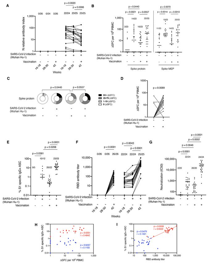
dose vaccination may act as a boost following natural infec-        timepoint and 3 weeks after vaccination, showed a signifi-
tion. Therefore, we aimed to test the impact of prior SARS-         cantly increased response (p = 0.0089) (Fig. 1D). Three indi-
CoV-2 infection on T cell and B cell responses to first dose        viduals who previously showed a response, despite lack of
vaccination.                                                        laboratory evidence for infection (therefore presumably a
    To do this, we analyzed T and B cell immunity after the         cross-reactive response to an endemic human coronavirus)
first 30 μg dose of the Pfizer/BioNTech mRNA vaccine                showed an unchanged or decreased response to spike after
BNT162b2, in a cohort of UK hospital healthcare workers             vaccination.
(HCW) (9–12). The COVIDsortium HCW cohort has been                      The size of the SARS-CoV-2 S1 specific memory B cell pool
studied longitudinally since the end of March 2020, provid-         was investigated by B cell ELISpot (Fig. 1E and fig. S2B). As
ing accurate infection and immune history in the context of         for T cell responses, the number of S1 specific IgG+ antibody
genotyping, including HLA imputation (10–12). Our aim was           secreting cells (ASC) was far greater in vaccinated post infec-
to compare T and B cell immunity after a first dose of vaccine      tion individuals compared with vaccinated naïve individuals
in December 2020, in individuals post-infection (after natural      (p < 0.0001). Prior infection generated a 63-fold increase in
infection), vaccinated post-infection (vaccination in the con-      S1 specific ASC. There were no pre-existing S1 specific ASC in
text of prior SARS-CoV-2 infection) and vaccinated naïve (sin-      uninfected HCW pre-vaccination. Twenty of 22 vaccinated

                                                                                                                                         Downloaded from http://science.sciencemag.org/ on May 29, 2021
gle dose vaccination). We wished to explore if there is             naïve individuals had detectable S1 specific ASC comprising
evidence for altered T cell recognition of the B.1.1.7 and          0.02% to 1.54% of the memory B cell (MBC) pool. By compar-
B.1.351 variants, and in particular, of the N501Y mutation          ison, all vaccinated post infection individuals had detectable
shared by several VOC.                                              S1 specific ASCs (1.90–50% of MBC pool). We previously re-
    The UK has deployed a heterodox vaccination regimen to          ported (14), spike receptor binding domain (RBD) enhanced
maximize immune protection and slow spread of the B.1.1.7           Ab responses in the vaccinated post infection group. In this
lineage, giving an initial 30 μg dose of BNT162b2, followed by      work, the vaccination naïve group attained similar antibody
boosting up to 12 weeks later (13). A cross-sectional sub-study     titers to the post infection group at 16–18 and 28–30 weeks
(n = 51) of the existing longitudinal HCW cohort (9–12) was         (Fig. 1F). Vaccinated naïve individuals made a lower nAb re-
recruited 22d (±2) after the first dose. After the start of the     sponse to WT virus than seen following natural infection at
study, the majority of acute infections had already occurred        16–18 weeks although this did not achieve statistical signifi-
among this cohort (11). At the time of receiving their first vac-   cance. In line with the findings for MBC and RBD binding,
cine dose in December 2020, 25 individuals were approxi-            there was a significantly enhanced nAb response in vac-
mately 39 weeks post SARS-CoV-2 infection with the Wuhan            cinated post infection individuals compared with the vac-
Hu-1 strain and prior to the emergence of VOC and 26 were           cinated naïve group (Fig. 1G), the mean value being 25,273
confirmed uninfected, having tested negative in longitudinal        compared to 420, that is, a 60-fold increase. To put this in
serology for spike and nucleocapsid (N) (table S1 and fig. S1).     context, these values are 43-fold higher than the values rec-
    We first measured SARS-CoV-2 N antibody longitudinally          orded after 2 vaccine doses in the Phase 1 trial (7). There was
up to 16–18 weeks, then at 28–30 weeks and finally 42 weeks         no correlation between the magnitude of the spike protein T
post-recruitment to confirm that there was no laboratory ev-        cell response and the percentage of S1 specific ASC (Fig. 1H).
idence of new infection at the time of drawing blood for the        As expected, there was a positive correlation between the per-
vaccine study at 42 weeks: none of the previously uninfected        centage of S1 specific ASC and the serum titer of RBD anti-
HCW had become seropositive (Fig. 1A). T cell responses to          body in the vaccinated post infection individuals (r = 0.6502;
spike protein and MEP (mapped epitope peptides) in either           p = 0.0008) (Fig. 1I). After vaccination, two previously in-
post infection, vaccinated post infection or vaccinated naïve       fected individuals showed lower percentages of S1 specific
individuals were compared (Fig. 1B). Ninety six percent             memory B cells and reduced serum RBD specific antibody
(22/23) of vaccinated post infection individuals made a T cell      levels than the rest of the group; prior infection involving
response to spike protein compared to 70% (16/23) of vac-           case-definition symptoms tended to be associated with a
cinated naïve individuals, with a 4-fold increase in magnitude      higher specific B cell frequency than milder disease (Fig. 1F
of T cell response. Furthermore, while the T cell response to       and fig. S2C). These individuals who, despite infection, had
spike protein in vaccinated naïve individuals increased (p =        also not shown a detectable T cell response (one never sero-
0.0440), it was lower than those of vaccinated post infection       converted and the other rapidly became seronegative during
individuals (p = 0.0557) (Fig. 1C). As expected, there was no       longitudinal follow up) had a poor or absent response to in-
significant change in T cell response to N (a measure of im-        fection that was only minimally overcome by vaccination.
munity to natural infection) (fig. S2A).                                The data in Fig. 1 indicate that there is a strong prime-
    Paired analysis of T cell immunity to spike protein in pre-     boosting effect of prior infection on single dose vaccination.
viously uninfected individuals, analyzed at the 16–18 week          Augmentation is seen more strongly in MBC frequency, anti-

First release: 30 April 2021                            www.sciencemag.org       (Page numbers not final at time of first release)   2
RBD, and nAb responses than for T cell response frequency. cell pool after single dose vaccination (Fig. 2C). We looked at
Furthermore, there was no correlation between S1 ASC fre- correlations between RBD binding antibodies, B cell re-
quency and T cell response frequency (Fig. 1H). There is, how- sponses, T cell responses and IC50, comparing neutralization
ever, a correlation between S1 ASC and RBD antibody titers, of Wuhan Hu-1, B.1.1.7 and B.1.315 live virus (Fig. 2D). Despite
indicating that individuals with higher numbers of MBC the lower neutralization of B.1.1.7 and B.1.315 variants, the
make higher antibody responses, and individuals who had ex- pattern was retained of strong correlation between RBD an-
perienced infection clustered at the higher end of this re- tibody titer. S1 specific B cell frequency and neutralization
sponse (Fig. 1I). and somewhat weaker correlation between T cell response
Shortly before the vaccination program was initiated, sev- and neutralization.
eral VOC emerged, including the B.1.1.7 VOC. This variant has A lack of Ab-mediated protection in single-dose vaccinees
nine mutations in the spike protein. Several studies have re- could be mitigated by a broader repertoire of T cell responses
ported weaker nAb responses to B.1.1.7 relative to the previ- (18). To investigate differences in T cell recognition, we de-
ously circulating Wuhan-Hu-1 strain (2–6, 15–18). The signed peptide pools covering the affected regions of Wuhan-
majority of SARS-CoV-2 immune naïve individuals made no Hu-1, B.1.1.7, and B.1.351 variant sequence (table S2). We com-
nAb response to the B.1.1.7 (18/20) and B.1.351 (17/20) vari- pared T cell responses to these peptide pools in PBMC from
Downloaded from http://science.sciencemag.org/ on May 29, 2021
ants after single dose vaccination. In contrast, almost all vac- individuals vaccinated post infection and vaccinated naïve
cinated post infection individuals made a strong nAb (Fig. 2E). Responses in post infection vaccinees were in gen-
response to the B.1.1.7 (24/24) and B.1.351 (23/24) variants af- eral higher than in the vaccinated naïve individuals: note an
ter a single dose vaccination with a 46-fold (B.1.1.7) and 63- enhanced response to the B.1.1.7 peptide pool. T cell re-
fold (B.1.351) increase in mean nAb IC50 in vaccinated post sponses were heterogeneous; responses to variant pools
infection compared to vaccinated naïve individuals. In a could be either higher or lower than to Wuhan Hu-1 pools.
paired analysis, we observed in vitro significantly reduced Alterations in affinity for the T cell receptor can lead to al-
nAb potency to authentic B.1.1.7 variant virus (>24.7-fold tered peptide ligand effects and differential polarization of
lower than response to Wuhan Hu-1, p < 0.0001) in sera from cytokine effector programs, as we have previously observed
individuals with a past medical history of natural infection in Zika virus infection (19). We wondered if this was also oc-
(Fig. 2B). Worryingly, after single dose vaccination, 90% curring for SARS-CoV-2; however, we found no evidence for
(18/20) of vaccinated naïve individuals showed no detectable immune deviation to interleukins (IL)-4, -5, -10,–13, 17A or 23
nAbs (IC50 < 50) against B.1.1.7 (mean IC50 37, range 0 to (fig. S3).
184; p = 0.2090), but did show demonstrable nAb responses For B.1.1.7 and B.1.351, attention has centered on the
to Wuhan Hu-1 SARS-CoV-2 virus (mean IC50 420, range 80 N501Y mutation, as this is implicated in altered ACE2 bind-
to 2,004; p = 0.0046). In contrast, all vaccinated post infec- ing and enhanced infectivity and transmission but is also a
tion individuals responded to single dose vaccination with target for B and T cell recognition. We initially looked at T
substantially enhanced nAb responses, neutralizing not just cell responses following natural infection and found that at
Wuhan Hu-1 SARS-CoV-2 (mean IC50 25,273; range IC50 581 16–18 weeks post infection, the N501Y mutation appeared to
to 76,369), but also the B.1.1.7 (mean IC50 1717; range: IC50 have no substantial differential impact on the T cell response
52 to 4919) and B.1.351 (mean IC50 5451; range: IC50 41 to (Fig. 2F), unlike nAb recognition (5).
20,411) variants (Fig. 2, A and B, and fig. S3). We show a 14.7- The specific impact of any T cell epitope changes on the
fold reduction in neutralization (IC50) responses in SARS- immune response against VOC depends on changes in pep-
CoV-2 B.1.1.7 variant compared to Wuhan Hu-1 virus in vac- tide binding to the peptide-presenting HLA molecules. Since
cinated post infection individuals. However, despite this fall the HLA complex is the most polymorphic part of the human
the majority (22/24) remain within a “protective threshold.” genome, any alteration to core HLA binding motifs will dif-
This was not the case for vaccinated naïve individuals. There ferentially impact people with some HLA alleles over others.
was a 11.4-fold reduction in neutralization (IC50) responses We performed in silico analysis (NetMHCIIpan) to predict
against SARS-CoV-2 B.1.1.7 variant (mean, 37) compared to which of the B.1.1.7 and B.1.351 mutations were found in HLA
Wuhan Hu-1 virus (mean, 420) resulting in the majority of core binding motifs and how this might impact binding to
individuals (19/20) falling below the “protective threshold.” common HLAII alleles (DRB1*0101, DRB1*0301, DRB1*0401,
This result was mirrored in the SARS-CoV-2 S1 specific DRB1*0701, DRB1*1101, DRB1*1301, and DRB1*1501) (tables
memory B cell pool where reduced numbers of S1 specific S3 and S4). Some of the mutations did not fall in a region
IgG+ ASC are seen (in vaccinated naïve individuals compared predicted to bind the HLAII alleles tested (D3L, T716I,
to vaccinated post infection individuals) responding to S1 an- T1001I, A1708D and 3675-7 SGF del). Although several muta-
tigen containing the N501Y, K417N and E484K mutations. tions were not predicted to significantly change affinity for
Prior infection substantially enhances the specific memory B the HLAII alleles, others did show predicted differential
First release: 30 April 2021 www.sciencemag.org (Page numbers not final at time of first release) 3affinities depending on host HLAII type (tables S3 and S4). In contrast, nAb responses in individuals several months on Analyzing altered responses to the D1118H mutation, we from mild infection show much lower IC50s against B.1.1.7 noted that individuals who carried DRB1*0301 and and B.1.351, often
3. D. M. Altmann, R. J. Boyton, R. Beale, Immunity to SARS-CoV-2 variants of concern.               Fontana, A. Smit, A. Semper, B. O’Brien, B. Chain, T. Brooks, C. Manisty, T. Treibel,
     Science 371, 1103–1104 (2021). doi:10.1126/science.abg7404 Medline                             J. C. Moon, M. Noursadeghi, D. M. Altmann, M. K. Maini, Á. McKnight, R. J. Boyton;
4. A. Rambaut, N. Loman, O. Pybus, W. Barclay, J. Barrett, A. Carabelli, T. Connor, T.              COVIDsortium investigators; COVIDsortium immune correlates network,
     Peacock, D. L. Robertson, E. Volz, on behalf of COVID-19 Genomics Consortium                   Discordant neutralizing antibody and T cell responses in asymptomatic and mild
     UK, (CoG-UK), Preliminary genomic characterisation of an emergent SARS-CoV-                    SARS-CoV-2          infection.      Sci.     Immunol.      5,    eabf3698        (2020).
     2 lineage in the UK defined by a novel set of spike mutations, virological.org                  doi:10.1126/sciimmunol.abf3698 Medline
     (2020); https://virological.org/t/preliminary-genomic-characterisation-of-an-              12. C. Manisty, T. A. Treibel, M. Jensen, A. Semper, G. Joy, R. K. Gupta, T. Cutino-
     emergent-sars-cov-2-lineage-in-the-uk-defined-by-a-novel-set-of-spike-                         Moguel, M. Andiapen, J. Jones, S. Taylor, A. Otter, C. Pade, J. Gibbons, J. Lee, J.
     mutations/563.                                                                                  Bacon, S. Thomas, C. Moon, M. Jones, D. Williams, J. Lambourne, M. Fontana, D.
5. Z. Wang, F. Schmidt, Y. Weisblum, F. Muecksch, C. O. Barnes, S. Finkin, D. Schaefer-              M. Altmann, R. Boyton, M. Maini, A. McKnight, B. Chain, M. Noursadeghi, J. C.
     Babajew, M. Cipolla, C. Gaebler, J. A. Lieberman, T. Y. Oliveira, Z. Yang, M. E.                Moon, Time series analysis and mechanistic modelling of heterogeneity and sero-
     Abernathy, K. E. Huey-Tubman, A. Hurley, M. Turroja, K. A. West, K. Gordon, K. G.               reversion in antibody responses to mild SARS-CoV-2 infection. EBioMedicine 65,
     Millard, V. Ramos, J. Da Silva, J. Xu, R. A. Colbert, R. Patel, J. Dizon, C. Unson-             103259 (2021). doi:10.1016/j.ebiom.2021.103259 Medline
     O’Brien, I. Shimeliovich, A. Gazumyan, M. Caskey, P. J. Bjorkman, R. Casellas, T.          13. COVID-19 vaccines: Acting on the evidence. Nat. Med. 27, 183 (2021).
     Hatziioannou, P. D. Bieniasz, M. C. Nussenzweig, mRNA vaccine-elicited                          doi:10.1038/s41591-021-01261-5 Medline
     antibodies to SARS-CoV-2 and circulating variants. Nature 592, 616–622 (2021).             14. C. Manisty, A. D. Otter, T. A. Treibel, Á. McKnight, D. M. Altmann, T. Brooks, M.
     doi:10.1038/s41586-021-03324-6 Medline                                                          Noursadeghi, R. J. Boyton, A. Semper, J. C. Moon, Antibody response to first
6. D. A. Collier, A. De Marco, I. A. T. M. Ferreira, B. Meng, R. P. Datir, A. C. Walls, S. A.        BNT162b2 dose in previously SARS-CoV-2-infected individuals. Lancet 397,
     Kemp, J. Bassi, D. Pinto, C. Silacci-Fregni, S. Bianchi, M. A. Tortorici, J. Bowen, K.          1057–1058 (2021). doi:10.1016/S0140-6736(21)00501-8 Medline

                                                                                                                                                                                                Downloaded from http://science.sciencemag.org/ on May 29, 2021
     Culap, S. Jaconi, E. Cameroni, G. Snell, M. S. Pizzuto, A. F. Pellanda, C. Garzoni, A.     15. X. Shen, H. Tang, C. McDanal, K. Wagh, W. Fischer, J. Theiler, H. Yoon, D. Li, B. F.
     Riva, A. Elmer, N. Kingston, B. Graves, L. E. McCoy, K. G. C. Smith, J. R. Bradley,             Haynes, K. O. Sanders, S. Gnanakaran, N. Hengartner, R. Pajon, G. Smith, G. M.
     N. Temperton, L. Ceron-Gutierrez, G. Barcenas-Morales, W. Harvey, H. W. Virgin,                 Glenn, B. Korber, D. C. Montefiori, SARS-CoV-2 variant B.1.1.7 is susceptible to
     A. Lanzavecchia, L. Piccoli, R. Doffinger, M. Wills, D. Veesler, D. Corti, R. K. Gupta;         neutralizing antibodies elicited by ancestral spike vaccines. Cell Host Microbe 29,
     CITIID-NIHR BioResource COVID-19 Collaboration; COVID-19 Genomics UK (COG-                      529–539.e3 (2021). doi:10.1016/j.chom.2021.03.002 Medline
     UK) Consortium, Sensitivity of SARS-CoV-2 B.1.1.7 to mRNA vaccine-elicited                 16. A. J. Greaney, A. N. Loes, K. H. D. Crawford, T. N. Starr, K. D. Malone, H. Y. Chu, J.
     antibodies. Nature 10.1038/s41586-021-03412-7 (2021). Medline                                   D. Bloom, Comprehensive mapping of mutations in the SARS-CoV-2 receptor-
7. E. E. Walsh, R. W. Frenck Jr., A. R. Falsey, N. Kitchin, J. Absalon, A. Gurtman, S.               binding domain that affect recognition by polyclonal human plasma antibodies.
     Lockhart, K. Neuzil, M. J. Mulligan, R. Bailey, K. A. Swanson, P. Li, K. Koury, W.              Cell Host Microbe 29, 463–476.e6 (2021). doi:10.1016/j.chom.2021.02.003
     Kalina, D. Cooper, C. Fontes-Garfias, P. Y. Shi, Ö. Türeci, K. R. Tompkins, K. E. Lyke,         Medline
     V. Raabe, P. R. Dormitzer, K. U. Jansen, U. Şahin, W. C. Gruber, Safety and                17. P. Supasa, D. Zhou, W. Dejnirattisai, C. Liu, A. J. Mentzer, H. M. Ginn, Y. Zhao, H. M.
     immunogenicity of two RNA-based Covid-19 vaccine candidates. N. Engl. J. Med.                   E. Duyvesteyn, R. Nutalai, A. Tuekprakhon, B. Wang, G. C. Paesen, J. Slon-
     383, 2439–2450 (2020). doi:10.1056/NEJMoa2027906 Medline                                        Campos, C. López-Camacho, B. Hallis, N. Coombes, K. R. Bewley, S. Charlton, T.
8. U. Sahin, A. Muik, E. Derhovanessian, I. Vogler, L. M. Kranz, M. Vormehr, A. Baum,                S. Walter, E. Barnes, S. J. Dunachie, D. Skelly, S. F. Lumley, N. Baker, I. Shaik, H. E.
     K. Pascal, J. Quandt, D. Maurus, S. Brachtendorf, V. Lörks, J. Sikorski, R. Hilker, D.          Humphries, K. Godwin, N. Gent, A. Sienkiewicz, C. Dold, R. Levin, T. Dong, A. J.
     Becker, A. K. Eller, J. Grützner, C. Boesler, C. Rosenbaum, M. C. Kühnle, U.                    Pollard, J. C. Knight, P. Klenerman, D. Crook, T. Lambe, E. Clutterbuck, S. Bibi, A.
     Luxemburger, A. Kemmer-Brück, D. Langer, M. Bexon, S. Bolte, K. Karikó, T.                      Flaxman, M. Bittaye, S. Belij-Rammerstorfer, S. Gilbert, D. R. Hall, M. A. Williams,
     Palanche, B. Fischer, A. Schultz, P. Y. Shi, C. Fontes-Garfias, J. L. Perez, K. A.              N. G. Paterson, W. James, M. W. Carroll, E. E. Fry, J. Mongkolsapaya, J. Ren, D. I.
     Swanson, J. Loschko, I. L. Scully, M. Cutler, W. Kalina, C. A. Kyratsous, D. Cooper,            Stuart, G. R. Screaton, Reduced neutralization of SARS-CoV-2 B.1.1.7 variant by
     P. R. Dormitzer, K. U. Jansen, Ö. Türeci, COVID-19 vaccine BNT162b1 elicits                     convalescent and vaccine sera. Cell 184, 2201–2211.e7 (2021).
     human antibody and TH1 T cell responses. Nature 586, 594–599 (2020).                            doi:10.1016/j.cell.2021.02.033 Medline
     doi:10.1038/s41586-020-2814-7 Medline                                                      18. A. Muik, A. K. Wallisch, B. Sänger, K. A. Swanson, J. Mühl, W. Chen, H. Cai, D.
9. T. A. Treibel, C. Manisty, M. Burton, Á. McKnight, J. Lambourne, J. B. Augusto, X.                Maurus, R. Sarkar, Ö. Türeci, P. R. Dormitzer, U. Şahin, Neutralization of SARS-
     Couto-Parada, T. Cutino-Moguel, M. Noursadeghi, J. C. Moon, COVID-19: PCR                       CoV-2 lineage B.1.1.7 pseudovirus by BNT162b2 vaccine-elicited human sera.
     screening of asymptomatic health-care workers at London hospital. Lancet 395,                   Science 371, 1152–1153 (2021). 10.1126/science.abg6105 Medline
     1608–1610 (2020). doi:10.1016/S0140-6736(20)31100-4 Medline                                19. C. J. Reynolds, O. M. Suleyman, A. M. Ortega-Prieto, J. K. Skelton, P. Bonnesoeur,
10. J. B. Augusto, K. Menacho, M. Andiapen, R. Bowles, M. Burton, S. Welch, A. N.                    A. Blohm, V. Carregaro, J. S. Silva, E. A. James, B. Maillère, M. Dorner, R. J. Boyton,
     Bhuva, A. Seraphim, C. Pade, G. Joy, M. Jensen, R. H. Davies, G. Captur, M.                     D. M. Altmann, T cell immunity to Zika virus targets immunodominant epitopes
     Fontana, H. Montgomery, B. O’Brien, A. D. Hingorani, T. Cutino-Moguel, Á.                       that show cross-reactivity with other Flaviviruses. Sci. Rep. 8, 672 (2018).
     McKnight, H. Abbass, M. Alfarih, Z. Alldis, G. L. Baca, A. Boulter, O. V. Bracken, N.           doi:10.1038/s41598-017-18781-1 Medline
     Bullock, N. Champion, C. Chan, X. Couto-Parada, K. Dieobi-Anene, K. Feehan, G.             20. F. Krammer, K. Srivastava, H. Alshammary, A. A. Amoako, M. H. Awawda, K. F.
     Figtree, M. C. Figtree, M. Finlay, N. Forooghi, J. M. Gibbons, P. Griffiths, M.                 Beach, M. C. Bermúdez-González, D. A. Bielak, J. M. Carreño, R. L. Chernet, L. Q.
     Hamblin, L. Howes, I. Itua, M. Jones, V. Jardim, V. Kapil, W.-Y. Jason Lee, V.                  Eaker, E. D. Ferreri, D. L. Floda, C. R. Gleason, J. Z. Hamburger, K. Jiang, G. Kleiner,
     Mandadapu, C. Mfuko, O. Mitchelmore, S. Palma, K. Patel, S. E. Petersen, B.                     D. Jurczyszak, J. C. Matthews, W. A. Mendez, I. Nabeel, L. C. F. Mulder, A. J. Raskin,
     Piniera, R. Raine, A. Rapala, A. Richards, G. Sambile, J. Couto de Sousa, M.                   K. T. Russo, A. T. Salimbangon, M. Saksena, A. S. Shin, G. Singh, L. A. Sominsky,
     Sugimoto, G. D. Thornton, J. Artico, D. Zahedi, R. Parker, M. Robathan, L. M.                  D. Stadlbauer, A. Wajnberg, V. Simon, Antibody responses in seropositive persons
     Hickling, N. Ntusi, A. Semper, T. Brooks, J. Jones, A. Tucker, J. Veerapen, M.                 after a single dose of SARS-CoV-2 mRNA vaccine. N. Engl. J. Med. 384, 1372–1374
     Vijayakumar, T. Wodehouse, L. Wynne, T. A. Treibel, M. Noursadeghi, C. Manisty,                 (2021). doi:10.1056/NEJMc2101667 Medline
     J. C. Moon, Healthcare Workers Bioresource: Study outline and baseline                     21. A. Bertoletti, A. T. Tan, N. Le Bert, The T-cell response to SARS-CoV-2: Kinetic and
     characteristics of a prospective healthcare worker cohort to study immune                       quantitative aspects and the case for their protective role. Oxford Open
     protection and pathogenesis in COVID-19. Wellcome Open Res. 5, 179 (2020).                      Immunology 2, iqab006 (2021). doi:10.1093/oxfimm/iqab006
     doi:10.12688/wellcomeopenres.16051.2 Medline                                               22. R. R. Goel, S. A. Apostolidis, M. M. Painter, D. Mathew, A. Pattekar, O. Kuthuru, S.
11. C. J. Reynolds, L. Swadling, J. M. Gibbons, C. Pade, M. P. Jensen, M. O. Diniz, N. M.            Gouma, P. Hicks, W. Meng, A. M. Rosenfeld, S. Dysinger, K. A. Lundgreen, L. Kuri-
     Schmidt, D. K. Butler, O. E. Amin, S. N. L. Bailey, S. M. Murray, F. P. Pieper, S.              Cervantes, S. Adamski, A. Hicks, S. Korte, D. A. Oldridge, A. E. Baxter, J. R. Giles,
     Taylor, J. Jones, M. Jones, W. J. Lee, J. Rosenheim, A. Chandran, G. Joy, C. Di                 M. E. Weirick, C. M. McAllister, J. Dougherty, S. Long, K. D’Andrea, J. T. Hamilton,
     Genova, N. Temperton, J. Lambourne, T. Cutino-Moguel, M. Andiapen, M.                           M. R. Betts, E. T. Luning Prak, P. Bates, S. E. Hensley, A. R. Greenplate, E. J.

First release: 30 April 2021                                                    www.sciencemag.org                 (Page numbers not final at time of first release)                       5
Wherry, Distinct antibody and memory B cell responses in SARS-CoV-2 naïve and Fellowship (FS/19/35/34374). M.N. is supported by the Wellcome Trust
recovered individuals following mRNA vaccination. Sci. Immunol. 6, eabi6950 (207511/Z/17/Z) and by NIHR Biomedical Research Funding to UCL and UCLH.
(2021). doi:10.1126/sciimmunol.abi6950 Medline M.K.M. is supported by UKRI/NIHR UK-CIC, Wellcome Trust Investigator Award
23. Public Health England, COVID-19: laboratory evaluations of serological assays, (214191/Z/18/Z) and CRUK Immunology grant (26603). A.M.V., Á.M., C.M. and
gov.uk, 16 March 2021; www.gov.uk/government/publications/covid-19- J.C.M. were supported by the UKRI/MRC Covid-19 Rapid response grant
laboratory-evaluations-of-serological-assays. COV0331 MR/V027883/1. The funders had no role in study design, data
24. Y. Peng, A. J. Mentzer, G. Liu, X. Yao, Z. Yin, D. Dong, W. Dejnirattisai, T. Rostron, collection, data analysis, data interpretation, or writing of the report. Author
P. Supasa, C. Liu, C. López-Camacho, J. Slon-Campos, Y. Zhao, D. I. Stuart, G. C. contributions: R.J.B. conceptualized the study reported. C.M., T.A.T., J.C.M.,
Paesen, J. M. Grimes, A. A. Antson, O. W. Bayfield, D. E. D. P. Hawkins, D. S. Ker, M.N., Á.M., D.M.A. and R.J.B. designed the study. R.J.B. and D.M.A. designed and
B. Wang, L. Turtle, K. Subramaniam, P. Thomson, P. Zhang, C. Dold, J. Ratcliff, P. supervised the T cell and B cell experiments. A.M.V. supervised HLA analysis.
Simmonds, T. de Silva, P. Sopp, D. Wellington, U. Rajapaksa, Y.-L. Chen, M. Salio, T.B. and A.Se. supervised S1 IgG and N IgG/IgM studies. Á.M. designed and
G. Napolitani, W. Paes, P. Borrow, B. M. Kessler, J. W. Fry, N. F. Schwabe, M. G. supervised the nAb experiments. C.J.R. and D.K.B. developed, performed and
Semple, J. K. Baillie, S. C. Moore, P. J. M. Openshaw, M. A. Ansari, S. Dunachie, E. analyzed the T cell and B cell experiments. J.M.G. and C.P. developed, performed
Barnes, J. Frater, G. Kerr, P. Goulder, T. Lockett, R. Levin, Y. Zhang, R. Jing, L.-P. and analyzed the nAb experiments. A.D.O. performed and A.Se. analyzed the
Ho, R. J. Cornall, C. P. Conlon, P. Klenerman, G. R. Screaton, J. Mongkolsapaya, A. RBD and N antibody assays. T.B., C.M., Á.M., T.T., J.C.M., and M.N.
McMichael, J. C. Knight, G. Ogg, T. Dong; Oxford Immunology Network Covid-19 conceptualized and established the HCW cohort. R.J.B., T.A.T., J.C.M., and C.M.
Response T cell Consortium; ISARIC4C Investigators, Broad and strong memory designed the vaccine sub-study recruitment. K.M., M.F., A.S., J.S.W., C.M.,
CD4+ and CD8+ T cells induced by SARS-CoV-2 in UK convalescent individuals T.A.T., and J.C.M. collected HCW samples. C.J.R. and D.K.B processed HCW
following COVID-19. Nat. Immunol. 21, 1336–1345 (2020). doi:10.1038/s41590- samples. R.J.B., C.J.R., D.K.B., J.M.G., C.P., Á.M. and D.M.A. analyzed the data.
Downloaded from http://science.sciencemag.org/ on May 29, 2021
020-0782-6 Medline D.M.A., C.J.R., M.K.M., Á.M., B.C., C.M., T.A.T., J.C.M., A.Se., T.B., M.N., A.M.V.
25. GISAID, Tracking of variants; www.gisaid.org/hcov19-variants/. and R.J.B. interpreted the data. R.J.B. and D.M.A. wrote the manuscript with
26. B. Reynisson, B. Alvarez, S. Paul, B. Peters, M. Nielsen, NetMHCpan-4.1 and input from all the authors. All the authors reviewed and edited the manuscript
NetMHCIIpan-4.0: Improved predictions of MHC antigen presentation by and figures. Competing interests: R.J.B. and D.M.A. are members of the Global
concurrent motif deconvolution and integration of MS MHC eluted ligand data. T cell Expert Consortium and have consulted for Oxford Immunotec outside the
Nucleic Acids Res. 48, W449–W454 (2020). doi:10.1093/nar/gkaa379 Medline submitted work. Data and materials availability: All data needed to evaluate the
27. K. J. Quigley, C. J. Reynolds, A. Goudet, E. J. Raynsford, R. Sergeant, A. Quigley, S. conclusions in the paper are present in the paper or the supplementary
Worgall, D. Bilton, R. Wilson, M. R. Loebinger, B. Maillere, D. M. Altmann, R. J. materials. SARS-CoV-2 nucleoprotein (100982) and SARS-CoV-2 spike
Boyton, Chronic infection by mucoid Pseudomonas aeruginosa associated with (100979) are available from Dr Peter Cherepanov, Francis Crick Institute, UK
dysregulation in T-cell immunity to outer membrane porin F. Am. J. Respir. Crit. under a material transfer agreement with Centre for AIDS Reagents (CFAR),
Care Med. 191, 1250–1264 (2015). doi:10.1164/rccm.201411-1995OC Medline National Institute for Biological Standards and Control (NIBSC), UK. The SARS-
28. C. Reynolds, A. Goudet, K. Jenjaroen, M. Sumonwiriya, D. Rinchai, J. Musson, S. CoV-2 B.1.1.7 isolate was obtained from NIBSC, thanks to the contribution of
Overbeek, J. Makinde, K. Quigley, J. Manji, N. Spink, P. Yos, V. Wuthiekanun, G. PHE Porton Down and Dr Simon Funnell. The nCoV19 isolate/UK ex South
Bancroft, J. Robinson, G. Lertmemongkolchai, S. Dunachie, B. Maillere, M. Holden, African/2021 lineage B.1.351 EVA catalog code 04V-04071 was obtained from
D. Altmann, R. Boyton, T cell immunity to the alkyl hydroperoxide reductase of European Virus Archive Global, PHE Porton Down. The SARS-CoV-2 Wuhan Hu-1
Burkholderia pseudomallei: A correlate of disease outcome in acute melioidosis. J. Human 2019-nCoV Isolate EVA catalog code 026V-03883 was obtained from
Immunol. 194, 4814–4824 (2015). doi:10.4049/jimmunol.1402862 Medline European Virus Archive Global, Charité - Universitätsmedizin Berlin. This work is
licensed under a Creative Commons Attribution 4.0 International (CC BY 4.0)
ACKNOWLEDGMENTS license, which permits unrestricted use, distribution, and reproduction in any
The authors thank all the HCW participants for donating their samples and data for medium, provided the original work is properly cited. To view a copy of this
these analyses, and the research teams involved in consenting, recruitment and license, visit https://creativecommons.org/licenses/by/4.0/. This license does
sampling of the HCW participants. The COVIDsortium Healthcare Workers not apply to figures/photos/artwork or other content included in the article that
bioresource was approved by the ethical committee of UK National Research is credited to a third party; obtain authorization from the rights holder before
Ethics Service (20/SC/0149) and registered on ClinicalTrials.gov using such material.
(NCT04318314). The study conformed to the principles of the Helsinki SUPPLEMENTARY MATERIALS
Declaration, and all subjects gave written informed consent. The authors thank science.sciencemag.org/cgi/content/full/science.abh1282/DC1
Stuart Astbury for help imputing HLA genotypes from GWAS data, Sam Murray, Materials and Methods
Franziska Pieper and Kai-Min Lin for help processing HCW PBMC and serum Figs. S1 to S4
samples and the James Wigg Practice, London, UK for support. Funding: The Tables S1 to S5
COVIDsortium is supported by funding donated by individuals, charitable Trusts, UK COVIDsortium Investigators Collaborator List
and corporations including Goldman Sachs, Citadel and Citadel Securities, The UK COVIDsortium Immune Correlates Network Collaborator List
Guy Foundation, GW Pharmaceuticals, Kusuma Trust, and Jagclif Charitable References (23–28)
Trust, and enabled by Barts Charity with support from UCLH Charity. Wider MDAR Reproducibility Checklist
support is acknowledged on the COVIDsortium website. Institutional support
from Barts Health NHS Trust and Royal Free NHS Foundation Trust facilitated 21 February 2021; accepted 26 April 2021
study processes, in partnership with University College London and Queen Mary Published online 30 April 2021
University of London. R.J.B. and D.M.A. are supported by MRC (MR/S019553/1, 10.1126/science.abh1282
MR/R02622X/1 and MR/V036939/1), NIHR Imperial Biomedical Research
Centre (BRC):ITMAT, Cystic Fibrosis Trust SRC (2019SRC015), and Horizon
2020 Marie Skłodowska-Curie Innovative Training Network (ITN) European
Training Network (No 860325). Á.M. is supported by Rosetrees Trust, The John
Black Charitable Foundation, and Medical College of St Bartholomew’s Hospital
Trust. J.C.M., C.M. and T.A.T. are directly and indirectly supported by the
University College London Hospitals (UCLH) and Barts NIHR Biomedical
Research Centres and through the British Heart Foundation (BHF) Accelerator
Award (AA/18/6/34223). T.A.T. is funded by a BHF Intermediate Research
First release: 30 April 2021 www.sciencemag.org (Page numbers not final at time of first release) 6Downloaded from http://science.sciencemag.org/ on May 29, 2021 First release: 30 April 2021 www.sciencemag.org (Page numbers not final at time of first release) 7
Fig. 1. Impact of prior natural infection with SARS-CoV-2 during the first wave on T and B cell responses to
    a single dose of the mRNA COVID-19 vaccine, BNT162b2. (A) N Ab measured by electrochemiluminescence
    immunoassay analyzer (ECLIA) in serum samples from HCW with (n = 25) and without (n = 26) laboratory
    confirmed SARS-CoV-2 infection (Wuhan Hu-1, during the first wave) 3 weeks after a single dose of the mRNA
    COVID-19 vaccine, BNT162b2. (B) Magnitude of T cell response to spike protein and spike mapped epitope
    peptides (MEP) in HCW with and without laboratory confirmed SARS-CoV-2 infection (n = 23 per group). Data
    are shown pre-vaccination (16–18 weeks after infection) and 3 weeks after the first dose vaccination (week 42)
    with line at geo mean. (C) Proportion of HCW with (n = 23) and without (n = 23) laboratory confirmed SARS-
    CoV-2 infection (during the first wave) with a T cell response to Spike protein within the range (0, 1–19, 20–79,
    >80 ΔSFC/106 PBMC) before and 3 weeks after first dose vaccination. (D) Magnitude of T cell response to spike
    protein in HCW without a history of SARS-CoV-2 infection, plotted pairwise at 16–18 weeks and 42 weeks (3
    weeks after first dose vaccination). (E) Percentage of S1 specific IgG+ antibody secreting cells (ASC) in
    vaccinated HCW with (n = 23) and without (n = 22) prior SARS-CoV-2 infection and in unvaccinated HCW with
    (n = 12) and without prior infection (n = 5). Line at geo mean. (F) RBD Ab titers measured by ECLIA in serum
    samples from HCW with (n = 25) and without (n = 26) laboratory confirmed SARS-CoV-2 infection following first
    dose vaccination. (G) Neutralizing antibody titer (IC50) against Wuhan Hu-1 authentic virus in HCW with (n =
    24) and without (n = 20) laboratory confirmed SARS-CoV-2 infection. Line at arithmetic mean. (H) Correlation

                                                                                                                                     Downloaded from http://science.sciencemag.org/ on May 29, 2021
    between percentage of S1 specific ASC and magnitude of T cell response to spike protein in vaccinated HCW
    with (n = 21, red) and without (n = 19, blue) a history of SARS-CoV-2 infection during the first wave. (I) Correlation
    between percentage of S1 specific ASC and RBD Ab titer in HCW with (n = 23, red) and without (n = 23, blue) a
    history of SARS-CoV-2 infection. [(A), (B), (E), and (F)] Numbers of HCW in each group with detectable
    responses are shown. [(F) and (G)] Data are shown pre-vaccination (16–18 weeks after infection) and 3 weeks
    after the first dose vaccination (week 42). [(A), (D), and (F)] Wilcoxon matched-pairs signed rank test. [(B), (C),
    (E), and (G)] Kruskal Wallis multiple comparison ANOVA with Dunn’s correction. [(H) and (I)] Spearman’s rank
    correlation. Ab, antibody; HCW, health care workers; RBD, receptor binding domain; S1, spike subunit 1; SFC,
    spot forming cells.

First release: 30 April 2021                         www.sciencemag.org      (Page numbers not final at time of first release)   8
Downloaded from http://science.sciencemag.org/ on May 29, 2021 First release: 30 April 2021 www.sciencemag.org (Page numbers not final at time of first release) 9
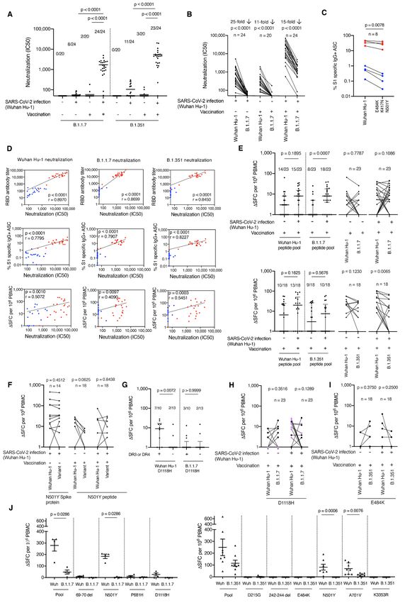
Fig. 2. Impact of vaccination and prior natural infection with SARS-CoV-2 during the first wave
            on T and B cell responses to the UK B.1.1.7 and South African B.1.315 variants. (A) Neutralizing
            antibody (nAb) titer (IC50) against B.1.1.7 and B.1.351 authentic virus in HCW with (n = 24) and
            without (n = 20) laboratory confirmed SARS-CoV-2 infection (Wuhan Hu-1). Lines at arithmetic
            mean. Data are shown pre-vaccination (16–18 weeks after infection) and 3 weeks after the first
            dose vaccination (week 42). (B) nAb (IC50) titers against Wuhan Hu-1 and B.1.1.7 authentic viruses
            plotted pairwise by individual. (C) Percentage of Wuhan Hu-1 S1 and S1 containing variant
            mutations (E484K, K417N and N501Y) specific IgG+ antibody secreting cells (ASC) in vaccinated
            HCW with (n = 4) and without (n = 4) prior SARS-CoV-2 infection. (D) Correlations between nAb
            (IC50) titers of Wuhan Hu-1, B.1.1.7 or B.1.351 authentic virus and RBD Ab titer, percentage of S1
            specific ASC and magnitude of T cell response to S1 protein in vaccinated HCW with (n = 22–24,
            red) and without (n = 18–20, blue) a history of SARS-CoV-2 infection. (E) Magnitude of T cell
            response to Wuhan Hu-1, B.1.1.7 or B.1.351 peptide pools in vaccinated HCW with (n = 23 or 18) and
            without (n = 23 or 18) SARS-CoV-2 infection (Wuhan Hu-1), plotted as grouped data (median plus
            interquartile range) and pairwise for each individual. (F) Magnitude of T cell response to Wuhan
            Hu-1 S1 protein and N501Y variant spike RBD protein in unvaccinated HCW with laboratory
            confirmed SARS-CoV-2 infection (n = 14) or to Wuhan Hu-1 and N501Y mutated peptide in

                                                                                                                                  Downloaded from http://science.sciencemag.org/ on May 29, 2021
            vaccinated HCW with (n = 18) and without (n = 18) a history of SARS-CoV-2 infection, plotted
            pairwise by individual. (G) Magnitude of T cell response to Wuhan Hu-1 or B.1.1.7 D1118H peptide in
            vaccinated HCW with a history of SARS-CoV-2 infection (n = 23), plotted by
            DRB1*0301/DRB1*0401 status. Lines at median plus interquartile range. (H) Magnitude of T cell
            response to Wuhan Hu-1 or B.1.1.7 D1118H peptide in vaccinated HCW with (n = 23) and without (n
            = 23) a history of SARS-CoV-2 infection plotted pairwise by individual and with individuals carrying
            DRB1*0301 or DRB1*0401 alleles marked in purple. (I) Magnitude of T cell response to Wuhan Hu-
            1 or B.1.351 E484K mutated peptide in vaccinated HCW with (n = 18) and without (n = 18) a history
            of SARS-CoV-2 infection, plotted pairwise by individual. (J) Magnitude of T cell response to Wuhan
            Hu-1 (Wuh), B.1.1.7 or B.1.315 peptide pools and individual peptides in Wuhan Hu-1 peptide
            immunized HLA-DRB1*04:01 transgenic mice (left-hand panel n = 4, right-hand panel n = 8. Lines
            at arithmetic mean + SEM). (A) Kruskal Wallis multiple comparison ANOVA with Dunn’s correction.
            [(B), (C), (E) – right-hand panels, (F), (H), and (I)] Wilcoxon matched-pairs signed rank test. (D)
            Spearman’s rank correlation. [(E) – left-hand panels, (G), and (J)] Mann-Whitney U test. ASC,
            antibody secreting cells; HCW, health care workers; RBD, receptor binding domain; S1, spike
            subunit 1; SFC, spot forming cells.

First release: 30 April 2021                        www.sciencemag.org     (Page numbers not final at time of first release) 10
Prior SARS-CoV-2 infection rescues B and T cell responses to variants after first vaccine dose
Catherine J. Reynolds, Corinna Pade, Joseph M. Gibbons, David K. Butler, Ashley D. Otter, Katia Menacho, Marianna Fontana,
Angelique Smit, Jane E. Sackville-West, Teresa Cutino-Moguel, Mala K. Maini, Benjamin Chain, Mahdad Noursadeghi, UK
COVIDsortium Immune Correlates Network, Tim Brooks, Amanda Semper, Charlotte Manisty, Thomas A. Treibel, James C. Moon,
UK COVIDsortium Investigators, Ana M. Valdes, Áine McKnight, Daniel M. Altmann and Rosemary Boyton
published online April 30, 2021
Downloaded from http://science.sciencemag.org/ on May 29, 2021
ARTICLE TOOLS http://science.sciencemag.org/content/early/2021/04/29/science.abh1282
SUPPLEMENTARY http://science.sciencemag.org/content/suppl/2021/04/29/science.abh1282.DC1
MATERIALS
RELATED http://stm.sciencemag.org/content/scitransmed/13/590/eabf7517.full
CONTENT
http://stm.sciencemag.org/content/scitransmed/13/578/eabd6990.full
http://stm.sciencemag.org/content/scitransmed/13/577/eabd2223.full
http://stm.sciencemag.org/content/scitransmed/13/577/eabf1555.full
REFERENCES This article cites 25 articles, 5 of which you can access for free
http://science.sciencemag.org/content/early/2021/04/29/science.abh1282#BIBL
PERMISSIONS http://www.sciencemag.org/help/reprints-and-permissions
Use of this article is subject to the Terms of Service
Science (print ISSN 0036-8075; online ISSN 1095-9203) is published by the American Association for the Advancement of
Science, 1200 New York Avenue NW, Washington, DC 20005. The title Science is a registered trademark of AAAS.
Copyright © 2021 The Authors, some rights reserved; exclusive licensee American Association for the Advancement of Science.
No claim to original U.S. Government Works. Distributed under a Creative Commons Attribution License 4.0 (CC BY).You can also read