COVID-19 comorbidity and metabolic syndrome: is there a Molecular basis? - Preprints.org
←
→
Page content transcription
If your browser does not render page correctly, please read the page content below
Preprints (www.preprints.org) | NOT PEER-REVIEWED | Posted: 21 June 2020                          doi:10.20944/preprints202006.0245.v1

                             COVID-19 comorbidity and metabolic syndrome: is there a Molecular basis?

                     Madhurima Basua#, Chinmay Sahaa,b#$, Kamalika Roy Choudhuryc , Susmita Duttaa , Sujoy Ghosha,
                           Subhankar Chowdhurya, Satinath Mukhopadhyaya*, and Nitai P. Bhattacharyyaa*

             a
                 Department of Endocrinology and Metabolism, Institute of Post Graduate Medical Education & Research and Seth
             Sukhlal Karnani Memorial Hospital, Kolkata 700020, West Bengal, India.
             b
                 Department of Genome Science, School of Interdisciplinary Studies, University of Kalyani, Kalyani, Nadia 741235,
             West Bengal, India.
             c
                 Cell Biology & Physiology Division, CSIR-IICB (TRUE campus), Kolkata-700091, India

             #
                 These authors contributed equally to this work

             $
                 Current address: Department of Genome Science, School of Interdisciplinary Studies, University of Kalyani, Kalyani,
             Nadia 741235, West Bengal, India.

             *Correspondence should be addressed either to
             Nitai P. Bhattacharyya, Ph.D, Department of Endocrinology and Metabolism, Institute of Post Graduate Medical
             Education & Research and Seth Sukhlal Karnani Memorial Hospital, Kolkata 700020, West Bengal, India, Email Id:
             nitai.bhattacharyya@gmail.com
             or
             Satinath Mukhopadhyay, DM, FRCP (London), Department of Endocrinology and Metabolism, Institute of Post
             Graduate Medical Education & Research and Seth Sukhlal Karnani Memorial Hospital, Kolkata 700020, West Bengal,
             India, email Id: satinath.mukhopadhyay@gmail.com

             Abstract

             The risk factors associated with COVID-19 related severity, morbidity, and mortality, i.e., obesity
             (often associated with NAFLD), hyperglycemia, hypertension and dyslipidemia all cluster together as
             metabolic syndrome (MetS). Instead of studying association of these risk factors with COVID-19, it
             makes sense studying the association between MetS on one hand and COVID-19 on the other. This
             study explores a molecular basis underpinning the above association. Severity of COVID-19 patients
             with MetS could be due to functional alterations of host proteins due to their interactions with viral
             proteins. We collected data from Enrichr (https://amp.pharm.mssm.edu/Enrichr/), DisGeNET
             (https://www.disgenet.org/) and others and carried out enrichment analysis using Enrichr. Various
             biological processes and pathways associated with viral protein interacting partners are known to
             involve in metabolic diseases. The molecular pathways underlying insulin resistance, insulin signaling
             and insulin secretion are not only involved in diabetes but also in CVD and obesity (associated with

                                                                              1

            © 2020 by the author(s). Distributed under a Creative Commons CC BY license.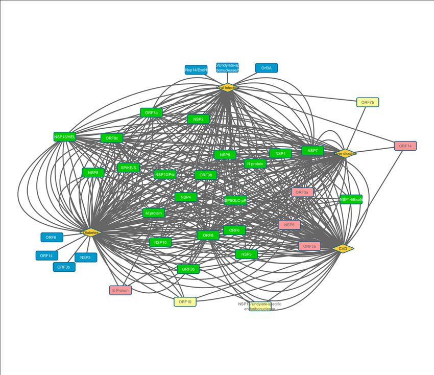
Preprints (www.preprints.org) | NOT PEER-REVIEWED | Posted: 21 June 2020       doi:10.20944/preprints202006.0245.v1

             non-alcoholic fatty liver disease; NAFLD). Lipid metabolism/lipogenesis, fatty acid oxidation and
             inflammation are associated with MetS. Viral interacting host proteins are associated and enriched
             with terms like hyperglycemia, coronary artery disease, hypertensive disease related to CVD and liver
             diseases in DisGeNET. Association of viral interacting proteins with disease-relevant biological
             processes, pathways and disease-related terms suggests that altered host protein function following
             interaction with viral proteins might contribute to frequent occurrence and/or severity of COVID-19 in
             subjects with MetS. Such analysis not only provides a molecular basis of comorbidity but also
             incriminates host proteins in viral replication, growth and identifies possible drug targets for
             intervention.

             Keywords

             COVID-19, Metabolic syndrome, Comorbidity, Enrichment analysis, biological processes and
             pathways

                                                                 2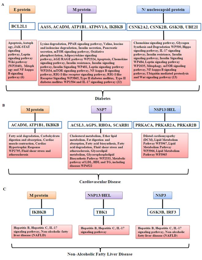
Preprints (www.preprints.org) | NOT PEER-REVIEWED | Posted: 21 June 2020         doi:10.20944/preprints202006.0245.v1

             Graphical Abstract

             Highlights
                 •   Viral protein interacting host proteins are enriched with diverse biological processes (BPs) and
                     pathways
                 •   These interacting host proteins are also enriched with disease/trait in DisGeNET
                 •   Enriched BPs and pathways are relevant for viral growth and survival
                 •   Enriched BPs, pathways and diseases are relevant for MetS
                 •   Functional changes of host proteins in infection and MetS might explain the severity of
                     COVID-19

                                                                 3
Preprints (www.preprints.org) | NOT PEER-REVIEWED | Posted: 21 June 2020          doi:10.20944/preprints202006.0245.v1

             1. Introduction
             Coronavirus disease 19 (COVID-19) is caused by the infection with severe acute respiratory syndrome
             corona virus 2 (SARS-CoV-2). Starting in the last quarter of 2019 in the Wuhan City of China, it
             spread rapidly to affect populations in almost all countries worldwide [8061550 people infected
             worldwide and causing death of 440290 (WHO Situation report 149, 17th June 2020)]. Comorbid
             conditions like hypertension, acute myocardial infarction, obesity, liver and kidney injuries,
             cardiovascular disease (CVD), type 2 diabetes (T2D), chronic obstructive pulmonary disease (COPD)
             and gastrointestinal diseases are over represented among COVID-19 patients. In severe cases, patients
             quickly progress to acute respiratory distress syndrome (ARDS), septic shock, metabolic acidosis, and
             coagulopathy. Studies have shown that older (>60 years) COVID-19 patients with hypertension,
             diabetes, COPD, CVD, liver and gastrointestinal disease and kidney disease to be susceptible to severe
             and rarely fatal outcomes [1-10]. Result presented in these publications showed that (i) age (ii) gender
             and (iii) comorbidity are risk factors for severity and/ or death.

             1.1. Metabolic syndrome and comorbidity of COVID-19
             Metabolic syndrome (MetS) consists of a cluster of inherited metabolic traits that center on obesity,
             insulin resistance, hypertension and atherogenic dyslipidemia and contribute to the prevalence of
             CVD, T2D, NAFLD and cancer (Figure 1). These traits were more prevalent among people with
             severe COVID-19 compared to their non-severe counterparts [3, 6, 11, 12]. Detailed information of
             the co-existence of obesity, CVD, hypertension, diabetes and NAFLD is shown in the supplementary
             Text (Supplementary text 1.1). It is thus pertinent to consider whether metabolic syndrome may be
             considered as co-morbid condition for COVID-19.

                                                                  4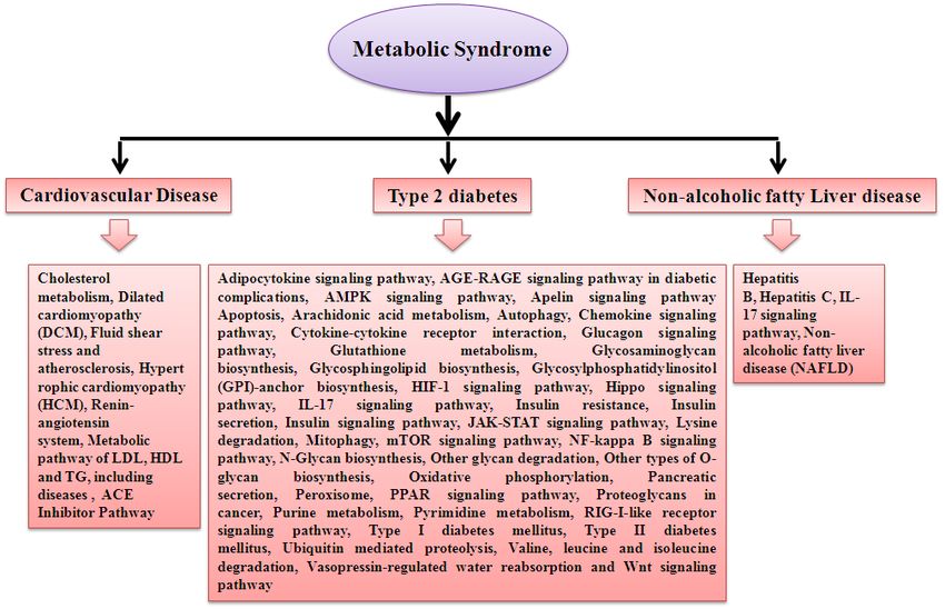
Preprints (www.preprints.org) | NOT PEER-REVIEWED | Posted: 21 June 2020         doi:10.20944/preprints202006.0245.v1

             Figure 1 Components of metabolic syndrome and long term outcome of metabolic syndrome ([13]
             [14]). HDL: high-density lipoprotein, Tg: triglyceride.

             1.2. Altered expression of host genes in virus infected cells
             Altered expression of host genes in vitro following infection with SARS-CoV-2 has been observed
             [15, 16]. Analysis of host transcriptional response to SARS-CoV-2 and other respiratory infections
             through in vitro, ex vivo and in vivo model systems reveal that infection deregulates many genes that
             serve as unique transcriptional footprints for the virus. Comparison with influenza A and respiratory
             syncytial viruses reveals an altered response that lacks robust induction of cytokine and chemokine.
             Transcriptional profiling in lung biopsy specimens from COVID-19 subjects reveal a unique and
             inappropriate inflammatory response defined by elevated chemokine expression [16]. Altered
             expressions of host genes on infection with many viruses including SARS-CoV are catalogued in
             Enrichr at https://amp.pharm.mssm.edu/Enrichr/ [17, 18].

             1.3.   Interactions between host proteins and SARS-CoV-2 coded viral proteins
             Viral proteins, including SARS-CoV and other human corona virus coded proteins interact with host
             proteins [1, 19-21]. Such interactions hijack host proteins and modify cellular processes and pathways
             to evade immune response of the host and survival of viruses. Interaction of viral protein with the host
             proteins might modulate the functions of the host proteins by (i) changing cellular localization,
             transport and interaction with other host proteins, (ii) changing activation of the host proteins by post
             translational modifications and (iii) degradation of the host proteins. SARS-CoV coded ORF6 interacts
             with STAT1 and prevents entry of STAT1 into the nucleus [22]. M protein of SARS-CoV interacts
             with TRF3 and inhibits TBK1/IKKepsilon complex assembly [23]. NSP1 interacts with 40S ribosomal
             subunit and inactivates the translational activity of the 40S subunits. NSP1-40S ribosome complex can
             modify the 5' region of capped mRNA template inhibiting the translation of the modified mRNA [24]
             as well as degradation of mRNA [25]. NSP1 inhibits phosphorylation of STAT1 and represses target
             genes like ISG15, ISG54, and ISG56 of STAT1 [26]. Inhibition of phosphorylation of IRF3 by
             ORF3b, ORF6 and N proteins of SARS-CoV and alters the activation of IRF3 [27]. Interaction and
             inhibition of phosphorylation and nuclear translocation of IRF3 by viral Papain-like protease (PLpro)
             have been observed [28]. Host protein IRF3 by PLproinhibits de-ubiquitination and changes the
             stability of IRF3 [29]. These interactions of different viral proteins with IRF3 and its downstream

                                                                 5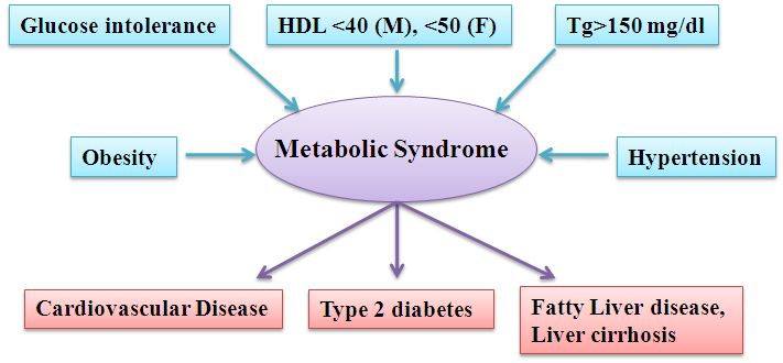
Preprints (www.preprints.org) | NOT PEER-REVIEWED | Posted: 21 June 2020       doi:10.20944/preprints202006.0245.v1

             STAT1 evade the host immune response. Host-viral protein interactions for SARS-CoV and MERS-
             CoV are reviewed extensively [19, 30].
             Interactions between SARS-CoV-2 proteins and the host proteins are not studied extensively. Very
             recently interactions of proteins coded by SARS-CoV-2 have been identified. Based on the functions
             of host interacting partners, it has been proposed that viral proteins coded by SARS-CoV-2 are
             associated with various functions like DNA replication (NSP1), epigenetic regulation and regulation of
             gene expression (NSP5, NSP8, NSP13 and E), vesicle trafficking (NSP2, NSP6, NSP7, NSP10,
             NSP13, NSP15, ORF3a, E, M, ORF8), lipid modification (S), RNA processing and regulation (NSP8,
             N), ubiquitin ligases (ORF10), signaling (NSP8, NSP13, N, ORF9b), nuclear transport machinery
             (NSP9, NSP15, ORF6), cytoskeleton function (NSP1, NSP13), mitochondrial functions (NSP4, NSP8,
             ORF9c) and modification of extracellular matrix (NSP9). In addition, host partners were also
             associated with various innate immune function, protein synthesis and proteasomal degradation [31] .

             1.4. Knowledge gap
             COVID-19 patients with MetS appear to have severe disease with higher mortality. When infected
             with SARS-CoV-2, people with MetS have unmasking and/or aggravation of some of its features.
             Alternatively, pre-existing metabolic conditions might allow infected SARS-CoV-2 to grow
             aggressively or prevent clearance of the virus due to immune modulation, leading to more severe
             disease manifestations. Increased viral load and slow clearance of the virus have been reported in
             severe COVID-19 patients [32, 33]. Severity being associated with MetS, there might be correlation
             between viral load and slow clearance. SARS-CoV-2 infection triggers stress conditions leading to
             release of glucocorticoids and catecholamines that increase blood glucose levels [34]. COVID-19
             patients with uncontrolled T2D had higher mortality than those with good glycemic control [35]. Two
             receptors ACE2 and DPP4, necessary for entry of SARS-CoV-2 and MERS-CoV respectively are
             involved in inflammatory pathways, cardiovascular physiology and glucose homeostasis [36] showing
             overlap of viral entry, growth and MetS. However, molecular interconnection between COVID-19
             severity and MetS remains largely unknown.

             1.5.   Hypothesis
             Severity of COVID-19 patients with MetS could be due to functional alterations of host proteins by
             interacting with viral proteins.

                                                                 6
Preprints (www.preprints.org) | NOT PEER-REVIEWED | Posted: 21 June 2020 doi:10.20944/preprints202006.0245.v1
2. Material and Methods
2.1. SARS viral protein interacting partners of host proteins
From published data, we collected 609 host proteins that interact with corona virus proteins; specific
viral proteins are known for 410 host proteins [19-21, 31, 37]. Detailed sources are shown in the
Supplementary Text Table ST2 and Supplementary Table S1.
2.2. Enrichment analysis
To find the functional associations of viral interacting proteins, enrichment analysis for the genes was
carried out using online facility at Enrichr (https://amp.pharm.mssm.edu/Enrichr/) [17, 18]. Enrichr is
an integrative web-based software application for analysis of a gene-set comparing with various gene-
set libraries. Given an input list of genes, it provides enrichment for different libraries like KEGG
pathways, Gene Ontology (GO) terms for biological processes (BPs). DisGeNET, a discovery
platform containing publicly available collections of genes and variants associated to human diseases
and is also available at the same site.
2.3. Altered expression of host genes in infected cultured cells
Altered expression of host genes on infection with virus in different conditions are downloaded from
Gene Expression Omnibus (GEO) database, a public functional genomics data repository
(https://www.ncbi.nlm.nih.gov/geo/) and catalogued in Enrichr at
https://amp.pharm.mssm.edu/Enrichr/ [17, 18]. We collected altered expression of genes for SARS-
CoV from this database, the details of which are shown in the Supplementary Table S6
2.4. Functions of the genes whenever necessary were taken from
https://www.ncbi.nlm.nih.gov/gene/ and provided by RefSeq.
2.5. Genes/proteins elevated in organs/tissues
Datasets for elevated genes and genes expressed in all tissues/organ were collected for 8 organs
namely brain, heart, kidney, liver, lung, pancreas, spleen, thyroid and adipose tissue as reported by
[38]. Datasets for tissue/organ-specific proteins also collected from data reported by Wilhelm, et al.
[39]. Combing these two datasets, unique proteins/genes were determined and designated as elevated
7Preprints (www.preprints.org) | NOT PEER-REVIEWED | Posted: 21 June 2020 doi:10.20944/preprints202006.0245.v1
genes in organ/tissue as well as the housekeeping genes. Expression of genes elevated in different
tissues and housekeeping genes are shown in the Supplementary Table S7.
For comparison of different set of genes/proteins, we used online facility at
http://bioinformatics.psb.ugent.be/webtools/Venn/
3. Results
3.1. Enriched Biological processes (BPs) with host proteins interacting with SARS virus
coded proteins.
To understand the functional implications of host protein- viral protein interactions, we collected 609
host proteins that interact with proteins coded by SARS-CoV-2 and other corona viruses. Enrichment
analysis revealed that 295 BPs enriched significantly (adjusted p≤0.05). The top 10 most significant
enriched BPs are shown in Figure 2 and the detail is shown in the Supplementary Table S2.
Figure 2: Top 10 significantly enriched biological processes. Golgi vesicle transport (GO:0048193),
viral life cycle (GO:0019058), intracellular protein transport (GO:0006886), viral process
8Preprints (www.preprints.org) | NOT PEER-REVIEWED | Posted: 21 June 2020 doi:10.20944/preprints202006.0245.v1
(GO:0016032), ER to Golgi vesicle-mediated transport (GO:0006888), Endoplasmic Reticulum-
Associated Degradation (ERAD) pathway (GO:0036503), endosomal transport (GO:0016197),
cellular protein localization (GO:0034613), ubiquitin-dependent ERAD pathway (GO:0030433) and
transport of virus (GO:0046794) are enriched with 42, 27, 45, 34, 28, 19, 31, 37, 16 and 15 viral
protein interacting host protein respectively.
3.2. Enrichment of KEGG and Wiki Pathways
Enrichment analysis with 609 interacting proteins revealed that all together 250 KEGG pathways were
associated with viral protein interacting host proteins (Supplementary Table S3A); 17 pathways were
significantly (adjusted p≤0.05) enriched with viral protein interacting host proteins (Figure 3). In
addition to KEGG pathways, Enrichr also provides enrichment for Wiki Pathways
(https://www.wikipathways.org/index.php/WikiPathways). Twenty Wiki pathways like TGF-beta
Signaling Pathway (WP366), IL-3 Signaling Pathway (WP286), Hepatitis C and Hepatocellular
Carcinoma (WP3646), Viral Acute Myocarditis (WP4298), Sterol Regulatory Element-Binding
Proteins (SREBP) signaling (WP1982) and IL-5 Signaling Pathway (WP127) were enriched
significantly (adjusted p≤0.05). Result is shown in the Supplementary Table S3B.
9Preprints (www.preprints.org) | NOT PEER-REVIEWED | Posted: 21 June 2020 doi:10.20944/preprints202006.0245.v1
Figure 3: Pathways enriched with SARS-virus interacting proteins.
3.3. Association of interacting host partners of viral proteins with various metabolic
disease/trait catalogued in DisGeNET database
To identify whether interacting partners of viral proteins are associated with different diseases or
disease related conditions/trait, we explored DisGeNET database. This database catalogs gene-disease
relations from diverse experimental data like Genome Wide association studies (GWAS) and others
[40] as catalogued in Enrichr at https://amp.pharm.mssm.edu/Enrichr/ [17, 18]. Such analysis with
viral interacting protein partners revealed that 55 diseases/conditions were significantly (adjusted
p≤0.05) associated with viral protein interacting partners; 5308 diseases/conditions were associated
with host proteins (Supplementary Table S5A-5C, Figure 4). Many viral interacting host proteins
were associated with infection, viruses, metabolic diseases and other diseases including cancers.
Figure 4: Proportion of genes coding for host protein that interact with proteins coded by SARS-CoV-
2 and in different categories of diseases/conditions in DisGeNET. For example, total 787 genes are
associated in cardiovascular disease. Among 609 protein coded by host genes, 39 proteins are
associated with Cardiovascular disease, giving the frequency as 0.05. Panel A over representation is
10Preprints (www.preprints.org) | NOT PEER-REVIEWED | Posted: 21 June 2020 doi:10.20944/preprints202006.0245.v1
significantly higher (adjusted p value ≤0.05), while the values for the bar diagram in panel B, level of
significance is ≤0.05 without correction for multiple testing.
3.4. Relevant biological processes, pathways and disease terms associated with MetS and
enriched with viral protein interacting host proteins
Various biological processes, pathways and terms in DisGeNET enriched with viral protein interacting
host proteins (Supplementary Tables, S2, S3 and S5A) are relevant and known for their involvement
in different metabolic diseases. Insulin resistance, insulin signaling pathway, insulin secretion are not
only involved in diabetes [41-44] but also in cardiovascular diseases [45] and obesity [46]. The
metabolic products of angiotensin I and II play an important role in cardiovascular pathophysiology
[47]. Deregulation of lipid metabolism/lipogenesis, fatty acid oxidation (FAO), triglyceride levels,
inflammation (adiponectin, TNFα IL6), transcription regulation of lipogenic genes by PPARG in the
activation of lipogenic FoxO1 signaling pathway changing glycogenolysis and gluconeogenesis
contribute to the development of NAFLD [48] [Figure 5].
11Preprints (www.preprints.org) | NOT PEER-REVIEWED | Posted: 21 June 2020 doi:10.20944/preprints202006.0245.v1
Figure 5 Pathways associated with CVD, diabetes and NAFLD. It is to be noted that even though
pathways are shown separately, connected with the diseases, same gene may be associated with
different biological processes and pathways.
Biological pathways associated with host genes that interact with different viral proteins were relevant
for CVD, diabetes and NAFLD. For example, host protein IKBKB, SLC27A2 and GSK3B interact
with coronavirus protein M, NSP2 and (NSP3, N/nucleocapsid protein) respectively and associated
with insulin resistance pathway. Similarly, host protein ATP1B1 interacts with M protein and
PRKACA interacts with Nsp13/HEL; both the proteins are associated with insulin secretion. GSK3B
interacts with viral NSP3 and N/nucleocapsid protein is associated with insulin signaling pathway.
Representative result of interactions of viral proteins with host proteins associated with disease
relevant pathways is shown in the Figure 6A-6C and Supplementary Text Table ST3. Detailed result
is shown in the Supplementary Tables S4A-S4D.
12Preprints (www.preprints.org) | NOT PEER-REVIEWED | Posted: 21 June 2020 doi:10.20944/preprints202006.0245.v1
Figure 6 Representative result of viral proteins, their interacting host proteins; host protein associated
pathways and involvement in different diseases
13Preprints (www.preprints.org) | NOT PEER-REVIEWED | Posted: 21 June 2020 doi:10.20944/preprints202006.0245.v1
In summary, 27 protein coded by SARS-CoV and SARS-CoV-2 interact with 168 host protein
associated with 101 biological processes and pathways related to MetS.
Various host proteins were associated with the diseases or terms linked with diseases in DisGeNET
database. Supplementary Figure STF2 shows the representative result of relations of the metabolic
diseases, the proteins associated and their interacting partners of viral proteins. Detail result is shown
in the supplementary Table S5C.
3.5. Cytoscape representation of the association of host proteins and viral proteins with
diseases in different categories
A protein categorized in a biological process in Gene Ontology database or in a KEGG pathway that is
relevant and known to be involved in a disease is considered to be associated with disease. Similarly,
associations of gene/protein with diseases, conditions/traits are catalogued in DisGeNET. Based on
our analysis, we categorized biological processes, pathways and disease terms into (i) CVD, (ii)
diabetes, (iii) liver disease and (iv) viral infection. To identify the overlap among the various MetS
comorbidities mentioned above with host proteins, the free software Cytoscape (version 3.2.1) was
used. Their relationships are shown in the Supplementary Tables S8A and S8B and represented
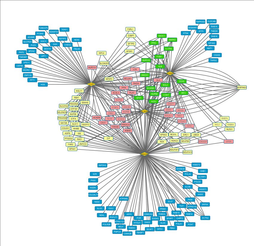
using cytoscape in Figures 7 and 8. Result in Figure 7 shows that 16 proteins shown in Green nodes
(viz. ACE2, ALB, BCL2, COMT, COX2, IRF3, SMOC1, PPIG, MARK2, IL17RA, NUP210, ITGB1,
HMOX1, SCARB1, NUP88 and GPX1) are common for all 4 diseases. In addition, 29 proteins
(represented in Pink) are in common for 3 disease nodes, 44 proteins (light yellow squares) overlap 2
disease nodes and the rest (Blue squares) are unique for a given disease.
14Preprints (www.preprints.org) | NOT PEER-REVIEWED | Posted: 21 June 2020 doi:10.20944/preprints202006.0245.v1
Figure 7: Cytoscape representation of involvement of host protein (rectangle) with CVD, diabetes,
Liver disease and viral infection (diamond shape). Green colored host proteins are interacting partners
of viral proteins are common for all four diseases, pink colored rectangles represents proteins which
are connected with 3 disease, yellow colored rectangles and blue colored rectangles represent viral
protein linked to 2 and one disease respectively.
By virtue of interactions of the viral proteins with host proteins, viral proteins are also associated with
different diseases. Twenty viral proteins(N protein, M protein, SPIKE/S, NSP1, NSP2, NSP3, NSP4,
NSP5, NSP7, NSP8, NSP9, NSP10, NSP12, NSP13, NSP14, ORF3b, ORF6, ORF8, ORF7a, ORF9b,
15Preprints (www.preprints.org) | NOT PEER-REVIEWED | Posted: 21 June 2020 doi:10.20944/preprints202006.0245.v1
OR 9c) were observed to overlap with all four disease modules viz. viral infection, CVD, Diabetes and
Liver disease (Figure 8). The viral proteins common for 3 diseases (viz. E proteins, ORF3a, ORF14,
and NSP6) are shown in Pink. Light yellow squares represent the viral proteins associated with 2
diseases (NSP15, ORF7b and ORF10). The rest of the viral proteins (Blue squares) are unique for a
particular disease. This analysis has strengthened our hypothesis that the frequently occurring
comorbidity of MetS could be due to interactions of viral protein with the host proteins.
Figure 8: Cytoscape representation of involvement of viral proteins (rectangles) with CVD, diabetes,
Liver disease and infectious disease (diamond shape). Green colored viral protein coded by corona
virus including MERS-CoV, SARS-CoV and SARS-CoV-2 are common for all four diseases, pink
color rectangles represents proteins which are connected with 3 disease, yellow and blue colored
rectangles represent viral protein linked to 2 and one disease respectively
3.6. Organ/tissue enriched expression of host protein interacting partners of SARS-CoV
coded protein
16Preprints (www.preprints.org) | NOT PEER-REVIEWED | Posted: 21 June 2020 doi:10.20944/preprints202006.0245.v1
To identify the enriched genes, defined as genes whose mRNA level, determined by RNA sequencing,
in a particular tissue is at least five times higher than those in all other tissues, in different organ/tissue
like brain, heart, kidney, liver lung, pancreas, spleen and thyroid together with adipose tissue, we used
result reported by Uhlén et al. [38]. Gene whose mRNA level is detected in all tissue with fragments
per kilobase of exon model per million reads mapped greater than1 is considered to be housekeeping
genes [38]. We used these organ/tissue enriched and compared with viral protein interacting partners,
to identify whether viral protein interacting partner of the host gene enhanced in any particular
tissue/organ. The result is shown in the Supplementary Text Table ST4. Majority of genes coded for
viral interacting proteins are housekeeping gene and expressed in all tissues. Only a small portion of
the genes are enriched with different tissues like heart, lung, pancreas, adipose tissue, kidney, and
liver. It requires further investigations whether this small number of organ/tissue enriched proteins
contributes to the organ/tissue specific pathogenic effects of COVID-19.
3.7. Altered expression of host genes after infection with SARS-CoV
Combined proteome and transcriptome datasets from infection of SARS-CoV in human lung epithelial
cells in culture are utilized to predict regulatory genes involved in the host response. After infection
expression of genes were determined at different time points [49]. Increased and decreased
expressions of the genes are taken from Enrchr. Summary of the result is shown in the Table and the
detail is shown in the supplementary Text Table T2. Overall about 5% of the increased expression and
similar proportion of decreased expression of host genes in SARS-CoV infected cells also interact
with viral proteins. Out of 609 viral protein interacting host proteins, expression of about 28% genes
coded for the viral protein interacting partners increased and expression of 36% genes decreased;
overall expression of 64% of genes altered (Table 3). Mechanism of deregulation of host genes by
viral infection remains largely unknown. Deregulation and physical interaction of deregulated host
genes, support the general notion that deregulated genes products interact with each other and carry
out biological functions.
17Preprints (www.preprints.org) | NOT PEER-REVIEWED | Posted: 21 June 2020 doi:10.20944/preprints202006.0245.v1
Table 3: Deregulated genes after infection with SARS-CoV and interactions of viral proteins
with the host proteins
Time after Total number of genes deregulated No. Viral protein interacting protein
infection Increased Decreased Increased (%) Decreased (%)
0h 581 694 28 (5.8) 41 (5.9)
12h 637 609 27 (4.2) 41(6.7)
24h 763 898 32 (4.2) 75 (8.3)
36h 197 218 18 (9.1) 15 (6.9)
48h 740 822 28 (3.8) 39 (4.7)
60h 616 662 25 (4.1) 37 (5.6)
72h 579 581 31 (5.4) 43 (7.4)
84h 587 585 25 (4.2) 30 (5.1)
96h 622 587 38 (6.1) 34 (5.8)
Total 3465 3598 170 (4.9) 220 (6.1)
4. Discussion
To find out the molecular basis of severity of COVI-19 with MetS, we collected data from various
sources for host protein interacting partners of proteins coded by SARS-CoV, SARS-CoV-2 and other
corona viruses and analyzed using various online tools. Such analysis revealed (i) possible role of host
proteins in life cycle of the virus and (ii) viral protein interacting host proteins are enriched with
various biological processes and pathways relevant for CVD, diabetes and NAFLD. Besides many
host genes coding for viral interacting partners are associated with MetS or related traits in
DisGeNET. This result shows that COVID-19 and MetS might be associated through the proteins
involved in MetS and are also targets of viral proteins.
4.1 Mechanism (s) of severity of COVID-19 in patients with MetS
Severity and mortality of COVID-19 is associated with MetS. Increased viral load and decreased
clearance of viruses are major contributors to COVID-19 related morbidity and mortality [32, 33].
Viral load for COVID-19 patient comorbid with MetS is not known. Possible increased viral load and
slow clearance in COVID-19 patients with MetS, if observed, might explain the higher death rate and
poor outcomes in them. Infection with SARS-CoV-2 in subjects with MetS, a state of chronic low
18Preprints (www.preprints.org) | NOT PEER-REVIEWED | Posted: 21 June 2020 doi:10.20944/preprints202006.0245.v1
grade inflammation, may trigger the release of catecholamines and glucocorticoids that further
increase blood glucose levels and increase glucose variability [34]. Hyperglycemia and insulin
resistance are known to promote enhanced synthesis of glycation end products (AGEs), pro-
inflammatory cytokines and adhesion molecules that may interact with viral proteins and worsen
outcome in COVID-19 [50, 51]. Thus, infection enhances the metabolic conditions which in turn
increase pathogenicity of the virus by modulating AGEs and pro-inflammatory cytokines.
In MetS, immune system is compromised due to defects in inflammation. This condition might delay
in clearing viruses and recovery. MERS-CoV infection to diabetic mice results in immune
deregulation and enhances disease severity of the infection [52]. Virus RNA was detected in lungs of
100% of the COVID-19 patients, while in about 40% of the patients viral RNA was detected in heart,
liver and kidney, possibly reached through blood [53]. Increased levels of troponin levels, a sensitive
marker for acute cardiac injury, is commonly observed in severe cases of COVID-19 and is strongly
associated with mortality [54]. Acute coronary syndrome and myocardial infarction were noted to
occur after SARS [55]. So far no convincing reports are available to show that SARS-CoV-2 infects
pancreatic cells [56] or causes acute pancreatitis by the infection [57]. However, effects of direct
infection of SARS-CoV in pancreas inducing diabetes in non-diabetic persons are known [58].
4.2 Common genes in COVID-19 and MetS
Sixteen genes are related to infectious diseases, CVD, diabetes and liver diseases (Figure 7).
Relevance of ACE2, ALB, ITGB1, IRF3, MARK2, IL17RA, HMOX1, SCARB1, GPX1 and BCL2
genes for infections and MetS are known. ACE2 has been implicated in obesity, hypertension,
diabetes, myocardial infarction, heart failure and inflammatory lung disease [59-61]. ACE2 has been
proposed to be the receptor for entry into the host cells by interacting with the spike protein of SARS-
CoV-2 [62] and SARS-CoV, possibly through renin-angiotensin system as shown for SARS-CoV
[63]. Serum albumin levels are decreased in infection, liver dysfunction, obesity, diabetes and CVD
due to its ability to act as antioxidant [64, 65], modify inflammation, cytokine production [66] and
maintain capillary membrane stability and fluid balance across the capillary [67]. ITGB1, a member of
integrin beta chain family, promotes entry of different viruses [68], although its role in SARS-CoV-2
remains unknown. ITGB activation enhances secretion of insulin and inhibited by excess glucose [69].
Interferon regulatory factor 3 (IRF3) modulates innate immune response to viral and bacterial
infections [70], inflammation [71], glucose and lipid homeostasis [72, 73] by interacting with viral
proteins and regulating interferon genes. IL17RA by altering downstream cytokines and chemokine
19Preprints (www.preprints.org) | NOT PEER-REVIEWED | Posted: 21 June 2020 doi:10.20944/preprints202006.0245.v1
involves in infectious disease and reviewed [74], diabetes [75] and heart failure [76]. Evidences that
the IL-17 signaling pathway contributes to the pathogenesis of liver diseases by inhibiting fatty acid β-
oxidation and altering cell death [77], ERK1/2/p65 signaling pathway [78], obesity, inflammation and
reviewed [79] are available. Modified IL-17 signaling pathway has been observed in chronic viral
hepatitis [80]. SCARB1, a receptor for high density lipoproteins, facilitates uptake of cholesteryl esters
from HDL to the liver. It has been implicated in entry of HCV [81]. Level of serum HDL-Cholesterol
is risk factor for CVD [82, 83], hypertension, hyperlipidemia, insulin resistance and obesity [84].
Microtubule Affinity Regulating Kinase 2 (MARK2) involves in motility and uncoatng HIV-1 by
locally phosphorylating the viral cores [85]. It is one of the kinases that phosphorylates AMPK and
involves in epithelial mesenchymal transition [86], metabolic rate, adiposity, and insulin
hypersensitivity [87]. HMOX1 possess cytoprotective, anti-inflammatory and antioxidant properties
and modulates HBV infection [88], fatty liver, NASH, NAFL, insulin resistance, hypertension,
vascular dysfunction, diabetes, atheroscerosis and others [89]. Glutathione peroxidase 1 (GPX-1) is an
enzyme having antioxidant properties and involves in coxsackie virus induced myocarditis [90],
influenza A virus-induced lung inflammation [91], HCV induced liver fibrosis and heptocellular
carcinoma [92] and HBB replication [93]. Increased oxidative stress in MetS and a decreased anti-
oxidative defense were correlated with triglycerides, high-density lipoprotein cholesterol, waist
circumference, blood pressure components of MetS [94]. There are reports to show that mortality due
to CVD is associated with low serum level selenium. Low selenium levels also correlate with risk of
myocardial infarction. Contradictory result of no effect or increased risk of ischemic heart disease
((reviewed in Bellinger, et al. [95]) has also been reported. Even though various proteins identified
here have common role in viral infections and MetS, whether these are relevant for SARS-CoV-2
infection remains unknown and further studies are necessary. Taking together, our analysis provides
molecular clues for over representation of MetS in COVID-19 patients, especially for severe disease
and/or or non-survivors.
Altered gene expression from peripheral blood in MetS shows a general systemic inflammation
condition mediated by the innate immune system. NF-κB activation, activation of T cells,
macrophages and increased expression of CXCL14, IL4R, GPX1 and many others are observed in
MetS, diabetes and coronary heart disease [96]. In pre-existing MetS condition, when infection with
SARS-CoV-2 occurs, viral coded proteins interact with host proteins that have already been
compromised or activated. Such interactions might result in functional interactions synergistically
20Preprints (www.preprints.org) | NOT PEER-REVIEWED | Posted: 21 June 2020 doi:10.20944/preprints202006.0245.v1
between the infection and pre-existing conditions. This condition might delay in clearing viruses and
recovery as observed for MERS-CoV infection to diabetic mice [52]. It remains unknown whether
infection with SARS-CoV-2 in individual without any comorbidity induces acute injury to heart, liver
or pancreas either due to direct infection in these organ or by systemic changes in inflammation and
immune modulation. Acute cardiac injury is strongly associated with mortality and severity [54].
SARS-CoV coded structural protein 7a can cause direct liver injury by inducing apoptosis in vitro
studies [97]. Liver injury has also been observed in COVID-19 patients [98, 99].
5. Conclusion
SARS-CoV-2 coded protein interacting host proteins are associated with CVD, diabetes, or NAFLD
relevant biological processes, pathways and diseases/traits. Interaction of the viral proteins alters the
functions of their host partners for the survival of viruses. In absence of infection, functional changes
in these proteins, possibly through expression, result in MetS. SARS-CoV-2 infection in patients with
metabolic diseases like CVD, diabetes or NAFLD, might synergistically modify systemic functions of
these common genes resulting in severity and mortality of COVID-19 patients. Direct infection of
SARS-CoV-2 in heart and liver might also result in localized as well as systemic functional changes of
the proteins giving rise severity of the disease. Induction of acute injury to liver, heart or pancreas
either due to direct infection of the virus or systemic damage by SARS-CoV-2 infection of normal
individuals and their long term effects, if any, remain unknown. Such analysis not only provided
molecular basis of comorbidity but also knowledge about the role of host proteins in viral replication
and growth and possible drug targets for intervention. Further studies are necessary to correlate with
altered gene expression in diabetes, CVD, hypertension, obese and other metabolic conditions with
altered gene expression in viral infected tissues. It also remains unknown whether people with MetS
are susceptible to SARS-CoV-2 infection or infection with SARS-CoV-2 made people prone to MetS.
Funding
This research did not receive any specific grant from funding agencies in the public, commercial, or
not-for-profit sectors.
Disclosure of potential conflicts of interest
The authors declare no conflict of interest.
21Preprints (www.preprints.org) | NOT PEER-REVIEWED | Posted: 21 June 2020 doi:10.20944/preprints202006.0245.v1
References
[1] F. Zhou, T. Yu, R. Du, et al., Clinical course and risk factors for mortality of adult inpatients with COVID-19 in
Wuhan, China: a retrospective cohort study, The lancet, 395 (2020) 1054–1062, doi: https://doi.org/10.1016/S0140-
6736(20)30566-3.
[2] M. Yuan, W. Yin, Z. Tao, et al., Association of radiologic findings with mortality of patients infected with 2019 novel
coronavirus in Wuhan, China, Plos one, 15 (2020) e0230548, doi: https://doi.org/10.1371/journal.pone.0230548.
[3] J. Yang, Y. Zheng, X. Gou, et al., Prevalence of comorbidities and its effects in patients infected with SARS-CoV-2: a
systematic review and meta-analysis, International journal of infectious diseases, 94 (2020) 91-95, doi:
10.1016/j.ijid.2020.03.017.
[4] T. Chen, Z. Dai, P. Mo, et al., Clinical characteristics and outcomes of older patients with coronavirus disease 2019
(COVID-19) in Wuhan, China (2019): a single-centered, retrospective study, The journals of gerontology. Series A,
Biological sciences and medical sciences (2020), doi: 10.1093/gerona/glaa089.
[5] C. Huang, Y. Wang, X. Li, et al., Clinical features of patients infected with 2019 novel coronavirus in Wuhan, China,
The lancet, 395 (2020) 497-506, doi: https://doi.org/10.1016/S0140-6736(20)30183-5.
[6] S. Garg, L. Kim, M. Whitaker, et al., Hospitalization Rates and Characteristics of Patients Hospitalized with
Laboratory-Confirmed Coronavirus Disease 2019 - COVID-NET, 14 States, March 1-30, 2020, MMWR Morb Mortal
Wkly Rep, 69 (2020) 458-464, doi: 10.15585/mmwr.mm6915e3.
[7] G. Onder, G. Rezza, S. Brusaferro, Case-Fatality Rate and Characteristics of Patients Dying in Relation to COVID-19
in Italy, Jama, 323 (2020) 1775-1776, doi: 10.1001/jama.2020.4683.
[8] D. Wang, B. Hu, C. Hu, et al., Clinical Characteristics of 138 Hospitalized Patients With 2019 Novel Coronavirus–
Infected Pneumonia in Wuhan, China, JAMA, 323 (2020) 1061-1069, doi: 10.1001/jama.2020.1585.
[9] R.E. Jordan, P. Adab, K.K. Cheng, Covid-19: risk factors for severe disease and death, BMJ, 368 (2020) m1198, doi:
10.1136/bmj.m1198.
[10] C.C.f.D.C. Epidemiology Working Group for Ncip Epidemic Response, Prevention, [The epidemiological
characteristics of an outbreak of 2019 novel coronavirus diseases (COVID-19) in China], Zhonghua liu xing bing xue za
zhi = Zhonghua liuxingbingxue zazhi, 41 (2020) 145-151, doi: 10.3760/cma.j.issn.0254-6450.2020.02.003.
[11] J. Lighter, M. Phillips, S. Hochman, et al., Obesity in Patients Younger Than 60 Years Is a Risk Factor for COVID-19
Hospital Admission, Clinical Infectious Diseases, (2020), doi: 10.1093/cid/ciaa415.
[12] Y. Dong, H. Chen, J. Gao, et al., Molecular machinery and interplay of apoptosis and autophagy in coronary heart
disease, Journal of molecular and cellular cardiology, 136 (2019) 27-41, doi: 10.1016/j.yjmcc.2019.09.001.
[13] S. O'Neill, L. O'Driscoll, Metabolic syndrome: a closer look at the growing epidemic and its associated pathologies,
Obesity reviews : an official journal of the International Association for the Study of Obesity, 16 (2015) 1-12, doi:
10.1111/obr.12229.
[14] C.A. Aguilar-Salinas, T. Viveros-Ruiz, Recent advances in managing/understanding the metabolic syndrome,
F1000Research, 8 (2019), doi: 10.12688/f1000research.17122.1.
[15] S.A. Heialy, M. Hachim, A. Senok, et al., Regulation of angiotensin converting enzyme 2 (ACE2) in obesity:
implications for COVID-19, bioRxiv, (2020) 2020.2004.2017.046938, doi: 10.1101/2020.04.17.046938.
22Preprints (www.preprints.org) | NOT PEER-REVIEWED | Posted: 21 June 2020 doi:10.20944/preprints202006.0245.v1
[16] D. Blanco-Melo, B.E. Nilsson-Payant, W.-C. Liu, et al., Imbalanced Host Response to SARS-CoV-2 Drives
Development of COVID-19, Cell, 181 (2020) 1036-1045.e1039, doi: https://doi.org/10.1016/j.cell.2020.04.026.
[17] E.Y. Chen, C.M. Tan, Y. Kou, et al., Enrichr: interactive and collaborative HTML5 gene list enrichment analysis tool,
BMC Bioinformatics, 14 (2013) 128, doi: 10.1186/1471-2105-14-128.
[18] M.V. Kuleshov, M.R. Jones, A.D. Rouillard, et al., Enrichr: a comprehensive gene set enrichment analysis web server
2016 update, Nucleic Acids Res, 44 (2016) W90-97, doi: 10.1093/nar/gkw377.
[19] T.S. Fung, D.X. Liu, Human Coronavirus: Host-Pathogen Interaction, Annu Rev Microbiol, 73 (2019) 529-557, doi:
10.1146/annurev-micro-020518-115759.
[20] P. V'kovski, M. Gerber, J. Kelly, et al., Determination of host proteins composing the microenvironment of
coronavirus replicase complexes by proximity-labeling, Elife, 8 (2019) e42037, doi: 10.7554/eLife.42037.
[21] S. Pfefferle, J. Schöpf, M. Kögl, et al., The SARS-coronavirus-host interactome: identification of cyclophilins as
target for pan-coronavirus inhibitors, PLoS pathogens, 7 (2011), doi: 10.1371/journal.ppat.1002331.
[22] M. Frieman, B. Yount, M. Heise, et al., Severe acute respiratory syndrome coronavirus ORF6 antagonizes STAT1
function by sequestering nuclear import factors on the rough endoplasmic reticulum/Golgi membrane, Journal of virology,
81 (2007) 9812-9824, doi: 10.1128/jvi.01012-07.
[23] K.L. Siu, K.H. Kok, M.H. Ng, et al., Severe acute respiratory syndrome coronavirus M protein inhibits type I
interferon production by impeding the formation of TRAF3.TANK.TBK1/IKKepsilon complex, The Journal of biological
chemistry, 284 (2009) 16202-16209, doi: 10.1074/jbc.M109.008227.
[24] W. Kamitani, C. Huang, K. Narayanan, et al., A two-pronged strategy to suppress host protein synthesis by SARS
coronavirus Nsp1 protein, Nature structural & molecular biology, 16 (2009) 1134-1140, doi: 10.1038/nsmb.1680.
[25] W. Kamitani, K. Narayanan, C. Huang, et al., Severe acute respiratory syndrome coronavirus nsp1 protein suppresses
host gene expression by promoting host mRNA degradation, Proceedings of the National Academy of Sciences, 103 (2006)
12885-12890, doi: 10.1073/pnas.0603144103.
[26] M.G. Wathelet, M. Orr, M.B. Frieman, et al., Severe acute respiratory syndrome coronavirus evades antiviral
signaling: role of nsp1 and rational design of an attenuated strain, Journal of virology, 81 (2007) 11620-11633, doi:
10.1128/jvi.00702-07.
[27] S.A. Kopecky-Bromberg, L. Martínez-Sobrido, M. Frieman, et al., Severe acute respiratory syndrome coronavirus
open reading frame (ORF) 3b, ORF 6, and nucleocapsid proteins function as interferon antagonists, Journal of virology 81
(2007) 548-557, doi: 10.1128/jvi.01782-06.
[28] S.G. Devaraj, N. Wang, Z. Chen, et al., Regulation of IRF-3-dependent innate immunity by the papain-like protease
domain of the severe acute respiratory syndrome coronavirus, The Journal of biological chemistry, 282 (2007) 32208-
32221, doi: 10.1074/jbc.M704870200.
[29] K. Matthews, A. Schäfer, A. Pham, et al., The SARS coronavirus papain like protease can inhibit IRF3 at a post
activation step that requires deubiquitination activity, Virology Journal, 11 (2014) 209, doi: 10.1186/s12985-014-0209-9.
[30] Y. Chen, Q. Liu, D. Guo, Emerging coronaviruses: genome structure, replication, and pathogenesis, Journal of
medical virology, 92 (2020) 418-423, doi: https://doi.org/10.1002/jmv.25681.
23Preprints (www.preprints.org) | NOT PEER-REVIEWED | Posted: 21 June 2020 doi:10.20944/preprints202006.0245.v1
[31] D.E. Gordon, G.M. Jang, M. Bouhaddou, et al., A SARS-CoV-2 protein interaction map reveals targets for drug
repurposing, Nature, (2020), doi: 10.1038/s41586-020-2286-9.
[32] Y.-Y. Zheng, Y.-T. Ma, J.-Y. Zhang, et al., COVID-19 and the cardiovascular system, Nature Reviews Cardiology, 17
(2020) 259-260, doi: https://doi.org/10.1038/s41569-020-0360-5.
[33] Y. Liu, L.M. Yan, L. Wan, et al., Viral dynamics in mild and severe cases of COVID-19, Lancet Infect Dis, 20 (2020)
656-657, doi: 10.1016/s1473-3099(20)30232-2.
[34] A. Wang, W. Zhao, Z. Xu, et al., Timely blood glucose management for the outbreak of 2019 novel coronavirus
disease (COVID-19) is urgently needed, Diabetes research and clinical practice, 162 (2020) 108118, doi:
10.1016/j.diabres.2020.108118.
[35] L. Zhu, Z.G. She, X. Cheng, et al., Association of Blood Glucose Control and Outcomes in Patients with COVID-19
and Pre-existing Type 2 Diabetes, Cell metabolism, 31 (2020) 1068-1077, doi: 10.1016/j.cmet.2020.04.021.
[36] D.J. Drucker, Coronavirus Infections and Type 2 Diabetes-Shared Pathways with Therapeutic Implications, Endocrine
reviews, 41 (2020), doi: 10.1210/endrev/bnaa011.
[37] Y. Zhou, Y. Hou, J. Shen, et al., Network-based drug repurposing for novel coronavirus 2019-nCoV/SARS-CoV-2,
Cell discovery, 6 (2020) 1-18, doi: https://doi.org/10.1038/s41421-020-0153-3.
[38] M. Uhlén, L. Fagerberg, B.M. Hallström, et al., Proteomics. Tissue-based map of the human proteome, Science, 347
(2015) 1260419, doi: 10.1126/science.1260419.
[39] M. Wilhelm, J. Schlegl, H. Hahne, et al., Mass-spectrometry-based draft of the human proteome, Nature, 509 (2014)
582-587, doi: 10.1038/nature13319.
[40] J. Piñero, J.M. Ramírez-Anguita, J. Saüch-Pitarch, et al., The DisGeNET knowledge platform for disease genomics:
2019 update, Nucleic Acids Res, 48 (2020) D845-d855, doi: 10.1093/nar/gkz1021.
[41] R. Taylor, Insulin Resistance and Type 2 Diabetes, Diabetes, 61 (2012) 778-779, doi: 10.2337/db12-0073.
[42] J. Boucher, A. Kleinridders, C.R. Kahn, Insulin receptor signaling in normal and insulin-resistant states, Cold Spring
Harb Perspect Biol, 6 (2014) a009191, doi: 10.1101/cshperspect.a009191.
[43] B.I. Posner, Insulin Signalling: The Inside Story, Canadian journal of diabetes, 41 (2017) 108-113, doi:
10.1016/j.jcjd.2016.07.002.
[44] P.J. Guillausseau, T. Meas, M. Virally, et al., Abnormalities in insulin secretion in type 2 diabetes mellitus, Diabetes
& Metabolism, 34 (2008) S43-S48, doi: https://doi.org/10.1016/S1262-3636(08)73394-9.
[45] Z. Bloomgarden, Y. Handelsman, World congress on insulin resistance, diabetes and cardiovascular disease, Journal
of diabetes, 10 (2018) 776-777, doi: 10.1111/1753-0407.12787.
[46] E.J. Gallagher, D. Leroith, E. Karnieli, Insulin resistance in obesity as the underlying cause for the metabolic
syndrome, The Mount Sinai journal of medicine, 77 (2010) 511-523, doi: 10.1002/msj.20212.
[47] M. Paz Ocaranza, J.A. Riquelme, L. García, et al., Counter-regulatory renin–angiotensin system in cardiovascular
disease, Nature Reviews Cardiology, 17 (2020) 116-129, doi: 10.1038/s41569-019-0244-8.
[48] A. Berlanga, E. Guiu-Jurado, J.A. Porras, et al., Molecular pathways in non-alcoholic fatty liver disease, Clinical and
experimental gastroenterology, 7 (2014) 221-239, doi: 10.2147/ceg.s62831.
24Preprints (www.preprints.org) | NOT PEER-REVIEWED | Posted: 21 June 2020 doi:10.20944/preprints202006.0245.v1
[49] H.D. Mitchell, A.J. Eisfeld, A.C. Sims, et al., A network integration approach to predict conserved regulators related
to pathogenicity of influenza and SARS-CoV respiratory viruses, PLoS One, 8 (2013), doi: 10.1371/journal.pone.0069374.
[50] S. Knapp, Diabetes and infection: is there a link?--A mini-review, Gerontology, 59 (2013) 99-104, doi:
10.1159/000345107.
[51] J.R. Petrie, T.J. Guzik, R.M. Touyz, Diabetes, Hypertension, and Cardiovascular Disease: Clinical Insights and
Vascular Mechanisms, The Canadian journal of cardiology, 34 (2018) 575-584, doi: 10.1016/j.cjca.2017.12.005.
[52] K.A. Kulcsar, C.M. Coleman, S.E. Beck, et al., Comorbid diabetes results in immune dysregulation and enhanced
disease severity following MERS-CoV infection, JCI Insight, 4 (2019) e131774, doi: 10.1172/jci.insight.131774.
[53] D. Wichmann, J.-P. Sperhake, M. Lütgehetmann, et al., Autopsy findings and venous thromboembolism in patients
with COVID-19: a prospective cohort study, Annals of Internal Medicine, (2020), doi: https://doi.org/10.7326/M20-2003w.
[54] M. Madjid, P. Safavi-Naeini, S.D. Solomon, et al., Potential Effects of Coronaviruses on the Cardiovascular System:
A Review, JAMA Cardiology, (2020) E1-E10, doi: 10.1001/jamacardio.2020.1286.
[55] J.S.M. Peiris, C.-M. Chu, V.C.-C. Cheng, et al., Clinical progression and viral load in a community outbreak of
coronavirus-associated SARS pneumonia: a prospective study, The Lancet, 361 (2003) 1767-1772, doi: 10.1016/S0140-
6736(03)13412-5.
[56] X. Yao, T. Li, Z. He, et al., A pathological report of three COVID-19 cases by minimally invasive autopsies,
Zhonghua bing li xue za zhi= Chinese journal of pathology, 49 (2020) E009-E009, doi: 10.3760/cma.j.cn112151-
20200312-00193
[57] W. Thaweerat, Current evidence on pancreatic involvement in SARS-CoV-2 infection, Pancreatology, (2020), doi.
[58] J.-K. Yang, S.-S. Lin, X.-J. Ji, et al., Binding of SARS coronavirus to its receptor damages islets and causes acute
diabetes, Acta diabetologica, 47 (2010) 193-199, doi: 10.1007/s00592-009-0109-4.
[59] S.D. Crowley, S.B. Gurley, M.J. Herrera, et al., Angiotensin II causes hypertension and cardiac hypertrophy through
its receptors in the kidney, Proc Natl Acad Sci U S A, 103 (2006) 17985-17990, doi: 10.1073/pnas.0605545103.
[60] N.S. Kalupahana, N. Moustaid-Moussa, The renin-angiotensin system: a link between obesity, inflammation and
insulin resistance, Obesity reviews, 13 (2012) 136-149, doi: 10.1111/j.1467-789X.2011.00942.x.
[61] S. Sibley, Hypertension, obesity, and the renin-angiotensin system: a tale of tight associations, Minnesota medicine,
86 (2003) 46-48, doi: https://pubmed.ncbi.nlm.nih.gov/12585560/.
[62] M. Hoffmann, H. Kleine-Weber, S. Schroeder, et al., SARS-CoV-2 cell entry depends on ACE2 and TMPRSS2 and is
blocked by a clinically proven protease inhibitor, Cell, 52 (2020) 731-733, doi: https://doi.org/10.1016/j.cell.2020.02.052.
[63] K. Kuba, Y. Imai, S. Rao, et al., A crucial role of angiotensin converting enzyme 2 (ACE2) in SARS coronavirus–
induced lung injury, Nature Medicine, 11 (2005) 875-879, doi: 10.1038/nm1267.
[64] M. Roche, P. Rondeau, N.R. Singh, et al., The antioxidant properties of serum albumin, FEBS letters, 582 (2008)
1783-1787, doi: 10.1016/j.febslet.2008.04.057.
[65] M. Anraku, V.T. Chuang, T. Maruyama, et al., Redox properties of serum albumin, Biochimica et biophysica acta,
1830 (2013) 5465-5472, doi: 10.1016/j.bbagen.2013.04.036.
[66] O. Zirafi, P.C. Hermann, J. Münch, Proteolytic processing of human serum albumin generates EPI-X4, an endogenous
antagonist of CXCR4, Journal of leukocyte biology, 99 (2016) 863-868, doi: 10.1189/jlb.2MR1115-521RR.
25Preprints (www.preprints.org) | NOT PEER-REVIEWED | Posted: 21 June 2020 doi:10.20944/preprints202006.0245.v1
[67] S. Arques, P. Ambrosi, Human serum albumin in the clinical syndrome of heart failure, Journal of cardiac failure, 17
(2011) 451-458, doi: 10.1016/j.cardfail.2011.02.010.
[68] L. Shuai, J. Wang, D. Zhao, et al., Integrin β1 Promotes Peripheral Entry by Rabies Virus, Journal of virology 94
(2020), doi: 10.1128/jvi.01819-19.
[69] W.J. Gan, O.H. Do, L. Cottle, et al., Local Integrin Activation in Pancreatic β Cells Targets Insulin Secretion to the
Vasculature, Cell reports, 24 (2018) 2819-2826.e2813, doi: 10.1016/j.celrep.2018.08.035.
[70] N. Li, H. Zhou, H. Wu, et al., STING-IRF3 contributes to lipopolysaccharide-induced cardiac dysfunction,
inflammation, apoptosis and pyroptosis by activating NLRP3, Redox biology, 24 (2019) 101215, doi:
10.1016/j.redox.2019.101215.
[71] P.M. Becher, S. Hinrichs, N. Fluschnik, et al., Role of Toll-like receptors and interferon regulatory factors in different
experimental heart failure models of diverse etiology: IRF7 as novel cardiovascular stress-inducible factor, PLoS One, 13
(2018) e0193844, doi: 10.1371/journal.pone.0193844.
[72] X.A. Wang, R. Zhang, Z.G. She, et al., Interferon regulatory factor 3 constrains IKKβ/NF-κB signaling to alleviate
hepatic steatosis and insulin resistance, Hepatology (Baltimore, Md.), 59 (2014) 870-885, doi: 10.1002/hep.26751.
[73] M. Kumari, X. Wang, L. Lantier, et al., IRF3 promotes adipose inflammation and insulin resistance and represses
browning, J Clin Invest, 126 (2016) 2839-2854, doi: 10.1172/JCI86080.
[74] O. Pacha, M.A. Sallman, S.E. Evans, COVID-19: a case for inhibiting IL-17?, (2020) 1-2, doi: 10.1038/s41577-020-
0328-z.
[75] L.A. Zúñiga, W.-J. Shen, B. Joyce-Shaikh, et al., IL-17 regulates adipogenesis, glucose homeostasis, and obesity,
Journal of immunology (Baltimore, Md. : 1950), 185 (2010) 6947-6959, doi: 10.4049/jimmunol.1001269.
[76] C. Sandip, L. Tan, J. Huang, et al., Common variants in IL-17A/IL-17RA axis contribute to predisposition to and
progression of congestive heart failure, Medicine, 95 (2016) e4105, doi: 10.1097/md.0000000000004105.
[77] T. Shen, X. Chen, Y. Li, et al., Interleukin-17A exacerbates high-fat diet-induced hepatic steatosis by inhibiting fatty
acid β-oxidation, Biochimica et Biophysica Acta (BBA) - Molecular Basis of Disease, 1863 (2017) 1510-1518, doi:
https://doi.org/10.1016/j.bbadis.2017.01.027.
[78] Y.J. Pan, X. Ren, Y.Y. Zhang, et al., IL-17A-mediated ERK1/2/p65 signaling pathway is associated with cell
apoptosis after non-alcoholic steatohepatitis, 71 (2019) 302-309, doi: 10.1002/iub.1960.
[79] T. Kisseleva, Does interleukin-17 play the villain in nonalcoholic steatohepatitis?, Hepatology (Baltimore, Md.), 59
(2014) 1671-1672, doi: 10.1002/hep.26955.
[80] F.C. Paquissi, Immunity and Fibrogenesis: The Role of Th17/IL-17 Axis in HBV and HCV-induced Chronic Hepatitis
and Progression to Cirrhosis, Front Immunol, 8 (2017) 1195-1195, doi: 10.3389/fimmu.2017.01195.
[81] S. Westhaus, M. Deest, A.T.X. Nguyen, et al., Scavenger receptor class B member 1 (SCARB1) variants modulate
hepatitis C virus replication cycle and viral load, J Hepatol, 67 (2017) 237-245, doi: 10.1016/j.jhep.2017.03.020.
[82] S. Samadi, Z. Farjami, Z.S. Hosseini, et al., Rare P376L variant in the SR-BI gene associates with HDL dysfunction
and risk of cardiovascular disease, Clinical biochemistry, 73 (2019) 44-49, doi: 10.1016/j.clinbiochem.2019.06.014.
26Preprints (www.preprints.org) | NOT PEER-REVIEWED | Posted: 21 June 2020 doi:10.20944/preprints202006.0245.v1
[83] J. Rejeb, A. Omezzine, I. Boumaiza, et al., Association of three polymorphisms of scavenger receptor class BI gene
(exon8, exon1, intron5) with coronary stenosis in a coronary Tunisian population, Gene, 511 (2012) 383-388, doi:
10.1016/j.gene.2012.09.070.
[84] C.K. Roberts, K. Liang, R.J. Barnard, et al., HMG-CoA reductase, cholesterol 7alpha-hydroxylase, LDL receptor, SR-
B1, and ACAT in diet-induced syndrome X, Kidney international 66 (2004) 1503-1511, doi: 10.1111/j.1523-
1755.2004.00914.x.
[85] V. Malikov, M.H. Naghavi, Localized Phosphorylation of a Kinesin-1 Adaptor by a Capsid-Associated Kinase
Regulates HIV-1 Motility and Uncoating, Cell reports, 20 (2017) 2792-2799, doi: 10.1016/j.celrep.2017.08.076.
[86] G. Xu, Y. Ge, X. Tao, et al., MARK2 inhibits the growth of HeLa cells through AMPK and reverses epithelial-
mesenchymal transition, Oncology reports, 38 (2017) 237-244, doi: 10.3892/or.2017.5686.
[87] J.B. Hurov, M. Huang, L.S. White, et al., Loss of the Par-1b/MARK2 polarity kinase leads to increased metabolic rate,
decreased adiposity, and insulin hypersensitivity in vivo, Proceedings of the National Academy of Sciences,
104 (2007) 5680-5685, doi: 10.1073/pnas.0701179104.
[88] J.A. Espinoza, P.A. González, A.M. Kalergis, Modulation of Antiviral Immunity by Heme Oxygenase-1, The
American journal of pathology, 187 (2017) 487-493, doi: 10.1016/j.ajpath.2016.11.011.
[89] G.S. Drummond, J. Baum, M. Greenberg, et al., HO-1 overexpression and underexpression: Clinical implications,
Archives of biochemistry and biophysics, 673 (2019) 108073, doi: 10.1016/j.abb.2019.108073.
[90] M.A. Beck, R.S. Esworthy, Y.S. Ho, et al., Glutathione peroxidase protects mice from viral-induced myocarditis,
FASEB journal, 12 (1998) 1143-1149, doi: 10.1096/fasebj.12.12.1143.
[91] S. Yatmaz, H.J. Seow, R.C. Gualano, et al., Glutathione peroxidase-1 reduces influenza A virus-induced lung
inflammation, Am J Respir Cell Mol Biol, 48 (2013) 17-26, doi: 10.1165/rcmb.2011-0345OC.
[92] V.C. Sousa, R.F. Carmo, L.R. Vasconcelos, et al., Association of Catalase and Glutathione Peroxidase 1
Polymorphisms with Chronic Hepatitis C Outcome, Annals of human genetics, 80 (2016) 145-153, doi:
10.1111/ahg.12152.
[93] S. Moossavi, S. Besharat, M. Sharafkhah, et al., Inverse Association of Plasma Level of Glutathione Peroxidase with
Liver Fibrosis in Chronic Hepatitis B: Potential Role of Iron, Middle East J Dig Dis, 8 (2016) 122-130, doi:
10.15171/mejdd.2016.17.
[94] L. Vávrová, J. Kodydková, M. Zeman, et al., Altered activities of antioxidant enzymes in patients with metabolic
syndrome, Obesity facts, 6 (2013) 39-47, doi: 10.1159/000348569.
[95] F.P. Bellinger, A.V. Raman, M.A. Reeves, et al., Regulation and function of selenoproteins in human disease,
Biochem J, 422 (2009) 11-22, doi: 10.1042/BJ20090219.
[96] B.L. Grayson, L. Wang, T.M. Aune, Peripheral blood gene expression profiles in metabolic syndrome, coronary artery
disease and type 2 diabetes, Genes & Immunity, 12 (2011) 341-351, doi: 10.1038/gene.2011.13.
[97] Y.-J. Tan, B.C. Fielding, P.-Y. Goh, et al., Overexpression of 7a, a protein specifically encoded by the severe acute
respiratory syndrome coronavirus, induces apoptosis via a caspase-dependent pathway, Journal of virology, 78 (2004)
14043-14047, doi: 10.1128/JVI.78.24.14043-14047.2004.
27You can also read