What is follicular lymphoma? Let us explain it to you.
←
→
Page content transcription
If your browser does not render page correctly, please read the page content below
Follicular Lymphoma

                            What is
                           follicular
                         lymphoma?

                                        Let us explain
                                           it to you.

www.anticancerfund.org   www.esmo.org

                                                ESMO/ACF Patient Guide Series
                                            based on the ESMO Clinical Practice Guidelines
FOLLICULAR LYMPHOMA: A GUIDE FOR PATIENTS
     PATIENT INFORMATION BASED ON ESMO CLINICAL PRACTICE GUIDELINES
This guide for patients has been prepared by the Anticancer Fund as a service to patients, to help
patients and their relatives better understand the nature of follicular lymphoma and appreciate the
best treatment choices available according to the subtype of follicular lymphoma. We recommend
that patients ask their doctors about what tests or types of treatments are needed for their type and
stage of disease. The medical information described in this document is based on the clinical practice
guidelines of the European Society for Medical Oncology (ESMO) for the management of newly
diagnosed and relapsed follicular lymphoma. This guide for patients has been produced in
collaboration with ESMO and is disseminated with the permission of ESMO. It has been written by a
medical doctor and reviewed by two oncologists from ESMO including the lead author of the clinical
practice guidelines for professionals, as well as two oncology nurses from the European Oncology
Nursing Society (EONS). It has also been reviewed by patient representatives from ESMO’s Cancer
Patient Working Group.

More information about the Anticancer Fund: www.anticancerfund.org

More information about the European Society for Medical Oncology: www.esmo.org

For words marked with an asterisk, a definition is provided at the end of the document.

Follicular Lymphoma: a guide for patients - Information based on ESMO Clinical Practice Guidelines – v.2014.1         Page 1
This document is provided by the Anticancer Fund with the permission of ESMO.
The information in this document does not replace a medical consultation. It is for personal use only and cannot be modified,
reproduced or disseminated in any way without written permission from ESMO and Reliable Cancer Therapies.
Table of contents

Fact sheet about Follicular Lymphoma ................................................................................................... 3

Definition of Follicular Lymphoma .......................................................................................................... 4

Is Follicular Lymphoma frequent? ........................................................................................................... 5

What causes Follicular Lymphoma? ........................................................................................................ 6

How is Follicular Lymphoma diagnosed? ................................................................................................ 7

What is important to know to get the optimal treatment? .................................................................... 9

What are the treatment options? ......................................................................................................... 12

What are the possible side effects of the treatment? .......................................................................... 17

What happens after the treatment? ..................................................................................................... 20

Definitions of difficult words ................................................................................................................. 22

This text was written by Dr. Holbrook E.K. Kohrt and Dr. Ana Ugarte (the Anticancer Fund) and reviewed by Dr. Gauthier
Bouche (the Anticancer Fund), Dr. Svetlana Jezdic (ESMO), Prof. Martin Dreyling (ESMO), Anita Margulies BSN RN (EONS),
Matthew Fowler RN Dip HE, BNurs, PG Cert (Advanced Cancer Nursing Practice) (EONS), Anita Waldman (ESMO Cancer
Patient Working Group), Guy Bouguet (France Lymphome Espoir), and Prof. Marco Ladetto (ESMO).

Follicular Lymphoma: a guide for patients - Information based on ESMO Clinical Practice Guidelines – v.2014.1                              Page 2
This document is provided by the Anticancer Fund with the permission of ESMO.
The information in this document does not replace a medical consultation. It is for personal use only and cannot be modified,
reproduced or disseminated in any way without written permission from ESMO and Reliable Cancer Therapies.

FACT SHEET ABOUT FOLLICULAR LYMPHOMA
Definition of follicular lymphoma
Follicular lymphoma is a cancer that develops in the white blood cells, lymphatic system and
bone marrow.
Follicular lymphoma is a subtype of Non-Hodgkin Lymphoma (NHL). Cells of the lymphoid tissues
in the lymphatic system multiply uncontrollably to eventually result in tumours.
Diagnosis
Common symptoms of follicular lymphoma are painless swelling of the lymph nodes, fever for no
apparent reason, drenching night sweats, fatigue, infections and bleeding. Sometimes the
diagnosis in patients occurs with no symptoms as a result of imaging or laboratory tests.
The number of red blood cells, white blood cells and platelets can be lower than normal and
white blood cells could also look larger.
The diagnosis can only be confirmed with a lymph node* biopsy* (removal of a piece of tissue to
be analysed in a laboratory).
Treatment according to the extension of the disease (classified into stages)
Stage I and stage II follicular lymphomas involve one and two groups of lymph nodes
respectively. They are located on the same side of the diaphragm*.
o When the lesions are smaller than 7.5 cm in diameter, radiotherapy is usually curative.
o Close monitoring of the patient’s condition through watchful waiting is possible instead of
using active administration of treatment.
o In all other cases, treatment with chemotherapy* and rituximab* before radiotherapy* is
recommended.
Stage III follicular lymphomas affect lymph nodes on both sides of the diaphragm* or spread to a
nearby organ. Stage IV follicular lymphomas have spread to the bone marrow or other organs. At
these stages, an “induction” treatment aiming to reduce the size of the tumour(s) is followed by
a “consolidation” treatment to increase the chance of a cure.
o The induction treatment consists of intensive chemotherapy together with rituximab*. In
some cases, the induction treatment can be more or less intensive depending on the
features of the disease.
o The consolidation treatment starts after completion of the induction treatment and consists
of rituximab* for 2 years.
Relapsed disease means that after initial elimination or control of the tumour(s), the disease
becomes active again. Depending on the time between completion of treatment and relapse,
and on the type of treatment previously administered, various effective options are available.
They include chemotherapy, rituximab, radioimmunotherapy* and stem cell transplantation.
Follow-up
Consultations are scheduled regularly with the purpose of either detecting a relapse as a
transformation into an aggressive form or a new cancer as early as possible. These consultations
are more frequent the first few years.
During this period, blood tests are performed to evaluate anything abnormal.
Doctors are also vigilant to monitor and if necessary address potential side effects of the
therapies administered.
Follicular Lymphoma: a guide for patients - Information based on ESMO Clinical Practice Guidelines – v.2014.1 Page 3
This document is provided by the Anticancer Fund with the permission of ESMO.
The information in this document does not replace a medical consultation. It is for personal use only and cannot be modified,
reproduced or disseminated in any way without written permission from ESMO and Reliable Cancer Therapies.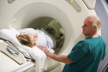
DEFINITION OF FOLLICULAR LYMPHOMA
Follicular lymphoma is a cancer that develops in the white blood cells, lymphatic system and bone
marrow. Follicular lymphoma is a well-defined subtype of Non-Hodgkin Lymphoma (NHL)* with cells
of the lymphoid tissues in the lymphatic system multiplying uncontrollably to eventually cause
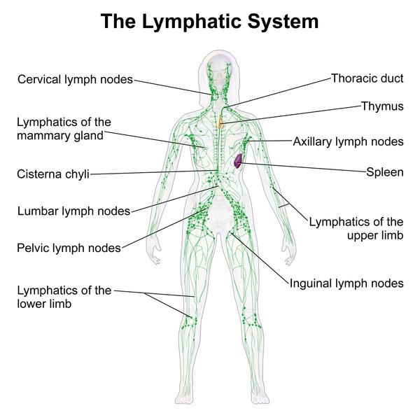
tumours to grow. The lymphatic system is constituted of lymph vessels that branch through the body
of the veins and arteries* as well as lymph glands or lymph nodes along the lymph vessels. Lymphoid
tissue is consisted of several types of system cells that help the body fight infection. Most of the cells
in lymphoid tissue are cells called lymphocytes (a type of white blood cells) with two main types of
lymphocytes, specifically B- and T-lymphocytes. Different types of lymphoma can develop from each
type of lymphocyte, but follicular lymphoma arises in particular from B-lymphocytes. The malignant
cells in lymphoma grow in clusters to form nodules. Some organs are also part of the lymphatic
system and partially constructed by lymphoid tissue with the spleen, thymus, tonsils and adenoids.
The lymphatic system filters blood, lymph (the liquid that circulates in lymph vessels), drains fluid
from tissues back to the bloodstream and fights infections. Since lymphoid tissue is found throughout
the body, follicular lymphoma can begin in almost any part of the body. The bone marrow may
become invaded by lymphocytes* that does not function properly. As the bone marrow also
produces platelets to critically stop bleeding and red blood cells to deliver oxygen to all cells in the
body, excess accumulation lymphocytes prevents the normal production of red blood cells and
platelets. Follicular lymphoma is usually slow-growing.
The Lymphatic System
Photo credit: Bruce Blaus (Creative Commons)
Follicular Lymphoma: a guide for patients - Information based on ESMO Clinical Practice Guidelines – v.2014.1 Page 4
This document is provided by the Anticancer Fund with the permission of ESMO.
The information in this document does not replace a medical consultation. It is for personal use only and cannot be modified,
reproduced or disseminated in any way without written permission from ESMO and Reliable Cancer Therapies.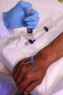
IS FOLLICULAR LYMPHOMA FREQUENT?

Compared to breast cancer in women or prostate cancer in men, NHLs* are not common. But,
nevertheless they are the sixth most common cancer in Europe. They account for around 3% of all
cancers and follicular lymphomas represent approximately 25% of all NHLs*. In Western Europe,
follicular lymphoma is the second most frequent subtype of lymphoma.

The number of patients diagnosed with follicular lymphoma every year has increased from 2-3 cases
per 100,000 people in the 1950s, to 5-7 cases per 100,000 people now.

In general, the risk of getting NHL* increases with age. There is a 5-7 fold increase in the number of
cases among patients older than 65 years.

Follicular Lymphoma: a guide for patients - Information based on ESMO Clinical Practice Guidelines – v.2014.1         Page 5
This document is provided by the Anticancer Fund with the permission of ESMO.
The information in this document does not replace a medical consultation. It is for personal use only and cannot be modified,
reproduced or disseminated in any way without written permission from ESMO and Reliable Cancer Therapies.

WHAT CAUSES FOLLICULAR LYMPHOMA?

Today, the cause of follicular lymphoma is not understood. Some risk factors have been identified. A
risk factor increases the risk that cancer occurs, but it is neither necessary nor sufficient to cause
cancer. A risk factor is not a cause in itself.

Some people with these risk factors will never develop follicular lymphoma and some people with
none of these risk factors will develop it.

Lifestyle, enviromental factors and previous medical conditions have been linked to the ocurrence of
follicular lymphoma, but their influence is not clear yet.

    -    Lifestyle factors:
         o Diet: A link between follicular lymphoma and consumption of meat and milk was found,
             as well as nitrates* and nitrites* frequently present in our diet. Cured meat, food
             preservatives, and those naturally occuring in some fruits are some sources. Protective
             effects have been suggested with the consumption of polyunsaturated fatty acids*,
             vitamin D, fruits and vegetables, among others. The association with
             obesity is not clear.

         o    Alcohol: Alcohol intake has been associated with cancer in humans.
              Wine consumption has been found to increase the risk of developing
              follicular lymphoma, particularly for drinkers who started before the
              age of 20 and/or when the alcohol consumption is higher than 19
              grams per day (a glass of wine has approximately 14 grams of
              alcohol).

         o    Smoking: Current smokers have higher risks of developing follicular
              lymphoma according to study results in comparison to former
              smokers Heavy smokers are also at higher risk.

    -    Enviromental factors:
             Pesticides allegedly cause a specific genetic mutation that has a role
             in the development of follicular lymphoma. Yet the specific mutation
             has been found in healthy individuals that never developed follicular
             lymphoma. Hair dyes have been linked with follicular lymphoma in
             some studies. Other chemicals such as solvents with benzene need to be confirmed as
             risk factors. But their association to follicular lymphoma has already been suggested in
             different studies. Moderate sun exposure, on the other hand, has been associated with
             reduced risk.

    -    Other medical conditions:
             Conditions with a suppressed immune system are also linked to follicular lymphoma
             such as HIV/AIDS, autoimmune diseases and medication.

Follicular Lymphoma: a guide for patients - Information based on ESMO Clinical Practice Guidelines – v.2014.1         Page 6
This document is provided by the Anticancer Fund with the permission of ESMO.
The information in this document does not replace a medical consultation. It is for personal use only and cannot be modified,
reproduced or disseminated in any way without written permission from ESMO and Reliable Cancer Therapies.
HOW IS FOLLICULAR LYMPHOMA DIAGNOSED?

Patients may be diagnosed on the basis of their symptoms or sometimes in patients with no
symptoms as a result of imaging and laboratory tests.
Symptoms and signs of follicular lymphoma may include:
    1. Painless swelling of the lymph nodes in the neck, arm pits, and/or groin. If follicular
        lymphoma arises mostly in deep lymph nodes, it might present with compression of some
        vital organ and causing symptoms. Among others, some include chronic cough, problems to
        breath, chest pain, abdominal pain or back pain depending on the organ involved.
    2. Fever for no known reason.
    3. Drenching night sweats.
    4. Undesired or unintentional weight loss.
    5. Fatigue. Fatigue is a common symptom of anaemia*. Patients who are physically active may
        not notice the effects of being anaemic until it is severe.
    6. Infections. Due to replacement of an important part of the healthy immune system that
        protects us from infections and by cancer, patients can experience recurrent infections or
        infections unusually difficult to treat.
    7. Bleeding. Rarely a low platelet count resulting from replacement of the normal bone marrow
        with cancerous cells results in easy bruising, bleeding from the nose or gums, and
        appearence of small red spots on the skin commonly over the shins and ankles.

Symptoms 2-4 are known as B symptoms, which are taken into account when staging the disease.

Patients with the above symptoms will have a complete blood count test, which is a laboratory test
done to check the three types of cells made in the bone marrow: 1) white blood cells, 2) red blood
cells, and 3) platelets. Occasionally a patient may have a complete blood count done for another
reason to show the first indication of a possible lymphoma based on laboratory findings alone. In
addition to identifying a low red cell count or platelet count, as part of the white blood cell count the
complete blood count may find abnormal cells circulating in the blood. Abnormal white blood cells
multiplying at a high rate are larger than the more mature normal white blood cells found in
circulation.

If a diagnosis of follicular lymphoma becomes suspected based on symptoms and the white blood
cell count, a lymph node biopsy* is performed.

Exact diagnosis of follicular lymphoma can only be based on a lymph node biopsy*. It involves the
removal of a lymph node when the patient may be under anaesthesia (excisional biopsy). The
removed lymph node tissue will be examined in the laboratory. This examination is called
histopathology*and mainly consists of visually assessing the tissue under a microscope to look for
lymphoma cells. In contrast, the removal of only a part of a lymph node
using a wide needle (core biopsy) should only be performed in patients
without easily accessible lymph nodes due to their position in the body.
Of note, the results of the biopsy examination may not be clear since
the lymphoma cells could be different from one part of the lymph node
to another (known as heterogeneity). The removal of tissue or fluid
using a thin needle (fine needle biopsy) is not recommended for a
reliable lymphoma diagnosis.

Follicular Lymphoma: a guide for patients - Information based on ESMO Clinical Practice Guidelines – v.2014.1         Page 7
This document is provided by the Anticancer Fund with the permission of ESMO.
The information in this document does not replace a medical consultation. It is for personal use only and cannot be modified,
reproduced or disseminated in any way without written permission from ESMO and Reliable Cancer Therapies.
The World Health Organisation (WHO) has classified lymphoma as Grade 1, 2, 3A or 3B. This grade is
dependent upon the number of lymphoma cells (called blasts*) seen under the microscope. Grade
3B has the greatest number of blasts and considered to be more aggressive. Therefore, Grade 3B
must be treated differently to other types of lymphoma. When possible, additional biopsy material
should be stored fresh frozen; this means that additional tests could be performed later into the
future if needed.

Follicular Lymphoma: a guide for patients - Information based on ESMO Clinical Practice Guidelines – v.2014.1         Page 8
This document is provided by the Anticancer Fund with the permission of ESMO.
The information in this document does not replace a medical consultation. It is for personal use only and cannot be modified,
reproduced or disseminated in any way without written permission from ESMO and Reliable Cancer Therapies.

WHAT IS IMPORTANT TO KNOW TO GET THE OPTIMAL TREATMENT?
Doctors will need to gather different elements to decide what would be the best
treatment.
Relevant information about the patient
General well-being.
Personal medical history.
History of cancer in relatives.
Results from the clinical examination by the doctor. Your doctor will look
for enlarged lymph nodes and organs, as well as general appearance of the skin and inside
the mouth particularly. The physical examination should be detailed.
Results of imaging studies* of the body, such as a computed tomography (CT)* scan, includes
the neck, chest, abdomen and pelvis. These studies will identify the location and extent of
the lymphoma. The scans are critical to stage the lymphoma in one of four stages based on
the extent of spread. These stages are described further on the next page. Positron emission
tomography (PET)* scan may be useful in confirming a localised form of disease (stage II).
PET* results allow doctors to see the locations of cancerous cells since the radiolabeled
glucose is concentrated in the cancerous cells and they can be seen in the PET image.
A complete blood count, routine blood chemistry including lactate deshydrogenase* (LDH)
and uric acid, screening tests for human immunodeficiency (HIV) and hepatitis B and C are
required. These tests are very important when deciding on the best treatment. If the results
of these tests show to be positive, a specific therapy for these conditions is recommended.
A bone marrow biopsy is a procedure to take a bone marrow sample from the hip to
evaluate whether the bone marrow is affected. After the disease diagnosis, the bone marrow
biopsy must be done to identify the stage of the disease
adequately. It is performed under local anesthesia.
In addition to clinical examination, others may be performed
to assess the risks of complications due to the treatment. For
example, to assess the function of the heart, it is
recommended to perform an echocardiogram (ultrasound of
the heart).
If severe headaches, problems with vision, sensation, or muscle function are present, an
assessment of the cerebrospinal fluid* (the fluid surrounding the brain and spinal cord) may
be necessary. This is performed by doing a lumbar puncture (spinal tap) with a fine needle
passed into the spine. After anaesthetic is used to numb the area in order to obtain some of
this fluid, the next step occurs with the fluid sent to the pathologist for examination under
the microscope. A CT scan or magnetic resonance imaging (MRI)* of the head may be
necessary if the lumbar puncture does not identify any cancer cells. The CT scan* or MRI*
could show an area of lymphoma inside the brain that requires additional treatment specific
to that location of tumour.
Follicular Lymphoma: a guide for patients - Information based on ESMO Clinical Practice Guidelines – v.2014.1 Page 9
This document is provided by the Anticancer Fund with the permission of ESMO.
The information in this document does not replace a medical consultation. It is for personal use only and cannot be modified,
reproduced or disseminated in any way without written permission from ESMO and Reliable Cancer Therapies.Relevant information about the cancer
Staging
After the diagnosis of follicular lymphoma, tests must be done to find out if lymphoma cells have
spread within the lymph system or to other parts of the body. Staging is the process of determining
whether the tumour has spread, and if so, how far. It is extremely important to know the stage of the
disease in order to plan the treatment.
The staging system used to describe the spread of follicular lymphoma is called the Ann Arbor
Staging System. It uses Roman numerals (I-IV) for different stages.
Stage Definition
Stage I The lymphoma is in one group of the body's lymph nodes such as the groin or
neck, or in one organ of the lymph system.
Stage II Two or more groups of lymph nodes, or one organ close to the affected lymph
nodes and one or more groups of lymph nodes on the same side of the diaphragm
contain lymphoma cells. The diaphragm is the muscle that divides the chest and
abdomen.
For example, lymphoma might be above the diaphragm in lymph
nodes in the neck and arm pits. Or lymphoma might be below the
diaphragm in lymph nodes in the groin and abdomen.
Stage III The lymphoma is in lymph nodes on both sides of the diaphragm. It may have also
spread into an organ close to the affected lymph nodes or spleen.
Stage IV The lymphoma is in stage IV if the lymphoma involves the bone marrow or distant
organs.
Follicular lymphoma stages are also noted by the presence or absence of certain findings and/or
symptoms of the disease:
A lymphoma that affects organs or tissues other than the lymph nodes has an "E", for
extranodal, added to its staging nomenclature.
If a nodal mass is at least 7.5 cm of diameter, it translates as a bulky disease.
If it affects the spleen, an "S" is added.
If the patient has fever, night sweats, or unexplained weight loss, the letter "B" is added.
If none of these is present, an "A" is added.
Grade (WHO classification) and other histopathological characteristics
The grading, as explained previously, could be 1, 2, 3A and 3B to reflect the number of
lymphoma cells or blasts under the microscope using maximal magnification. Grade 3B is the
highest grade and considered an aggresssive lymphoma.
Prognosis and risk classification
A Follicular Lymphoma-specific International Prognostic Index (FLIPI) should be determined
for prognostic purposes. FLIPI allows to identify the risk of progression of the disease after
treatment and to adapt treatment and follow-up accordingly. The parameters used in the
original version FLIPI 1 are > 4 involved nodal sites, age >60 years, elevated LDH* level, stage
Follicular Lymphoma: a guide for patients - Information based on ESMO Clinical Practice Guidelines – v.2014.1 Page 10
This document is provided by the Anticancer Fund with the permission of ESMO.
The information in this document does not replace a medical consultation. It is for personal use only and cannot be modified,
reproduced or disseminated in any way without written permission from ESMO and Reliable Cancer Therapies.III or IV disease and haemoglobin*
WHAT ARE THE TREATMENT OPTIONS?
The treatment should take place only in centres used to treat patients with
lymphoma and offering an adequate multidisciplinary approach. You are also
encouraged to ask about any clinical trials* for your current situation.

Treatment of follicular lymphoma is tailored to each individual based on the
stage and patient characteristics, including age and other diseases that the
patient may have such as diabetes, coronary heart disease or chronic
obstructive pulmonary disease. Although this usually occurs in the management of solid tumours,
surgery does not play a major role in the treatment of follicular lymphoma since the nature of the
lymphatic system lymphoma is not often confined to only one area of the body.

Treatment may control or cure the lymphoma. It can also improve your quality of life by controlling
symptoms of the disease. The goal of follicular lymphoma treatment is to apply one or more of these
strategies, including killing the lymphoma cells as quickly as possible, stopping the growth of new
lymphoma cells, treating side effects caused by lymphoma such as pain, fevers, chills and night
sweats, and/or maintaining a sense of control over your treatment choices and life.

In general, several approaches are considered when treatment decisions are made for the
management of follicular lymphoma. These include watchful waiting, radiotherapy*, chemotherapy*
and targeted therapy* with monoclonal antibody*. Except for watchful waiting, these therapies may,
and typically are, used in combination with each other.

Watchful waiting is a term used for a strategy featuring close monitoring of each
patient’s condition without giving any treatment until symptoms appear or change.
This aproach is meaningful in low tumour burden follicular lymphoma since
spontaneus partial regression have been observed in
some individuals. Moreover, evidence suggests that
normal T cells* are capable of controlling the
lymphoma in some individuals. Some patients may be
watched closely for over 10 years before treatment is
needed. During watchful waiting, patients are still
being “treated” to track their situation in spite of no drugs or radiotherapy being used.

External radiotherapy is a cancer treatment that uses high-energy x-rays* or other types of radiation
to kill cancer cells and keep them from growing. External radiotherapy uses a machine that focuses
the radiation from outside the body and targets it towards the cancer.

Chemotherapy is anticancer treatment that uses drugs to stop the growth of cancer cells either by
killing the cells or by stopping them from dividing. When chemotherapy
is taken by mouth or injected into a vein, the drugs enter the
bloodstream and can reach cancer cells throughout the body (systemic
chemotherapy). Rarely when follicular lymphoma has spread to the
brain, chemotherapy can be administered into the cerebrospinal fluid* to
kill lymphoma cells in the brain or spinal cord. The way chemotherapy is
given depends on the stage of the disease. Combination chemotherapy

Follicular Lymphoma: a guide for patients - Information based on ESMO Clinical Practice Guidelines – v.2014.1        Page 12
This document is provided by the Anticancer Fund with the permission of ESMO.
The information in this document does not replace a medical consultation. It is for personal use only and cannot be modified,
reproduced or disseminated in any way without written permission from ESMO and Reliable Cancer Therapies.
provides treatment using two or more anticancer drugs. Steroid drugs may be added to kill the
lymphoma cells as well.

Immunotherapy is a type of treatment that either boosts the patients own immune system, or uses
man made drugs called monoclonal antibodies* that attack a specific target on the surface of
lymphocytes (cells in which lymphoma starts). The monoclonal antibody rituximab* is a drug used to
treat follicular lymphoma and administered by infusion into veins.

Radioimmunotherapy refers to a type of treament where a radioactive substance is attached to the
monoclonal antibody*. When the monoclonal antibody* is delivered to the cells’ targets, the
radioactice source acts on the lymphoma cells and potentially some nearby cells. This treatment is
known as yttrium 90-ibritumomab tiuxetan*.

Bone marrow transplantation either with patients’ own bone marrow cells or a donor’s may be a
part of lymphoma treatment. Before performing the bone marrow transplantation, the radiation and
chemotherapy are applied with the aim to reduce the cells in bone marrow affected by the disease.

The treatment mainly depends on the stage of the disease. For more advanced stages (stage III and
IV), the goal of the treatment involves two major strategies. Firstly, induce a regression of the
tumour     (induction    phase).   Secondly,   consolidate     or   maintain    this     regression
(consolidation/maintenance phase). Treatment details are presented below stage by stage.

Treatment plan for stage I-II follicular lymphoma
Stage I means that one lymph node group or lymphatic* organ is affected.
Stage II means that either two or more lymph node groups on the same side of the diaphragm* are
affected, or one or more lymph node groups together with a lymphatic* organ close to the lymph
nodes involved on the same side of the diaphragm* are affected.

         In the small proportion of patients with limited non-bulky stage I-II disease, the
         administration of radiotherapy targeting the site of the involved lymph nodes has curative
         potential.

         In selected cases, watchful waiting may be discussed to avoid the side effects of radiation
         and it could be of the same efficacy as active treatment.

         The presence of bulky, large tumour, or with two or more sites involved and other risks help
         doctors to identify patients who could benefit from treatment with chemotherapy* and
         monoclonal antibody* rituximab*. In this case the role of radiotherapy may be considered
         after this initial treatment if the sites of involved lymph nodes are located in a way that
         radiotherapy can be given wihout major side effects.

Treatment plan for Stage III-IV
Stage III means that the lymphoma is in lymph nodes on both sides of the diaphragm. It may have
also spread into a nearby organ.

Stage IV means that the lymphoma is in the bone marrow, or distant organs.

Follicular Lymphoma: a guide for patients - Information based on ESMO Clinical Practice Guidelines – v.2014.1        Page 13
This document is provided by the Anticancer Fund with the permission of ESMO.
The information in this document does not replace a medical consultation. It is for personal use only and cannot be modified,
reproduced or disseminated in any way without written permission from ESMO and Reliable Cancer Therapies.
In up to 10-20% of lymphoma cases, the disease could disappear or regress without any
treatment. Early initiation of therapy in asymptomatic patients did not show any
improvement of survival in different studies. Therefore in that case, watchful waiting is
recommended. Treatment should only be utilized due to symptoms, including B-symptoms
(fever for an unknown reason, drenching night sweats and undesired or unintentional weight
loss), impairment in blood cells formation, bulky disease, compression of important organs,
abnormal presence of fluid in abdominal cavity (ascites), or in the space between the lungs
an thoracic* wall (pleural effusion) and rapid progression of the disease. The treatment
strategies are explained later in the section on induction and consolidation/maintenance
treatment.
The induction treatment is the first step to start reducing the number of cancer cells. From
there, a consolidation phase further reduces the number of cancer cells and increases the
probability of the lymphoma not coming back. This is followed by a maintenance phase with
goals to maintain the remission and prevent a relapse.
Complete remission is achieved when the tumours disappear completely and no further signs
of disease exist. Partial remission comes from a shrinkage of the tumours with some
remaining. This is shown through a physical examination or by imaging, as well as disease
symptoms that might persist.
Induction treatment
In the majority of patients with advanced stage III and IV disease, chemotherapy does not
result in a definite cure. But, remissions usually last for years and the average survival is
longer than 20 years.
Combination of monoclonal antibody* rituximab* and chemotherapy* such as CHOP
(cyclophosphamide, doxorubicin, vincristine and prednisone) or bendamustine* are
recommended for disease remission and long-term maintenance of achieved response.
When a physical examination or new biopsy indicates results to suspect that the disease
turned into an aggressive course, a regimen such as CHOP is preferred. Studies also showed
when monoclonal antibody* rituximab* is added to chemotherapy, improvements occur
with tumour shrinkage, keeping the disease stable and prolonging patients’ lives in general.
The chemotherapy regimen CVP (cyclophosphamide*, vincristine* and prednisone*) in
combination with rituximab* may be given in case of contraindications for chemotherapy
drug doxorubicin*.
Full courses of fludarabine* and cyclophosphamide* or fludarabine* and mitoxantrone* are
not recommended due to higher haemathological toxicities*.
In symptomatic patients with low volume tumour mass, and in case of slow disease
progression, therapy with rituximab* alone may be considered.
In low-risk elderly patients, and elderly patients with contraindications for more intensive
combination of chemotherapy* and rituximab*, the treatment with rituximab* plus
chlorambucil*, rituximab* alone or radioimmunotherapy* are alternatives.
Follicular Lymphoma: a guide for patients - Information based on ESMO Clinical Practice Guidelines – v.2014.1 Page 14
This document is provided by the Anticancer Fund with the permission of ESMO.
The information in this document does not replace a medical consultation. It is for personal use only and cannot be modified,
reproduced or disseminated in any way without written permission from ESMO and Reliable Cancer Therapies.There are specific recommendations for monitoring and treatment of patients with hepatitis
B virus infection. Often, blood tests will be taken to monitor activity of hepatitis B virus, as
well as antiviral* medications recommended.
Consolidation/maintenance treatment
After induction treatment achieves complete or partial remission, rituximab* as maintenance
therapy is administered in a single application every two months for up to two years. This
strategy delays the progression of the disease.
Radioimmunotherapy* as consolidation delays progression of the disease only after
chemotherapy, but the benefit seems inferior in comparison to rituximab* maintenance for 2
years.
Stem cell transplant using a patient’s own stem cells may delay disease progression only if
chemotherapy was administered before. However, such an approach is not recommended
for patients that respond to first-line therapy. The benefit from a stem cell transplant would
be limited after rituximab*.
Relapsed disease
Relapse is the reappearance of the disease. A repeated biopsy is strongly recommended to know if the
lymphoma that relapsed turned into an aggressive form.
The treatment given when the disease relapsed is called salvage treatment and selection
depends on effectiveness from the previous treatments administered. In early relapse (< 12-
24 months disease-free) the disease might be resistant to the drugs used previously. As a
result, a regimen of different drugs to overcome resistance is preferred. An example comes
from bendamustine* after CHOP and vice versa. Rituximab* could be used again for the
patient if previously it achieved a disease-free period of more than 6-12 months.
Radioimmunotherapy* (radioactive substance combined/attached to a monoclonal
antibody*) represents an effective approach, especially in patients over 65 years of age with
other diseases present. In those cases, they are not suitable for chemotherapy and therefore
need other types of treatment such as radioimmunotherapy.
Rituximab* maintenance for up to 2 years as a single treatment every 3 months can be
administered in patients who received it as a part of induction and did not receive it in the
first-line treatment.
In young patients, high-dose chemotherapy with a stem cell transplantation using the
patient’s own stem cells could be considered. Research indicates that treatment combination
for young cancer patients delays the progression of the disease and prolongs survival.
However it is not always required now. Instead, rituximab is widely used for patients,
especially in patients experiencing late relapses.
Follicular Lymphoma: a guide for patients - Information based on ESMO Clinical Practice Guidelines – v.2014.1 Page 15
This document is provided by the Anticancer Fund with the permission of ESMO.
The information in this document does not replace a medical consultation. It is for personal use only and cannot be modified,
reproduced or disseminated in any way without written permission from ESMO and Reliable Cancer Therapies.Evaluation of response to treatment
In all cases, scans should be performed halfway through treatment and after completion of
the treatment. If insufficient or no response is detected, early treatment for relapsed disease
should be considered.
PET*/CT scan to evaluate response to treatment continues to be under investigation,
although it has shown some benefit as a predictor of the disease course outcome. However,
further studies are needed.
A laboratory test to see if any cancer cells remain in the body (known as minimal residual
disease analysis) after completion of treatment is known to be highly predictive. But, the test
should not guide the treatment outside of clinical trials.
Follicular Lymphoma: a guide for patients - Information based on ESMO Clinical Practice Guidelines – v.2014.1 Page 16
This document is provided by the Anticancer Fund with the permission of ESMO.
The information in this document does not replace a medical consultation. It is for personal use only and cannot be modified,
reproduced or disseminated in any way without written permission from ESMO and Reliable Cancer Therapies.WHAT ARE THE POSSIBLE SIDE EFFECTS OF THE TREATMENT? Watchful waiting This strategy becomes stressful for patients as they have to wait until the disease has turned aggressive to get active treatment. However, watchful waiting is valid considering the good prognosis of this disease compared to other malignances. You must discuss any concerns about this situation with your doctor. Radiotherapy Most people will have a few side effects, but often they are mild. As radiotherapy affects people in different ways, it is difficult to predict exactly how the patient will react to the treatment. Some strategies are available to prevent or relieve some of these side effects. There have been important improvements in application of radiotherapy. As a result, severe side effects are now very rare. Most of the side effects of radiotherapy disappear gradually once the course of treatment is over. But for some people, side effects may continue for a few weeks. During radiotherapy, side effects may occur in organs that are directly targeted, but also in healthy tissues or organs close to the region that needs to be irradiated and cannot be protected from the X- rays. Side effects may be increased when radiotherapy* is administered with chemotherapy*. The symptoms and signs of the side effects vary according to the part of the body irradiated. For example, if neck irradiation occurs, then salivary glands may be affected. Also, dysfunction reflected in dry mouth might be persistant. Yet in most cases any side effects are transient. Strategies to prevent and relieve post-radiation reactions must be provided by the radiation oncologist or the nurse. There are some long‐term side effects that can take months and sometimes years to develop. The skin can feel different or may be more pigmented than before. Red ‘spidery’ marks (telangiectasia) may appear on the skin when small blood vessels are damaged. Radiotherapy itself can sometimes cause cancer and a small number of people will develop a second cancer due to the treatment. However, the chance of a second cancer developing is overall rare and the risks of getting radiotherapy are minimal compared to the benefits. Immunotherapy Monoclonal antibody/Rituximab Although rituximab* is expected to affect only cancer cells, some side effects can occur and they should be reported to your doctor immediately. Although sometimes the side effects seem severe, generally approved drugs’ benefits outweigh potential risks. Rituximab may cause severe infusion-related side effects particularly during the first infusion or within 24 hours after the infusion. Later side effects are also possible. Your healthcare team will inform you about these possibilities. Infusion-related side effects Follicular Lymphoma: a guide for patients - Information based on ESMO Clinical Practice Guidelines – v.2014.1 Page 17 This document is provided by the Anticancer Fund with the permission of ESMO. The information in this document does not replace a medical consultation. It is for personal use only and cannot be modified, reproduced or disseminated in any way without written permission from ESMO and Reliable Cancer Therapies.
Allergic reactions with flu-like symptoms, breathing problems like shortness of breath,
difficulty breathing, or wheezing, fever, body aches, redness and bumps on the skin, itching,
swelling of lips, tongue, throat and face, low blood pressure, and/or chest pain.
In case of infusion-related side effects, the infusion should be discontinued and reinitiated when all
symptoms have stopped. Your doctor should give you some medications before the infusion to
decrease the chance of a severe infusion reaction.
Later side effects
Heart problems.
Feeling sick.
Low red blood cell counts leading to anaemia*.
Low white blood cell counts that could lead to infections.
Low platelet counts impairing coagulation.
If you have had hepatitis B infection or carry hepatitis B virus, receiving rituximab* could cause the
infection to become active again. Hepatitis B reactivation may cause serious liver problems. During
the active phase of hepatitis B infection, you should not receive rituximab*. Please remember to tell
your healthcare team before starting therapy that you have had hepatitis B.
Rituximab* may also cause tumour lysis syndrome* caused by a fast breakdown of cancerous cells,
and characterised by kidney failure and abnormal heart rhythm. But this only occurs under certain
circumstances depending on the size of the tumour.
Treatment with rituximab* may lower the ability of your immune system to fight infections.
In very rare cases, rituximab* may cause a serious brain virus infection. If you feel, or if anyone close
to you notices confusion or problems thinking, loss of balance, changes in how you walk or talk,
weakness on one side of your body, blurred vision or loss of vision, report it to your doctor
immediately.
Radiolabelled monoclonal antibodies (Radioimmunotherapy)
All side effects described for rituximab are potential side effects of radioimmunotherapy as well.
However, the most common side effects of yttrium 90-ibritumomab tiuxetan* are a low platelet
count, bleeding, anaemia and low number of white blood cells.
Chemotherapy
Side effects of chemotherapy are very frequent. They will depend on the drug(s) administered, the
doses and other individual factors.
Combinations of different drugs can lead to more side effects than taking a single drug. The nature,
frequency and severity of the side effects vary for any chemotherapy drug combination used.
Effective supportive therapies for some of these side effects exist and treatments are possible.
Follicular Lymphoma: a guide for patients - Information based on ESMO Clinical Practice Guidelines – v.2014.1 Page 18
This document is provided by the Anticancer Fund with the permission of ESMO.
The information in this document does not replace a medical consultation. It is for personal use only and cannot be modified,
reproduced or disseminated in any way without written permission from ESMO and Reliable Cancer Therapies.Listed below are side effects known to occur with one or several of the chemotherapy drugs
currently used for follicular lymphoma.
The most frequent side effects are:
o Decreased blood cell counts that may lead to anaemia, bleeding and bruising, and infections.
o Lack of energy.
o Fever, chills, body aches, and flu symptoms.
o Sores in mouth and throat.
o Feeling sick, vomiting and diarrhoea.
o Some of the chemotherapy drugs used can cause urination problems with little or no urine
elimination and painful urination. The urine can also change colour and depends on the drug.
Anthracyclines* (doxorubicin) could turn urine’s color into reddish-orange. However, it is not
harmful and disappears after one day or two.
o In women menstrual periods may also be affected. They may completely cease during and/or
after the treatment, or you may miss some periods (for example, in case of chemotherapy
drugs doxorubicin*, cyclophosphamide*, and mitoxantrone*) or bleeding may increase (for
example, in case of mitoxantrone*). In men there is a risk of infertility as well. Your doctor
will discuss with you about all the options to preserve fertility and offer available support
before your treatment.
Other side effects that may occur frequently are:
o Numbness or tingling feeling around your mouth.
o Yellowing of the skin and the white part of your eyeballs (for example, in case of side effects
from chemotherapy drugs that are metabolised though the liver, or liver
damage induced by hepatitis B infection).
o Temporary or complete hair loss and/or thinning of the hair.
o Skin changes or reactions.
o Memory deficiencies and difficulties in concentrating.
Occasional side effects:
o Bone pain, constipation (in case of vincristine). Constipation may also
appear as a result of some drugs given to prevent nausea and vomiting.
o Doxorubicin* can cause damage to heart muscles. Therefore, the
assessment of heart function is important before its administration.
o Rarely, doxorubicin* can cause leukaemia or secondary cancers later on.
In general, all side effects should be reported to your physician as soon as they appear.
Follicular Lymphoma: a guide for patients - Information based on ESMO Clinical Practice Guidelines – v.2014.1 Page 19
This document is provided by the Anticancer Fund with the permission of ESMO.
The information in this document does not replace a medical consultation. It is for personal use only and cannot be modified,
reproduced or disseminated in any way without written permission from ESMO and Reliable Cancer Therapies.WHAT HAPPENS AFTER THE TREATMENT?
It is not unusual to continue to experience some treatment-
related symptoms once the treatment is over.
It is not rare that anxiety, sleep problems or depression
take place during the post-treatment phase. Patients
dealing with those symptoms need support and even
from psychological professionals.
Memory deficiencies with difficulty in concentrating are not uncommon chemotherapy side
effects and generally reversible within a few months.
Follow-up with doctors
After the treatment has been completed, doctors will propose a follow-up aiming to:
detect a possible relapse as soon as possible
evaluate adverse effects of the treatment and treat them
provide psychological support and information to enhance returning to normal life
Under general consensus follow-up visits with the doctor should include:
History-taking, monitoring of symptoms and physical examination every three months for
two years, every four to six months for additional three years and subsequently once a year.
Your doctor will be vigilant to the possibility that lymphoma may come back, or the disease
considered under control then transformed into more an aggressive course, or that a new
cancer or leukaemia may appear.
Blood count and other routine blood analyses every six months for two years and then only if
suspicious symptoms appear.
Evaluation of thyroid function at one, two and five years if the patient received irradiation to
the neck.
Radiological* and ultrasound studies* every six months for two years and annually
thereafter. However, CT scans are not mandatory outside clinical trials.
Minimal residual disease analysis may also be performed within clinical trials.
Return to normal life
Returning to life after cancer offers good perspectives. Yet it can be challenging to live with the
potential idea that the disease can come back. From what is known today, no specific ways to reduce
the risk of recurrence after completion of the treatment can be recommended. As a consequence of
the disease itself and treatment, depending upon the individual, returning to normal life may not be
easy for some people. Questions related to body-image, sexuality, fatigue, work, emotions or lifestyle
may be a concern. Discussing these questions with relatives, friends, other patients or the healthcare
team may be helpful. Support from patient organisations providing advice on treatment effect
management, as well as psycho-oncologist services, telephone conversations and email exchanges
are available in many countries.
Follicular Lymphoma: a guide for patients - Information based on ESMO Clinical Practice Guidelines – v.2014.1 Page 20
This document is provided by the Anticancer Fund with the permission of ESMO.
The information in this document does not replace a medical consultation. It is for personal use only and cannot be modified,
reproduced or disseminated in any way without written permission from ESMO and Reliable Cancer Therapies.What if the lymphoma comes back? If the lymphoma returns, it is called a relapse and the treatment depends on the age of the patient, prior treatment, and the possibility of a bone marrow transplant. A new biopsy is strongly recommended to determine whether the relapsed lymphoma became aggressive. The treatment given after relapse called salvage treatment depends on how effective the treatments previously administered were. Different combinations of chemotherapy and rituximab can be given. For further details, please explore various sections in this guide about treatment. In elderly patients not suitable for these regimens, radioimmunotherapy* is an option. In case of relapse in selected younger patients with a high-risk profile, potentially curative stem cell transplantation from a donor may be considered. Follicular Lymphoma: a guide for patients - Information based on ESMO Clinical Practice Guidelines – v.2014.1 Page 21 This document is provided by the Anticancer Fund with the permission of ESMO. The information in this document does not replace a medical consultation. It is for personal use only and cannot be modified, reproduced or disseminated in any way without written permission from ESMO and Reliable Cancer Therapies.
DEFINITIONS OF DIFFICULT WORDS

Abdomen
Part of the body between the thorax and pelvis. The muscles corresponding to this area enclose a
cavity containing the stomach, intestines, liver, spleen, and pancreas. It is also known as belly.

Anaemia
Condition characterised by the shortage of red blood cells or haemoglobin, the iron that contains the
hemoglobin carries oxygen from the lungs to the whole body, this process is diminished in this
condition.

Anthracyclines
Antibiotic drug used in chemotherapy to treat a wide range of cancers.

Antiviral
Agent that kills a virus or that suppresses its ability to replicate.

Artery/Arteries
A blood vessel that carries blood from the heart to tissues and organs in the body.

Ascites
Abnormal buildup of fluid in the abdomen that may cause swelling. In late-stage cancer, tumour cells
may be found in the fluid in the abdomen. Ascites also occurs in patients with liver disease.

Bendamustine
The active ingredient in a drug that is used to treat chronic lymphocytic leukaemia (CLL), to treat
slow-growing B-cell non-Hodgkin lymphoma (NHL) that has gotten worse within 6 months of
treatment with other anticancer drugs, and is being studied in the treatment of other types of
cancer. Bendamustine may damage the DNA in cancer cells and cause them to die. It is a type
ofalkylating agent and a type of antimetabolite.

Biopsy
The removal of cells or tissues for examination by a pathologist. The pathologist may study the tissue
under a microscope or perform other tests on the cells or tissue. There are many different types of
biopsy procedures. The most common types include: (1) incisional biopsy, in which only a sample of
tissue is removed; (2) excisional biopsy, in which an entire lump or suspicious area is removed; and
(3) needle biopsy, in which a sample of tissue or fluid is removed with a needle. When a wide needle
is used, the procedure is called a core biopsy. When a thin needle is used, the procedure is called a
fine-needle aspiration biopsy.

Cerebrospinal fluid
The fluid that surrounds and bathes the spinal cord and brain. Its main function is to protect the
brain and the spinal cord.

Follicular Lymphoma: a guide for patients - Information based on ESMO Clinical Practice Guidelines – v.2014.1        Page 22
This document is provided by the Anticancer Fund with the permission of ESMO.
The information in this document does not replace a medical consultation. It is for personal use only and cannot be modified,
reproduced or disseminated in any way without written permission from ESMO and Reliable Cancer Therapies.
Chemotherapy
A type of cancer treatment using drugs that kill cancer cells and/or limit their growth. These drugs
are usually administered to the patient by slow infusion into a vein but can also be administered
orally, by direct infusion to the limb or by infusion to the liver, according to cancer location.

Chlorambucil
A drug used to treat several types of leukaemias and lymphomas. Chlorambucil blocks cell growth by
damaging the cell’s DNA and may kill cancer cells. It is a type of alkylating agent.

Clinical trial
A type of research study that tests how well new medical approaches work in people. These studies
test new methods of screening, prevention, diagnosis, or treatment of a disease. Also called clinical
study.

Computed tomography CT
A form of radiography in which body organs are scanned with X-rays and the results synthesized by a
computer to generate images of parts of the body.

Cyclophosphamide
A drug that is used to treat many types of cancer and is being studied in the treatment of other types
of cancer. It is also used to treat some types of kidney disease in children. Cyclophosphamide
attaches to DNA in cells and may kill cancer cells. It is a type of alkylating agent.

Diaphragm
The thin muscle below the lungs and heart that separates the chest from the abdomen *.

Doxorubicin
A drug that is used to treat many types of cancer and is being studied in the treatment of other types
of cancer. Doxorubicin comes from the bacterium Streptomyces peucetius. It damages DNA and may
kill cancer cells. It is a type of anthracycline antitumor antibiotic.

Extralymphatic
It refers to any organ or structure outside of the lymph nodes and lymphatic system.

Fludarabine
The active ingredient in a drug used to treat B-cell chronic lymphocytic leukaemia (CLL) that has not
responded to treatment with other anticancer drugs or that has gotten worse. Fludarabine blocks
cells from making DNA and may kill cancer cells. It is a type of purine antagonist and a type of
ribonucleotide reductase inhibitor.

Haemoglobin
The substance inside red blood cells* that binds to oxygen in the lungs and carries it to the tissues.

Hematological toxicities
The extent to which something is poisonous or harmful to blood cells with, i.e. red blood cells, white
blood cells and platelets.

Follicular Lymphoma: a guide for patients - Information based on ESMO Clinical Practice Guidelines – v.2014.1        Page 23
This document is provided by the Anticancer Fund with the permission of ESMO.
The information in this document does not replace a medical consultation. It is for personal use only and cannot be modified,
reproduced or disseminated in any way without written permission from ESMO and Reliable Cancer Therapies.
Histopathology
The examination and study of tissue and cells using a microscope. Tissue obtained from the body
by biopsy or surgery is placed in a fixative and transported to the laboratory. Here, it is cut into thin
sections, stained with various dyes and then studied under the microscope.
A histopathologist is a doctor interpreting sections of tissue including tumour tissue.

Imaging studies/Imaging test/Imaging procedure
A type of test that makes detailed pictures of areas inside the body. Imaging tests use different forms
of energy such as X-rays (high-energy radiation), ultrasound (high-energy sound waves), radio waves,
and radioactive substances. They may be used to help diagnose disease, plan treatment, or find out
how well the treatment is working. Examples of imaging tests are computed tomography (CT),
ultrasonography, magnetic resonance imaging (MRI), and nuclear medicine tests. Also called imaging
procedure.

Lactate deshydrogenase (LDH)
One of a group of enzymes found in the blood and other body tissues and involved in energy
production in cells. An increased amount of lactate dehydrogenase in the blood may be a sign of
tissue damage and some types of cancer or other diseases. Also called lactic acid dehydrogenase and
LDH.
Lymphocyte
A type of white blood cell that is essential in the immune system. The three major types
of lymphocyte are T cells, B cells and natural killer (NK) cells specific roles in the immune system.

Lymph node
A rounded mass of lymphatic tissue that is surrounded by a capsule of connective tissue. Lymph
nodes filter lymph and they store lymphocytes. They are located along lymphatic vessels. Also called
lymph gland.

Magnetic resonance imaging MRI
An imaging technique that is used in medicine. It uses magnetic resonance. Sometimes a fluid is
injected that enhances the contrast between different tissues to make structures more clearly
visible.

Minimal residual disease analysis
Laboratory test to detect small numbers of cancerous cells that remain in the patient’s body during
or after treatment when other indicators show remission in patients (no signs or symptoms of
disease).

Mitoxantrone
A drug used to treat advanced prostate cancer that does not respond to hormones, adult acute non-
lymphocytic leukaemia, and advanced or multiple sclerosis. It is also being studied in the treatment
of other cancers. It belongs to the family of drugs called antitumour antibiotics.

Monoclonal antibody
Monoclonal antibodies are antibodies exactly the same since they are produced by clones of the
same parent cell.

Follicular Lymphoma: a guide for patients - Information based on ESMO Clinical Practice Guidelines – v.2014.1        Page 24
This document is provided by the Anticancer Fund with the permission of ESMO.
The information in this document does not replace a medical consultation. It is for personal use only and cannot be modified,
reproduced or disseminated in any way without written permission from ESMO and Reliable Cancer Therapies.
You can also read