MALIGNANT SKIN CANCER DETECTION USING CONVOLUTIONAL NEURAL NETWORKING
←
→
Page content transcription
If your browser does not render page correctly, please read the page content below
ISSN No.: 2454- 2024 (online)

 International Journal of Technical Research & Science
 MALIGNANT SKIN CANCER DETECTION
 USING CONVOLUTIONAL NEURAL
 NETWORKING
 Rachakonda Hrithik Sagar1, Abhishek Bingi2, Aashray Pola3, Krishna Sai Raj Goud4, Tuiba Ashraf5,
 Subrata Sahana6
 Email: hrithiksagar36@gmail.com , bingi.abhishek@yahoo.com2, aashraypolaa@gmail.com3,
 1

2018006226.krishna@ug.sharda.ac.in4, 2018003670.tuiba@ug.sharda.ac.in5, 1subrata.sahana@gmail.com6
 Department of Computer Science Engineering, School of Engineering and Technology, Sharda
 University, Greater Noida, India
 Department of Electronics and Computer Engineering, Sreenidhi Institute of Science and Technology,
 Ghatkesar, Hyderabad, India
Abstract- The incidence of skin cancer is increasing by epidemic proportions. According to WHO, Skin Cancer
is the world’s 6th most common cancer. It can be classified into Basal cell carcinoma, Squamous cell carcinoma
and Melanoma among which Melanoma is more difficult to predict. By using this method, we can assist
dermatologists to detect at an early stage as Computer Vision plays a vital role in diagnosis.
In this paper, to detect skin cancer we are using machine learning-based algorithms. Traditionally classification
algorithms are Convolutional neural networking which Consists of initialization, adding a convolutional layer,
summing pooling layer, summing flattening layer, summing a dense layer, then compiling Convolutional neural
networks and fitting the CNN model to a dataset. We used machine learning model architecture to determine if
the skin images of the patients are harmful or harmless via using machine learning libraries provided in python.
We have chosen this approach to be more precise and specific in recognizing about cancer and ultimately declining
the mortality rate caused by it.
Keywords: Convolutional Neural Networking, Skin cancer, Malignant, Melanoma, Basal cell carcinoma,
Squamous cell carcinoma.
1. INTRODUCTION
The main objectives of this research are:
 ➢ To develop a prototype of a skin cancer-detecting system for Non-melanoma and melanoma skin
 cancer so that anyone who is reading this can develop their model.
 ➢ To learn and Implement Convolutional neural networking model of deep learning to extract features
 from images that get trained and show output with great accuracies.
 ➢ To help patients to prevent melanoma in early stages so that, that won't be leading to further skin
 problems.
Skin cancer is one of the diseases where it can put an individual's life at death door. At least 5.4 million people
get diagnosed each year only in the US compared to lung cancer, breast cancer, colorectal cancer and prostate
cancer [22]. The main parts which are detected for the cancer are the skin cells which do not grow at a normal rate
on our body. A person falling prey for skin cancer may be in different forms but predominantly when exposed to
Sunlight, its Ultraviolet radiation is the prime reason. The rate of being infected is inclining due to toxic substances
present in our surroundings and a weakened immune system [6].
Skin cancer is one of the world's 5th common cancer according to the WHO. Skin cancer is divided into melanoma
skin cancer and Non-Melanoma skin cancer. Basal cell carcinoma and squamous cell carcinoma comes under
Non-Melanoma skin, compared to melanoma these are not dangerous if detected early. If not detected early twice
as many people die as that of melanoma deaths. Last year 12000 people died of skin cancer out of which 9940
people died of melanoma and rest are Non-Melanoma skin cancer [7].
In the first category, we have basal and squamous cell carcinoma which are less likely to spread in different parts
of the body. A gigantic number of more than 3 million people in America are being treated every year with this
problem. 1 in 5 Americans is affected by skin cancer by the age of 70. Sunburns are also one of the risk factors
for developing Melanoma to a large extent. The use of indoor tanning material is one of the pivotal cause for both
melanoma and non-melanoma is calculated that a 10 per cent decrease in ozone levels will result in an additional
300,000 non-melanoma and 4,500 melanoma skin cancer cases. About 80% of non-melanoma skin cancers are
basal cell carcinoma [8].
1.1 Non-Melanoma Skin Cancer
Non-Melanoma skin cancer can be found time and again and is curable. Every year 3 million-plus cases are
reported for non-melanoma in the US and the number is rising every year. Organ transplant patients are more or
less100 times plausible than the general public to develop squamous cell carcinoma. The rate of deaths gradually
decreasing every year as we are detecting at early levels and getting advanced medical facilities [9].

DOI Number: https://doi.org/10.30780/IJTRS.V06.I05.001 pg. 1
 www.ijtrs.com, www.ijtrs.org
Paper Id: IJTRS-V6-I4-001 Volume VI Issue V, May 2021
 @2017, IJTRS All Right Reserved
ISSN No.: 2454- 2024 (online)

 International Journal of Technical Research & Science
Basal cell carcinoma, Squamous cell carcinoma and Merkel cell carcinoma are common types of Non-melanoma
skin cancer. Basal cell is being diagnosed 80% more every year compared to rest. About 2000 people die every
year with it. The latest statistical figures tell us that the number of deaths in America who are effected with
squamous cell carcinoma is twice the number when compared to melanoma [10].
The risk factor for NMSC is:
 ➢ About 95% of skin cancer patients can be treated if it is detected early for Non-Melanoma skin
 cancer.
 ➢ Sun exposure: The Ultraviolet rays of the sun also damage skin, the more we expose to the sun the
 more we are likely to get exposed to skin cancer.
 ➢ Use of tanning booths and sun lamps such as artificial tanning agents or ultraviolet rays if used
 before 30 years would lead to skin cancer too.
 ➢ Certain colours of skin, hair, and eyes: In Skin, people with blond hair or red hair and pale skin and
 green, grey or blue eyes tend to attract more towards Skin cancer.
 ➢ People who had skin cancer before and whose family might have had it previously which would
 pass in DNA have increased risk of getting skin cancer. For example Actinic Keratosis.
 ➢ Skin cancer depends on Age too, the older we get more we are prone to skin cancer.
 ➢ Men's are more likely to get Non-Melanoma skin cancer than women.
 ➢ For people such as who had organ transplantation might have weak immune system those people
 are at higher risk and their skin cancer is more likely to be serious.
 ➢ People who work in mining and are exposed to larger contents of arsenic tend to develop skin
 cancer.
 ➢ People with a history of radiation treatment are at higher risk of skin cancer.
 ➢ Skin cancer can develop in the areas where there might be scars, burns and inflamed skin.
 ➢ Smoking also leads to skin cancer on the lips.
 ➢ “Human papillomavirus infection” is also known as HPV infection. Some types of HPV can infect
 skin near the genital area and increase the risk of skin cancer around that place.
 ➢ Risk of skin cancer is more at an early age for people who are with conditions such as basal cell
 nervous syndrome or xeroderma pigmentosum.
 ➢ Medicines and some of the steroids that make the skin more likely to “sunburn” and that weaken
 the immune system and more probably to increase the risk of skin cancer. Such as voriconazole,
 vandetanib and vemurafenib. Medicines such as BRAF can increase risk of getting new Non-
 Melanoma Skin Cancers.
 ➢ If the anatomic distribution is more than 80% then skin cancer is more prone to the upper limb, head
 and neck.

 Fig. 1.1 Skin Cancerous Cells
80% of all NMSC are basal cell carcinoma, which makes it a more common type. This is Low risk but highly
disfigured by invading local tissue whereas other 20% is Squamous cell carcinoma and 2nd most cutaneous
malignant and most directly correlated with chronic sun exposure, burn injuries scars, HPV virus [11].
Some of the symptoms of Non-Melanoma Skin cancer are:
 ➢ Sore that doesn't heal doesn't come back after healing.
 ➢ Yellow or pale white areas that look like scars.
 ➢ Scaly and raised red patches.
 ➢ Shiny, smooth and small pearly white or pink or red lumps.
 ➢ Growth with small blood vessels on the surface.
 ➢ Bleeding sore.
 ➢ Itchy sore.

DOI Number: https://doi.org/10.30780/IJTRS.V06.I05.001 pg. 2
 www.ijtrs.com, www.ijtrs.org
Paper Id: IJTRS-V6-I4-001 Volume VI Issue V, May 2021
 @2017, IJTRS All Right Reserved
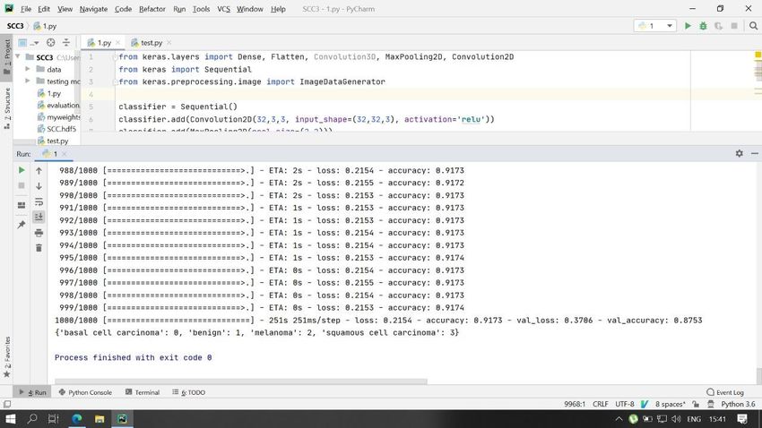
ISSN No.: 2454- 2024 (online)

 International Journal of Technical Research & Science
 ➢ Scaly or rough red patches with irregular borders.
 ➢ Wart-like growth.
 ➢ Sore that bleeds easily.
1.2 Melanoma Skin Cancer [MSC]
This is more threatening and prominent in men and women. As per net statistics of the year 2019, around 100,400
people are diagnosed with melanoma in the USA out of this 60,000 are men and 40,400 are women. This type is
ranked 6th most occurring type among men and the 5th most occurring type of cancer among women.
Unbelievably people that are diagnosed with skin cancer are remarkably older than 70 years. 90% per cent of
white people are diagnosed with Merkel cell cancer. Women are twice less likely to be diagnosed than men [12].
The melanoma type has a drastic increase in the past few decades, although it depends on the age factor. If this is
diagnosed at an early stage, it is cured by initial surgery. The number of people who are already living with skin
cancer for about 5 years is identified by a “5-year survival rate”. According to the reports people who got
diagnosed in an initial stage their survival rate is 5 years is almost 90% but this depends on the thickness of the
melanoma, the spread of melanoma to different parts of the body Lymph node involvement [13].
Increase of melanoma is over 700% per cent since the past 50 years. This is a lifetime risk. One person dies with
melanoma each hour. Mostly happens to young women and the mortality rate is quite high. People who are
detected with melanoma early indeed has 5 years of survival at least and 99% curable [14].
Characteristics of melanoma are:
 ➢ Cells of melanoma are asymmetrical when we draw a line from the centre, they don't collide nor be
 symmetrical.
 ➢ Most of the cells have border irregularities and are recognized as the scalloped or notched edge.
 ➢ Colour variability matters here, generally affected cells turn to brown, tan, black, and as progressing
 they turn to red, white and blue.
 ➢ Moles are of higher diameter than normal moles and grow longer than common moles.
 ➢ Evolution, the evolution of cells is higher than regular there exist more and more moles at one point
 of skin.
Places where melanoma can exist without our notice:
 ➢ Melanoma can hide under fingernails or toenails, it knows as Acral-melanoma and is the rare form
 of melanoma. Most commonly present in people like Asian descent, black people and other people
 with dark skin.
 ➢ Melanoma can be in the nose, mouth, digestive tract, urinary tract or vagina this kind of melanoma
 is known as Mucosal Melanoma and these are very difficult to detect as they have similar conditions
 with other issues.
 ➢ It can be in the eye; eye melanoma is also called ocular melanoma. Generally, they occur in the
 layer beneath the white of the eye this can cause eyesight, vision problem and can be diagnosed
 during an eye test.

 Fig. 1.2 Images of Benign vs Malignant Skin Cancer
Risk factors for MSC are:
 ➢ Fair skin.
 ➢ History of Sunburn.
 ➢ Excessive Ultraviolet rays’ exposure.
 ➢ Generally, people who live close to the equator or who live at a higher temperature.
 ➢ Having many moles or unusual moles.
 ➢ Family history of Melanoma.
 ➢ Other inherited genetic conditions such as retinoblastoma, Li-Fraumeni syndrome, xeroderma
 pigmentosum.
DOI Number: https://doi.org/10.30780/IJTRS.V06.I05.001 pg. 3
 www.ijtrs.com, www.ijtrs.org
Paper Id: IJTRS-V6-I4-001 Volume VI Issue V, May 2021
 @2017, IJTRS All Right Reserved
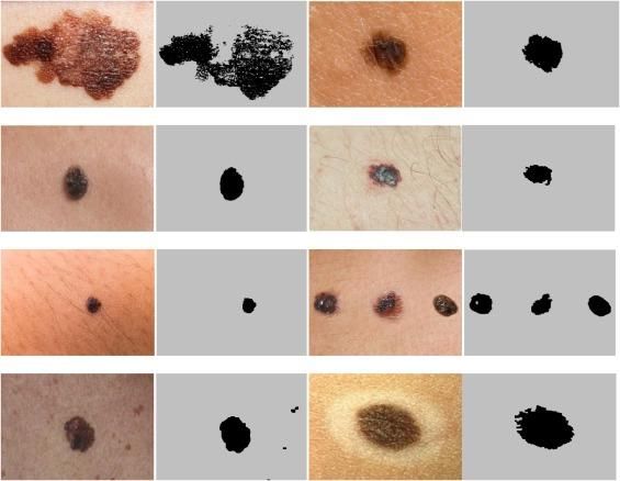
ISSN No.: 2454- 2024 (online)

 International Journal of Technical Research & Science
2. SIGNIFICANCE OF THE SYSTEM
The primary focus of this paper is, how Machine Learning Techniques are being implemented in its most archaic
nature applied in detecting skin cancerous cells before scathingly dangerous effects on the human's lives. Its study
on literature survey is written in section III, Ease of use is given in section IV, Section V describes Dataset that
we have used in the algorithm and for references, Background work of the paper is described in section VI,
Methodology is being elaborated in section VII, section VIII consists of the results that have been tested and
verified by the model, section IX has the conclusions on the paper and its methodology.
3. LITERATURE REVIEW
Although there are many types of Neural Networking architectures after a thorough analysis of these architectures,
Convolutional neural; networking works better in detecting Skin Cancer as it entirely deals with image processing
and has a lot of support for image analysis such as it’s able to help users extract features very distinctively, has
high accuracy, it follows a hierarchical model which works on building an entire network and then provide a fully
connected layer at the end where every neuron are connected and the outputs are processed. Most importantly
convolution layers are built for images processing which helps in detecting features that can be missed in simply
flattening images pixels values and is done automatically without any human supervision. It creates classes itself
without the help of human otherwise which is a big task and CNN is computationally efficient. The link between
adaptive filters and feed-forward neural networks are known as CNN. All these claims are being verified by
trustable sources [1].
Most of the other models that are detecting Skin cancer are using Support vector Machines (SVM), Artificial
Neural Networking (ANN), K-Means. Compared to these CNN was giving more better results as this is fully
based on Image processing and build for this sole purpose [2]. Both SVM and CNN are Supervised machine
learning algorithms where CNN is a parametric classifier which uses hyper parameters during the training phase
and SVM is a non-parametric classifier which finds a linear line to separate classes and mainly used for
classification and regression analysis whereas CNN is non-linear, this works well with visual image recognition
and increases model complexity by adding more and more layers. On huge data, CNN works far better than SVM
[3].
According to a study done by “Abien Fred M. Agarap” written in this paper “An Architecture Combining
Convolutional Neural Network (CNN) and Support Vector Machine (SVM) for Image Classification” [4] has
proven that Convolutional neural networking gives the accuracy of 99.23% while Support Vector Machine gives
out the accuracy of 99.04% which gets CNN lead over SVM. He has performed a detailed analysis of both the
techniques [5].
4. EASE OF USE
Generally, the goal of this paper is to achieve the maximum probability of predicting skin cancerous cells which
makes patients check within the app instead of visiting hospitals and going through conventional methods. Hence,
the final goal is to reach the maximum possibility and audience.
According to the survey we have done, we have tested our program on 20 people and trained over 2435 images
and tested on 450 test data, this program was giving 8 out of 10 right outputs
The conventional way would be long and costs a certain amount of money for a patient whereas if they use this
technique they will get to know whether they are suffering from skin cancer or not up to certain probability free
of cost, hence without this model it'll be hard for most of the below middle-class people to afford hospitals as it
costs around 7000 Rupees to 20000 Rupees per person for testing. Therefore it will be good to visit hospitals if
we are confirmed that we are positive with skin cancer.
To access these users just need to run the “SSC_Main_2” file and put their skin image and run the file. Users can
use this through their laptop at home or anywhere just with the help for laptop. Users don’t need internet
connectivity to access it.
Users need little knowledge of running the model even if they don’t have they can learn it in simple steps.
5. DATASET
There are four sub-datasets for this dataset. The first dataset has basal cell carcinoma cancerous cells samples,
then squamous cell carcinoma cells samples photos come melanoma cancerous cells samples then samples of
skin cells which aren’t cancerously known as benign these are samples of healthy cell tissues. This division
helps us in getting an indication and for diving classes to train machine learning models. The dataset is taken
from minimum 7 to 95 years old age range of people having cancerous cells on the skin.
6. BACKGROUND
Hence considering the loss and sacrifices due to Skin cancer and analyzing how to overcome the issue, the answer
would be detecting it early. Most of the time for most of the people sin cancer might be in the form of normal
Mole which we obviously think is mole without knowing the risk behind it we ignore which leads us to the bringer
of death [15].

DOI Number: https://doi.org/10.30780/IJTRS.V06.I05.001 pg. 4
 www.ijtrs.com, www.ijtrs.org
Paper Id: IJTRS-V6-I4-001 Volume VI Issue V, May 2021
 @2017, IJTRS All Right Reserved
ISSN No.: 2454- 2024 (online)

 International Journal of Technical Research & Science
The possible solution to this upcoming fatal incident would be by detecting it early, of course, there are many
ways for this but most of the ways take a lot of time and needs to be travelled from one place to other such as
Home clinic/hospital. There are other ways of self-checking for skin cancer to spot it early, and that’s done in the
following ways [16]:
Checking whole body thoroughly (including genital areas, toe/fingernails, back…etc.), new skin growth, irregular
shapes, bumps, expanding of the skin or changing growth, rough red patches, bleeding skin, wart-like growing,
odd-shaped moles/ changing in size of them/colour [17].
This is a long process and can’t say accurately whether we are diagnosed with it or now. After considering this
process it's good to detect skin cancer early so that the treatment would be given once it's late it'll be a problem.
Thus, we need to do something which detects automated skin cancer cells of humans, so the diagnosis and
treatment can be given before it grows dangerous and that is to detect skin cancer using machine learning and
subdomain as image processing using neural networking this model is consistent, efficient and cost-effective as it
was done purely from the open-source image processing techniques [18]. Although there are many ways of using
image processing such as:
 ➢ K-means algorithms.
 ➢ Support Vector Machines algorithm.
 ➢ Neural networking.
 ➢ K-Nearest Neighbours algorithm.
 ➢ Scale-Invariant feature transform algorithm.
 ➢ Random sample consensus algorithm.
 ➢ Cross-Validation algorithm.
6.1 Neural Networks
Let us understand the basic types of neural networks and their applications so that we will know which neural
networking model to use of all the pre-present architectures:
6.1.1 Feedforward Neural Networks:
These are known as artificial neurons. Generally used for supervised learning. These are the simplest form of
artificial neural networks and it has one direction input/output/data. These might have hidden layers to justify it
just has front-propagation wave and no sign of backpropagation wave.
Summation of products of the inputs and weights are being calculated and fed to the output. If the threshold value
is 0 then the output is considered to be above a particular value and neuron fires with the activated output of value
1. If the neuron does not fire then it usually emits -1 which means it’s deactivated
Applications:
 ➢ Computer vision.
 ➢ Speech recognition.
 ➢ Pattern recognition.
 ➢ Speech recognition.
6.1.2 Radial basis function Neural Networks:
Mentioned as RBF. This has layers in first features are being combined with radial basis function in the inner layer
and output of it is taken into consideration. Which will be used while computing the same output in the next step
which I s memory.
Applications:
 ➢ Function approximation.
 ➢ Pattern recognition.
 ➢ Time series prediction.
 ➢ Universal approximation property.
 ➢ Mechanical Fault diagnosis.
6.1.3 Kohonen Self Organizing Neural Networks
Type of ANN used to train unsupervised learning to get low dimensional, discrete representation of input space
on training data called a MAP and it is known as “dimensionality reduction”. Has either 1 or 2 dimensions.
This process has different kinds of parts, in the very first phase of self-organization, every neuron value is
initialized with a small weight and the input vector. In 2nd phase, here comes “Winning Neuron” which is closest
to the neuron, and neuron connected to this neuron will move towards closest winning neuron and this distance is
calculated using Euclidian distance and the neuron with less distance wins.
Applications [19]:
 ➢ Pattern recognition.
 ➢ Medical analysis to clustering data into different categories.
 ➢ Tool monitoring.
 ➢ Static security assignment.
DOI Number: https://doi.org/10.30780/IJTRS.V06.I05.001 pg. 5
 www.ijtrs.com, www.ijtrs.org
Paper Id: IJTRS-V6-I4-001 Volume VI Issue V, May 2021
 @2017, IJTRS All Right Reserved
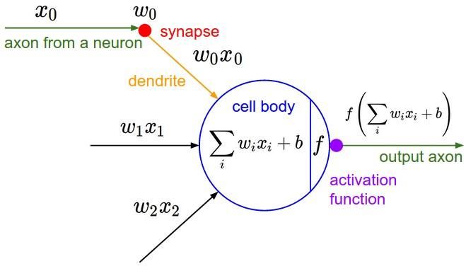
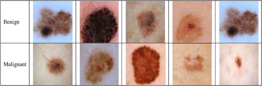
ISSN No.: 2454- 2024 (online)

 International Journal of Technical Research & Science
6.1.4 Recurrent Neural Network (RNN) – Long Short-Term Memory
RNN works by saving the output of a layer, then feeding same back to the input to help in the prediction of the
outcome of the layer.
In this 1st layer is formed similar to feed-forward NN with the product of the features and sum of weights. The
RNN starts once this is computed, i.e. one-time step to the next each neuron will remember some information it
had in the previous step. This makes every neuron act like a memory cell in performing computations. For that
process, we need to let RNN work on the forward propagation and store in memory what information it needs for
later use. Here, if the predicted output is wrong we use the learning rate or error correction to make small changes
so that it will gradually work towards making the right prediction during the backpropagation [20].
Applications of recurrent neural networks:
 ➢ Text to speech conversion.
 ➢ Text conversion of Human-like speech.
6.1.5 Convolutional Neural Network (CNN)
CNN's are very much similar to feed-forward neural networks, whose principles are having learnable weights
and biases.
CNN is a Deep Learning algorithm which takes input in form of an image, assigns importance such as biases and
learnable weights to various features/objects in the image and then can differentiate one from the other [18].
Applications:
 ➢ Signal and image processing.
 ➢ Computer vision.
 ➢ Image processing.
 ➢ Image classification.
 ➢ Image analysis and recognition.
 ➢ Facial recognition applications.
 ➢ Social media.
 ➢ Recommender systems.
 ➢ Document digitization.
6.1.6 Modular Neural Network:
These have different kinds of networks collections which work independently and contribute outputs. Each neuron
contributes a unique set of values of input compared to another kind of network-constructions and then performs
later tasks. While accomplishing tasks these don’t interact with each other networks [21].
Advantages of these are: Large computational process are breakdown into small thus decreasing complexities,
which helps in reducing the number of connections hence negotiating neural network interactions with others
which leads to computations speed fastening though this time depends on the involvement of neurons in
computations and their number.
Applications of the modular neural network are:
 ➢ Aircraft trajectory prediction.
 ➢ Pattern recognition.
 ➢ Grade estimations.
 ➢ Biometrics.
7. PROPOSED SYSTEM

 Preprocessing of
 Data Acquisition Pre-processing of data Classification using CNN
 data
 `

 Data Set Patient’s details Disease prediction

 Fig. 7.1 Flow Chart Representation of CNN
DOI Number: https://doi.org/10.30780/IJTRS.V06.I05.001 pg. 6
 www.ijtrs.com, www.ijtrs.org
Paper Id: IJTRS-V6-I4-001 Volume VI Issue V, May 2021
 @2017, IJTRS All Right Reserved
ISSN No.: 2454- 2024 (online)

 International Journal of Technical Research & Science
So let us see what happens with data in CNN in brief:
In the above flow chart Fig. 7.1, detailed explanation of titles:
7.1 Data Acquisition
This is the sampling real-world data then converting it to digital values which can be used by a computer to process
them, Generally waveforms are converted into digital form, in our case skin image in the format of JPEG are
converted into digital format and then used in the next step, pre-processing.
 ➢ Pre-processing of data: let us understand this by taking reference of the dataset images of skin used
 in the paper, this data is going to get encoded, transformed, bought to s state where the algorithm
 can understand it easily and the features of images are interpreted by it. Whose features might be
 vectors, patterns, matric values, entities, edges, corners, Scale-invariant Feature Transform,
 Speeded up robust feature.
 ➢ Classification using CNN: Though there are many other effective ways to classify data, CNN does
 it well with images and is preferred rather than other algorithms, this work with high dimensional
 pictures, deep learning framework like TensorFlow, which helps to get greater outputs at fewer
 lines of codes.
 ➢ Disease prediction: Prediction of the disease in this case Skin cancer, in this step the algorithm
 predicts if the input image is diagnosed with skin cancer of not.
 ➢ Patient details: This is the step where details of the patient are known to the algorithm and outputs
 them along with the disease prediction.
 ➢ Data set: Dataset is taken from various networks and some are taken by the survey that’s been done
 and this dataset is processed to the algorithm for going through processes written before this step.
7.1 History of Convolutional Neural Networking
Neural networking was first proposed by Warren McCullough and Walter Pitts in 1944, who were students of the
University of Chicago then moved into MIT in 1952.
Convolutional Neural Networks are generally known as “ConvNets” and are considered to be the backbone of
many computer vision systems. Aim of ConvNets is to find the correlation between adjacent inputs found in the
image. CNN consists of one or more convolutional layers which are being followed by a fully connected layer
and whose design is based on the 2D input image’s structure.
The first work on Neural networking was done by “Dr Kunihiko Fukushima” which was inspired by “Hubel and
Wiesel's” work on complex and simple cells and proposed “NEOCOGNITRON” model.
Then the first-ever work on CNN was done in the 1990s by “Yann leCun et al”, inspired by The Neocognitron,
though it was invented in 1980s the first official paper was launched in 1998 citing word deep convolutional
network about 6184 times. This paper was widely appreciated and regarded as one of the most influential
publications in the field of technology.
Later Alex Krizhevsky developed AlexNet around 2012 and that achieved “state-of-the-art performance” in
“ImageNet challenge” and dataset consisted of 1.2 Million images of high resolution.
As Human vision and biological neural networks, CNN also follow “Simple-to-Complex” hierarchical structure,
just that CNN’s are build using mathematical operations [1], [3], [12], [19].

 Fig. 7.2 Structure of a Neural Network
DOI Number: https://doi.org/10.30780/IJTRS.V06.I05.001 pg. 7
 www.ijtrs.com, www.ijtrs.org
Paper Id: IJTRS-V6-I4-001 Volume VI Issue V, May 2021
 @2017, IJTRS All Right Reserved
ISSN No.: 2454- 2024 (online)
International Journal of Technical Research & Science
Convolutional neural networking:
In a nutshell:
➢ CNN is a Deep Learning algorithm which takes input in form of the image, assigns learnable
weights and biases to various features/objects in the image and then can differentiate one from the
other.
➢ Similar to ANNs but more capable and advanced, CNNs are more complex around computation
and pre-processing.
➢ The input to the model is skin lesion image and then it’s applied by conventional image processing
techniques, it analyses those inputs to provide conclusion about the presence of cancerous cells.
➢ This Lesion Image analysing tools checks for the various Melanoma parameters Like Border,
Asymmetry, Diameter, colour etc. by size, texture and shape analysis for image segmentation and
feature stages.
In-depth:
Convolution neural networking is generally called as CNN/ ConvNets. In a nutshell, this is a Deep learning
algorithm where the input would be an Image then assign importance to it based on various features and which
will be able to differentiate one feature form other, which are known as weights and biases. CNN learns features,
characteristics of the training images.
The most important feature of CNN’s are time taken for “Pre-Processing” which would be much less when
compared to other algorithms which perform classification. The architecture of ConvNets is similar to human
brain neural networks pattern of connectivity and these are even inspired by visual cortex organization.
While coming to technical terms ConvNets train and test models each of the input images shall pass through series
of convolution layers using filters known as kernels then through pooling layer then after that they get flattened
then to fully connected layers and then applied with activation function known as Softmax whose probability
functional values range between 0 and 1.
The Softmax function is represented this way:
i i
( = | θ^(i)) = ( θ )/ ∑ =0 θ (1)
Where,
θ = W0 X0 + W1 X1 + ……+ Wk Xk =∑ =0 = (2)
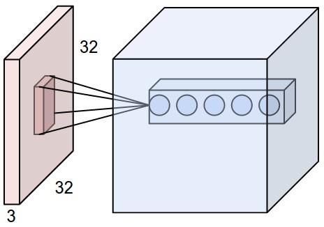
Generally, photos are scaled to (32*32*3) which corresponds to Width (W), Height (H) and Colour channels(C)
red, green, blue. Which means neurons are arranged in 3 dimensions (W, H, C). The final output layer would be
the dimension of (1*1*10) as by end image was pre-processed and flattened into a single vector arranged along
with depth dimension along with class scores. Weights on the neuron are also calculated this way:
Ex:32 ∗ 32 ∗ 3 = 3072 (3)
3072 total weights are being processed in the neural network in one hidden layer of it. when image size increases
the number of weights that needs to be processed increased which would take a lot more processing power, time
and several more neurons and finally leads to overfitting.
Simply ConvNets are a sequence of layers and the most important layers to build neural networking architectures
are Convolutional layer, pooling and Fully Connected layer. Then these layers are stacked upon each other to form
fully connected neural networking architecture.
7.2 Convolution Layer
The Convolution layer is the core method of a Convolutional Neural Network. Its parameters consist of different
learnable filters and every filter is small but gives a major feature that we need. ConvNets learns filters by which
it activates when it detects the features such as colour, edge…etc. by this process, we will have a set of filters in
each Convolution layer. Each one of them will create an activation layout by which we can detect output we need.
7.3 Local Connectivity
As Images are high dimensional inputs connecting neuron to neurons are impractical, instead, we connect each
neuron to a local region of the volume of input. The special extent of that connectivity is defined by “Respective
filed of neuron”. The depth axis along with the field of the neuron is equal to the depth of input volume. The
important part is to emphasize the asymmetry that occurs and how we treat that spatial dimensions such as height
and width along with depth dimensions. The connections are always full with the depth of input volumes but are
local inside space.
For example, let the size of input be [32x32x3] of volume, other examples might be RGB CIFAR-10 image. Let
us keep filter size as [5x5], then the weights of each neuron would be the size of [5x5x3] region of the part of the
input volume, which would be making 75 {5*5*3 = 75} weights then the bias of +1 would be added. Now change
DOI Number: https://doi.org/10.30780/IJTRS.V06.I05.001 pg. 8
www.ijtrs.com, www.ijtrs.org
Paper Id: IJTRS-V6-I4-001 Volume VI Issue V, May 2021
@2017, IJTRS All Right ReservedISSN No.: 2454- 2024 (online) International Journal of Technical Research & Science the notice in the depth axis along with the extent of the connectivity would turn out to be 3, as this would be the input volume’s depth. Fig. 7.3 Red, Example Input Volume Fig. 7.4 Neurons that Remained Unchanged 7.4 Spatial Arrangement Zero-padding, depth and Stride are the three main hyper-parameters which control the size of output volume. Refer above data for knowing more about them. Table-7.1 Tabular representation of usage of depth, Stride and Zero-Padding DEPTH STRIDE ZERO-PADDING The number of filters that are Performs sliding of a filter. Zero-padding controls the being used. spatial size of outputs volumes. Size of output volume is being calculated by the above formula: 1((W−F)+2P) ((W−F)+2P) OUTPUT VOLUME = +1+ (4) S S Where, ➢ Input volume size = W ➢ Receptive field size = F ➢ Stride = S ➢ Zero padding = P Fig. 7.5 Neurons using stride for clear Understanding 7.5 Parameter Sharing Controlling the number of parameters is known as parameter sharing. Let's see a real-world example here: We see that there are 40*40*85 = 136,000 neurons in the very first convent layer and each neuron weight 16*16*3 =768 (weights) and bias of value usually 1 together, they add up to 136,000*763=104,448,000 parameters in the very 1st layer itself and the number is very high that can be reduced by making depth slice which is the only valid DOI Number: https://doi.org/10.30780/IJTRS.V06.I05.001 pg. 9 www.ijtrs.com, www.ijtrs.org Paper Id: IJTRS-V6-I4-001 Volume VI Issue V, May 2021 @2017, IJTRS All Right Reserved
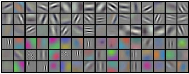
ISSN No.: 2454- 2024 (online)

 International Journal of Technical Research & Science
assumption. If every neuron in a single depth uses the same weight vectors and those are passed to the previous
layer i.e. convolution layer, and each depth slice is computed neuron weights convolution along with input volume.
Thus, that layer was given name as a convolution layer.
Hence, it’s commonly referred to as “Filters/Kernels” (Sets of weights) which are convoluted along with inputs.
Parameter sharing assumptions sometimes might not make any sense. In such types of cases it is common to relax
parameter sharing scheme this is called a locally connected layer.

 Fig. 7.6 Filters learnt by Krizhevsky et al in his paper, size= 55*55 in 1 Depth Slice
7.5 Pooling Layer
This layer progressively reduces the spatial size to reduce the parameters and calculations of the network. Pooling
layer uses MAX operations on every depth slice to reduce parameters. Filter size must be (2x2) with the value 2
as stride, downwards. Each sample depth is the input of 2 on the height and width which leads to discarding 73%
of activations is very common in pooling layer. In this case, MAX operation take a max 4 numbers. Pooling layer
also performs other functions like L2-norm pooling or average pooling.
Pooling layer does:
 ➢ Accepts a volume of size
 W1×H1×D1W1×H1×D1 (5)
 Where [w1 = H1 = D1]
 Where,
 W = width
 H = Height
 D = dimensions
 ➢ Requires two hyper parameters.
 1. Spatial extent.
 2. Stride.
 ➢ Produces a volume of size.
 W2×H2×D2W2×H2×D2 (6)
 Where
 H2=(H1−F)/S+1 (7)
 W2=(W1−F)/S+1 (8)
 D2=D1 (9)
7.6 Normalization Layer
This was proposed to use in CNN with intentions of implementing its inhibition schemes which are observed in a
biological brain. These will decrease models training time which makes a huge impact. Each feature is normalized
which makes it easy as many weights might be higher or lower/unbiased. It reduces internal covariate shift which
means a change of network parameters while training which Improves training.
7.7 Fully Connected Layer
This takes outputs of previous layers and turns that into a single vector layer which can be an input for the next
steps and backpropagation. Takes inputs from feature analysis then apply weights to predict correct labels. This
gives the final probability of each label. Their activations can be computed with matrix multiplication followed
with bias offset.
Layer pattern would be:
 INPUT -> [[CONV -> RELU]*N -> POOL?]*M -> [FC -> RELU]*K->FC (10)
What generally happens inside a neural network?
DOI Number: https://doi.org/10.30780/IJTRS.V06.I05.001 pg. 10
 www.ijtrs.com, www.ijtrs.org
Paper Id: IJTRS-V6-I4-001 Volume VI Issue V, May 2021
 @2017, IJTRS All Right Reserved
ISSN No.: 2454- 2024 (online)

 International Journal of Technical Research & Science

 Fig. 7.7 Block diagram for predicting Skin Cancer
In the above Fig. 7.7, explanation of terms:
Skin cancer images to be predicted: These consists of the images of the skin that a user wants to detect the
probability of diagnosed with what type of cancer.
7.8 Segmentation
This is the most crucial part of image processing, as it divides the image into visual input then into segments to
analysis image i.e. identifying parts of images understanding them which object they belong to, this is the basic
step for object detecting. There are types in segmentations they are Semantic segmentation, Instance segmentation.
Semantic segmentation means it classifies objects into different classes and correspond them to real-world
categories and then colour them to identify by the model and also known as a dense prediction as this predicts
each pixel’s meaning. And whereas instance segmentation means it recognizes each instance of an object from the
image. This classifies every pixel.
7.9 Feature Extraction
This describes detailed shapes information contained in pattern and classifies those and make them easy to
recognize, this helps in finding relevant information about an object from original data then it represents same
information in reduced data such as dimensionality reduction.
Classification: This whole paper is related to classification hence we will learn more about it further. Classification
means extracting class’s information from an image as it consists of many different objects and then classifying
it, this is used to create “thematic maps. There are 2 types of classifications namely supervised and unsupervised.
8. MODEL PIPELINE
To Build the CNN Model which was used to detect skin cancer, we go through the above-written process of the
pipeline:
 ➢ Importing required packages such as Keras Packages, Libraries and TensorFlow.
 ➢ Initializing the Convolutional neural networks.
 ➢ Adding the Convolution Layers.
 ➢ Adding the Max-Pooling layer.
 ➢ Flattening those above layers.
 ➢ Fully connecting above layers.
 ➢ ConvNets compilation.
 ➢ Model fitting to the dataset.
Let us see how these are used in the current algorithm for s=detecting skin cancer:
8.1 Import Keras Packages and Libraries, TensorFlow
From Keras.layers import Dense, Flatten, Convolution3D, MaxPooling2D, Convolution2D
from Keras import Sequential
from keras.preprocessing.image import ImageDataGenerator
8.2 Initializing the CNN
First, we arrange all the matrices of the image into a sequence and perform an operation on that and to do that we
use keyword Sequential()
From Keras. models
Model = sequential()

DOI Number: https://doi.org/10.30780/IJTRS.V06.I05.001 pg. 11
 www.ijtrs.com, www.ijtrs.org
Paper Id: IJTRS-V6-I4-001 Volume VI Issue V, May 2021
 @2017, IJTRS All Right Reserved
ISSN No.: 2454- 2024 (online)

 International Journal of Technical Research & Science
8.3 Adding the Convolution Layer
Then we convolute the matrices or arrays using conv2d which is available in Keras. layer.
model.add(convolution2d(64,3,3,input_shape=(64,64,3),activation=’relu’)
The first 64 represents a bit of the computer which generally should be 64.
The next 3, 3 are the size or dimensions.
The next 64, 64 in input shape are the matrix pixels.
The next 3 is colour channels, 3 means red blue and green channels.
ReLu is the activation function, which is defined as Rectifier Unit. Which activates the neurons of the CNN, it
adds non-linearity to improve the efficiency of the NN it only keeps Positive values and neglects Negative values.
8.4 Adding the Pooling Layer
Then for the convoluted data, we apply max-pooling using keyword maxpooling2d from Keras. layers.
In max-pooling, all the training is done to the dataset: model.add(MaxPooling2D(pool_size=(2,2)))
Here the attributed (2, 2) describe the factor by which to downscale the images.
8.5 Adding Flattening to the Layer
After training it, we flatten all the matrices using flatten from Keras. layers.
8.6 Adding the Fully Connected Layer
Now, this is the most important part of the code, the dense layer is known as a fully connected layer whose output
layer which is similar to hidden layers in ANNs.
model.add(Dense(output_dim=128,activation = ‘relu’)
Here output dimensions are 128 because of the computer, it makes code more accurate and it should not be too
small such as 32 or too big and thus 128 is accurate.
Activation function ReLu.
model.add(Dense(output_dim=1,activation = ‘sigmoid’)
Here in this output dimensions are 1 because we need to get one output that is either yes or no (0 or 1)
And the activation function here is sigmoid which is a type of activation function and defined as squashing
function whose values would lie between 0 and 1.
8.7 Compiling the CNN
Next step is to compile the CNN using compiler keyword with attributes optimizer, loss and metrics.
model. compile(optimizer=’adam’,loss=’binary_crossentropy’,metrices=[’accuracy’])
Adam is a stochastic gradient descent algorithm which is the fastest algorithms of all available.
The model will evaluate metrics.
Generally, metrics = true prediction/total prediction.
8.8 Fitting the CNN to my image dataset
The model was fitted to the proposed dataset by using ImageDataGenerator function. This is used to expand
dataset size artificially for creating modified images versions in the dataset and its application is for data
Augmentation. It replaces original batch with new random batches of data. Then takes the new image and write it
back to the model and keeps repeating this model until all the epochs are completed.
For example, If the dataset has 100 images it turns them into 1000 images.
Step 1:
Training batch is presented to it.
train_datagen=ImageDataGenerator (rescale=1./255,shear_range=0.2,zoom_range=0.2,horizontal_flip=True)
Where,
Rescale= reshaping images into different pixels here it is 1./255 size as 255 is the maximum pixel value.
Shear range = image will be distributed along the axis to create perception angles basically to view image from
various angles as of human viewing structure.
Zoom range= the value up to which the images can be zoomed and reformed
Horizontal flip= rotating images into various angles.
Step 2:
Transforms every image to the batch of new random images, such as it rotates it, changes resolution..etc.
Step 3:
Now random batch is returned to model.
classifier.fit_generator(train_set,steps_per_epoch=10000,=30,validation_data=test_set,validation_steps=300000
)
Where,
A trainset is the training dataset location.
Steps per epoch are the number of batches of samples to be used in one iteration and the start of the next iteration.

DOI Number: https://doi.org/10.30780/IJTRS.V06.I05.001 pg. 12
 www.ijtrs.com, www.ijtrs.org
Paper Id: IJTRS-V6-I4-001 Volume VI Issue V, May 2021
 @2017, IJTRS All Right Reserved
ISSN No.: 2454- 2024 (online)

 International Journal of Technical Research & Science

 Fig. 8.1 Step by step image processing in a Neural Network
9. RESULTS
The present paper differentiates between 3 types of skin cancer and benign. They are Squamous cell carcinoma,
Basal cell carcinoma and malignant type of cancers.
If the value is 0 then its Basal cell carcinoma, if the output value is 1 it is benign, if the output value is 2 then its
melanoma and if the value of output is 3 then its Squamous cell carcinoma.
Model is shown an accuracy of 91.74% on trained data and it shows value accuracy of 87.33%. Here are the
screenshots that show the accuracy of the model.

 Fig. 9.1 The Output of the Model along with the Classification of types of Skin Cancer
These are not permanent values by the change in a dataset and by the amount of dataset value of accuracies change.
CONCLUSION
Skin cancer is a deadly disease which is safely diagnosed when detected early and for maintaining a healthy
lifestyle. One of the accurate ways to detect skin cancer early is by using a machine learning algorithm which is
Convolutional Neural Networking. This paper contains the methodology of detecting skin cancer with accuracy
greater than 90% as CNN is very powerful and solely made for image processing. We train different types of skin
cancer cells through image format and apply CNN over them so that it can perform well. Dataset is taken manually
from different websites and in real-world through surveys. We have compared different types of algorithms used
to detect skin cancer in this paper. Hence, after a comparison of different algorithms, CNN is turned out to be the
best algorithm to detect skin cancer.
REFERENCES
[1] A. A. Ali and H. Al-Marzouqi, “Melanoma detection using regular convolutional neural networks,” 2017
 Int. Conf. Electr. Comput. Technol. Appl. ICECTA 2017, vol. 2018-Janua, pp. 1–5, 2017.

DOI Number: https://doi.org/10.30780/IJTRS.V06.I05.001 pg. 13
 www.ijtrs.com, www.ijtrs.org
Paper Id: IJTRS-V6-I4-001 Volume VI Issue V, May 2021
 @2017, IJTRS All Right Reserved
ISSN No.: 2454- 2024 (online)

 International Journal of Technical Research & Science
[2] P. M. Amulya and T. V. Jayakumar, “A study on melanoma skin cancer detection techniques,” Proc. Int.
 Conf. Intell. Sustain. Syst. ICISS 2017, no. Iciss, pp. 764–766, 2018.
[3] A. Naeem, M. S. Farooq, A. Khelifi, and A. Abid, “Malignant Melanoma Classification Using Deep
 Learning: Datasets, Performance Measurements, Challenges and Opportunities,” IEEE Access, vol. 8, pp.
 110575–110597, 2020.
[4] A. F. Agarap, “An architecture combining convolutional neural network (CNN) and support vector machine
 (SVM) for image classification,” arXiv Prepr. arXiv1712.03541, 2017.
[5] E. Jana, R. Subban, and S. Saraswathi, “Research on Skin Cancer Cell Detection Using Image Processing,”
 2017 IEEE Int. Conf. Comput. Intell. Comput. Res. ICCIC 2017, 2018.
[6] R. L. Siegel, K. D. Miller, and A. Jemal, “Cancer statistics, 2016,” CA. Cancer J. Clin., vol. 66, no. 1, pp.
 7–30, 2016.
[7] M. A. Albahar, “Skin lesion classification using a convolutional neural network with novel regularizer,”
 IEEE Access, vol. 7, pp. 38306–38313, 2019.
[8] U.-O. Dorj, K.-K. Lee, J.-Y. Choi, and M. Lee, “The skin cancer classification using deep convolutional
 neural network,” Multimed. Tools Appl., vol. 77, no. 8, pp. 9909–9924, 2018.
[9] M. A. Kassem, K. M. Hosny, and M. M. Fouad, “Skin lesions classification into eight classes for ISIC 2019
 using deep convolutional neural network and transfer learning,” IEEE Access, vol. 8, pp. 114822–114832,
 2020.
[10] P. Tschandl et al., “Expert-level diagnosis of non-pigmented skin cancer by combined convolutional neural
 networks,” JAMA Dermatology, vol. 155, no. 1, pp. 58–65, 2019.
[11] C. Barata and J. S. Marques, “Deep learning for skin cancer diagnosis with hierarchical architectures,” Proc.
 - Int. Symp. Biomed. Imaging, vol. 2019-April, no. Isbi, pp. 841–845, 2019.
[12] R. S. Sanketh, M. Madhu Bala, P. V. N. Reddy, and G. V. S. Phani Kumar, “Melanoma Disease Detection
 Using Convolutional Neural Networks,” Proc. Int. Conf. Intell. Comput. Control Syst. ICICCS 2020, no.
 Iciccs, pp. 1031–1037, 2020.
[13] H. R. Mhaske and D. A. Phalke, “Melanoma skin cancer detection and classification based on supervised
 and unsupervised learning,” 2013 Int. Conf. Circuits, Control. Commun. CCUBE 2013, pp. 1–5, 2013.
[14] F. K. Nezhadian and S. Rashidi, “Melanoma skin cancer detection using colour and new texture features,”
 19th CSI Int. Symp. Artif. Intell. Signal Process. AISP 2017, vol. 2018-Janua, pp. 1–5, 2018.
[15] R. Suganya, “An automated computer aided diagnosis of skin lesions detection and classification for
 dermoscopy images,” in 2016 International Conference on Recent Trends in Information Technology
 (ICRTIT), 2016, pp. 1–5.
[16] H. N. Pham et al., “Lesion Segmentation and Automated Melanoma Detection using Deep Convolutional
 Neural Networks and XGBoost,” Proc. 2019 Int. Conf. Syst. Sci. Eng. ICSSE 2019, pp. 142–147, 2019.
[17] D. H. Murphree and C. Ngufor, “Transfer learning for melanoma detection: Participation in ISIC 2017 skin
 lesion classification challenge,” arXiv Prepr. arXiv1703.05235, 2017.
[18] M. Ashcroft, L. Kaati, and M. Meyer, “A Step Towards Detecting Online Grooming-Identifying Adults
 Pretending to be Children,” Proc. - 2015 Eur. Intell. Secur. Informatics Conf. EISIC 2015, pp. 98–104, 2016.
[19] E. Chabi Adjobo, A. T. Sanda Mahama, P. Gouton, and J. Tossa, “Proposition of convolutional neural
 network-based system for skin cancer detection,” Proc. - 15th Int. Conf. Signal Image Technol. Internet-
 Based Syst. SISITS 2019, pp. 35–39, 2019.
[20] S. Pathan, K. G. Prabhu, and P. C. Siddalingaswamy, “Techniques and algorithms for computer-aided
 diagnosis of pigmented skin lesions—A review,” Biomed. Signal Process. Control, vol. 39, pp. 237–262,
 2018.
[21] K. Ramlakhan and Y. Shang, “A mobile automated skin lesion classification system,” in 2011 IEEE 23rd
 International Conference on Tools with Artificial Intelligence, 2011, pp. 138–141.

DOI Number: https://doi.org/10.30780/IJTRS.V06.I05.001 pg. 14
 www.ijtrs.com, www.ijtrs.org
Paper Id: IJTRS-V6-I4-001 Volume VI Issue V, May 2021
 @2017, IJTRS All Right Reserved
You can also read