Invivo confocal microscopy in dermatology: from research to clinical application - SPIE Digital Library
←
→
Page content transcription
If your browser does not render page correctly, please read the page content below
In vivo confocal microscopy in
                dermatology: from research to clinical
                application

                Martina Ulrich
                Susanne Lange-Asschenfeldt

Downloaded From: https://www.spiedigitallibrary.org/journals/Journal-of-Biomedical-Optics on 27 May 2020
Terms of Use: https://www.spiedigitallibrary.org/terms-of-use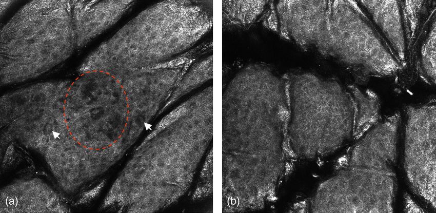
Journal of Biomedical Optics 18(6), 061212 (June 2013)

                In vivo confocal microscopy in dermatology:
                from research to clinical application
                Martina Ulrich and Susanne Lange-Asschenfeldt
                Charité Universitätsmedizin Berlin, Charitéplatz 1, 10117 Berlin, Germany

                              Abstract. Confocal laser scanning microscopy (CLSM) represents an emerging technique for the noninvasive
                              histomorphological analysis of skin in vivo and has shown its applicability for dermatological research as well
                              as its value as an adjunct tool in the clinical management of skin cancer patients. Herein, we aim to give an over-
                              view on the current clinical indications for CLSM in dermatology and also highlight the diverse applications of
                              CLSM in dermatological research. © 2013 Society of Photo-Optical Instrumentation Engineers (SPIE). [DOI: 10.1117/1.JBO.18.6.061212]
                              Keywords: cosmetics; fluorescence confocal microscopy; reflectance confocal microscopy; skin aging; skin cancer.
                              Paper 12620SS received Sep. 18, 2012; revised manuscript received Oct. 25, 2012; accepted for publication Oct. 26, 2012; published
                              online Jan. 21, 2013.

                1    Introduction                                                                  of the objective obtains parallel sections of the skin. Endoge-
                Confocal laser scanning microscopy (CLSM) represents an                            neous chromophores in the skin are responsible for the differ-
                emerging technique for the noninvasive histomorphological                          ences in reflectivity, which results in gray scale images. Of
                analysis of skin in vivo. In the past decade, various studies                      these, melanin represents the skin structure with the highest
                have shown the applicability of in vivo reflectance CLSM for                       refractive index and therefore provides the highest contrast
                the noninvasive diagnosis of melanocytic skin lesions as well                      by CLSM. On CLSM images, melanin-containing cells (both
                as nonmelanoma skin cancer. Furthermore, CLSM has been                             pigmented keratinocytes and melanocytes) appear white. But
                used for the evaluation of benign inflammatory skin diseases.                      also other skin structures such as keratin, collagen, or haemo-
                   Recently, novel devices combining the standard confocal                         globin are natural contrast agents.
                laser scanning microscopy in reflectance mode with multi-laser
                fluorescence techniques have been used for clinical studies.1
                   In addition, ex vivo set-ups allow for the evaluation of biopsy
                                                                                                   3       Application of In Vivo CLSM for Skin
                specimens enabling rapid evaluation of tumor margins in Mohs’                              Cancer Diagnosis
                micrographic surgery.                                                              Early diagnosis of skin cancer represents the most important fac-
                   Herein, we aim to give an overview of established CLSM                          tor in the management of the increasing skin cancer burden and
                techniques for in vivo diagnosis of skin disease, including a                      may significantly reduce mortality and morbidity of the patients.
                brief outlook on emerging CLSM systems currently under                             Most skin cancers can be cured by simple excision when diag-
                investigation.                                                                     nosed at an early evolving stage. This particularly applies to
                                                                                                   melanoma as its prognosis is directly correlated with anatomic
                2    History and Technical Principle of CLSM                                       depth of invasion.4 However, also in nonmelanoma skin can-
                In 1955, Marvin Minsky described the optical principle of                          cers (NMSC), such as basal cell carcinoma and squamous cell
                confocal laser scanning microscopy at Harvard University in                        carcinoma, early diagnosis is essential for positive therapeutic
                Boston, Massachusetts.2 However, this innovative technology                        outcome, prognosis, and cosmesis. Although these tumors
                was only published as a patent and remained more or less                           rarely metastasize, they are often located in cosmetic sensitive
                unrecognized until the 1990s, when Rajadhyaksha et al. first                       areas of the face and scalp where larger excisions may give rise
                conceived reflectance mode CLSM for skin imaging in its                            to disfiguring scars and healing impairment. In the past decade,
                current design.3 Since then, continued technical innovations                       a number of noninvasive treatment modalities have become
                decreased the size of the machine, thus facilitating the imaging                   available, thus optimizing the therapeutic management for
                process. Nowadays, handheld devices are available, which can                       selected subtypes of NMSC. In that regard, it has been suggested
                be used for rapid in vivo examination, facilitating evaluations in                 that noninvasive diagnostic techniques such as in vivo CLSM
                difficult anatomic areas and also at the bedside.                                  may be applied for therapeutic monitoring and follow-up.
                    The technical principle of CLSM is based on the illumination                       Despite the great potential of CLSM, it also has limitations,
                of a small spot within the tissue using a point light source.                      mainly including the restricted penetration depth. Furthermore,
                Reflected light is guided through a pinhole in front of the detec-                 the evaluation of a skin lesion by CLSM requires 5 to 10 min
                tor. Thus, only reflected light from the tissue spot, which is                     and is therefore more time consuming than clinical evaluation
                in-focus, is used for image generation, whereas light coming                       or dermoscopy alone. Due to the horizontal images of CLSM,
                from out-of focus planes is eliminated. Horizontal movement                        early invasion of skin tumors may be very difficult to diagnose
                                                                                                   as the basement membrane cannot be visualized. Lastly, the in-
                                                                                                   terpretation of CLSM images requires training and experience
                Address all correspondence to: Martina Ulrich, Charité Universitätsmedizin
                Berlin, Charitéplatz 1, 10117 Berlin, Germany. Tel: +49-30 450618276; Fax:
                +49-30450 518945; E-mail: martina.ulrich@charite.de                                0091-3286/2013/$25.00 © 2013 SPIE

                Journal of Biomedical Optics                                              061212-1                                            June 2013   •   Vol. 18(6)

Downloaded From: https://www.spiedigitallibrary.org/journals/Journal-of-Biomedical-Optics on 27 May 2020
Terms of Use: https://www.spiedigitallibrary.org/terms-of-use
Ulrich and Lange-Asschenfeldt: In vivo confocal microscopy in dermatology . . .
Fig. 1 Representative RCM images (500 × 500 μm) illustrating specific patterns of different skin tumors. (a) An RCM image of a melanoma with large
atypical nucleated cells (black arrows) in the epidermis, representing widespread pagetoid spread of atypical melanocytes. (b) An RCM image of a
benign nevus with the presence of small round cells of bright reflectance at the dermo-epidermal junction. A ringed pattern with well-edged papillae
(white arrows) and a cobble stone pattern of the overlying epidermis can be noted. (c) The typical cerebriform pattern of the epidermis in seborrheic
keratosis. Areas of epidermis with pigmented keratinocytes (black arrows) and invaginations filled with keratin can be noted (white asterisks). (d) A
typical pattern of a basal cell carcinoma with the presence of tumor islands (white dashed circle) consisting of elongated cells with palisading in the
periphery. Between the BCC tumor islands and the surrounding fibrous stroma small cleft-like spaces (white asterisk) can be noted that correspond to
mucin deposition. Dilated blood vessels (BV) can be seen in the surrounding stroma. (e) Illustrates the atypical honeycomb pattern of an actinic
keratosis, different size and shape of keratinocytes can be noted (pleomorphism). (f) Shows an RCM image of an invasive squamous cell
carcinoma illustrating tumor islands with central bright center (black dashed circle) located in the upper dermis corresponding to keratinization
(black asterisk).
of the observer as it is the case with every diagnostic techni- diagnosis of melanoma and equivocal pigmented lesions9–13
que (Fig. 1). and it has been shown by Pellacani et al. that CLSM may
improve the diagnostic specificity and may thus reduce the num-
ber of unnecessary biopsies of benign lesions. In 2007, Pellacani
3.1 Melanoma et al. described the RCM features of epidermal disarray, page-
toid cells in the epidermis, nonedged papillae, cellular atypia at
Different types of melanoma can be distinguished, including
the junction, atypical nests, and bright nucleated cells in the der-
superficial spreading melanoma (SSM), lentigo maligna mela-
mis as being associated with melanoma.9 Based on these criteria,
noma (LMM), nodular melanoma (NM), and acrolentiginous
melanoma (ALM). Of these, SSM represents the most common a score was developed that included two major criteria, each
type, accounting for about 70% of all melanomas diagnosed. scored 2 points (nonedged papillae, cellular atypia), and 4 minor
NM is the second most common form and diagnosis is often criteria, each scored one point (roundish pagetoid cells, wide-
challenging as the tumor arises on normal skin and clinical algo- spread pagetoid infiltration, cerebriform nests and nucleated
rithms like the “ABCD” rule usually fail to obtain the diagnosis cells in the dermis). A total RCM score ≥3 showed a sensitivity
in early stages. Furthermore, the tumors lack a horizontal growth of 91.6% and a specificity of 69.3% for the diagnosis of mel-
phase and vertical spread occurs at an early stage, resulting in anoma. Furthermore, the two step method was proposed by
higher metastatic potential. In contrast, LMM occurs on chroni- Segura et al.14 The two-step method described four features
cally sun-exposed skin of the elderly often showing a prolonged that distinguished melanocytic lesions from nonmelanocytic
horizontal growth phase. However, clinical as well as histo- lesions including cobblestone pattern of epidermal layers, page-
pathological diagnosis of LMM may be difficult to obtain in toid spread, mesh appearance of the dermoepidermal junction,
the background of actinic damage with the presence of multiple and the presence of dermal nests as criteria for melanocytic
solar lentigines. As in vivo CLSM offers the possibilty of eval- lesions. In the second step of the algorithm, two protective cri-
uating skin tumors noninvasively and with near-histological teria were identified (typical basal cells, edged papillae) and
resolution, the use of CLSM for melanoma and its differential scored minus one and two high risk criteria (roundish pagetoid
diagnosis has been the subject of extensive research in this field. cells, atypical nucleated dermal cells) were scored plus one. A
Earlier studies have focused on the correlation of CLSM fea- lesion with total score of 0 to 2 was indicative to be most prob-
tures of melanoma and naevi with histology5–8 and showed the able melanoma, with a sensitivity and specificity of 86.1%
reproducibility of histological criteria such as pagetoid spread and 95.3%, respectively. Very recently, a large study on 710 con-
and typical nests of melanocytes by RCM. Since then, multiple secutive cases analyzed the sensitivity and specificity of two
studies have evaluated the applicability of RCM for the algorithms for the diagnosis of basal cell carcinoma (BCC)
Journal of Biomedical Optics 061212-2 June 2013 • Vol. 18(6)
Downloaded From: https://www.spiedigitallibrary.org/journals/Journal-of-Biomedical-Optics on 27 May 2020
Terms of Use: https://www.spiedigitallibrary.org/terms-of-useUlrich and Lange-Asschenfeldt: In vivo confocal microscopy in dermatology . . .
and melanoma (MM).15 In this study, the diagnostic accuracy for performed by three independent groups have shown the applic-
the BCC algorithm was 100% sensitivity and 88.5% specificity. ability of RCM for the diagnosis of nonhyperkeratotic AKs.24–26
The MM algorithm showed a sensitivity of 87.6% and a speci- Ulrich et al. analyzed 46 AKs and reported the presence of archi-
ficity of 70.8%. Interestingly, the four melanomas that were mis- tectural disarray and cellular pleomorphism as the best predictor
diagnosed by the RCM method were of nevoid subtype on for AK; sensitivity and specificity values in this study ranged
histopathological examination. In conclusion, RCM is applic- from 80% to 98.6%. In the study by Ahlgrimm-Siess et al.,
able for the diagnosis of melanoma, showing good sensitivity 30 actinic keratoses were analyzed and a sensitivity of
and specificity values. However, rare subtypes of melanoma 93.34% and specificity of 88.34% could be achieved by two
(nevoid melanoma, spitzoid melanoma) represent a potential clinical dermatooncologists (positive predictive value 88.94%,
pitfall that needs to be considered. Careful clinical and dermo- negative predictive value 93.15%). A one-step algorithm was
scopy correlation of the lesion is beneficial and doubtful proposed by the authors, which was based on only one criterion
cases need to be excised for histopathological examination (irregular keratinocyte cell borders) and allowed a correct diag-
[Fig. 2(a)]. nosis in 86.67% of actinic keratoses and 85% of normal skin. In
a further study, Rishpon et al. analyzed 7 AKs, 25 SCCs in situ,
3.2 Basal Cell Carcinoma 3 invasive SCCs, and 3 keratoacanthomas and described the pre-
sence of an atypical honeycomb or a disarranged pattern of the
Basal cell carcinoma (BCC) represents the most common skin spinous-granular layer, round nucleated cells at the spinous-
cancer in humans and typically arises on chronically sun- granular layer, and round blood vessels traversing through
exposed skin. A number of different subtypes can be distin- the dermal papilla as key RCM features of SCC. The features
guished that vary regarding the clinical presentation and include of architectural disarray and cellular polymorphism that has
among others nodular, superficial, pigmented, and sclerosing been used in earlier studies correlate to the feature of atypical
type of BCC. On histological examination BCCs are character- honeycomb pattern that represents the most important feature
ized by tumor aggregates consisting of basophilic cells that typi- for RCM diagnosis of AK. Recent studies have also evaluated
cally show a rim of elongated palisading cells in the periphery RCM for monitoring of AKs during treatment and for the assess-
and that are separated from the surrounding stroma by clefts. ment of treatment response,27 indicating the applicability of
In vivo RCM has early been used to investigate features of basal RCM for this indication [Fig. 2(b)].
cell carcinoma16,17 and in 2004 a multicenter study evaluated
the sensitivity and specificity of five independent parameters,
including architectural alteration and cellular pleomorphism 5 Application of In Vivo CLSM for
of the overlying epidermis, areas of refratile tumor cells with Dermatological Research
longated, monomorphic nuclei, nuclear polarization, increased The noninvasive nature of CLSM allows evaluations of skin
dermal vasulature, and prominent inflammatory infiltrate.18 In sites over time, thus enabling the investigation of dynamic phy-
this study, the identification of two or more criteria showed a siological or pathological skin processes in vivo and at different
sensitivity for BCC diagnosis of 100%, whereas the presence timepoints. A number of investigations of normal and diseased
of four or more criteria showed a sensitivity of 82.9% and a spe- skin in vivo have analyzed the reaction patterns to exogenous
cificity of 95.7%. Further studies have described RCM features stimuli such as UV-radiation, contact irritants, and superficial
of pigmented BCC that includes the additional presence of den- skin wounds. Owing to the advantage of CLSM to analyze skin
dritic cells within tumor nodules of BCC19 and have analyzed and its appendages in its native state, any assessment may be
the applicability of RCM for monitoring of treatment response made with no further tissue alteration or processing.
to imiquimod and cryotherapy as well as the applicability of
RCM as an adjunct tool to Moh’s micrographic surgery.20,21
As already mentioned above, a recent study confirmed the high 5.1 Contact Dermatitis
diagnostic accuracy of RCM for the diagnosis of BCC, showing Contact allergies are of high relevance in occupational and clin-
a 100% sensitivity and 88.5% specificity for the proposed BCC ical dermatology as well as cosmetic research, such that accurate
algorithm by Guitera et al.15 diagnosis is of utmost importance. The gold standard of diag-
nosis is the clinical evaluation of patch tests, however, accuracy
4 Actinic Keratosis, Bowen’s Disease, and of test results ranges from 75% to 85%, still yielding a substan-
Invasive Squamous Cell Carcinoma tial number of false positive/negative test results. Furthermore,
Actinic keratosis (AK), Bowen’s disease (BD), and invasive there is a limited availability and suitability of experimental
squamous cell carcinoma (SCC) represent a continuum of a dis- in vitro or ex vivo test kits to increase diagnostic accuracy.
ease process in actinically damaged skin. AK and BD share his- This has prompted further research for noninvasive, diagnostic
tological and molecular genetic features of invasive SCC, but are tools for in vivo analysis of patch-test sites. González and his
limited to the epidermis.22 As both AK and BD represent super- group were the first to describe the CLSM features of contact
ficial forms of skin cancer they seem most suitable for RCM dermatitis (CD) in vivo and noninvasively.28 Thereby, features
evaluation. However, these lesions are also often characterized such as spongiosis, vesicle formation, exocytosis, and paraker-
by the presence of hyperkeratotic scale that may impair image atosis have been defined and correlated with routine histology.
resolution and diagnostic accuracy. In invasive SCC, prominent It was shown that CLSM may aid in the distinction of allergic
hyperkeratosis may significantly interfere with the imaging of (ACD) and irritant contact dermatitis (ICD) in vivo based on
underlying structures. features seen upon CLSM evaluation.29 The most characteristic
A preliminary study by Aghassi et al. in 2000 already de- feature of ICD was the disruption of the corneal layer by exo-
scribed the presence of hyperkeratosis, lower epidermal nuclear genously applied irritants. Epidermal spongiosis and exocytosis
enlargement and pleomorphism, and architectural disarray as on the other hand were comparable for both ACD and ICD after
features of AK in a limited subset of patients.23 Larger studies correcting for clinical score.
Journal of Biomedical Optics 061212-3 June 2013 • Vol. 18(6)
Downloaded From: https://www.spiedigitallibrary.org/journals/Journal-of-Biomedical-Optics on 27 May 2020
Terms of Use: https://www.spiedigitallibrary.org/terms-of-useUlrich and Lange-Asschenfeldt: In vivo confocal microscopy in dermatology . . .
(a)
melanocytic lesion
CLSM evaluation
cobble stone pattern disarranged epidermal pattern/atypical
honeycomb/atypical cobble stone
edged papillae
round pagetoid cells
dense nests
widespread pagetoid infiltration
pagetoid cells around follicle
atypical cells at the DEJ
non edged papillae
dishomogeneous nests
cerebriform nests
most likely most likely
benign lesion malignant lesion
(nevus) (melanoma in situ,
invasive melanoma)
Management
any clinical or
no treatment Excision and
dermoscopic
follow-up or confocal histological evaluation
suspicion/
uncertainty
Excision and
histological evaluation
(b)
non melanocytic lesion
CLSM evaluation
typical honeycomb atypical honeycomb/parakeratosis/ atypical honeycomb with marked cellular
(normal skin) superficial disruption/ atypia/disarranged pattern/frequent
solar elastosis dyskeratosis/round to oval or s-shaped
cord-like rete ridges/bulbous
(actinic keratosis) blood vessels
projections/keratin-filled
invaginations/horn cysts (SCC in situ/Bowen`s disease)
(seborrheic keratosis)
lobules consisting of cells with
central hyperkeratosis/atypical
bright speckled cytoplasma
honeycomb/disarranged pattern/presence of
arranged around central
tumour islands with large bright center
opening
within the dermis/dilated vessels/hairpin
(sebaceous hyperplasia)
vessels
(invasive SCC)
atypical honeycomb/ elongated keratinocytes
at the DEJ with streaming/ presence of
dark silhouettes and/or basaloid tumour
islands with peripheral palisading and
clefting
(BCC)
Management
benign lesion, topical treatment/curettage/cryotherapy excision with histological
no treatment etc. evaluation/ biopsy
any clinical or dermoscopy or confocal suspicion/uncertainty
biopsy to exclude malignancy
Fig. 2 (a) The CLSM criteria of melanocytic and (b) nonmelanocytic skin tumors including diagnostic and therapeutic algorithms for the most common
entities.
Journal of Biomedical Optics 061212-4 June 2013 • Vol. 18(6)
Downloaded From: https://www.spiedigitallibrary.org/journals/Journal-of-Biomedical-Optics on 27 May 2020
Terms of Use: https://www.spiedigitallibrary.org/terms-of-useUlrich and Lange-Asschenfeldt: In vivo confocal microscopy in dermatology . . .
Investigations were continued to evaluate the kinetic evolu- sun-care, and UV-protection, there is an increased need for
tion of ACD and ICD over time.30 The main differences in- noninvasive analysis for their efficacy, tolerabilibity, and physio-
cluded a prolonged activity of ACD compared to ICD, whereas logical properties in human skin. Such tests involve an assess-
ICD presented with features of intraepidermal necrosis as ment of general toxicity, eye and skin irritancy, phototoxicity,
well as superficial disruption.30 Furthermore, ethnic variability and mutagenicity. The majority of products rely on a series
in skin response following contact with a common household of ex vivo test models, including artificial skin models, cell cul-
irritant has been analyzed. Following exposure to increasing tures, or in vitro assays. While their results may be reproducible,
concentrations of a common household irritant (Ivory Soap™ their actual applicability to live human skin has been a subject of
dishwashing liquid) serial clinical and CLSM evaluations were controversial discussion. At the same time, animal studies for
performed at 24 and 48 h. Thereby, individual irritancy levels the purpose of cosmetic industry related research are to be
were determined for the two subgroups, being 10% for Cauca- avoided for ethical reasons. In 2009, the European Union
sians and 25% for African-Americans. Interestingly, it was (EU) agreed to initiate a near-total ban on the sale of animal-
shown that CLSM was able to detect features of irritancy (i.e., tested cosmetics throughout the EU, and to interdict all cos-
parakeratosis, detachment of keratinocytes) in patients with no metics-related animal testing. In that context it has been pro-
clinical signs of ICD. Following CLSM analysis, selected posed that CLSM may be suitable for in vivo assessment of
features such as spongiosis, parakeratosis, and disruption of topographic, morphological assessment of physiological and
the stratum corneum were more severe for Caucasians, indicat- pathological skin response to topically applied products.
ing a higher susceptibility to irritants compared to African- Since CLSM offers the opportunity to determine the kinetics
Americans.31,32 Overall these findings suggest a superior barrier of topically applied substances with an immediate assessment
function in African-Americans compared to Caucasians.
of local cutaneous changes, CLSM may be a useful tool for der-
Another study performed a sensitivity/specificity analysis of
matological cosmetic research.34,35
CLSM compared to the gold standard, i.e., the clinical evalua-
tion of patch tests.33 Following the clinical evaluation of allergic
patch test sites at 72 h, specificity/sensitivity values for respec- 6 Skin Aging
tive CLSM features ranged from 92.6% to 100%. The highest
sensitivity/specificity were determined for spongiosis and exo- First investigations using CLSM for analysis of skin aging were
cytosis of the granular and spinous layer. Interestingly, CLSM performed by Sauermann et al. comparing volar forearm skin
was able to visualize features of ACD in the absence of clinical of young and older individuals.36 Thereby, a significant increase
findings, thereby detecting subclinical disease (Fig. 3). in epidermal thickness was shown for the older group, asso-
ciated with a significant decrease in the number of dermal papil-
lae per area. The younger group on the other hand demonstrated
5.2 CLSM Application for Cosmetic Research
an increased thickness of the granular layer, while the overall
With an increasing interest in the research and development of thickness of the stratum corneum showed no difference between
cosmetic and skin care products for the purpose of anti-aging, the two groups.
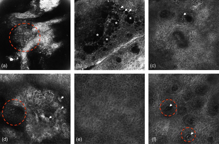
Fig. 3 Representative RCM images of (a) to (c) biopsy proven eczema and (d) to (f) psoriasis. Part (a) was obtained at the stratum corneum showing areas
of parakeratosis (red dashed circle). (b) The presence of spongiotic microvesicles corresponding to dark nonrefractive spaces on RCM (white asterisk).
Furthermore. small bright cells can be noted that correspond to inflammatory cells (white arrow). (c) RCM images at the DEJ with the presence
of inflammatory cells (white arrow). (d) Respective images of parakeratosis (red dashed circle) and single detached corneocytes (white arrows) in
a psoriatic plaque. (e) The honeycomb pattern of the epidermis. (f) Papillomatosis (red dashed circle) as present in psoriasis at the DEJ. Dilated
blood vessels can be observed in the center of the papillae (white arrows).
Journal of Biomedical Optics 061212-5 June 2013 • Vol. 18(6)
Downloaded From: https://www.spiedigitallibrary.org/journals/Journal-of-Biomedical-Optics on 27 May 2020
Terms of Use: https://www.spiedigitallibrary.org/terms-of-useUlrich and Lange-Asschenfeldt: In vivo confocal microscopy in dermatology . . .
In the same line of research, Wurm et al.37 have performed a using CLSM. Aghassi et al. have also used CLSM for monitor-
comprehensive analysis of chronological and photoaged skin ing of PDL treatment for sebaceous hyperplasia.47 A cohort of
using CLSM. They have shown, that CLSM permits a charac- 10 patients with a total of 29 lesions were treated by PDL fol-
terization and quantification of histomorphometric features lowed by serial confocal evaluations at baseline and follow-up
of epidermal and papillary dermis as a sign of skin aging. up to eight weeks post treatment. CLSM was able to visualize
Among 21 features analyzed, 15 were shown to be significant the morphological and vascular aspects of dynamic treatment
for quantification of skin aging. Among others, dermal papillary response, including a coagulation of blood vessels and conse-
index (number of papillae∕mm2 ), the thickness of the basal cutive (partial) regression of sebacious lesions.48
layer, and an increased thickness of the granular layer permit Yamashita et al. have reported on a series of subjects with
a semiquantitative grading.37 These changes were found to be benign, superficial pigmented skin lesions on the face of Asian
more pronounced with increasing age, and were also associated volunteers, treated by serial sessions of intense pulsed light
with changes in the underlying microvasculature. (IPL) therapy.49 CLSM was used to document lesions at base-
Another study by Lagarrigue et al.38 used CLSM to quantify line, as well as monitor treatment response including crusting,
epidermal pigmentation and density of dermal papilla in corre- migration of melanosomes, and overall melanocyte activity. The
lation with age and skin phototype. Following an analysis of 111 authors have come to the conclusion that CLSM is suitable to
healthy female volunteers, it was shown that papillary contrast at monitor the removal of superficial melanosomes from the basal
the level of the dermo-epidermal junction correlated strongly layer, while leaving overall melanocyte activity intact.
with skin pigmentation assessed clinically using the Fitzpatrick
skin phototype classification. While the first studies on the 8 Conditions with Exogenous/Endogenous
detection and distribution of melanin and melanocytes have Pigment Alterations/Decorative Tattoo
been performed as early as 2005,39,40 these findings are also con-
CLSM may be useful for evaluation of tattoo-related skin altera-
firmatory of a preliminary study by Antoniou et al., first analyz-
tions. CLSM was able to visualize subepidermal deposits of
ing pigment distribution in correlation with the Fitzpatrick
dense, clustered pigment granules of up to about 3 μm in size
classification.41 With respect to skin pigmentation, a number
corresponding to black tattoos, while red, blue, and green tattoos
of studies have been aimed at analyzing the process of immedi-
present on CLSM with a more scarce and diffuse appearance.
ate and delayed pigment darkening reactions and their respective
Thereby, it was postulated that CLSM may be a potentially
association with skin microvasculature.42
useful tool for evaluation of laser tattoo removal as well as
Further analyses have focused on the therapeutic effects of
monitoring skin repair after laser therapy.34,50
topically applied antioxidants such as Vit-C for skin aging.43
These investigations have shown that five weeks of topical
Vit-C application resulted in a significant increase in dermal 9 Melasma
papillary density, compared to vehicle treated control. RCM Melasma is a common pigmentary facial skin disorder caused
was able to show a restoration of the dermoepidermal junction by abnormal melanin deposits. Kang et al. were the first to
in young skin, and an increase in density of capillaries within analyze a total of 29 patients with a clinical diagnosis of mel-
dermal papillae in aged skin compared with untreated control. asma using reflectance confocal microscopy,51,52 describing a
These longitudinal studies illustrate the opportunity of CLSM to set of morphological criteria in correlation with routine histol-
monitor treatment response and local skin reaction as well as ogy. All patients showed a significant increase in hyperrefractile
dynamic events of skin repair and regeneration. basal cells, corresponding to hyperpigmented basal keratino-
cytes. Selected patients showed dendritic cells in the epidermis
corresponding to activated melanocytes. At the level of the der-
7 Cosmetic and Medical Laser Treatment mal layer, CLSM identified plump bright cells corresponding to
Longo et al.44 performed a systematic analysis of local effects of melanophages, a feature that has previously also been described
epidermal and dermal changes following laser skin rejuvenation in conjunction with melanocytic skin lesions such as benign and
using a fractionated CO 2 laser in an ablative mode including 10 dysplastic nevi.
female volunteers. Following a single treatment with a radiofre- A preliminary study by Ardigo et al. included a total of
quency excited ultrapulsed CO 2 laser device, four serial CLSM (n ¼ 15) patients previously diagnosed with facial melasma
evaluations were performed at baseline, 3, 6, and 12 weeks followed by CLSM analysis of their pigment distribution.52
after treatment. After blinded image analysis, it was shown Furthermore, therapeutic monitoring of skin response to treat-
that CLSM was able to document a disappearance of mottled pig- ment with pyruvic acid and hydroquinone was performed in
mentation as a feature of skin dyspigmentation in photoaged skin. seven of these patients. The results of this study suggest that by
Furthermore, collagen remodeling was described by the appear- localizing the site and extent of pigment deposits CLSM is a
ance of newly formed collagen at week 3, seen as bright, straight useful technique for the diagnosis and therapeutic follow-up
fibers in parallel arrangement throughout the entire CLSM mosaic for melasma.
and persisting at the 12 weeks evaluation timepoint. A large cohort of 210 Asian patients was evaluated by CLSM
Xu et al. have used CLSM for monitoring and follow-up of studying the correlation of histological classification of mel-
laser treatment for infraorbital dark circles using a low-fluence asma.53 It was shown that CLSM classification of melasma
Q-switched 1,064-nm laser.45 The results of this investigation corresponded well with routine histology. CLSM evaluation
showed a dramatic decrease of melanin deposition in the upper alone showed the predominant subtype to be epidermal (n ¼
dermis as seen by CLSM evaluation. 143) followed by the mixed type yielding a total of n ¼ 57
Other studies using laser treatment have evaluated the applic- patients. These results suggest that CLSM may be a useful tool
ability of CLSM for follow up of cherry angioma treated by for the classification of melasma, ultimately facilitating the
pulsed dye laser (PDL) in vivo.46 Thereby, the disappearance therapeutic management, follow-up, and prognosis on the ther-
of dilated capillary loops after PDL treatment was shown apeutic outcome. Pilot studies evaluating the efficacy of novel
Journal of Biomedical Optics 061212-6 June 2013 • Vol. 18(6)
Downloaded From: https://www.spiedigitallibrary.org/journals/Journal-of-Biomedical-Optics on 27 May 2020
Terms of Use: https://www.spiedigitallibrary.org/terms-of-useUlrich and Lange-Asschenfeldt: In vivo confocal microscopy in dermatology . . .
formulations for treatment of melasma have shown promising increase in the thickness of the stratum corneum compared to
results as to the clinical applicability of CLSM in monitoring unexposed control. There was a trend towards increased epi-
treatment response.54 dermal thickness, however, findings did not reach levels of
significance. The size of granular keratinocytes was signifi-
10 CLSM for Evaluation of UV-Induced Skin cantly larger in exposed versus unexposed skin and epidermal
melanin content was significantly higher for UVA-exposed skin.
Response
In that regard, a number of research studies have been aimed
Acute and repeated exposure of the skin to ultraviolet radiation at the prevention of sun-induced skin changes, employing pri-
results in a series of biological responses, including erythema, mary, secondary, and tertiary prevention strategies. Topically
immediate and delayed pigment darkening, tanning, and skin applied protective formulations include chemical compounds
color changes among the major ones. Chronic overexposure as well as physical absorbers, while systemic chemopreventive
to UV radiation gives rise to genetic alterations, cell cycle arrest, strategies have evaluated protective properties of natural anti-
and apoptosis of keratinocytes, resulting in the development of oxidants, and the administration of supplemental vitamins, plant
photocarcinogenesis, photoaging, and the occurence of photo- extracts, and nutritional compounds.
allergic and photoimmunologic skin responses. For the assessment of protective properties of sunscreen
While comprehensive research has been performed on UVA- formulations, the evaluation of minimal erythema dose (MED)
induced skin pigmentation and associated vascular changes, the
has been established in dermatological research, allowing to
specific events remain to be elucidated. Due to procedural
grade the ability of a compound to protect against sunburns.
limitations of serial histological studies, it was suggested that
Since MED is assessed by visual inspection, there is a high
noninvasive imaging techniques may aid in the investigation of
inter-, and intraobserver variability even when implementing
dynamic skin changes following UVA exposure including the
reflectometric and chromometric methods.56–58
evaluation of associated changes in blood flow.39,42,55
Gambichler et al. have performed a correlative study evalu-
Yamashita et al. performed an evaluation with 27 volunteers
(Fitzpatrick skin phototype SPT II-VI).42 Following irradiation ating the dynamic skin changes following exposure to a solar
with a solar simulator, UVA-induced tanning response was simulator at 1 × MED and 3 × MED using CLSM in a total of
evaluated by CLSM. The influence of reactive vasodilatation 10 volunteers.59 The findings of this study showed an increase
on immediate pigment darkening (IPD) development was exam- in epidermal thickness, the presence of spongiosis and an
ined with the addition of epinephrine this inducing vasocon- increased pigmentation of the basal layer in a dose/time-
striction. Thereby, the authors have shown that CLSM detects dependent manner. In addition, CLSM allowed the visualization
increased capillary flow immediately after UVA radiation. One of reactive vasodilatation of dermal capillaries.
week after irradiation an increased melanization of the basal Ulrich et al. performed an evaluation on cutaneous changes
layer was observed, whereby CLSM allowed the visualization following UVB-irradiation, including the evaluation a com-
of activated melanocytes in vivo. The addition of epinephrine mercially available sunscreen compared to untreated control.60
was able to partially inhibit the delayed darkening response, A total of five patients Fitzpatrick SPT II and III were enrolled in
suggesting a role for reactive vasodilation in the consecutive this pilot study, followed by irradiation of the volar forearm
events following UVA exposure. using a commercially available UVB light source with five gra-
Gambichler et al. used CLSM to assess the photoadaptive dually increasing dosages, whereby test sites with sunscreens
effects of repeated sunbed exposures on epidermal thickness, were compared to contralateral controls receiving no sunscreen.
cell size, and epidermal pigmentation.55 Eight volunteers were Clinical and CLSM evaluations were performed at four conse-
included in this study, receiving repeated sunbed exposures cutive time points.
within a three-week period and compared to an unexposed/ The findings of this study showed that CLSM permits the
protected control site followed by CLSM evaluation 24 h after visualization of inflammatory response in vivo, including the
the last UVA-exposure. The results showed a significant visualization of single apoptotic cells (sunburn cells) as well as
Fig. 4 Representative RCM images of the granular/spinous layer 24 h after irradiation with UV-B (240 J∕cm2 ) (a) without sunscreen and (b) after use of
sunscreen. (a) The RCM image (500 × 500 μm) illustrates the presence of cells with bright center and dark periphery, corresponding to sunburn cells
(white arrow). Furthermore, small microvesicles can be noted (red dashed circle). Part (b) was obtained at the contralateral arm of the same patient after
sunscreen application. The RCM image (500 × 500 μm) shows regular honeycomb pattern of the epidermis without any signs of sun damage.
Journal of Biomedical Optics 061212-7 June 2013 • Vol. 18(6)
Downloaded From: https://www.spiedigitallibrary.org/journals/Journal-of-Biomedical-Optics on 27 May 2020
Terms of Use: https://www.spiedigitallibrary.org/terms-of-useUlrich and Lange-Asschenfeldt: In vivo confocal microscopy in dermatology . . .
microvesicle formation. The presence of inflammatory changes consecutive clinically equivocal cases,” J. Invest. Dermatol. 132(10),
and sunburn cells correlated with respective UVB doses and the 2386–2394 (2012).
16. K. Sauermann et al., “Investigation of basal cell carcinoma [correction
respective susceptibility (skin phototype) Lastly, it was shown
of carcinoma] by confocal laser scanning microscopy in vivo,” Skin Res.
that following the topical application of sunscreen formulations Technol. 8(3), 141–147 (2002).
at standard dosage no UVB-induced skin changes were detected 17. S. González and Z. Tannous, “Real-time, in vivo confocal reflectance
using CLSM (Fig. 4). microscopy of basal cell carcinoma,” J. Am. Acad. Dermatol. 47(6),
869–874 (2002).
11 Conclusions and Future Perspectives 18. S. Nori et al., “Sensitivity and specificity of reflectance-mode confocal
microscopy for in vivo diagnosis of basal cell carcinoma: a multicenter
Reflectance confocal microscopy represents a promising techni- study,” J. Am. Acad. Dermatol. 51(6), 923–930 (2004).
que which has shown its usefulness for a wide range of scientific 19. A. L. Agero et al., “Reflectance confocal microscopy of pigmented
as well as clinical applications. In the past years, in vivo CLSM basal cell carcinoma,” J. Am. Acad. Dermatol. 54(4), 638–643
has developed from a research tool to a valuable adjunct diag- (2006).
nostic tool providing the opportunity for noninvasive evaluation 20. A. Torres et al., “5% imiquimod cream and reflectance-mode confocal
of skin lesions with histological detail. Thereby, CLSM has microscopy as adjunct modalities to Mohs micrographic surgery for
treatment of basal cell carcinoma,” Dermatol. Surg. 30(12, Pt. 1),
become an important part in the evaluation of skin cancer in 1462–1469 (2004).
academic clinical settings as well as specialized outpatient and 21. V. Ahlgrimm-Siess et al., “Monitoring efficacy of cryotherapy for
research centers. Furthermore it allows the noninvasive monitor- superficial basal cell carcinomas with in vivo reflectance confocal
ing of topical therapies as well as an assessment of efficacy for microscopy: a preliminary study,” J. Dermatol. Sci. 53(1), 60–64
cosmetic products and laser procedures. (2009).
22. R. S. Padilla et al., “Gene expression patterns of normal human skin,
actinic keratosis, and squamous cell carcinoma: a spectrum of disease
References progression,” Arch. Dermatol. 146(3), 288–293 (2010).
23. D. Aghassi, R. R. Anderson, and S. González, “Confocal laser micro-
1. E. Sattler et al., “How long does protection last?—in vivo fluorescence scopic imaging of actinic keratoses in vivo: a preliminary report,” J. Am.
confocal laser scanning imaging for the evaluation of the kinetics of Acad. Dermatol. 43(1, Pt 1), 42–48 (2000).
a topically applied lotion in an everyday setting,” Skin Res. Technol. 24. M. Ulrich et al., “Clinical applicability of in vivo reflectance confocal
18(3), 370–377 (2012). microscopy for the diagnosis of actinic keratoses,” Dermatol. Surg.
2. M. Minsky, “Memoir on inventing the confocal scanning microscope,” 34(5), 610–619 (2008).
Scanning 10(4), 128–138 (1988). 25. M. Horn et al., “Discrimination of actinic keratoses from normal skin
3. M. Rajadhyaksha et al., “In vivo confocal scanning laser microscopy with reflectance mode confocal microscopy,” Dermatol. Surg. 34(5),
of human skin: melanin provides strong contrast,” J. Invest. Dermatol. 620–625 (2008).
104(6), 946–952 (1995). 26. A. Rishpon et al., “Reflectance confocal microscopy criteria for squa-
4. C. M. Balch et al., “Final version of 2009 AJCC melanoma staging and mous cell carcinomas and actinic keratoses,” Arch. Dermatol. 145(7),
classification,” J. Clin. Oncol. 27(36), 6199–6206 (2009). 766–772 (2009).
5. Z. S. Tannous et al., “In vivo examination of lentigo malign and malig- 27. M. Ulrich et al., “Reflectance confocal microscopy for noninvasive
nant melanoma in situ, lentigo maligna type by near-infrared reflectance monitoring of therapy and detection of subclinical actinic keratoses,”
confocal microscopy: comparison of in vivo confocal images with his- Dermatology 220(1), 15–24 (2010).
tologic sections,” J. Am. Acad. Dermatol. 46(2), 260–263 (2002). 28. S. González et al., “Allergic contact dermatitis: correlation of in vivo
6. G. Pellacani, A. M. Cesinaro, and S. Seidenari, “In vivo assessment of confocal imaging to routine histology,” J. Am. Acad. Dermatol. 40(5),
melanocytic nests in nevi and melanomas by reflectance confocal 708–713 (1999).
microscopy,” Mod. Pathol. 18(4), 469–474 (2005). 29. K. Swindells et al., “Reflectance confocal microscopy may differentiate
7. G. Pellacani, A. M. Cesinaro, and S. Seidenari, “In vivo confocal reflec- acute allergic and irritant contact dermatitis in vivo,” J. Am. Acad.
tance microscopy for the characterization of melanocytic nests and Dermatol. 50(2), 220–228 (2004).
correlation with dermoscopy and histology,” Br. J. Dermatol. 152(2),
30. S. Astner et al., “Non-invasive evaluation of the kinetics of allergic
384–386 (2005).
and irritant contact dermatitis,” J. Invest. Dermatol. 124(2), 351–359
8. G. Pellacani, A. M. Cesinaro, and S. Seidenari, “Reflectance-mode con-
(2005).
focal microscopy for the in vivo characterization of pagetoid melano-
31. S. P. Hicks et al., “Confocal histopathology of irritant contact dermatitis
cytosis in melanomas and nevi,” J. Invest. Dermatol. 125(3), 532–537
in vivo and the impact of skin color (black vs. white),” J. Am. Acad.
(2005).
Dermatol. 48(5), 727–734 (2003).
9. A. Gerger et al., “Diagnostic applicability of in vivo confocal laser scan-
32. S. Astner et al., “Irritant contact dermatitis induced by a common
ning microscopy in melanocytic skin tumors,” J. Invest. Dermatol.
household irritant: a noninvasive evaluation of ethnic variability in
124(3), 493–498 (2005).
skin response,” J. Am. Acad. Dermatol. 54(3), 458–465 (2006).
10. G. Pellacani, A. M. Cesinaro, and S. Seidenari, “Reflectance-mode con-
33. S. Astner, S. González, and E. Gonzalez, “Noninvasive evaluation of
focal microscopy of pigmented skin lesions—improvement in melanoma
allergic and irritant contact dermatitis by in vivo reflectance confocal
diagnostic specificity,” J. Am. Acad. Dermatol. 53(6), 979–985 (2005).
microscopy,” Dermatitis 17(4), 182–191 (2006).
11. G. Pellacani et al., “The impact of in vivo reflectance confocal micros-
34. S. González and Y. Gilaberte-Calzada, “In-vivo refelectance confocal
copy for the diagnostic accuracy of melanoma and equivocal melano-
microscopy in clinical dermatology and cosmetology,“ Int. J. Cosmet.
cytic lesions,” J. Invest. Dermatol. 127(12), 2759–2765 (2007).
Sci. 30(1), 1–17 (2008).
12. A. Scope et al., “Remodeling of the dermoepidermal junction in super-
ficial spreading melanoma: insights gained from correlation of dermo- 35. “Cosmetics directive,” 76/768/EEC, European Commission Consumer
scopy, reflectance confocal microscopy, and histopathologic analysis,” Affairs, (2010), http://ec.europa.eu/consumers/sectors/cosmetics/
Arch. Dermatol. 144(12), 1644–1649 (2008). documents/directive/index_en.htm.
13. P. Guitera et al., “In vivo reflectance confocal microscopy enhances sec- 36. K. Sauermann et al., “Age related changes of human skin investigated
ondary evaluation of melanocytic lesions,” J. Invest. Dermatol. 129(1), with histometric measurements by confocal laser scanning microscopy
131–138 (2009). in vivo,” Skin Res. Technol. 8(1), 52–56 (2002).
14. S. Segura et al., “Development of a two-step method for the diagnosis of 37. E. M. Wurm et al., “In vivo assessment of chronological ageing and
melanoma by reflectance confocal microscopy,” J. Am. Acad. Dermatol. photoageing in forearm skin using reflectance confocal microscopy,”
61(2), 216–229 (2009). Br. J. Dermatol. 167(2), 270–279 (2012).
15. P. Guitera et al., “In vivo confocal microscopy for diagnosis of mela- 38. S. G. Lagarrigue et al., “In vivo quantification of epidermis pigmenta-
noma and basal cell carcinoma using a two-step method: analysis of 710 tion and dermis papilla density with reflectance confocal microscopy:
Journal of Biomedical Optics 061212-8 June 2013 • Vol. 18(6)
Downloaded From: https://www.spiedigitallibrary.org/journals/Journal-of-Biomedical-Optics on 27 May 2020
Terms of Use: https://www.spiedigitallibrary.org/terms-of-useUlrich and Lange-Asschenfeldt: In vivo confocal microscopy in dermatology . . .
variations with age and skin phototype,” Exp. Dermatol. 21(4), 281–286 50. K. O’goshi, C. Suihko, and J. Serup, “In vivo imaging of intradermal
(2012). tattoos by confocal scanning laser microscopy,” Skin Res. Technol.
39. M. A. Middelkamp-Hup et al., “Detection of UV-induced pigmentary 12(2), 94–98 (2006).
and epidermal changes over time using in vivo reflectance confocal 51. H. Y. Kang et al., “In vivo reflectance confocal microscopy detects
microscopy,” J. Invest. Dermatol. 126(2), 402–407 (2006). pigmentary changes in melasma at a cellular level resolution,” Exp.
40. T. Yamashita et al., “Non-invasive visualization of melanin and mela- Dermatol. 19(8), 228–233 (2010).
nocytes by reflectance-mode confocal microscopy,” J. Invest. Dermatol. 52. M. Ardigo et al., “Characterization and evaluation of pigment distri-
124(1), 235–244 (2005). bution and response to therapy in melasma using in vivo reflectance
41. C. Antoniou et al., “Analysis of melanin distribution in different eth- confocal microscopy: a preliminary study,” J. Eur. Acad. Dermatol.
nic groups by in vivo confocal microscopy,” Laser Phys. Lett. 6(5), Venereol. 24(11), 1296–1303 (2010).
393–398 (2009). 53. H. Liu et al., “Histological classification of melasma with reflectance
42. T. Yamashita et al., “In vivo assessment of pigmentary and vascular confocal microscopy: a pilot study in Chinese patients,“ Skin Res. Tech-
compartments changes in UVA exposed skin by reflectance-mode con- nol. 17(4), 398–403 (2011).
focal microscopy,” Exp. Dermatol. 16(11), 905–911 (2007). 54. K. Tsilika et al., “A pilot study using reflectance confocal microscopy
43. K. Sauermann et al., “Topically applied vitamin C increases the density (CLSM) in the assessment of a novel formulation for the treatment of
of dermal papillae in aged human skin,” BMC Dermatol. 4(1), 13 melasma,” J. Drugs Dermatol. 10(11), 1260–1264 (2011).
(2004). 55. T. Gambichler et al., “Effects of repeated sunbed exposures
44. C. Longo et al., “Laser skin rejuvenation: epidermal changes and col- on the human skin. In vivo measurements with confocal
lagen remodelling evaluated by in vivo confocal microscopy,” Lasers microscopy,” Photodermatol. Photoimmunol. Photomed. 20(1), 27–32
Med. Sci. (2012), [Epub ahead of print]. (2004).
45. T. H. Xu et al., “Treatment of infraorbital dark circles using a low- 56. J. Lock-Andersen and H. C. Wulf, “Threshold level for measurement of
fluence Q-switched 1,064-nm laser,” Dermatol. Surg. 37(6), 797–803 UV-sensitivity: reproducibility of phototest,” Photodermatol. Photo-
(2011). immunol. Photomed. 12(4), 154–161 (1996).
46. D. Aghassi, R. R. Anderson, and S. González, “Time-sequenced histo- 57. M. Falk, M. Ilias, and C. Anderson, “Interobserver variability in reading
logic imaging of laser-treated cherry angiomas with in vivo confocal of phototest reactions with sharply or diffusely delineated borders,”
microscopy,” J. Am. Acad. Dermatol. 43(1), 37–41 (2000). Skin Res. Technol. 14(4), 397–402 (2008).
47. D. Aghassi et al., “Elucidating the pulsed-dye laser treatment of sebac- 58. P. Clarys et al., “Skin color measurements: comparison between three
eous hyperplasia in vivo with real-time confocal scanning laser micros- instruments: the chromameter®, the DermaSpectrometer® and the
copy,” J. Am. Acad. Dermatol. 43(1), 49–53 (2000). Mexameter®,” Skin Res. Technol. 6(4), 230–238 (2000).
48. S. González et al., “Confocal imaging of sebaceous gland hyperplasia in 59. T. Gambichler et al., “A comparative pilot study on ultraviolet-induced
vivo to assess efficacy and mechanism of pulsed dye laser treatment,” skin changes assessed by noninvasive imaging techniques in vivo,”
Laser Surg. Med. 25(1), 8–12 (1999). Photochem. Photobiol. 82(4), 1103–1107 (2006).
49. T. Yamashita et al., “Intensed pulsed light therapy for superficial 60. M. Ulrich et al., “Comparison of UV-induced skin changes in
pigmented lesions evaluated by reflectance confocal microscopy and sun-exposed vs. sun-protected skin- preliminary evaluation by reflec-
optical coherence tomography,” J. Invest. Dermatol. 126(10), tance confocal microscopy,” Br. J. Dermatol. 161(S3), 46–53
2281–2286 (2006). (2009).
Journal of Biomedical Optics 061212-9 June 2013 • Vol. 18(6)
Downloaded From: https://www.spiedigitallibrary.org/journals/Journal-of-Biomedical-Optics on 27 May 2020
Terms of Use: https://www.spiedigitallibrary.org/terms-of-useYou can also read