Fostering a healthy culture: Biological relevance of in vitro and ex vivo skin models
←
→
Page content transcription
If your browser does not render page correctly, please read the page content below
DOI: 10.1111/exd.14296
EDITORIAL
Fostering a healthy culture: Biological relevance of in vitro and
ex vivo skin models
1 | B R I D G I N G TH E G A P B E T W E E N 2 | W H AT O RG A N OT Y PI C CU LT U R E S C A N
“ S I M PLI C IT Y ” O F I N V ITRO A N D AND CAN'T DO
CO M PLE X IT Y O F I N V I VO
Typically composed of primary cells isolated from patient skin
The field of experimental dermatology research has dramatically biopsies or surgical discard samples, the idea of studying 3D
benefited from the insights yielded by in vivo studies on animal skin equivalents in vitro is an attractive premise for investigative
models. Indeed, much of our understanding of the mechanisms dermatologists. From gaining molecular insight into essential
that regulate embryonic skin development, adult skin homeostasis aspects of skin development and homeostasis to preclinical testing
and physiological skin responses to stress, such as wound healing, of new drug candidates for skin diseases to their use as “tissue
has been educated by studies conducted in animal models, includ- farms” to grow new skin substitutes for burn and trauma patients,
ing mutant mice. These works become commonly published on the 3D cultures are becoming a mainstay approach both in basic and
pages of Experimental Dermatology and, in fact, represent one of the translational dermatological research (Figure 1). Current 3D culture
core interests of the journal. Highlighting in vivo mouse model stud- technologies include the following: (i) free-
floating cultures of
ies are recent works on hair follicle development and growth1-5 and spherical organoids initiated from pluripotent stem cells; (ii) layered
wound healing.6-8 Further, studies in animal models often help to constructs consecutively assembled by seeding primary skin cells
elucidate aspects of disease pathogenesis, and mouse models are into extracellular matrix scaffolds38 to contain stratified epidermis,
used to investigate mechanisms of human skin conditions such as sebocyte spheroids39 or hair pegs 40; (iii) freshly micro-
dissected
9-13 14-16 17,18 19
atopic dermatitis, contact dermatitis, psoriasis, rosacea skin and hair follicle explant cultures41-4 4; and (iv) organ-on-a-chip
20
and squamous cell carcinoma to name a few. cultures that incorporate capillary structures and allow active
On the other hand, not all human skin diseases or aspects of perfusion using microfluidics.45,46
human skin physiology can be reliably modelled in rodents. This However, so-called 3D skin “equivalent” cultures are not without
is not surprising, considering that humans and rodents are sepa- limitations. For instance, despite their morphological similarity to
rated by an estimated 96 million years of evolution. 21 Addressing native skin in vivo, their cellular composition is extremely simplified,
this fact, many studies are being conducted on patient-d erived and culture protocols can vary greatly, which affects reproducibil-
primary skin cells, including human keratinocytes, 22-25 melano- ity and interpretation. Depending on the protocol used, 3D cultures
cytes, 26-3 0 fibroblasts 31-3 4 and cell co-
c ultures. 35-37 However, may not have a normal stratum corneum, an intact epidermal bar-
typical in vitro culture conditions fail to replicate and, in fact, do rier, or proper lipid composition.47 Critically, 3D cultures, no matter
not come close to imitating the biomechanical and biochemical how sophisticated, lack system-level aspects of normal skin, such
complexity of the microenvironment in which cells exist and to as fully functioning vasculature, immune system and innervation.
which they respond to in native tissues. Further, ingredients in Even though this may be partially compensated for with ex vivo-
commonly used cell culture media and the two-d imensional con- reinnervation protocols,48 this puts limits on the maximum size that
straints of growth on plastic result in cells being exposed to a 3D cultures can achieve before experiencing hypoxia and simplifies
lot of artificial cues, to which they adapt but also prominently their responses to external and neurotrophic stimuli.
alter their gene expression profile and functional activities in the Yet, 3D cultures of the same primary skin cells49 are still far more
process. Therefore, behaviours displayed by skin cells in two- complex than their monolayer counterparts. For instance, a recent
dimensional cultures need to be comprehensively validated under genome-wide methylation study showed no correlation between
more native-like conditions in order to be deemed biologically methylation status and transcriptome changes during keratinocyte
and physiologically relevant. Helping to bridge the gap, three- differentiation in a monolayer culture, 22 despite other data show-
dimensional (3D) organotypic cultures and organ culture tech- ing significant methylation changes during in vivo skin morphogen-
niques have long been a highly instructive tool for investigating esis.50,51 Likewise, normal expression patterns of terminal marker
complex, tissue-level behaviours by skin cells. genes are significantly impaired in Ca2+ induced differentiation of
© 2021 John Wiley & Sons A/S. Published by John Wiley & Sons Ltd
Experimental Dermatology. 2021;00:1–6. wileyonlinelibrary.com/journal/exd | 12 | EDITORIAL
primary keratinocytes. 23 Yet, the biological relevance of monolayer function.56,57 Considering the relative simplicity of 3D equivalents,
cultures can be substantially increased by co-culturing two or more full-thickness skin biopsy explants are commonly used in follow-up
cell types. For instance, a recently reported co-culture study of se- validation experiments (Figure 1C). Recent examples of using skin
nescent dermal fibroblasts and macrophages identified macrophage- explants include studies on penetration and metabolism of vitamin
derived TNFα as an important trigger of senescent cell apoptosis A derivatives 41 and the effect of cigarette smoke exposure on skin
and clearance.35 Also, the co-culture of dermal fibroblasts and ke- barrier permeability.42
ratinocytes can model particulate matter exposure, where factors Where 3D skin equivalents and skin explants really shine is in
released by keratinocytes after heavy metal and hydrocarbon expo- their ability to model aspects of human skin disorders, including dry
sure can mediate dermal collagen degradation.36 In other examples, skin,58 radiodermatitis59 or hidradenitis suppurativa60 to name but
co-cultures of vitiligo patient-derived CD4+ and CD8+ T cells,52 or a few. In this issue of Experimental Dermatology, Yoshida et al. re-
keratinocytes and T cells from psoriasis patients,37 can be used to port on modelling hand-foot skin reaction (HFSR),61 a common side
advance our understanding of skin disease immunopathogenesis. effect of anti-angiogenic therapies, where soles and palms experi-
ence hyperkeratosis, leading to debilitating pain, difficulties walk-
ing and object grasping.62 Small-molecule tyrosine kinase inhibitors,
3 | W H E R E O RG A N OT Y PI C S K I N such as sunitinib, are used as angiogenesis inhibitors against various
CU LT U R E S “ S H I N E ” types of cancer, and the occurrence of HFSR is often correlated with
successful treatment.63 Sunitinib-induced HFSR causes epidermal
3D equivalent cultures are commonly used to study both basic thickening, with spinous and granular layers thinning and, at times,
and translational aspects of skin biology (Figure 1B). For example, separating from the reactive basal layer.64
relatively simple skin equivalents, featuring only keratinocytes and Yoshida et al. analysed both monolayer cultures of keratino-
fibroblasts, have been recently used to study the effects of Dead cytes and 3D skin equivalents treated with sunitinib and revealed
Sea water minerals on terminal epidermal differentiation.53 More decreased expression of basal gene KRT6A and terminal differen-
complex 3D equivalents that also contain melanocytes can be tiation genes SERPINB1 and SPINK6, suggesting the skin barrier is
used in studies on pigment-modulating components of cosmetic perturbed in HFSR.61 p38 MAPK and ERK1/2 phosphorylation was
products54 or to test the effect of organic environmental toxins also inhibited upon sunitinib treatment, suggesting that the MAPK
55
on hyperpigmentation. Recently, 3D equivalent cultures have pathway may control skin barrier gene expression. As subsequent
also been used to evaluate the impact of microbiota on skin cell MAPK signalling inhibition in monolayer keratinocyte cultures
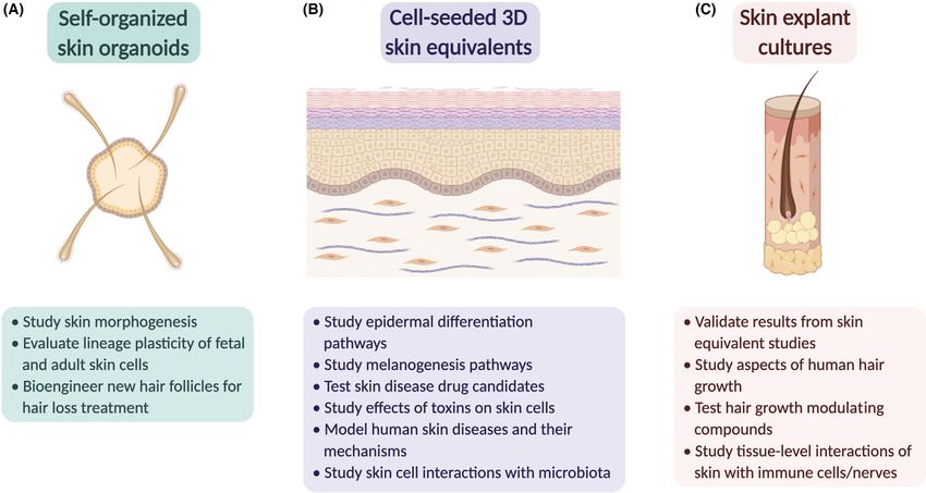
F I G U R E 1 In vitro and ex vivo skin culture models in dermatological research. Common applications are listed for (A) self-organized skin
organoids, (B) skin equivalents produced by seeding primary cells into extracellular matrix scaffolds and (C) explant cultures of freshly micro-
dissected skin and/or hair folliclesEDITORIAL | 3
phenocopied sunitinib treatment, the authors reasoned that acti- peg-like structures matured towards functional human hair follicles.
vation of MAPK signalling could rescue the sunitinib-induced gene To this end, recent advances in pluripotent stem cell differentiation
expression changes. Nuclear CTNNB1 can promote both p38 MAPK protocols have enabled production of human hair follicle-bearing or-
and ERK1/2 activity, whereas GSKB suppresses nuclear CTNNB1. ganoids fully in vitro (Figure 1A).75 Progress on this and similar in vitro
Using an inhibitor to GSK3B with and without sunitinib, the au- hair-bearing organoid systems is discussed in a recent Experimental
thors showed that MAPK signalling and skin barrier gene expression Dermatology Viewpoint article.
changes can both be rescued, suggesting that GSK3B inhibitors may Cultures of freshly micro-dissected human scalp hair follicles,
be a clinically relevant treatment to HFSR. which are a special variant of skin explant culture, have been suc-
Another common skin disorder ripe for in vitro modelling is acne cessfully used to study aspects of human hair biology not dis-
vulgaris, where genetic predispositions, hormonal changes, stress played in the animal models and have been instructive for our
and environmental factors lead to the formation of microcomedo- understanding of human hair follicle physiology and pathology
nes,65-67 many of which can become inflamed.68 Inflammatory mi- (Figure 1C).76,77 Studies on human hair follicle organ cultures are
crocomedones are caused by hyperseborrhoea, where fatty acid commonly featured in this journal, including recent works that
composition of sebum becomes altered, leading to accumulation of examined hair growth-p romoting effect of dermal papilla-d erived
peroxidized squalene and favouring expansion of C. acnes.66,69,70 The exosomes,78 regulation of hair follicle immune privilege79 and
condition is aggravated by hair duct clogging with overproduced and modulation of hair growth by the blue light-sensitive circadian
abnormally differentiated epidermis. In this issue of Experimental clock factor CRY1. 80
Dermatology, Laclaverie et al. developed and characterized a 3D skin While explant cultures of freshly dissected skin and hair follicles
equivalent model for acne vulgaris.71 The authors used primary kera- are arguably the most physiologically complete type of ex vivo cul-
tinocyte-and fibroblast-seeded 3D skin equivalents that were sub- ture models, they still suffer from being cut-off from numerous mi-
sequently treated with peroxidized squalene and/or C. acnes. They croenvironmental and systemic inputs that regulate their physiology
showed that combined treatment results in inflammatory, skin de- in vivo. To this end, a recent study showed that growth and pigmen-
fense and remodelling gene expression changes analogous to those tation parameters of scalp hair follicles in vitro are improved if they
seen in acneic skin. The authors then tested different phylotypes of are cultured with the adjacent piece of dermal adipose tissue that
C. acnes strains isolated from healthy and acneic patients and found secretes hepatocyte growth factor.81 Sensory nerves are another
that strain IA1, commonly found in acne-prone skin, causes the important source of regulatory signalling molecules, from which skin
major hallmarks of acne including hyper-keratinization, inflammation explants become severed in culture. To this end, a co-culture with rat
and altered barrier function. dorsal root ganglions (rDRGs) permits partial sensory reinnervation
of human skin explants in vitro,48 where “trophic” effects of rDRGs
have been shown to positively affect epidermal proliferation and in-
4 | G ROW I N G H A I R S I N A PE TR I D I S H duce activation of resident mast cells.82
Taken together, the above examples clearly show that organo-
The dynamic biology of hair follicles poses a particular challenge typic cultures have already evolved to a level of technical sophisti-
for their organotypic culture, yet in vitro approaches have been cation that permits studying aspects of normal skin physiology and
actively pursued in the field driven by many differences between conducting meaningful disease modelling. As the citations listed
human scalp and mouse pelage hairs and by the unmet demand to here demonstrate, Experimental Dermatology prides itself for being
bioengineer new hair follicles to treat hair loss. With regard to hair at the forefront of this area in skin biology research. At present, ex-
bioengineering, competent hair-fated epithelial and mesenchymal perimental dermatologists have the “luxury” to choose from several
skin cells, such as those derived from neonatal mice, can models, ranging from self-organizing skin organoids to cell-seeded
efficiently self-organize into many new hair follicles when injected extracellular matrix scaffolds to freshly micro-dissected skin or ap-
subcutaneously into host mice. However, such injections produce pendage explants. Looking into the future, technical efforts aimed at
“hairy cysts” that lack proper follicle orientation and are, thus, not better imitating system-level inputs will eventually advance organo-
therapeutically viable.72,73 typic cultures towards true organ-on-a-chip level, when complex
To overcome this fundamental limitation, Paik et al. recently re- skin functions, such as hair growth cycling, and progression of major
ported an approach in which neonatal mouse dermal and epidermal skin diseases will be modelled fully ex vivo.
74
cells are cultured within the collagen scaffold prior to being grafted.
This approach produces high densities of hair follicles that maintain AC K N OW L E D G E M E N T S
near-normal orientation perpendicular to the skin surface. In an ef- S.X.A. is supported by NIH grant R01-
C A237563 and ACS
fort to translate similar tissue engineering approaches to the human grant RSG-
19-
0 89-
01-
DDC. M.V.P. is supported by NIH grants
system, Weber et al. developed a 3D model for co-culturing human U01-
AR073159, R01-
AR069653, NSF grant DMS-
1951144, LEO
neonatal foreskin keratinocytes with human foetal scalp dermal cells foundation grants LF-OC-20-0 00611 and LF-AW_RAM-19-4 00008
that permits their self-organization into hair peg-like structures.40 and Pew Charitable Trust. Additional support to S.X.A. and M.V.P. is
When grafted after in vitro self-assembly to nude mice, at least some provided by P30-AR075047.4 | EDITORIAL
15. Fukuyama T, Nakamura Y, Kanemaru K, et al. Phospholipase
Scott X. Atwood1,2,3 Cgamma1 is required for normal irritant contact dermatitis re-
Maksim V. Plikus1,2,3,4 sponses and sebaceous gland homeostasis. Exp Dermatol.
2019;28(9):1051-1057.
1 16. Frempah B, Luckett-Chastain LR, Gallucci RM. IL6Ralpha function
Department of Developmental and Cell Biology, University of
in myeloid cells modulates the inflammatory response during irri-
California, Irvine, Irvine, CA, USA
tant contact dermatitis. Exp Dermatol. 2019;28(8):948-955.
2
Center for Complex Biological Systems, University of California, 17. Kiss B, Szanto M, Hegedus C, et al. Poly(ADP-ribose) polymerase-1
Irvine, Irvine, CA, USA depletion enhances the severity of inflammation in an imiquimod-
3
NSF-Simons Center for Multiscale Cell Fate Research, induced model of psoriasis. Exp Dermatol. 2020;29(1):79-85.
18. Yi F, Zheng X, Fang F, Zhang J, Zhou B, Chen X. ALA-PDT alleviates
University of California, Irvine, Irvine, CA, USA
the psoriasis by inhibiting JAK signalling pathway. Exp Dermatol.
4
Sue and Bill Gross Stem Cell Research Center, University of 2019;28(11):1227-1236.
California, Irvine, Irvine, CA, USA 19. Son M, Park J, Oh S, et al. Radiofrequency irradiation attenuates
angiogenesis and inflammation in UVB-induced rosacea in mouse
skin. Exp Dermatol. 2020.
Correspondence
20. Yang X, Daifallah AEM, Shankar S, et al. Topical kinase inhibitors
Scott X. Atwood and Maksim V. Plikus, Department of induce regression of cutaneous squamous cell carcinoma. Exp
Developmental and Cell Biology, University of California, Dermatol. 2019;28(5):609-613.
Irvine, Irvine, CA 92697, USA 21. Nei M, Xu P, Glazko G. Estimation of divergence times from multipro-
tein sequences for a few mammalian species and several distantly
Email: satwood@uci.edu (S.X.A.) and plikus@uci.edu (M.V.P.)
related organisms. Proc Natl Acad Sci USA. 2001;98(5):2497-2502.
22. Smits JPH, Dirks RAM, Qu J, et al. Terminal keratinocyte differ-
REFERENCES entiation in vitro is associated with a stable DNA methylome. Exp
1. Zhang B, He M, Rachmin I, et al. Melanocortin 1 receptor is dispens- Dermatol. 2020.
able for acute stress induced hair graying in mice. Exp Dermatol. 23. Jeriha J, Kolundzic N, Khurana P, et al. Markers for Ca(++) -induced
2020. terminal differentiation of keratinocytes in vitro under defined
2. Chovatiya G, Ghuwalewala S, Walter LD, Cosgrove BD, Tumbar T. conditions. Exp Dermatol. 2020;29(12):1238-1242.
High-resolution single-cell transcriptomics reveals heterogeneity 24. Emmert H, Fonfara M, Rodriguez E, Weidinger S. NADPH oxi-
of self-renewing hair follicle stem cells. Exp Dermatol. 2020. dase inhibition rescues keratinocytes from elevated oxidative
3. Wang Z, Chen Y, Chen M, Zhang Y. Overexpression of Fgf8 in the stress in a 2D atopic dermatitis and psoriasis model. Exp Dermatol.
epidermis inhibits hair follicle development. Exp Dermatol. 2020. 2020;29(8):749-758.
4. Nicu C, Wikramanayake TC, Paus R. Clues that mitochondria 25. Chen J, Liu K, Liu Y, Wang X, Zhang Z. Targeting mTORC1/2 with
are involved in the hair cycle clock: MPZL3 regulates entry into OSI-027 inhibits proliferation and migration of keloid keratino-
and progression of murine hair follicle cycling. Exp Dermatol. cytes. Exp Dermatol. 2019;28(3):270-275.
2020;29(12):1243-1249. 26. Xu P, Xue YN, Ji HH, Tan C, Guo S. H2 O2 -induced oxidative
5. Swanson JB, Vagnozzi AN, Veniaminova NA, Wong SY. Loss of stress disrupts mitochondrial functions and impairs migra-
Gata6 causes dilation of the hair follicle canal and sebaceous duct. tory potential of human epidermal melanocytes. Exp Dermatol.
Exp Dermatol. 2019;28(4):345-3 49. 2020;29(8):733-741.
6. Jiang TX, Harn HI, Ou KL, Lei M, Chuong CM. Comparative regen- 27. Lv J, Fu Y, Cao Y, et al. Isoliquiritigenin inhibits melanogenesis, me-
erative biology of spiny (Acomys cahirinus) and laboratory (Mus lanocyte dendricity and melanosome transport by regulating ERK-
musculus) mouse skin. Exp Dermatol. 2019;28(4):442-4 49. mediated MITF degradation. Exp Dermatol. 2020;29(2):149-157.
7. Maden M, Brant JO. Insights into the regeneration of skin from 28. Jimbo H, Nagai H, Fujiwara S, Shimoura N, Nishigori C. Fas-FasL
Acomys, the spiny mouse. Exp Dermatol. 2019;28(4):436-4 41. interaction in cytotoxic T cell-mediated vitiligo: the role of lesional
8. Kaymakcalan OE, Abadeer A, Goldufsky JW, et al. Topical alpha- expression of tumor necrosis factor-alpha and interferon-gamma in
gal nanoparticles accelerate diabetic wound healing. Exp Dermatol. Fas-mediated melanocyte apoptosis. Exp Dermatol. 2020;29(1):61-70.
2020;29(4):404-413. 29. Tam I, Dzierzega-Lecznar A, Stepien K. Differential expression of
9. Gilhar A, Reich K, Keren A, Kabashima K, Steinhoff M, Paus R. Mouse inflammatory cytokines and chemokines in lipopolysaccharide-
models of atopic dermatitis: a critical reappraisal. Exp Dermatol. 2020. stimulated melanocytes from lightly and darkly pigmented skin. Exp
10. Smith L, Gatault S, Casals-Diaz L, et al. House dust mite-treated Dermatol. 2019;28(5):551-560.
PAR2 over-expressor mouse: a novel model of atopic dermatitis. 30. Yoshimoto S, Ohagi Y, Yoshida M, et al. Placental extracts regu-
Exp Dermatol. 2019;28(11):1298-1308. late melanin synthesis in normal human melanocytes with alter-
11. Fu X, Hong C. Osthole attenuates mouse atopic dermatitis by inhib- ations of mitochondrial respiration. Exp Dermatol. 2019;28(Suppl
iting thymic stromal lymphopoietin production from keratinocytes. 1):50-5 4.
Exp Dermatol. 2019;28(5):561-567. 31. Arndt S, Lissner C, Unger P, Baumler W, Berneburg M, Karrer S.
12. Tang L, Cao X, Li X, Ding H. Topical application with conjugated Biological effects of a new ultraviolet al prototype based on light-
linoleic acid ameliorates 2, 4-dinitrofluorobenzene-induced atopic emitting diodes on the treatment of localized scleroderma. Exp
dermatitis-like lesions in BALB/c mice. Exp Dermatol. 2020. Dermatol. 2020;29(12):1199-1208.
13. Kake T, Imai M, Takahashi N. Effects of beta- c arotene on 32. Ham S, Harrison C, de Kretser D, Wallace EM, Southwick G,
oxazolone-induced atopic dermatitis in hairless mice. Exp Dermatol. Temple-S mith P. Potential treatment of keloid pathogenesis with
2019;28(9):1044-1050. follistatin 288 by blocking the activin molecular pathway. Exp
14. Porras M, Gomez LA, Perez JJ. Anti- inflammatory effect of Dermatol. 2020.
amygdalin analogs following topical administration on the TPA- 33. Macarak EJ, Wermuth PJ, Rosenbloom J, Uitto J. Keloid disorder:
induced irritant contact dermatitis model in mice. Exp Dermatol. Fibroblast differentiation and gene expression profile in fibrotic
2021. skin diseases. Exp Dermatol. 2021;30(1):132-145.EDITORIAL | 5
34. Lee YS, Liang YC, Wu P, et al. STAT3 signalling pathway is implicated vitiligo due to reduced NFATC1 and FOXP3 proteins. Exp Dermatol.
in keloid pathogenesis by preliminary transcriptome and open chro- 2020;29(8):759-775.
matin analyses. Exp Dermatol. 2019;28(4):480-484. 54. Portugal-Cohen M, Cohen D, Ish-Shalom E, Laor-Costa Y, Ma'or Z.
35. Ogata Y, Yamada T, Hasegawa S, et al. SASP-induced macrophage Dead Sea minerals: new findings on skin and the biology beyond.
dysfunction may contribute to accelerated senescent fibroblast ac- Exp Dermatol. 2019;28(5):585-592.
cumulation in the dermis. Exp Dermatol. 2021;30(1):84-91. 55. Bae IH, Lee ES, Yoo JW, et al. Mannosylerythritol lipids inhibit
36. Kim M, Kim JH, Jeong GJ, Park KY, Lee MK, Seo SJ. Particulate melanogenesis via suppressing ERK-CREB-MiTF-t yrosinase signal-
matter induces pro-inflammatory cytokines via phosphorylation of ling in normal human melanocytes and a three-dimensional human
p38 MAPK possibly leading to dermal inflammaging. Exp Dermatol. skin equivalent. Exp Dermatol. 2019;28(6):738-741.
2019;28(7):809-815. 56. Mi T, Dong Y, Santhanam U, Huang N. Niacinamide and
37. de Jesus-Gil C, Ruiz-Romeu E, Ferran M, et al. IL-15 and IL-23 syn- 12-hydroxystearic acid prevented benzo(a)pyrene and squalene
ergize to trigger Th17 response by CLA(+) T cells in psoriasis. Exp peroxides induced hyperpigmentation in skin equivalent. Exp
Dermatol. 2020. Dermatol. 2019;28(6):742-746.
38. Lee J, Koehler KR, Skin organoids: a new human model for develop- 57. Emmert H, Rademacher F, Glaser R, Harder J. Skin microbiota anal-
mental and translational research. Exp Dermatol. 2021. https://doi. ysis in human 3D skin models-"Free your mice". Exp Dermatol. 2020.
org/10.1111/exd.14292. 58. Khmaladze I, Butler E, Fabre S, Gillbro JM. Lactobacillus reuteri
39. Lee V, Singh G, Trasatti JP, et al. Design and fabrication of human DSM 17938-A comparative study on the effect of probiotics and
skin by three-dimensional bioprinting. Tissue Eng Part C Methods. lysates on human skin. Exp Dermatol. 2019;28(7):822-828.
2014;20(6):473-484. 59. Katsuyama Y, Taira N, Tsuboi T, Yoshioka M, Okano Y, Masaki H.
40. Zouboulis CC, Yoshida GJ, Wu Y, Xia L, Schneider MR. Sebaceous 3-O-L aurylglyceryl ascorbate improves the development of sensi-
gland: milestones of 30-year modelling research dedicated to the tive skin through the reduction of oxidative stress. Exp Dermatol.
"brain of the skin". Exp Dermatol. 2020. 2019;28(Suppl 1):64-68.
41. Weber EL, Woolley TE, Yeh CY, Ou KL, Maini PK, Chuong CM. Self- 60. Huth S, Marquardt Y, Huth L, et al. Molecular effects of photon
organizing hair peg-like structures from dissociated skin progeni- irradiation and subsequent aftercare treatment with dexpanthenol-
tor cells: New insights for human hair follicle organoid engineering containing ointment or liquid in 3D models of human skin and non-
and Turing patterning in an asymmetric morphogenetic field. Exp keratinized oral mucosa. Exp Dermatol. 2021.
Dermatol. 2019;28(4):355-366. 61. Sanchez J, Le Jan S, Muller C, et al. Matrix remodelling and MMP
42. Bjerke DL, Li R, Price JM, et al. The vitamin A ester retinyl pro- expression/activation are associated with hidradenitis suppurativa
pionate has a unique metabolic profile and higher retinoid-related skin inflammation. Exp Dermatol. 2019;28(5):593-600.
bioactivity over retinol and retinyl palmitate in human skin models. 62. Yoshida A, Yamamoto K, Ishida T, et al. Sunitinib decreases the ex-
Exp Dermatol. 2020. pression of KRT6A and SERPINB1 in 3D human epidermal models.
43. Percoco G, Patatian A, Eudier F, et al. Impact of cigarette smoke on Exp Dermatol. 2020.
physical-chemical and molecular proprieties of human skin in an ex 63. Lee WJ, Lee JL, Chang SE, et al. Cutaneous adverse effects in pa-
vivo model. Exp Dermatol. 2020. tients treated with the multitargeted kinase inhibitors sorafenib
44. Olah A, Alam M, Cheret J, et al. Mitochondrial energy metabolism and sunitinib. Br J Dermatol. 2009;161(5):1045-1051.
is negatively regulated by cannabinoid receptor 1 in intact human 64. Wang P, Tan G, Zhu M, Li W, Zhai B, Sun X. Hand-foot skin reaction
epidermis. Exp Dermatol. 2020. is a beneficial indicator of sorafenib therapy for patients with hepa-
45. Nicu C, Hardman JA, Pople J, Paus R. Do human dermal adi- tocellular carcinoma: a systemic review and meta-analysis. Expert
pocytes switch from lipogenesis in anagen to lipophagy and li- Rev Gastroenterol Hepatol. 2018;12(1):1-8.
polysis during catagen in the human hair cycle? Exp Dermatol. 65. Lacouture ME, Reilly LM, Gerami P, Guitart J. Hand foot skin re-
2019;28(4):432-4 35. action in cancer patients treated with the multikinase inhibitors
46. Marconi A, Quadri M, Saltari A, Pincelli C. Progress in melanoma sorafenib and sunitinib. Ann Oncol. 2008;19(11):1955-1961.
modelling in vitro. Exp Dermatol. 2018;27(5):578-586. 66. Dreno B. What is new in the pathophysiology of acne, an overview.
47. Groeber F, Engelhardt L, Lange J, et al. A first vascularized skin J Eur Acad Dermatol Venereol. 2017;31(Suppl 5):8-12.
equivalent as an alternative to animal experimentation. Altex. 67. Zouboulis CC. Endocrinology and immunology of acne: two sides of
2016;33(4):415-422. the same coin. Exp Dermatol. 2020;29(9):840-859.
48. Niehues H, Bouwstra JA, El Ghalbzouri A, Brandner JM, Zeeuwen P, 68. Josse G, Mias C, Le Digabel J, et al. High bacterial coloniza-
van den Bogaard EH. 3D skin models for 3R research: The potential tion and lipase activity in microcomedones. Exp Dermatol.
of 3D reconstructed skin models to study skin barrier function. Exp 2020;29(2):168-176.
Dermatol. 2018;27(5):501-511. 69. Moradi Tuchayi S, Makrantonaki E, Ganceviciene R, Dessinioti
49. Cheret J, Ponce L, Le Gall- Ianotto C, Bertolini M, Paus R. Re- C, Feldman SR, Zouboulis CC. Acne vulgaris. Nat Rev Dis Primers.
innervation of human skin by rat dorsal root ganglia permits to 2015;1:15029.
study interactions between sensory nerve fibres and native human 70. Jasson F, Nagy I, Knol AC, Zuliani T, Khammari A, Dreno B. Different
dermal mast cells ex vivo. Exp Dermatol. 2020. strains of Propionibacterium acnes modulate differently the cuta-
50. Sciezynska A, Nogowska A, Sikorska M, et al. Isolation and culture neous innate immunity. Exp Dermatol. 2013;22(9):587-592.
of human primary keratinocytes-a methods review. Exp Dermatol. 71. Ottaviani M, Alestas T, Flori E, Mastrofrancesco A, Zouboulis CC,
2019;28(2):107-112. Picardo M. Peroxidated squalene induces the production of inflam-
51. Bock C, Beerman I, Lien WH, et al. DNA methylation dynamics matory mediators in HaCaT keratinocytes: a possible role in acne
during in vivo differentiation of blood and skin stem cells. Mol Cell. vulgaris. J Invest Dermatol. 2006;126(11):2430-2437.
2012;47(4):633-6 47. 72. Laclaverie M, Rouaud-Tinguely P, Grimaldi C, et al. Development
52. Sen GL, Reuter JA, Webster DE, Zhu L, Khavari PA. DNMT1 main- and characterization of a 3D in vitro model mimicking acneic skin.
tains progenitor function in self-renewing somatic tissue. Nature. Exp Dermatol. 2020.
2010;463(7280):563-567. 73. Zheng Y, Du X, Wang W, Boucher M, Parimoo S, Stenn K. Organogenesis
53. Giri PS, Dwivedi M, Begum R. Decreased suppression of CD8(+) from dissociated cells: generation of mature cycling hair follicles from
and CD4(+) T cells by peripheral regulatory T cells in generalized skin-derived cells. J Invest Dermatol. 2005;124(5):867-876.6 | EDITORIAL
74. Morris RJ, Liu Y, Marles L, et al. Capturing and profiling adult hair hair follicles and augment the hair-inductive capacity of cultured
follicle stem cells. Nat Biotechnol. 2004;22(4):411-417. dermal papilla spheres. Exp Dermatol. 2019;28(7):854-857.
75. Paik SH, Choi SJ, Jang S, Jo SJ, Kim KH, Kwon O. Skin equivalent 80. Kim JE, Oh JH, Woo YJ, Jung JH, Jeong KH, Kang H. Effects of mes-
assay: an optimized method for testing for hair growth recon- enchymal stem cell therapy on alopecia areata in cellular and hair
stitution capacity of epidermal and dermal cells. Exp Dermatol. follicle organ culture models. Exp Dermatol. 2020;29(3):265-272.
2019;28(4):367-373. 81. Buscone S, Mardaryev AN, Westgate GE, Uzunbajakava NE,
76. Lee J, Rabbani CC, Gao H, et al. Hair- bearing human skin Botchkareva NV. Cryptochrome 1 is modulated by blue light in
generated entirely from pluripotent stem cells. Nature. human keratinocytes and exerts positive impact on human hair
2020;582(7812):399-4 04. growth. Exp Dermatol. 2020.
77. Philpott MP. Culture of the human pilosebaceous unit, hair follicle 82. Nicu C, O’Sullivan JDB, Ramos R, et al. Dermal adipose tissue se-
and sebaceous gland. Exp Dermatol. 2018;27(5):571-577. cretes HGF to promote human hair growth and pigmentation. J
78. Wang ECE, Higgins CA. Immune cell regulation of the hair cycle. Invest Dermatol.
Exp Dermatol. 2020;29(3):322-333. 83. Nakashima C, Ishida Y, Kitoh A, Otsuka A, Kabashima K. Interaction
79. Kwack MH, Seo CH, Gangadaran P, et al. Exosomes derived from of peripheral nerves and mast cells, eosinophils, and basophils in
human dermal papilla cells promote hair growth in cultured human the development of pruritus. Exp Dermatol. 2019;28(12):1405-1411.You can also read