Giraffes and hominins: reductionist model predictions of compressive loads at the spine base for erect exponents of the animal kingdom
←
→
Page content transcription
If your browser does not render page correctly, please read the page content below
RESEARCH ARTICLE

Giraffes and hominins: reductionist model
predictions of compressive loads at the spine
base for erect exponents of the animal kingdom
Michael Günther1 and Falk Mörl2,*

1
 Institut für Modellierung und Simulation Biomechanischer Systeme, Computational Biophysics and Biorobotics,
Universität Stuttgart, Nobelstraße 15, 70569 Stuttgart, Germany
2
 Forschungsgesellschaft für Angewandte Systemsicherheit und Arbeitsmedizin mbH, Biomechanik & Ergonomie,
Lucas-Cranach Platz 2, 99097 Erfurt, Germany
† Authors for correspondence: (s7gumi@uni-jena.de; falk.moerl@fsa.de)

Received 07/10/2020; revised 3/12/2020

    ABSTRACT

In humans, compressive stress on intervertebral discs is commonly deployed as a
measurand for assessing the loads that act within the spine. Examining this physical quantity
is crucially beneficial: the intradiscal pressure can be directly measured in vivo in humans,
and is immediately related to compressive stress. Hence, measured intradiscal pressure data
are utterly useful for validating such biomechanical animal models that have the spine
incorporated, and can, thus, compute compressive stress values. Here, we utilise human
intradiscal pressure data to verify the predictions of a reductionist spine model, which has in
fact only one joint degree of freedom. We calculate the pulling force of one lumped
anatomical structure that acts past this (intervertebral) joint at the base of the spine—lumbar
in hominins, cervical in giraffes—to compensate the torque that is induced by the weight of
all masses located cranially to the base. Given morphometric estimates of the human and
australopith trunks, respectively, and the giraffe’s neck, as well as the respective structures’
lever arms and disc areas, we predict, for all three species, the compressive stress on the
intervertebral disc at the spine base, while systematically varying the angular orientation of
the species’ spinal columns with respect to gravity. The comparison between these species
demonstrates that hominin everyday compressive disc stresses are lower than such in big
quadrupedal animals. Within each species, erecting the spine from being bent forward by,
for example, thirty degrees to fully upright posture reduces the compressive disc stress
roughly to a third. We conclude that erecting the spine immediately allows to carry extra
loads of the order of body weight, and yet the compressive disc stress is lower than in a
                                                                                                                      Biology Open • Accepted manuscript

moderately forward-bent posture with none extra load.

KEYWORDS: biomechanics, intradiscal pressure, frustum, morphometry, homo sapiens,
australopith

                                  © 2020. Published by The Company of Biologists Ltd.
         This is an Open Access article distributed under the terms of the Creative Commons Attribution License
        (http://creativecommons.org/licenses/by/4.0), which permits unrestricted use, distribution and reproduction
                           in any medium provided that the original work is properly attributed.

                             Downloaded from http://bio.biologists.org/ by guest on January 28, 2021
INTRODUCTION
In the extant animal kingdom, a permanently upright (fully erect) posture of the whole spinal
column is a rare exception, namely, performed solely by Homo sapiens. By ‘upright’ or ‘fully
erect’, respectively, we mean the alignment of the spine with gravity in a terrestrial way of
life. Immediately connected to the notion of upright posture is that the weight of all cranially
located body parts may be the crucial factor that determines the mechanical loads on the
spine. Accordingly, the focus of this paper is on examining the loads at the base of the spine,
where the weights accumulate. In humans, this is the intervertebral disc (IVD) at lumbar level
L4/L5.
A measure of mechanical loads on the spine is the compressive stress on an IVD, exerted
through the endplates of the adjacent vertebrae. This measure has at least two advantages:
First, in humans, measured data of intradiscal pressure are available
(Nachemson, 1960; 1966; Brinckmann and Grootenboer, 1991; Sato et al., 1999; Wilke
et al., 1999; Takahashi et al., 2006). These data strongly correlate to compressive stress
(external pressure) on IVDs (Nachemson, 1960; Brinckmann and Grootenboer, 1991).
Second, stress and pressure allow comparisons across body dimensions and species. In this
paper, we put the available human data of everyday values of compressive stress the whole
IVD is exposed to into context of the animal kingdom. This is as internal loads definitely
guide the mechanical design of animals, for example, the base angle setting of leg joints
(Biewener, 1989), which entails functional implications to be reflected in terms of (muscle-)
mechanics (Seyfarth et al., 2001; Günther and Blickhan, 2002; Günther et al., 2004) and
consequently metabolism (Biewener, 1990).
For comparison, one animal species stands out from the extant terrestrial animal
kingdom: Giraffes do likewise not have leg support for a cranially located part of the spine
that contains a significant portion of the body mass. The vertebrae in giraffes, like in all
mammals’ spines, are contacting via true intervertebral discs, which consist of an annulus
fibrosus and a nucleus pulposus. Thus, a giraffe’s neck seems the most adequate counterpart
for assessing the loads on the human lumbar spine in a comparative approach, with the base
of the giraffe’s neck at the C7/T1 level being the analogue to the lumbar L4/L5 region in
humans.
As a start, we have implied that the direct effect of body weight is a mechanically
plausible key factor that determines spinal loads. However, this only holds in case the spine
Biology Open • Accepted manuscript
approximately aligns with the direction of gravity. If spinal parts that are only supported at
their base are deflected away from upright positioning, then, moreover, a weight-
compensating torque is required from structures forming the joint at which the cranial spine
parts are suspended at the base: in humans, at the vertebra L5, which is itself attached to S1
and, thus, the pelvis, or, in giraffes, at T1 constituting the terminal of the thoracal spine.
Generally, these compensatory torque-generating structures are predominantly ligaments and
muscles, which pull past the IVD with their respective lever arms, as well as the IVD itself,
and the facet joints. Any force exerted by a structure that pulls past an IVD induces in
reaction an additional compressive stress to the IVD, with the added stress crucially
depending on the structure’s anatomical arrangement (lever arm). Helmuth (1985) had
demonstrated in principle, while focusing on australopiths, that these pulling-force-induced
contributions have a pronounced impact on compressive lumbar IVD stress at thirty degree
Downloaded from http://bio.biologists.org/ by guest on January 28, 2021forward flexion of australopiths’ spines. Unfortunately, the description of his biomechanical
model and his calculations were unintelligible, and the predicted compressive stress values
both partly irreproducible and, what is more, about ten times higher in australopiths than in
humans, partly due to then known values of australopith IVD areas too small. Also, stress
values in forward flexion scenarios were not quantitatively compared with such in fully erect
posture. Methodically in line with the mechanical analyses by
Helmuth (1985); Alexander (1985); Christian and Dzemski (2007), here, we predict, by a
reductionist biomechanical model, the compressive stresses on the base IVDs of the human
lumbar spine and the giraffe’s neck, respectively, while systematically varying the angular
orientation of the spinal columns with respect to gravity. Since the relation between
intradiscal pressure and compressive stress is empirically known in humans, we then verify
the model predictions of compressive stress, and can prove the model valid. Moreover, we
compare values of compressive lumbar stress in humans with those likewise predictable by
our model for australopiths. The latter calculations are now based on a recent literature source
of an australopith’s lower lumbar IVD endplate area, which differs significantly from data
available almost four decades ago (Helmuth, 1985; Johansson et al., 1982).
Determining mechanical measurands is a fundamental prerequisite for investigating
biological tissue build-up, wear and tear, fatigue, damage, and recovery, that is, responses of
living tissue to mechanical loads, as well as the laws and principles of structural organisation
of matter in general. It is also generally a means of searching for evolutionary boundary
conditions, rules, and design criteria, most notably when putting these data in context across
size scales, i.e., examining allometric relations. In this study, we carve out the (few as we
think) parameters—and distinguish and weigh two mechanisms—that essentially determine
the compressive loads on base structures of erect body parts, in particular, when the degree of
erection (spine or neck inclination) is varied.
MODEL FORMULATION
To calculate the compressive loads on the base IVD of the human lumbar spine and the
giraffe’s neck, we first computed the masses and centre of mass (CoM) positions of the
human head-arms-trunk (HAT) and the giraffe’s neck-head (HENE) segment assemblies,
respectively, the latter by geometrical modelling. In a second step, these and additional data
of the IVDs’ geometrical dimensions are then used in a mechanical model for a load analysis.
Anthropometry
We estimated the human HAT mass and the position of the HAT’s CoM for a male human of
Biology Open • Accepted manuscript
1.75 m height and 75 kg weight, using in-house software calcman2d (Hahn, 1993). The
distance of the HAT CoM position from the centre of the L4/L5 IVD joint, which
approximately coincides with the line connecting the hip joint centres, is LCoM , HAT 0.265 m,
the HAT mass was estimated as M HAT 51 kg . Dimensions of human vertebrae and IVDs
are well documented in the literature. Sagittal depth and frontal width values of lumbar
endplates have been determined by Nachemson (1960); Brinckmann and
Grootenboer (1991); Gilad and Nissan (1985); Zhou et al. (2000). From these data (Table 1),
we calculated the compressed area of the lumbar IVD at level L4/5 by again assuming an
elliptic form: AL 4 L 5 15 cm2 . The (mean) lever arm for pulling structures (muscles and
ligaments) past the L4/L5 IVD was extracted from Gilad and Nissan (1985): Rpull 0.049 m .
This set of human model parameters is used to predict, at any trunk (HAT) angle, the
Downloaded from http://bio.biologists.org/ by guest on January 28, 2021compressive force FL 4 L 5,|| on the human L4/L5 IVD according to Eq. (5), and the
corresponding compressive stress PL 4 L5 (with AL 4 L5 ) according to Eq. (6). That is, all these
model calculations are in full analogy to predicting the compressive force and stress on the
giraffe’s neck base, with just the human parameter values replacing the corresponding ones
(see Table 1) of the giraffe’s neck, which are gathered in the next section.
The survey of a giraffe
We computed the masses and CoM positions of a giraffe’s (Giraffa camelopardalis) neck and
head by modelling them each as an elliptic and circular frustum, respectively. By combining
two different sources in the literature, we related the dimensions of neck, head, and other
body parts in giraffe’s sagittal plane to body mass. Mass and overall body length from the
nose to the tip of the tail (without the tassels) have been documented (van Sittert et al., 2010)
for thirty-nine young and adult individuals. As a basis for our computations, we measured off
the dimensions depicted in Fig. 1 from the contour drawing of a giraffe in figure 1 of Vidal
et al. (2020), including lengths and depths of the neck and the head at different body
locations. These dimensional data were calibrated by the body height of a true-to-scale
contour of a human depicted in the same figure 1 of Vidal et al. (2020) given to be 1.70 m.
With all this, we estimated that the giraffe contour in figure 1 of Vidal et al. (2020) represents
a female specimen with an overall body length (“total length” (Mitchell et al., 2009)) of about
4.30 m and, thus (van Sittert et al., 2010, tab. 1), weight of about 800 kg. For a 800 kg
female, a cross-check using the “total height” (Mitchell et al., 2009) measure reveals that our
0.34 m
number (Fig. 1: 2.45 m 1.55 m 4.17 m ) is close to what can be read off from
2
Mitchell et al. (2009, fig. 1a) (4.30 m).
Dimensions in the frontal plane of a giraffe are not documented in the literature. As an
alternative, photos from the internet provided us with a rough guess of neck and head widths:
we estimated the ratio of frontal width to sagittal depth values of the neck being two to three,
which enabled us to approximate the neck geometry by a frustum with an elliptic base area.
For the head, we assumed sagittal depths and frontal widths in a cross-section to be
approximately the same, with its geometry hence being roughly representable by a frustum
with a circular base area. All parameters are summarised in Table 1.
To predict values of compressive stress (external pressure) on the IVD at the giraffe’s
cervical spine level C7/T1, depth and width values of the endplates were taken from van
Sittert et al. (2010, fig. 2C,D) (Table 1), and used to calculate the endplate areas assuming
Biology Open • Accepted manuscript
that half of the depth and width, respectively, represent the half-axes of an ellipse. They also
documented lengths of spinous processes. Based thereon, we set the (mean) lever arm for
structures (like the nuchal ligament) that exert pulling forces past the C7/T1 IVD in a
giraffe’s neck to the endplate depth of T1 plus half of its spinous process length (van Sittert
et al., 2010, fig. 2E), i.e., Rpull 0.155 m .
CoM position of a giraffe’s HENE: combining two frustums
The CoM position of a giraffe’s HENE segment was calculated by assembling the positions
of a neck segment, modelled as an elliptic frustum, and the head segment, modelled as a
circular frustum (the upper, dashed part of Fig. 2 depicts the HENE assembly). In a frustum
with an elliptic base (half-axes a and b ), the area of its base, its volume , its mass
Downloaded from http://bio.biologists.org/ by guest on January 28, 2021distribution along its longitudinal axis, and, thus, its CoM position on this axis, expressed as
distance from its base, are the same as in a frustum with a circular base of the
equivalent radius R a b : base area a b R 2 and volume
R 2 R r r 2 ) . Hereby,
is the height, above the base, of the frustum’s
3
smaller, top area of the (equivalent) radius r . The CoM position (assuming homogeneous
mass density) is (see, e.g., Stöcker (2008))
R2 2 r R 3 r 2 1 3 3
1 ) . (1)
4 R2 r R r 2 4 1 1 2
r
The right equality arises from defining the ratio , with 0 1 . Given the
R
dimensions in Table 1 and assuming a homogeneous body mass density of approximately
water-like body 1000 kg m 3 , the mass of the giraffe’s neck and head segments are
calculated as the product of body and their respective (frustum) volume: approximately
M neck 120 kg and M head 27 kg, respectively. The respective CoM distances from their
bases are calculated by Eq. (1): LCoM ,neck 0.60 m and LCoM ,head 0.215 m,respectively. As a
result, the distance of the CoM of the overall HENE segment from the centre of the C7/T1
IVD joint located at its base (in common with the neck segment) is calculated by assembling
the neck and head segments:
M neck LCoM ,neck M head Lneck CoM ,head
LCoM , HENE 2 , (2)
M neck M head
with M neck M head M HENE 147 kg being the overall HENE mass, and
CoM ,head Dhead LCoM , head
Dhead dhead the head depth (height) at the head CoM position:
2 Lhead
LCoM , HENE 0.80 m .
The compressive load on the base IVD in static equilibrium
Biology Open • Accepted manuscript
We assume that HENE is suspended at a joint located at its base, thinking of a point in the
sagittal plane within or at the surface of the loaded C7/T1 IVD (a centre of rotation, force
transmission, pressure, or else: the centre of the circle at the base of HENE in Fig. 2). The
following calculation of both the bending torque induced by the HENE weight acting around
the base joint and its corresponding compensatory, body-internal pulling force (magnitude:
Fpull ; see Fig. 2) exerted by a lumped anatomical structure that passes this IVD joint is
analogous to Helmuth (1985); Alexander (1985); Christian and Dzemski (2007), but we
systematically expand this analysis to the whole angular range from fully erect ( 90 ) to
fully forward bent (flexed: 0 ) HENE postures ( : see Fig. 2). In a giraffe, we primarily
think of the lumped anatomical structure to be the nuchal ligament. It is assumed, for
simplicity of the model and again in line with Helmuth (1985); Alexander (1985); Christian
Downloaded from http://bio.biologists.org/ by guest on January 28, 2021and Dzemski (2007), that the structural pulling force vector Fpull aligns with the longitudinal
axis of the HENE segment (Fig. 2), which itself is meant to represent the neck’s longitudinal
axis. In the force equilibrium of the HENE segment, considered in only one dimension,
namely, in the direction along the longitudinal axis, the sum of the longitudinal projections of
the two forces acting on HENE—the HENE body weight vector M HENE g and the structural
force Fpull (the magnitude: Fpull ) pulling HENE to the trunk—are compensated by the
corresponding force FC 7 T 1 , with FC 7 T 1,|| being the (compressive) projection (see bottom,
greyed part of Fig. 2) by which the C7/T1 IVD, placed between HENE and trunk, counters:
FC 7 T 1,|| Fpull M HENE g sin . (3)
We assume thereby that forces acting in the facet joints are of minor significance in the
angle range investigated here ( 090 ), which seems justified due to the measured range
of motion of the C7/T1 joint alone being between 40 (Stevens and Parrish, 2005, fig. 6.13)
and 50 (Dzemski and Christian, 2007, fig. 9). Note that we do not analyse shear forces in
this study. In the most reduced model here, Fpull does not induce any shear force, thus, shear
force in our model is M HENE g cos : zero, if HENE is fully erect, and HENE weight, if
horizontal posture is adopted.
For also fulfilling static torque equilibrium of the HENE segment, the torque exerted by
Fpull via the lever arm Rpull around the IVD joint compensates the external (bending) torque
around this joint exerted by the HENE weight acting on HENE at its CoM:
LCoM , HENE M HENE g cos Rpull Fpull . (4)
Solving Eq. (4) for Fpull and inserting this into Eq. (3), we find the load the IVD joint has
to counter:
LCoM , HENE
FC 7 T 1,|| M HENE g cos sin ) . (5)
R pull
Biology Open • Accepted manuscript
We interpret FC 7 T 1,|| as the compressive load on the C7/T1 IVD, which implies that this
load is acting mainly perpendicular (normal) to the compensating structural (IVD) surface.
As the anatomy of the cervical joints in giraffes is ball-and-socket-like (van Sittert
et al., 2010; Vidal et al., 2020; Danowitz and Solounias, 2015), this implication generally
seems well justified in these animals, and referring to a giraffe’s C7/T1 joint, ‘shear force’
(see above) is a mathematical rather than a physical term.
For our comparing the giraffes’ compressive loads at the base of the neck with those in
humans at their lumbar L4/L5 level, we accordingly (i) apply Eq. (5) to humans, with solely
exchanging the giraffe’s model parameters by correspondingly human-like ones (Table 1).
Further, we (ii) imply that the angular orientation of the barely curved human L4/L5 IVD
surfaces (sandwiched between the two adjacent vertebrae’s endplates) is always
Downloaded from http://bio.biologists.org/ by guest on January 28, 2021approximately perpendicular to the longitudinal spine axis, whatever inclination angle
relative to gravity is analysed. That is, in this study, the orientation of the surface of
compressive load analysis is chosen the same, at any angle , in humans (and australopiths)
and giraffes. Only the dashed outline of a giraffe’s HENE in Fig. 2 must be replaced in our
minds’ eyes by a hominin’s HAT outline, with an accordingly changed weight and CoM
distance from the IVD joint.
To now predict the compressive stress (external pressure) Pi applied to any IVD in focus,
C7/T1 in giraffes and L4/L5 in humans—the index i refers to any species-specific spinal
level investigated, and can here be ‘ C 7 T1 ’, ‘ L4 L5 ’, or ‘ L4 ’; for regarding the latter,
see discussion Sect. “Morphological examinations: ”—, we eventually divide the
respective compressive joint force Fi ,|| (Eq. (5)) by the values (Table 1) of the area Ai of a
respective adjacent vertebra endplate, as extracted from literature:
Fi ,||
Pi . (6)
Ai
RESULTS
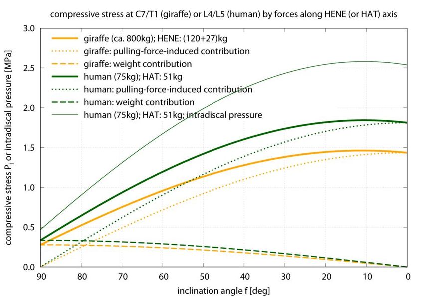
As can be seen in Fig. 3, the compressive stress values predicted by our model for the human
L4/L5 IVD ranges from 0.34 MPa in fully erect posture ( 90 ) to a maximum of 1.85 MPa
at about 10 , i.e., forward bending (flexion) of the HAT by 80 from upright posture. For
making these data immediately comparable with measured in vivo intradiscal pressure values
(Nachemson, 1966; Sato et al., 1999; Wilke et al., 1999; Takahashi et al., 2006), we
multiplied our predicted compressive stress (external pressure) values by a conservatively
estimated factor of 1.4, which has been determined in in vitro IVD preparations
(Nachemson, 1960; Brinckmann and Grootenboer, 1991). With this, we predict an intradiscal
pressure of 0.47 MPa for a fully erect posture, which perfectly corresponds to three sources
(Nachemson, 1966; Sato et al., 1999; Wilke et al., 1999) from which a fourth source
(Takahashi et al., 2006) deviates significantly but not dramatically (0.35 MPa). At the point
of maximum compressive stress, 10 , we would predict an intradiscal pressure of 2.58
MPa.
For standing with the HAT bent forward by 30 ( 60 ), we predict a compressive
stress of about 1.2 MPa (an increase from 90 by a factor of 3.5; intradiscal pressure
Biology Open • Accepted manuscript
about 1.7 MPa). In one (Wilke et al., 1999) of the more recent studies, an intradiscal pressure
value of 1.1 MPa (i.e., a factor of 2.2) was directly measured for some similar forward
bending (see their table 1 and figure 11), however, unfortunately, they did not give
quantitative data of the bending magnitude. In a later paper, Wilke et al. (2001) related their
1.1 MPa value to 36 bending, however, in terms of a local lumbar deflexion measure with
poorly reported marker position referencing and angle calculation. Again differently
quantifying the degree of bending, namely, by the local angular deflexion of L4 with respect
to L5, Sato et al. (1999) found an increase of intradiscal pressure during some forward
bending (their table 2: 5 local L4/L5 deflexion) from 0.54 MPa to 1.32 MPa, i.e., a factor of
2.5. For forward bending of a little less than 30 from fully erect, Takahashi et al. (2006)
found that their directly measured intradiscal pressure values increased by a factor of 3.7
Downloaded from http://bio.biologists.org/ by guest on January 28, 2021(from 0.35 MPa to 1.6 MPa, see their figure 4). Finally, for sitting either relaxed upright or
actively straightened, 0.46 MPa or 0.55 MPa, respectively, have been measured by Wilke
et al. (1999), consistent to their data of fully erect standing. We can establish that the
intradiscal pressure values predicted by our biomechanical model for quasi-static postural
conditions in human L4/L5 IVDs well match the respective data directly measured in vivo.
Our most reduced mechanical model—each just one degree of freedom and one torque-
compensating, pulling-force structure—is ‘co-contraction-free’ and, hence, provides a
minimum-IVD-compression estimate. Therefore, the good match with the currently available
measured intradiscal pressure data proved above allows a first inference from our results:
Active muscular stiffness modulation that might arise from stability requirements does not
seem determinative for the magnitude of compressive loads occurring in the human lumbar
spine. In other words: Co-contraction of muscles has minimal impact on everyday lumbar
compressive load scenarios.
Both in walking (Basu et al., 2019b) and galloping (Basu et al., 2019a), giraffe’s hold
their necks at about 35 . Values of the compressive stress on the giraffe’s neck base IVD
in their standing everyday postures (Fig. 3: from 0.95 MPa at 60 to 1.4 MPa at 30 )
are comparable to those on the L4/L5 IVD of a human who moderately deflects its spine from
its characteristic, fully erect posture ( 90 ), that is, within a range covering a human’s
daily activities: To let its compressive L4/L5 stress increase up to a giraffe’s everyday
maximum (1.4 MPa at 30 ), the human must flex its spine forward by 39 (i.e., to
51 ), with a corresponding intradiscal pressure of 1.95 MPa. The other way round,
humans who carry on their everyday activities like standing, sitting, or walking in a relaxed
way and close to fully erect posture, are exposed to about a third of the compressive stress
values on their lumbar discs as compared with giraffes in their everyday roaming (assumed to
be performed at 60 , the neutral posture (Stevens and Parrish, 2005, fig. 6.2)). The slope
of the pulling-force-induced contribution to the compressive stress Pi treated as a function of
the inclination angle (reduction means forward bending, see again Fig. 3) is proportional to
the species-specific multiplier (see Eq. (5) together with Eq. (6))
M i LCoM ,i
i , (7)
Ai Rpull ,i
with i indicating the species-specific spinal level analysed, M i the mass above the level, Ai
the level’s IVD area, and Ri the compensating structure’s lever arm. This multiplier S i is
Biology Open • Accepted manuscript
higher, thus, the slope steeper, in humans than in giraffes SL 4 L5 SC 7 T 1 .
DISCUSSION
Erecting the spine: a biomechanical design criterion
Our study has been initiated by a comparison of mechanical loads acting on comparable
anatomical structures in two species. The results of the comparisons across different degrees
of spine bending, which are common to the species examined, touch general evolutionary
issues across all species that developed erect body parts. Namely, the degree of erectness of a
species may be indicative of at least four design criteria being balanced during evolution: (i)
Downloaded from http://bio.biologists.org/ by guest on January 28, 2021(neural) control effort (Haeufle et al., 2014) for posture and movement (Haeufle et al., 2020),
(ii) loads of force-bearing structures, (iii) metabolic energy consumption, and (iv) functional
capabilities of the motor apparatus. Two of these are definitely costs (control effort,
metabolic energy), one may be indicative in itself of a balance of costs and basic limits of
physics (mechanical loads), and one is a gain (motor capabilities). Criterion (i) may even be
fundamental for itself, still, (i) and (iii) may be closely related in some respects (Niven
et al., 2007). Likewise, considering criteria (ii) and (iii) combined yields an example of a
design trade-off. On the one hand, there are energy costs of maintaining the body material:
Longer lever arms would usually imply being surrounded by increased volume of other tissue
to be maintained and moved. On the other hand, for both reducing structure loads and
energetic costs of near-isometric force production (Taylor et al., 1980; Taylor, 1985; Kram
and Taylor, 1990) longer lever arms and increased cross-sectional areas are desirable.
Concerning the erection of the human spine, a specification of these criteria of evolutionary
balancing would be: (i) Balancing the multiple inverted (unstable) pendulum HAT against
gravity likely increases the requirement for sensor deployment and their feedback signal
processing. (ii) The loads on the IVDs in particular must be limited by design, given that the
spine is exposed to, compared to other species, increased flexibility in multiple degrees of
freedom (even at once: think of throwing a stone, or a spear later in evolution) with partly
high deviations from being fully erect. (iii) Metabolic energy is required for continuous
balancing. Also, due to working with potentially high spine deviations, muscle deployment
must be limited. Realising (ii) and (iii) at once, may have favoured ample deployment of
ligaments. (iv) The gain of erecting the spine is in enabling humans to carry heavy loads,
combined with freeing their arms and hands from the demand to support weight, and all the
potential evolutionary consequences of freed hands.
We suspended our present story at the seemingly plausible notion that erecting a part of
the spine, that is, leaving a big portion of body mass (trunk or neck) from head downwards
unsupported against gravity by extremities, would pose a major mechanical challenge to body
material properties. As part of this notion, the challenge would grow with the size of the
animal, because the ratio of the weight of the erect spine part to its supporting area might be
expected to increase along with size. Our calculations have yielded an entirely different fact:
Erecting the cranial spine part immediately relieves the structures at its base from constantly
required mechanical loads, which can be measured particularly in terms of compressive stress
to IVDs. This allows to reduce the cross-sectional areas of the torque-compensating
ligaments and muscles, and maybe even their lever arms if slender spine design is a goal.
Additionally, the shear loads on the compressed material at the base may even be minimised:
The more erect the cranial spine part, the more can the pulling ligaments and muscles all
along it align with the ever-present external load due to gravity. As a consequence, the
Biology Open • Accepted manuscript
supporting weight-compensatory structures (IVDs) can be laid out to mainly resist axial
compression, even contribute themselves to low-torque compensation by inhomogeneous
compression, and still enable multiple degrees of spine movement by allowing moderate
shear forces instead of form fitting.
We can conclude, much in line with Helmuth (1985), that erecting the cranial spine
enables an animal to carry significant body-external extra load—at least, if hold close to the
spine axis—without having to withstand higher compressive IVD stresses than when just
supporting its cranial spine in moderately forward bent posture. Whether in a hominin or a
giraffe, the compressive stress value in the spine base at fully erect posture is only about a
third of the value at 30 forward flexion. In the latter neck posture, which occurs almost
permanently as a giraffe’s everyday biomechanical loading condition, the compressive IVD
Downloaded from http://bio.biologists.org/ by guest on January 28, 2021stress is about 1 MPa. Utilising empirical data in humans, this value would correspond to an
intradiscal pressure value of about 1.4 MPa, which can, hence, be considered a non-critical
norm for repeatedly occurring everyday activities of terrestrial vertebrates. For comparison
with a compressive stress value of 1 MPa, Alexander (1985) estimated dinosaurs’ spinal
everyday stress values to range from 2 MPa to 3 MPa, and Morris et al. (1961) calculated that
stress extremes as high as 6.2 MPa can be reached by humans in weight lifting.
In a nutshell, erecting the spine definitely opens, from a basic mechanical point of view,
an enormous potential to carry extra loads. The major biomechanical design and movement
challenges then are, of course, to handle such extra loads, that is, to ideally bring them into
carrying positions as close to the spine axis as possible, to actively balance (the trunk itself
plus the extra load) in unstable upright posture, and to discharge the extra load, as for these
tasks the spine must be somehow deflected from upright position, which may definitely and
immediately increase body-internal loads beyond critical material limits.
Morphological examinations: the significance of knowing anatomical and material properties
well
Another main conclusion of our reductionist, biomechanical model calculations, is that the
geometry—lever arms and IVD areas (see Eq. (7) and Eq. (5), with Eq. (6))—of the main
load-bearing body-internal structures (muscles, ligaments, IVDs, and vertebrae), strongly
dominates the magnitude of the compressive stress on the IVDs in the spine, as soon as the
spine is deflected from full erection: Already at 30 deflection from full erection, the pulling-
force-induced contribution to compressive stress has increased to more than two times HAT
or HENE weight, and to more than three times the weight contribution at this deflection, as
the weight contribution progressively decreases with deflection (Fig. 3). This conclusion
applies to as much the stress values occurring in the real, biological world as predictions
based on a model. By all means, it seems biomechanists all right agree that local load
distributions between various load-bearing body-internal structures somehow depend on the
local structures’ anatomical and material properties. Using intradiscal pressure values as one
example of how much internal loads depend on such properties, this study, in combination
with two similar ones (Helmuth, 1985; Jäger and Luttmann, 1989), can be understood as
highlighting the sensitivity of internal loads with respect to particularly anatomical
parameters.
For example, a human’s mean lever arm for a characteristic structure pulling past the
L4/L5 IVD was assumed in these three studies: 0.049m here, 0.05m in Helmuth (1985), and
0.065m in Jäger and Luttmann (1989). Thus, the data in the latter study deviate by about
Biology Open • Accepted manuscript
20%, which is immediately reflected by differences between our Fig. 3 and their calculated
compressive IVD stress values. Just one further model parameter included (here) or neglected
(there), namely the transmission factor of about 1.4 (Nachemson, 1960; Brinckmann and
Grootenboer, 1991) from (external) compressive stress to intradiscal pressure values,
amplifies to well over 40% lower pressure predictions by Jäger and Luttmann (1989, fig. 7)
than by us. Using the length of the spinous process as a caliper, the length of the lever arm of
this or that ligament or muscle passing the IVD can vary by at least a factor of three between
maximum and minimum possible values; with this, the magnitude of the torque contribution
of a respective structure may be uncertain accordingly. However, as all pulling structures
have to share the limited anatomical space, the assumption of a net lever arm length with a
corresponding net pulling force value will, thus, be uncertain on a similar level only at low
forces. With the force level increasing with forward flexion, the uncertainty of the predicted
Downloaded from http://bio.biologists.org/ by guest on January 28, 2021pulling force and, thus, the compressive stress will largely diminish, as all structures are
likely to contribute, which yields an averaging effect regarding lever arms.
Another example of model-based predictions of compressive stress values in the base
IVD area of hominins, namely, australopiths had been provided by Helmuth (1985). At 30
forward flexion (i.e., 60 ), he gave calculated values in the range from 3.8 MPa to 7.5
MPa in his paper text, which would be 3.2 to 6.3 times higher than predicted by us for a
human. A little confusing, taking the morphometric data of the biggest australopith specimen
considered by Helmuth (1985) (Australopithecus boisei: body height 1.63 m, body weight 50
kg, thus, M HAT 34 kg ), as well as both the highest lever arm Rpull 3.7 cm and the exactly
human-like LCoM , HAT 0.265 m given there, our re-calculation yielded compressive stress
values of only 2.4 to 3.3 times those in humans, i.e., 2.9 MPa to 4.0 MPa at 60 . These
compressive stress ranges are mainly due to the range of endplate areas, 3.6 cm 2 to 5.2 cm 2 ,
given by Helmuth (1985), which had been extracted there from even earlier australopith
literature (Johansson et al., 1982). As these values are much lower than in a human (about 15
cm 2 ), the compressive stress values calculated so far have been so much higher in
australopiths than in humans. Using the area values above according to
Helmuth (1985); Johansson et al. (1982), at fully erect posture ( 90 ), our re-calculated
compressive stress values would range from 0.65 MPa to 0.92 MPa . Assuming the lowest
lever arm value Rpull 2.6 cm, we would even predict 0.93 MPa to 1.31 MPa, very close to
giraffes’ everyday compressive stress values.
However, more data on australopiths have been collected since 1985. From the calibrated
figure 7.14 in a most recent source (Williams and Meyer, 2017), the dimensions of an
Australopithecus afarensis L4 endplate can be read off: a width of WL 4 4.8 cm and a depth
of DL 4 2.8 cm , i.e., an estimated area of AL 4 10.6 cm 2 . With these data, Rpull 3.7 cm,
and again the (likely adult) australopith body height and weight assumed above, we
eventually predict practically human-like compressive stress values of 0.32 MPa at 90
and 1.40 MPa at 60 in adult australopiths.
Accordingly, properly determining endplate areas, just like muscle and ligament lever
arms, in extant species or from fossil records are all key to well predicting compressive IVD
stresses. As a last example, referring to material properties, it has recently been shown that
widely scattering published data on spinal ligament properties, their stiffnesses (Mörl
Biology Open • Accepted manuscript
et al., 2020; Damm et al., 2019) and rest lengths (Mörl et al., 2020) in particular, are a source
of erroneous calculations of loads on all spinal structures. Consequently, our second major
conclusion is that the mechanical function(s) of an anatomical structure, as well as functional
interrelations between structures, are desirable to always be kept in mind when
experimentally examining and determining anatomical and material properties.
Our simple model for calculating compressive stress does only factor in a lumped
anatomy of all force generators passing the IVD and the IVD’s endplate area, but no further
geometrical, physical, or physiological knowledge of the load-bearing structures surrounding
the IVD. Nevertheless, it can reliably predict the compressive stress on an IVD, because,
among other things, this is the only joint structure that is compressed in flexion, and the
compressive stress acting on it, just like its intradiscal pressure, intrinsically represents an
Downloaded from http://bio.biologists.org/ by guest on January 28, 2021accumulative load quantity. However, to eventually understand, for example, damage to the
load-bearing structures occurring during movement, it seems almost trivial to punctuate that
structure-resolved modelling is absolutely essential. Even more, and in the spine in particular,
the structures’ non-linear interactions determine the load distribution among them (Mörl
et al., 2020). Thus, to resolve cause-effect chains, that is, to causally understand natural
processes, structure-resolved, mechanistic models are indispensable, beyond a simple
reductionist approach like the present.
On the distribution among passive and active pulling forces in the (human) spine
Accordingly, let us have a closer look at our present calculation results (for humans) with the
help of a complex model of the human lumbar spine that has incorporated all main spinal
load-bearing structures (Mörl et al., 2020). We verify the consistency of the load predictions
by both models, and deploy the complex model as a hand lens for decomposing the net
structural pulling force in our present, simple model into force contributions by the main
load-bearing structures. States of spine flexion in this complex, structure-resolved model can
be immediately compared with corresponding states of the present reductionist model,
because model values of body height and weight are similar. Whereas the spine flexion is
gravity-induced in the simple model here, a torque generated by a simulated machine motor
flexed the complex model of a human subject lying on its side, so that weight did not add a
compressive force to the spine.
The point of comparison shall be the state of maximum spine flexion in the complex
model, namely, its (steady) flexion response at about 30 N m IVD joint torque. This torque
value occurs in our present model when the spine is bent forward by 13 from upright
30 N m
posture ( 77 with Fpull 612 N from Fig. 4 and Rpull 0.049 m from Table 1).
R pull
At this 30 N m point, a compressive stress of 0.73 MPa (Fig. 3) is predicted by the present
model, while the directly weight-induced contribution is 0.33 MPa. At about the same 30 N
m IVD torque in the complex model (Mörl et al., 2020, fig. 5), a compressive IVD force of
pretty exactly 500 N has been predicted (Mörl et al., 2020, fig. 6), which corresponds to 0.33
MPa if a mean endplate area of 15 cm 2 (Table 1) at L4/L5 level is assumed. Adding 0.33
MPa due to the weight of our HAT mass (51 kg) for comparison with our present model
prediction, 0.66 MPa would be expected. The difference between 0.66 MPa and 0.73 MPa
can be largely explained: As predicted by the complex model, 75% of the IVD joint torque is
caused by forces due to structures (passive muscles and ligaments) pulling past the IVD
(Mörl et al., 2020, fig. 5), the remaining 25% are generated by the squeezed IVD itself. If,
accordingly, this 25% torque contribution by the IVD itself were non-existent and had instead
Biology Open • Accepted manuscript
to be additionally generated by structures pulling past the IVD, the compressive force would
be roughly a third higher than 500 N , therefore, the corresponding compressive stress
induced by these forces would be about 0.44 MPa, and adding 0.33 MPa due to bearing HAT
weight would yield 0.77 MPa, which is very close to 0.73 MPa predicted by the present,
simple model. It has also been found by Mörl et al. (2020) that ligament forces in particular
are certainly still moderately over-estimated by the complex model. Thus, the 500 N
compressive force predicted by the complex model, and 0.77 MPa compressive stress with it,
have to be still taken with some care. In any case, the consistency of load predictions in the
human lumbar spine by our present, simple model and a much more complex one is high.
Any co-contraction of pulling structures will, of course, increase the compressive stress on
the IVD. Therefore, the compressive stress values calculated by our simple model
Downloaded from http://bio.biologists.org/ by guest on January 28, 2021approximate the minimum to be expected from basic mechanics. If the values found in nature
are indeed measured to be close to this minimum, this may be an indication that co-
contraction in the human spine has been avoided as far as possible during its evolutionary
design, with the minimisation of co-contraction entailing minimised metabolic energy
consumption.
By means of our present, simple model, we predict the maximum compressive stress at
the base of HAT or HENE, respectively, to occur if they are bent forward by 80 from
upright posture ( 10 ; Fig. 3), and the maximum torque-compensating pulling force is
always required when being bent fully forward ( 0 ; Fig. 4). In humans, for example, the
corresponding compressive stress, IVD joint torque, and pulling force values are predicted as
1.81 MPa, 132.6 Nm, and 2706 N (Fig. 4), respectively, at 0 . Gracovetsky (1986)
estimated the maximum isometric force of all human lumbar back muscles to be about 2500
N. A more recent study documented a mean cross-sectional area of 87.4 cm 2 of all lumbar
muscles in men (Chang et al., 2016). The mean maximum isometric stress of skeletal muscle
at 37 C is about 25 N cm 2 (e.g., Mörl et al. (2020, sect. 2.5.7)), which would result in an
even lower maximum isometric force of 2185 N . There are, thus, strong indications that the
lumbar muscles alone of a human would not even be sufficient to lift its own upper body
masses, not to speak of external loads, at least if markedly bent forward but still straightened.
Muscles not located in the lumbar region, but reaching into it via an aponeurosis (e.g.,
actuated by m. latissimus), may aid in load lifting.
In any case, other passive pulling force generating structures like ligaments or the torque-
generating annulus fibrosus are essential for spinal functioning. In giraffes, there is likewise a
strong passive force involvement, and probably even stronger than in humans, as can be
inferred from their almost non-curved neck at a neutral posture (Christian and
Dzemski, 2007; Stevens and Parrish, 2005) of about 60 (Stevens and Parrish, 2005, fig.
6.2), and a marked nuchal ligament (Jouffroy, 1992; Taylor and Wedel, 2013), which
together make it very likely that muscle activity is required to lower the head down to their
toe level for drinking water (“browse by ventriflexion” (Stevens and Parrish, 2005)). In
humans, passive structures resist already moderate flexion of the lumbar spine (Mörl
et al., 2020). Holding significant parts of body mass in positions that are not fully erect, like
in everyday postures of giraffes, permanently requires static compensating pulling forces.
Perfectly upright postures, like in hominins, seem to be clearly less to non-demanding.
However, balancing these masses around full erection comes with the demand of struggling
with the inherent instability by constantly and dynamically loading and unloading the
compensating structures during all phases of everyday activity. Either way, fully erect or not,
Biology Open • Accepted manuscript
using active muscles for generating compensatory forces is metabolically demanding.
Therefore, relying strongly on passive compensatory contributions seems to be an appropriate
design by nature for balancing erect body portions.
Downloaded from http://bio.biologists.org/ by guest on January 28, 2021Acknowledgements
Formidable thanks from Michael to Maria Hammer for her sense of mathematical beauty, who therewith paved the way
to the frustum’s formulation.
Competing interests
We have no competing interest.
Contribution
MG and FM conjointly developed the story. MG performed the model calculations, prepared the stress and force plots,
and did the literature search on dinosaurs and australopiths. FM did the literature search on humans, giraffes, and other
animals, the survey of the giraffe, and its drawing. MG and FM constantly discussed the results and findings. MG and
FM conjointly drafted the introduction, MG wrote the initial draft of most of the results, and initially collected and
condensed the discussion points into written form. FM repeatedly reviewed both results and discussion. FM wrote the
initial draft of the abstract, MG revised it. The entire manuscript has been finally reviewed by both authors.
Funding
Michael Günther was supported by Deutsche Forschungsgemeinschaft (DFG: SCHM2392/5-2) granted to Syn Schmitt
(Universität Stuttgart).
Data availability
All relevant data are given in the text. All model calculations were done within ‘gnuplot’ (open source available under
GNU General Public License) in a flow with preparing the plots showing the results.
Supplementary
There is no supplementary material.
Biology Open • Accepted manuscript
Downloaded from http://bio.biologists.org/ by guest on January 28, 2021REFERENCES
Alexander, R. M., 1985. Mechanics of posture and gait of some large dinosaurs. Zoological Journal of the
Linnean Society 83 (1), 1–25.
Basu, C. K., Deacon, F., Hutchinson, J. R., Wilson, A. M., 2019a. The running kinematics of free-roaming
giraffes, measured using a low cost unmanned aerial vehicle (UAV). PeerJ 7, e6312 (21pp).
Basu, C. K., Wilson, A. M., Hutchinson, J. R., 2019b. The locomotor kinematics and ground reaction forces
of walking giraffes. The Journal of Experimental Biology 222 (Pt 2), jeb159277.
Biewener, A. A., 1989. Scaling body support in mammals: limb posture and muscle mechanics. Science
245 (4913), 45–48.
Biewener, A. A., 1990. Biomechanics of mammalian terrestrial locomotion. Science 250 (4984), 1097–
1103.
Brinckmann, P., Grootenboer, H., 1991. Change of disc height, radial disc bulge, and intradiscal pressure
from discectomy. An in vitro investigation on human lumbar discs. Spine 16 (6), 641–646.
Chang, Y. H., Healey, R. M., Snyder, A. J., Sayson, J. V., Macias, B. R., Coughlin, D. G., Bailey, J. F.,
Parazynski, S. E., Lotz, J. C., Hargens, A. R., 2016. Lumbar spine paraspinal muscle and intervertebral disc
height changes in astronauts after long-duration spaceflight on the international space station. Spine 41 (24),
1917–1924.
Christian, A., Dzemski, G., 2007. Reconstruction of the cervical skeleton posture of Brachiosaurus
brancai Janensch, 1914 by an analysis of the intervertebral stress along the neck and a comparison with
the results of different approaches. Fossil Record 10 (1), 38–49.
Damm, N., Rockenfeller, R., Gruber, K., 2019. Lumbar spinal ligament characteristics extracted from
stepwise reduction experiments allow for preciser modeling than literature data. Biomechanics and
Modeling in Mechanobiology 19 (3), 893–910.
Danowitz, M., Solounias, N., 2015. The cervical osteology of Okapia johnstoni and Giraffa
camelopardalis. PLoS One 10 (8), e0136552.
Dzemski, G., Christian, A., 2007. Flexibility along the neck of the ostrich (Struthio camelus) and
consequences for the reconstruction of dinosaurs with extreme neck length. Journal of Morphology 268 (8),
701–714.
Gilad, I., Nissan, M., 1985. Sagittal evaluation of elemental geometrical dimensions of human vertebrae.
Biology Open • Accepted manuscript
Journal of Anatomy 143, 115–120.
Gracovetsky, S. A., 1986. Determination of safe load. British Journal of Industrial Medicine (Occupational
and Environmental Medicine) 43 (2), 120–133.
Günther, M., Blickhan, R., 2002. Joint stiffness of the ankle and the knee in running. Journal of
Biomechanics 35 (11), 1459–1474.
Günther, M., Keppler, V., Seyfarth, A., Blickhan, R., 2004. Human leg design: optimal axial alignment
under constraints. Journal of Mathematical Biology 48 (6), 623–646.
Haeufle, D. F. B., Günther, M., Wunner, G., Schmitt, S., 2014. Quantifying control effort of biological and
technical movements: An information-entropy-based approach. Physical Review E 89, 012716.
Downloaded from http://bio.biologists.org/ by guest on January 28, 2021Haeufle, D. F. B., Wochner, I., Holzmüller, D., Driess, D., Günther, M., Schmitt, S., 2020. Muscles reduce
neuronal information load: quantification of control effort in biological vs robotic pointing and walking.
Frontiers in Robotics and AI – Soft Robotics 77 (13pp).
Hahn, U., 1993. Entwicklung mehrgliedriger Modelle zur realistischen Simulation dynamischer Prozesse in
biologischen Systemen. Master’s thesis, Eberhard-Karls-Universität, Tübingen.
Helmuth, H., 1985. Biomechanics, evolution and upright stature. Anthropologischer Anzeiger 43, 1–9.
Jäger, M., Luttmann, A., 1989. Biomechanical analysis and assessment of lumbar stress during load lifting
using a dynamic 19-segment human model. Ergonomics 32 (1), 93–112.
Johansson, D., Lovejoy, C., Kimbel, W., White, T., Ward, S., Bush, M., Latimer, B., Coppens, Y., 1982.
Morphology of the Pliocene partial hominid skeleton (AL 288-1) from the Hadar formation, Ethiopia.
American Journal of Physical Anthropology 57 (4), 403–451.
Jouffroy, F. K., 1992. Evolution of the Dorsal Muscles of the Spine in Light of Their Adaptation to Gravity
Effects. In: Berthoz, A., Graf, W., Vidal, P. P. (Eds.), The Head-Neck Sensory Motor System. Oxford
University Press, Oxford, UK, pp. 22–35.
Kram, R., Taylor, C., 1990. Energetics of running: a new perspective. Nature 346 (6281), 265–267.
Mitchell, G., van Sittert, S., Skinner, J., 2009. Sexual selection is not the origin of long necks in giraffes.
Journal of Zoology 278 (4), 281–286.
Mörl, F., Günther, M., Riede, J. M., Hammer, M., Schmitt, S., 2020. Loads distributed in vivo among
vertebrae, muscles, spinal ligaments, and intervertebral discs in a passively flexed lumbar spine.
Biomechanics and Modeling in Mechanobiology 19 (6), 2015–2045.
Morris, J. M., Lucas, D. B., Bresler, B., 1961. Role of the trunk in stability of the spine. The Journal of Bone
& Joint Surgery 43-A (3), 327–351.
Nachemson, A. L., 1960. Lumbar intradiscal pressure. Experimental studies on post-mortem material. Acta
Orthopaedica Scandinavica 31 (Suppl. 43), 1–104.
Nachemson, A. L., 1966. The load on lumbar disks in different positions of the body. Clinical Orthopaedics
and Related Research 45, 107–122.
NASA Reference Publication, 1978. Anthropometric Source Book. Tech. Rep. 1024, I-III, NASA Scientific
and Technical Information Office, Springfield.
Niven, J., Anderson, J., Laughlin, S., 2007. Fly photoreceptors demonstrate energy-information trade-offs in
neural coding. PLoS Biology 5 (4), e116. Biology Open • Accepted manuscript
Sato, K., Kikuchi, S., Yonezawa, T., 1999. In vivo intradiscal pressure measurement in healthy individuals
and in patients with ongoing back problems. Spine 24 (23), 2468–2474.
Seyfarth, A., Günther, M., Blickhan, R., 2001. Stable operation of an elastic three-segment leg. Biological
Cybernetics 84 (5), 365–382.
Stevens, K. A., Parrish, J. M., 2005. Digital Reconstructions of Sauropod Dinosaurs and Implications for
Feeding. In: Wilson, J., Curry-Rogers, K. (Eds.), The Sauropods: Evolution and Paleobiology. University of
California Press, Berkeley, CA, Ch. 6, pp. 178–200.
Stöcker, H. (Ed.), 2008. Taschenbuch mathematischer Formeln und moderner Verfahren, 21st Edition. Harri
Deutsch, Frankfurt, Deutschland.
Downloaded from http://bio.biologists.org/ by guest on January 28, 2021Takahashi, I., Kikuchi, S. I., Sato, K., Sato, N., 2006. Mechanical load of the lumbar spine during forward
bending motion of the trunk—a biomechanical study. Spine 31 (1), 18–23.
Taylor, C., 1985. Force development during sustained locomotion: a determinant of gait, speed and
metabolic power. The Journal of Experimental Biology 115, 253–262.
Taylor, C., Heglund, N., McMahon, T., Looney, T., 1980. Energetic cost of generating muscular force
during running: a comparison of large and small animals. The Journal of Experimental Biology 86, 9–18.
Taylor, M. P., Wedel, M. J., 2013. The effect of intervertebral cartilage on neutral posture and range of
motion in the necks of sauropod dinosaurs. PLoS One 8 (10), e78214.
van Sittert, S. J., Skinner, J. D., Mitchell, G., 2010. From fetus to adult—an allometric analysis of the giraffe
vertebral column. Journal of Experimental Zoology 314B (6), 469–479.
Vidal, D., Mocho, P., Páramo, A., Sanz, J. L., Ortega, F., 2020. Ontogenetic similarities between giraffe and
sauropod neck osteological mobility. PLoS One 15 (1), e0227537.
Wilke, H. J., Neef, P., Caimi, M., Hoogland, T., Claes, L. E., 1999. New in vivo measurements of
pressures in the intervertebral disc in daily life. Spine 24 (8), 755–762.
Wilke, H. J., Neef, P., Hinz, B., Seidel, H., Claes, L. E., 2001. Intradiscal pressure together with
anthropometric data – a data set for the validation of models. Clinical Biomechanics 16 (Suppl. 1), S111–
S126.
Williams, S. A., Meyer, M. R., 2017. The Spine of Australopithecus. In: Been, E., Gómez-Olivencia,
A., Kramer, P. (Eds.), Spinal Evolution. Springer, Cham, CH, Ch. 7, pp. 125–151.
Zhou, S. H., McCarthy, I. D., McGregor, A. H., Coombs, R. R. H., Hughes, S. P. F., 2000. Geometrical
dimensions of the lower lumbar vertebrae – analysis of data from digitised CT images. European Spine
Journal 9 (3), 242–248.
Biology Open • Accepted manuscript
Downloaded from http://bio.biologists.org/ by guest on January 28, 2021FIGURES
Biology Open • Accepted manuscript
Fig. 1.
Geometrical dimensions of an adult (female) giraffe with an overall body length (“total
length” (Mitchell et al., 2009): sum of tail, neck and head lengths, plus distance from tail base
to withers) of about 4.30 m and weight of 800 kg, respectively, with the dimensions given in
metres. The neck length is 1.55 m, and the “total height” (Mitchell et al., 2009) is the withers
height (2.45 m) plus neck length (1.55 m) plus half of the head height (0.34 m), i.e. 4.17 m.
These numbers have been measured in the original drawing of Vidal et al. (2020, fig. 1). Our
sketch here is a freehand drawing, so the numbers may slightly deviate from the distances
within our sketch.
Downloaded from http://bio.biologists.org/ by guest on January 28, 2021Fig. 2.
The geometry and mechanical structure of the model of a giraffe’s head-neck (HENE)
segment assembly, true to scale in all dimensions (Table 1), and the modelled forces acting
on HENE (condensed in grey at the bottom: only schematically, Fpull is plotted much too
short in relation to weight vector). The smaller filled circles are the CoMs of neck and head
parts, the thick one is HENE’s overall CoM, with the thick black line depicting its distance
(Eq. (2)) from the base of HENE (at the C7/T1 joint: open circle), and the thin line the head’s
distance. The thick short line labelled with Rpull and oriented perpendicular to the double
arrow that symbolises the pulling force vector Fpull is the lever arm of the Fpull -generating
structure that spans HENE’s base joint and, therewith, compensates the torque (Eq. (4))
Biology Open • Accepted manuscript
around the joint generated by HENE’s weight M HENE g . The compressive force FC 7 T 1,||
(Eq. (3) or Eq. (5), respectively) on HENE’s base is also depicted: the grey thick line
perpendicular to the dashed base line (surface) of HENE, i.e., the projection of the force
vector Fpull M HENE g on HENE’s longitudinal axis. For calculating compressive force
FL 4 L5,|| on a hominin L4/L5 IVD instead of FC 7 T 1,|| , HENE parameters are replaced in Eqs.
(5,6)—and the sketch here: only the erect body part’s mass and CoM position varies among
the species—by the corresponding human- or australopith-like ones (see Table 1).
Downloaded from http://bio.biologists.org/ by guest on January 28, 2021You can also read