Microbial hitchhiking: how Streptomyces spores are transported by motile soil bacteria
←
→
Page content transcription
If your browser does not render page correctly, please read the page content below
The ISME Journal (2021) 15:2591–2600
                                    https://doi.org/10.1038/s41396-021-00952-8

                                        ARTICLE

                                    Microbial hitchhiking: how Streptomyces spores are transported by
                                    motile soil bacteria
                                                         1,2                         1,2                        1,2
                                    Alise R. Muok              ●
                                                                   Dennis Claessen         ●
                                                                                               Ariane Briegel

                                    Received: 24 August 2020 / Revised: 19 February 2021 / Accepted: 25 February 2021 / Published online: 15 March 2021
                                    © The Author(s) 2021. This article is published with open access

                                    Abstract
                                    Streptomycetes are sessile bacteria that produce metabolites that impact the behavior of microbial communities. Emerging
                                    studies have demonstrated that Streptomyces spores are distributed through various mechanisms, but it remains unclear how
                                    spores are transported to their preferred microenvironments, such as plant roots. Here, we show that Streptomyces spores are
                                    capable of utilizing the motility machinery of other soil bacteria. Motility assays and microscopy studies reveal that
                                    Streptomyces spores are transported to plant tissues by interacting directly with the flagella of both gram-positive and gram-
                                    negative bacteria. Genetics experiments demonstrate that this form of motility is facilitated by structural proteins on the
1234567890();,:
                  1234567890();,:

                                    spore coat. These results demonstrate that nonmotile bacteria are capable of utilizing the motility machinery of other
                                    microbes to complete necessary stages of their lifecycle.

                                    Introduction                                                                spores over long distances through attachment to insects
                                                                                                                and nematodes, but it is unclear how they relocate over
                                    Bacteria belonging to the genus Streptomyces are an integral                short distances to their preferred microenvironments such as
                                    component of diverse ecosystems and are well-known to                       plant root systems [6, 7].
                                    produce chemically diverse metabolites, including the vast                     While Streptomyces are nonmotile, many other soil
                                    majority of all clinically relevant antibiotics [1, 2]. Soil-               microbes, such as Bacillus subtilis (Bs) and Pseudomonas
                                    dwelling Streptomycetes, such as Streptomyces coelicolor                    fluorescens (Pf), are motile and can move through their
                                    (Sc), colonize plant roots and provide the associated plant                 environment by regulating the rotation of flagella [8]. On
                                    protection from potential phytopathogens through antibiotic                 solid surfaces, flagellar rotation enables cell swarming.
                                    secretion [1]. The symbiosis of Streptomycetes with their                   During swarming, cells are densely packed and con-
                                    plant hosts has been shown to improve plant health and                      tinuously move outward toward unoccupied areas. In liquid
                                    productivity, and thereby provides a potential sustainable                  media, flagella enable cell swimming where the cells move
                                    solution to increase crop yields [2–5]. The lifecycle of                    independently and can rapidly change swimming direction.
                                    Streptomycetes is complex and involves stages of aerial                     In addition to flagellar motility, some bacteria can also
                                    hyphae formation on the soil surface to produce spores, and                 move through a passive diffusion on surfaces called sliding
                                    spore germination on plant roots to produce filamentous                      [8]. Sliding does not involve flagella but occurs when cells
                                    colonies [1]. Immotile Streptomyces bacteria distribute their               are pushed through the forces generated by the outward
                                                                                                                growing colony.
                                                                                                                   Recent reports have revealed that microbe transport by
                                                                                                                inter-species interactions can occur between motile and
                                    Supplementary information The online version contains
                                    supplementary material available at https://doi.org/10.1038/s41396-         immotile microbes [9]. These studies demonstrate that inter-
                                    021-00952-8.                                                                microbial transport occurs among organisms natively found
                                                                                                                on abiotic surfaces [10, 11], plant surfaces [12], and within
                                    * Ariane Briegel
                                      a.briegel@biology.leidenuniv.nl
                                                                                                                the soil [13, 14]. In some instances, the transportation of
                                                                                                                human pathogens are facilitated on abiotic surfaces,
                                    1
                                         Institute for Biology, Leiden University, Leiden, The Netherlands      including nonmotile Staphylococcal species that directly
                                    2
                                         Centre for Microbial Cell Biology, Leiden University, Leiden, The      adhere to their mobile partners [10], Aspergillus fumigatus
                                         Netherlands                                                            (Af) spores that interact with the flagella of motile bacteria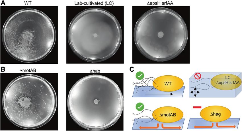
2592                                                                                                              A. R. Muok et al.

[14], various nonmotile human microbiome bacteria that are
carried by Capnocytophaga gingivalis [15], and Legionella
pneumophila that is transported internally by their amoebae
hosts [16].
   Here, we demonstrate that spores of the sessile Strep-
tomycetes, such as Sc, are transported by Bs to their
preferred microenvironment. Sc and Bs are both soil-
dwelling bacteria that utilize plant root exudate as a
nutritive source [1, 17]. Using microscopy methods,
motility assays, and genetics approaches, we demonstrate
that Bs transports Sc spores via direct attachment to
Bs flagella, a mode of transportation we call “hitchhik-
ing”. Hitchhiking is dependent on the conserved rodlin
proteins, which form a fibrous outer layer on the spore
coat of almost all Streptomycetes, but with a hitherto
unclear function [18, 19]. These results exemplify that
nonmotile bacteria are capable of utilizing the motility
machinery of other microbes to occupy advantageous
environments, and that this mode of transport may be
                                                              Fig. 1 S. coelicolor spores are transported by B. subtilis. A When
widespread in nature.                                         Sc and Bs are innoculated on the center of a swarm plate, visible
                                                              Sc colonies (white dots) are apparent and are only dispersed in the
                                                              presence of motile Bs. Left: Sc with Bs. Middle: Sc alone. Right: Bs
Results                                                       alone. B When Sc and Bs are innoculated in different positions on
                                                              swarm plates, the Sc colonies are dispersed in the swarming direction
                                                              of the Bs cells (black arrows). Asterisks denote the Bs innoculation
Bacillus subtilis disperses Streptomyces coelicolor           sites. C Bs moves spores toward plant tissues. Left: Sc with
spores                                                        Bs. Middle: Sc alone. Right: Bs alone, asterisk denotes the
                                                              Bs innoculation site.
Transportation of Sc spores by Bs was demonstrated by
mixing isolated Sc spores with a liquid culture of Bs         B. subtilis transports S. coelicolor spores to plant
followed by inoculation onto an agar swarm plate and          tissues
incubation. After 5 days, Sc colonies are visible on the
plate and are only dispersed in the presence of Bs in all     In nature, Sc and Bs thrive near plant roots that excrete
samples tested (n = 13) (Fig. 1A). Spore dispersal by Bs      exudates but only Bs can move toward the root systems. We
occurs across the entire surface of the plate and to the      conducted assays to determine if Bs can transport Sc spores
plate’s edge (4.5 cm from the inoculation point). To          to plant tissues. Assays with the Bs strain alone demonstrate
demonstrate that the Sc spores are being moved by Bs          that the plates become “cloudy” with Bs cells in areas
cells and not merely “floating” in the expanding Bs col-       around plant tissues, perhaps due to the presence of nutritive
ony, we conducted identical assays but inoculated the Sc      plant exudates that facilitates bacterial growth (Fig. 1C).
spores and Bs culture separately and onto different areas     Like previous experiments, the Sc spores alone do not
of the plate. The resulting Sc colonies form streaks across   exhibit movement unless they are co-inoculated with Bs
the plate that emanate from the Bs inoculation site in a      cells, and the dispersed spores preferentially establish
predictable manner in all samples tested (n = 10)             colonies near plant tissues in all instances (n = 5) (Fig. 1C).
(Fig. 1B). This experiment was repeated on 12 cm plates
and demonstrate that Sc spores are dispersed to the edge      Spore dispersal occurs with swarming and sliding Bs
of the plate, which is 10 cm from the spore innoculation      strains
point (n = 3) (Fig. S1A). To determine the effeciency of
transport, this experiment was conducted with dilutions of    Bs has two modes of flagellar-mediated motility, swimming
Sc spores so that the total number of Sc colonies             and swarming, that occur in liquid environments and on
would germinate without overlap and could be individu-        solid surfaces, respectively. When Bs senses that it is on a
ally counted. The results demonstrate that 86 ± 5.6% of       solid surface, it will differentiate into a swarmer cell that has
the apparent colonies are located outside of the initial      a significant increase in the number of flagella and produces
innoculation point under these conditions (n = 4)             hydrophobic surfactants, such as surfactin, to efficiently
(Fig. S1B).                                                   spread across the surface [20–22]. In our experiments, we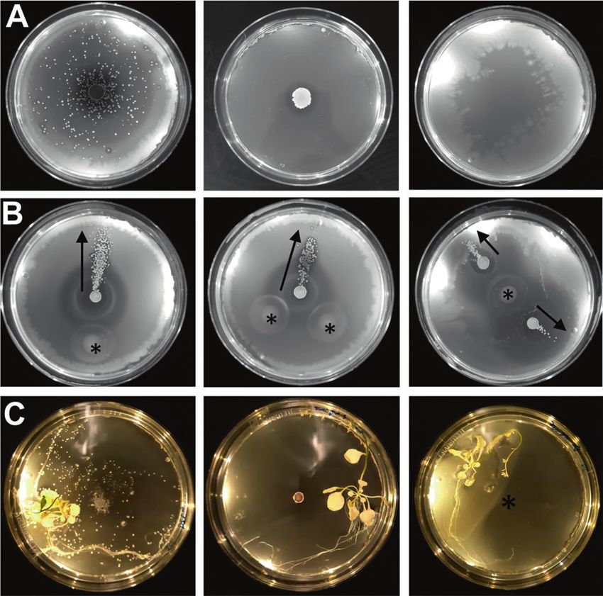
Microbial hitchhiking: how Streptomyces spores are transported by motile soil bacteria                                                       2593

Fig. 2 B. subtilis can transport spores via swarming and sliding.           Δhag strain that does not possess flagella cannot disperse the spores
A The WT (swarming strain) can transport spores on agar plates              via sliding at WT levels (n = 6). Quantification of results is shown in
(0.27–0.5%) (n = 13), but swimming only strains (laboratory-culti-          Fig. S2. All Δhag plates are shown in Fig. S3. C In summation, spores
vated WT strain or ΔepsH srfAA) cannot (0.25–0.3% agar) (n = 6 and          are dispersed by swarming (WT) and sliding (ΔmotAB) Bs cells in a
n = 8, respectively). Quantification of the results is shown in Fig. S2.     flagella-dependent manner. However, spores are not dispersed via
B A Bs ΔmotAB strain that possesses flagella but lacks flagellar              swimming (lab-cultivated or ΔepsH srfAA), and are significantly less
motility can disperse spores via sliding at WT levels (n = 6). A Bs         dispersed via sliding in the absence of flagella (Δhag).

utilized an undomesticated strain of Bs (NCIB3610) that can                 entire surface and to the edge of the agar plates in all
swarm, unlike common laboratory strains that lack                           samples (4.5 cm from the inoculation point) (n = 6)
the ability to differentiate into swarmer cells and fail to                 (Figs. 2B and S2). The number of dispersed spores cannot
produce surfactin to undergo swarming motility (Table S1)                   be ascertained due to overlapping colonies that result in
[20, 22–24]. To determine if the Sc spores are transported                  apparent smears across the plates, but each plate contains
by both swarming and swimming motilities, we repeated the                   over 100 dispersed Sc colonies. The Δhag strain shows
experiments with a laboratory-cultivated Bs strain that is                  severely reduced dispersal, where an average of 3.2 spores
incapable of swarming, and spore transport does not occur                   are transported with an average maximum distance of
on the agar swimming plates (0.27% agar) (Figs. 2A and                      0.95 ± 0.85 cm from the inoculation point (n = 6) (Figs. 2B,
S2) [20]. Importantly, the laboratory-cultivated strain has                 S2 and S3). Therefore, we surmise that spore dispersal can
accumulated many genetic defects in addition to defective                   also occur through sliding but is facilitated by the presence
swarming capabilities. To ensure that the decrease in spore                 of flagella (Fig. 2C). As an additional control, we also
dispersal can be attributed to limitations of bacterial                     conducted these experiments by first spreading the Bs cells
swimming, we utilized a Bs strain with an undomesticated                    across the surface of the plate and then inoculating the Sc
genetic background but has been genetically altered so that                 spores in the center. Spore dispersal was significantly
it is only capable of swimming motilities (Δepsh srfAA,                     reduced for both the Bs WT (2.06 ± 0.05 cm) and ΔmotAB
DK1484). Like the laboratory-cultivated strain, this strain                 (1.25 ± 0.16 cm) strains when compared to co-inoculation in
does not disperse Sc spores on swim plates (Figs. 2A and                    the center of the plate (Figs. S2 and S4). These data indicate
S2). Therefore, we conclude that spore transport can be                     that the Sc spores are transported by Bs cells that con-
accomplished by swarming motility but not swimming                          tinuously move away from the inoculation point and are not
motility.                                                                   transported after the motile Bs has covered the surface of
   In addition to flagellar motilities, Bs can also move                     the plate.
through sliding on surfaces. To determine if spores can be
dispersed through sliding, we utilized a Bs mutant that has                 S. coelicolor spores attach to B. subtilis flagella
flagella but the flagella cannot rotate (ΔmotAB, DS222)
[25], and a mutant that does not possess flagella (Δhag,                     We utilized several microscopy methods to elucidate a
DS1677) [26]. The ΔmotAB strain moves spores over the                       mechanism for Sc spore dispersal by Bs. Fluorescently
2594 A. R. Muok et al.
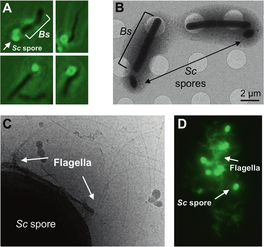
co-localizing with the spores in two-dimensional cryo-
EM images (Figs. 3C and S5B).
Sc spore adherence to Bs flagella was further confirmed
by visualization of a mixture of Sc spores with sheared
flagella via fluorescence microscopy. Fluorescent labeling
of Bs flagella was accomplished using a Bs strain (DS1919)
that is mutated in a single flagellin residue (T209C) [28].
The surface-exposed thiol allows for direct labeling with
dyes that possess a reactive maleimide group. This dye also
labels proteins on the surface of the Sc spore coat. Indeed,
when sheared Bs flagella are isolated and mixed with
spores, and unassociated flagella are washed from the
mixture, a majority of spores still retain associated flagella.
In total, ~64% of the spores are associated with flagella
(n = 130 spores). In some instances, large “clumps” of
spores entangled in flagella are observed (Figs. 3D and
S5C). In comparison, the flagella without spores added do
not form aggregates and are randomly dispersed (Fig. S5C).
Fig. 3 Microscopy methods indicate that Sc spores directly adhere
to Bs flagella. A Fluorescence microscopy of dye-labeled spores with
unlabeled Bs cells demonstrate that the spores localize to the cell poles Hitchhiking is conserved in Streptomycetes
of Bs. B Cryo-electron microscopy samples of mixed Sc spores and Bs
cells reveal that the spores do not directly adhere to the Bs cell body. To determine if spore dispersal by Bs also occurs in other
C Cryo-EM shows the Bs flagella colocalize with the Sc spore coat.
D Fluorescence microscopy of dye-labeled sheared flagella and spores Streptomyces species, we conducted Bs swarm plate assays
demonstrate that spores directly interact with Bs flagella to form with Streptomyces tendae (St), Streptomyces griseus (Sg),
extended associations of both components. Streptomyces scabies (Ss), and Streptomyces avermitilis
(Sa). To quantify spore dispersal, we prepared swarm plate
assays where Bs cells were inoculated at the center of the
labeled Sc spores were imaged under a fluorescence plate (9 cm in diameter) and the isolated spores are inocu-
microscope and were immotile as expected (Supplemen- lated in four equidistant positions around the Bs inoculation
tary Movie 1). However, when Bs cells were added to the site. The maximum dispersal distance, which is the distance
fluorescent spores, the spores localize near the Bs cell from the center of the spore inoculation site to the most
poles (Supplementary Movie 2 and Fig. 3A). In some distant dispersed colony, was measured for each of the four
instances, the spores are stationary on the surface of the spore samples. The wild-type (WT) Sc spores are dispersed
glass slide and an associated Bs cell is seen rotating by Bs in 100% of samples and are moved an average
around the spore (Movie 1). The observed Bs cell rotation maximum distance of 2.67 ± 0.43 cm from the initial
in these assays is reminiscent of rotations seen in Bs cells inoculation point (n = 20). Likewise, these assays demon-
that have their flagella chemically tethered to a solid strate that St (n = 12), Sg (n = 8), and Ss (n = 8) spores are
surface, whereby the torque generated by the immobilized dispersed in 100% of samples to similar distances as WT Sc
flagella induces rotation of the cell body [27]. This spores. However, the Sa spores are dispersed at significantly
observation suggests that the Sc spores adhere directly to shorter distances (n = 12) (Fig. 4A, B) and are dispersed in
the Bs flagella, and therefore effectively mimic a flagellar 83% of the samples. As the last common ancestor of Sc and
tether in these instances. To verify that the Sc spores do Sg existed more than 200 million years ago and both species
not directly interact with the Bs cell body, we imaged a are capable of hitchhiking, these data suggest that the
mixture of Sc spores and Bs cells with a cryo-electron ancestor also possessed this dispersal mechanism and it
microscope. Like the fluorescence microscopy images, remained conserved.
the Sc spores are localized near the Bs cell poles but do
not make direct contact with the Bs cell body (Figs. 3B Spore dispersal by B. subtilis is facilitated by the
and S5A). In total, ~77% of the spores are located within rodlin proteins
1 μm of a Bs cell poll (n = 35 spores). To determine if the
spores interact with Bs flagella, we utilized a Bs “mini- The outer surface of most Streptomyces spores is char-
cell” strain (minD::TnYLB) that lacks the excreted acterized by a fibrillar rodlet layer, which is a striated pat-
material inhibiting their direct visualization. Indeed, tern of pairwise aligned rodlets composed of the rodlin
when mixed with Sc spores, the flagella can be seen proteins [18, 19]. Scanning electron microscopy (SEM)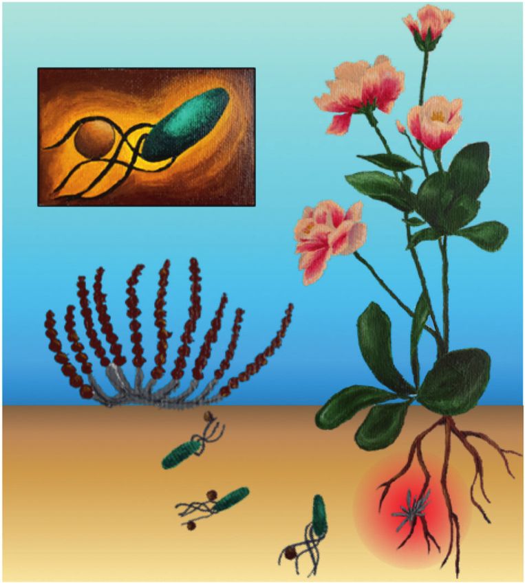
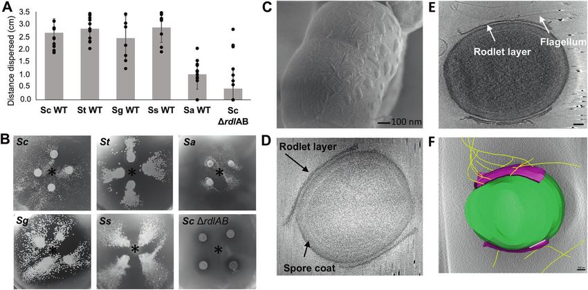
Microbial hitchhiking: how Streptomyces spores are transported by motile soil bacteria                                                          2595

Fig. 4 Hitchhiking of Streptomyces spores is facilitated by the             (p < 0.05). B Representative images of Bs/spore swarm plates from
presence of rodlin proteins. A Bs swarm assays with both wild-type          A. The Bs innoculation site is denoted with an asterisk. C SEM of WT
(WT) Streptomyces spores (S. coelicolor n = 20, S. tendae n = 12,           Sc spores shows the rodlet layer with pairwise rodlets of 20 nm spa-
S. griseus n = 8, S. scabies n = 8, S. avermitilis n = 12) and Sc spores    cing. D A representative cryo-ET image of isolated Sc WT spores
lacking the rodlin proteins (Sc ΔrdlAB n = 24) demonstrate that             shows that the rodlet layer does not cover the entire spore but leaves
spores are dispersed in all tested WT species but with S. avermitilis       the poles exposed (n = 22). E Cryo-ET reconstructions show that
dispersed the shortest distance, and dispersal is abrogated by the loss     flagella preferentially interact with the rodlet layer (n = 12). Scale bar
of the rodlins in Sc. Results are expressed as the mean of dispersal        100 nm. F Segmentation of the reconstruction from E clearly
distance ± standard error of the mean. Differences for Sc ΔrdlAB and        demonstrates the flagella:rodlin interaction. Spore body: green, rodlet
Sa dispersal compared to WT Sc, St, Sg, and Ss strains are statistically    layer: purple, flagella: yellow. Scale bar 100 nm (Color figure online).
significant using a two-tailed null hypothesis significance test

images of Sc and Ss spores show the striated rodlet layer                   dispersed with an average maximum distance of 0.46 ±
(Fig. 4C). In previous studies, the rodlets of Sc, S. lividans,             0.82 cm from the initial inoculation point and dispersal
St, Sg, and Ss were visually indistinguishable [19, 29].                    occurs in 33% of the samples (n = 24) (Fig. 2).
Using electron microscopy images from this and previous                         To characterize how rodlins interact with flagella in three
studies, we measured the spacing of the rodlets in these                    dimensions, we conducted cryo-ET experiments of samples
species, which is highly conserved and around ~20 nm                        containing Bs minicells and Sc spores. Reconstructions
(when measured from the center of the rodlet fibers)                         show that the Sc spores are oval shaped and possess a thick
(Table S2). Furthermore, the rodlin proteins from Sc, St, and               coat. The rodlet layer can be seen as a sheath around the
Sg have ~34% sequence identity despite the species’ distant                 lateral sides of the spore with frayed edges, leaving the
evolutionary relation (Fig. S6) [19].                                       poles exposed, and suggest that the rodlet sheath easily
   Intriguingly, Sa spores are less widely dispersed than the               peels away from the cell body (n = 22) (Fig. 4D). A similar
other Streptomyces species and it is the only tested species                spore morphology has also been observed in Streptomyces
that natively lacks rodlin proteins [19]. In agreement, an Sc               albus [32]. Bs flagella accumulate around and directly
mutant strain that lacks the rodlin proteins (ΔrdlAB) abro-                 interact with the rodlet layer (n = 12) (Fig. 4E, F and
gates hitchhiking by Bs (n = 3, Figs. S2 and S7). Impor-                    Movie 2). However, due to the thickness of the spores the
tantly, previous studies demonstrate that the Sc ΔrdlAB                     resolution is limited and we could not deduce if the flagella
strain is not delayed in germination, and does not exhibit                  preferentially bind specific features of the rodlet layer.
any behavioral or phenotypic change compared to the WT                      Collectively, these data suggest that the rodlet layer facil-
strain with the exception of the rodlet layer [18, 19, 30]. In              itates spore dispersal by interacting directly with flagella.
contrast, Sc mutants that lack proteins which produce
polysaccharides found on the surface of Streptomycetes                      Hitchhiking of S. coelicolor spores is not limited to
(ΔcslA and ΔmatAB) are unaffected (n = 3, Figs. S2 and                      Bacillus
S7) [31]. However, spore dispersal was not completely
abolished in the ΔrdlAB strain. Using identical swarm plate                 Although B. subtilis is ubiquitous in soil, other genera are
assays described in the section above, the ΔrdlAB strain is                 also flagellated and may also contribute to dispersal of Sc
2596                                                                                                                      A. R. Muok et al.

                                                                           soil microbes by directly attaching to their flagella. While
                                                                           these experiments demonstrate that Sc spores are dis-
                                                                           persed by Bs and Pf regardless of their destination,
                                                                           these motile bacteria are also known to associate with
                                                                           plant roots. Therefore, this mechanism of dispersal,
                                                                           called hitchhiking, may provide Streptomyces spores a
                                                                           mechanism for translocation to beneficial environments.
                                                                           Indeed, assays with A. thaliana plants demonstrate
                                                                           that Bs can transport Sc spores to plant tissues. This
                                                                           allows spores to germinate near nutrient-rich plant
                                                                           exudate to generate filamentous colonies that produce
                                                                           antibiotics, thereby protecting the plant from potential
                                                                           phytopathogens.
                                                                               Hitchhiking is facilitated by two spore coat proteins,
                                                                           RdlA and RdlB, which are conserved in most Streptomy-
                                                                           cetes. These proteins assemble into pairwise aligned fila-
                                                                           ments, called rodlets, on the outer surface of the spores and
                                                                           are spaced ~20 nm apart (Table S2). Until now, the function
                                                                           of the rodlets has remained elusive [30]. Interestingly, the
                                                                           diameter of the bacterial flagellar filament is also ~20 nm
                                                                           [22, 36]. Therefore, it is possible that the rodlet layer pro-
Fig. 5 An overview of the hitchhiking model. Aerial Streptomyces           vides a gripped surface for the flagella, which become
spores are transported on the cm-scale to plant root systems by directly
                                                                           “wrapped” in the grooves made by the rodlin proteins and
adhering to the flagella of motile bacteria (inset). Here, the spores
germinate and produce antibiotics (red gradient) to ward off microbial     thereby facilitates spore transport. However, our cryo-ET
competitors (Color figure online).                                          data can not support such speculations due to the limited
                                                                           resolution of the whole-cell reconstructions. Therefore, it is
                                                                           still unclear what properties of the rodlet layer encourage
spores. We therefore conducted swarm plate assays with a                   interactions with flagella.
P. fluorescens WT strain (R1SS101), which is also natively                      Emerging studies have demonstrated that flagella pre-
associated with plant roots [33]. Importantly, Pf disperses                ferentially interact with hydrophobic surfaces and fla-
spore similarly to Bs, but Sc colonies appear in patterns                  gellin can undergo methylation to increase flagella
that are reminiscent of Pseudomonas swarm patterns [34]                    hydrophobicity [37–39]. This increase in hydrophobicity
(n = 6) (Fig. S8). These data demonstrate that hitchhiking is              allows pathogenic bacteria to adhere to host cells [37–39],
a widespread mechanism that allows Streptomyces spores to                  and flagellar adherence to plant cells has also been
disperse at cm-scales (Fig. 5).                                            implicated in establishing colonization [40, 41]. Flagella
                                                                           hydrophobicity may facilitate interactions with hydro-
                                                                           phobic spores and may account for spore transport that is
Discussion                                                                 seen in the absence of rodlins, given that the spore surface
                                                                           without rodlins remains hydrophobic (in Sa WT and Sc
Sessile Streptomycetes have a complex lifecycle that                       ΔrdlAB strains) [18, 42, 43]. Therefore, the same fla-
involves formation of aerial hyphae that differentiate into                gellar interactions that facilitate adherence to plant roots
spores. The spores of some Streptomyces species,                           may also contribute to adherence to spores, and necessi-
including Sc, are dispersed over long distances by direct                  tate that motile bacteria not evolve to eliminate this
attachment to insects and nematodes [6]. Intriguingly,                     interaction. Since spore:flagella interactions are in-part
recent reports identify that specific volatile metabolites                  facilitated by hydrophobic interactions, this mode of
secreted by Streptomycetes attract arthropods as a                         transport may be influenced by environmental factors that
mechanism for spore dispersal [7], and they can induce                     impact Van der Waals screening distances such as salt
the formation of Streptomyces “explorer cells”, while                      concentrations and pH.
simultaneously starving microbial competitors [35].                            Although spore hitchhiking seems disadvantageous to
However, it is unclear how the spores are dispersed                        the motile partner, previous research has shown that fungal
specifically at the centimeter scale to plant root micro-                   hyphae can form so-called “fungal highways” that serve as
environments. Here, we demonstrate that Streptomyces                       bridges for motile bacteria over air gaps (such as air gaps
spores are able to utilize the motility machinery of motile                found in soil) [14, 44, 45]. Like fungi, Steptomycetes form
Microbial hitchhiking: how Streptomyces spores are transported by motile soil bacteria 2597
aerial hyphae that are structurally similar to fungal hyphae Pseudomonas fluorescens cultivation
[46]. Therefore, Streptomycetes may be able to form
“bacterial bridges” for their motile partner, but such inter- The Pf strain R1SS101 25% glycerol stock was placed into
actions have not been reported yet. If such “bacterial 5 ml of 50% TB and grown overnight at 30 °C. After 16 h of
bridges” do form, this would supply the system with a growth, 100 μl of the overnight culture was diluted into 5 ml
synergistic transport that has been previously observed of 50% TB and grown at 37 °C to an O.D. of 0.4–0.5.
between nonmotile fungal spores and motile bacteria [14].
The hitchhiking model is supported by a previous study Swarm and swim plate assays
that examines the interaction of two other root-colonizing
microbes: the immotile fungus Af and the motile bacterium Swarm and swim plates were conducted on nutrient broth
Paenibacillus vortex (Pv) [14]. Af spores are demonstrated plates (0.5% peptone, 0.3% yeast extract, 0.5% NaCl)
to be dispersed by Pv in a swarming-dependent manner via containing specific amounts of agar (0.27–0.5%). All
direct attachment to flagella; dispersal is abrogated by the components were mixed, autoclaved for 20 min, and 30 ml
addition of excess purified Pv flagella or perturbations to the of the media was poured into a plastic petri dish with a 9 cm
Af spore coat [14]. Furthermore, scanning EM micrographs diameter. The plates were cooled for 30 min in a sterile
show direct contact between Pv flagella and Af spores [14]. fume hood and then stored in a 4 °C fridge for a maximum
Although this study does not identify the spore coat com- of 1 week. To determine if Sc spores are dispersed by Bs,
ponent(s) responsible for adherence to flagella, Aspergillus 3 μl of Bs cells are inoculated onto the plate and 3 μl of Sc
spores also possess a rodlet layer [47]. Additionally, this spore stocks are either added on the Bs inoculation site or to
study demonstrates that some Penicillium species are also a separate inoculation site. The plates are incubated at 30 °C
transported by Pv, and these fungi possess a rodlet layer for 5 days and imaged on a light box.
[48]. Collectively, these data may suggest that hitchhiking For plates that first had Bs cells spread across the surface,
of spores onto motile bacteria via the formation of a striated 100 μl of Bs cells at an O.D. 0.4–0.5 were pipetted onto the
rodlet layer is a dispersal mechanism that convergently plate (0.5% agar) and then spread over the surface of the
evolved in both domains of life. plate using an L-spreader. Then 3 μl of Sc spore stock was
The colonization of plant roots by some Streptomycetes added to the center of the plate. Plates were incubated at
improves plant health and performance in a natural and sus- 30 °C for 5 days and imaged on a light box.
tainable manner [2–5]. Therefore, our data are applicable to For all plate images, the distance of spore dispersal
industrial initiatives that aim to improve soil conditions for was determined using Image J bundled with Java
Streptomyces root colonization. Likewise, many Aspergillus 1.8.0_172 software. To measure the distances, the diameter
fungi, like Af and A. niger, are human and plant pathogens. of the petri dish (9 cm) was used as a reference scale. Then,
Therefore, insights into hitchhiking of these sessile organisms a line was drawn from the inoculation point of the spores to
may elucidate unknown infection mechanisms. the Streptomyces colony that is furthest from the inoculation
point. The distance of the line was calculated based on the
value of the reference scale.
Methods and materials
Fluorescence microscopy
Streptomyces spore isolation
For fluorescent labeling of spores with unlabeled B. subtilis,
The following Streptomyces strains were used in this study: 10 μl of Sc spore stock was added to 1 ml of iced LB. The 1 μl
S. coelicolor M145 [49], S. coelicolor ΔrdlAB6 [42], S. of the fluorescent styryl dye, FM2–10 (Thermo Fisher Sci-
tendae Tü901/8c [50], S. griseus (ATCC 13273), S. aver- entific), was added to the 1 ml solution and inverted to mix.
mitilis (ATCC 31267), and S. scabies ISP5078. Spores were Excess dye was removed by rinsing the spores 4X with 1 ml
harvested from MS agar plates and quantified as described of iced LB via centrifugation and decanting. After the final
before [49]. decantation, 1 ml of Bs cells with an O.D. of 0.4 were added to
the spores, mixed via pipetting, and incubated at ambient
Bacillus subtilis cultivation temperatures for 5 min. Immediately before imaging, 5 μl of
the samples were placed on a glass slide and a glass coverslip
The undomesticated Bs strain NCIB3610 [23] 25% glycerol was placed on top. The sample was imaged on a Zeiss
stock was placed into 5 ml of LB and grown overnight at Axioscope A1 fluorescent microscope scope equipped with an
30 °C. After 16 h of growth, 100 μl of the overnight culture Axiocam Mrc5 camera (Zeiss) in the Institute of Biology
was diluted into 5 ml of LB and grown at 37 °C to an O.D. Microscopy Unit using a GFP filter. Images were collected
of 0.4–0.5. and processed using Axiovision software (Zeiss).2598 A. R. Muok et al.
For fluorescent labeling of sheared B. subtilis flagella protein A- treated 10-nm colloidal gold solution (Cell
with spores, B. subtilis (strain Δhag amyE::Phag- Microscopy Core, Utrecht University, Utrecht, The Nether-
hagT209C spec) was grown to an O.D. of 0.6, and 2 ml of lands) was added to the mixture and mixed by pipetting. The
the cells were pelleted via centrifugation and resuspended grids were prepared using an automated Leica EM GP system
in 1 ml of PBS buffer pH 7.5. The 1 ml suspension was (Leica Microsystems) with the sample chamber set at 20 °C
passed back-and-forth between two 5 ml syringes with 21 and at 95% humidity. In total, 3 μl of the sample mixture was
gauge needles that were connected by a plastic tube applied to a freshly glow-discharged copper R2/2 200 grid
(10 cm long with an inner diameter of 0.58 mm). The cell (Quantifoil Micro Tools), pre-blotted for 30 s, and then blotted
suspension was gently passed back and forth between the for 2 s. The grid was plunge frozen in liquid ethane and stored
syringes 50 times, with 1 min pauses every ten passes. in liquid nitrogen.
The cells were removed from the mixture via cen- Images were recorded with a Gatan K3 Summit direct
trifugation at 5000 × g for 5 min. The supernatant con- electron detector equipped with a Gatan GIF Quantum
taining the flagella were then centrifuged once again to energy filter with a slit width of 20 eV. Images were taken at
remove any residual cells. In total, 5 μg/ml Alexa Fluor a magnification of ×19,500, which corresponds to a pixel
488 C5 maleimide dye was added to the suspension and size of 4.4 Å. Tilt series were collected using SerialEM with
incubated for 5 min at room temperature. In total, 10 μl of a bidirectional dose-symmetric tilt scheme (−60° to 60°,
Sc WT spore stock was then added to the mixture. The starting from 0°) with a 2° increment. The defocus was set
tube was gently inverted 50 times and the spores with to −12 μm and the cumulative exposure per tilt series was
associated flagella were pelleted via centrifugation at 160 e-/A2. Bead tracking-based tilt series alignment and
5000 × g for 5 min. The pellet was washed with 1 ml PBS drift correcting were done using IMOD [51] and CTFplotter
buffer pH 7.5 and centrifuged once again. The resulting was used for contrast transfer function determination and
pellet was resuspended in 50 μl PBS buffer pH 7.5. The correction [52]. Tomograms were reconstructed using
sample was imaged on a Zeiss Axioscope A1 fluorescent simultaneous iterative reconstruction with iteration set to 4.
microscope scope equipped with an Axiocam Mrc5 Segmentation was done in IMOD.
camera (Zeiss) in the Institute of Biology Microscopy
Unit using a GFP filter. Images were collected and pro- Plant growth
cessed using Axiovision software (Zeiss).
Arabidopsis thaliana Col-0 strain was grown from sterilized
Cryo-electron microscopy seedlings on sterilized plant MS agar media. Harvested
A. thaliana seeds were sterilized in a sterile fume hood by
B. subtilis cells were grown to an O.D. of 0.5 and 1 ml of B. incubation in 10% bleach for 30 min, washed with sterile
subtilis cells were mixed with 5 μl S. coelicolor spores water, and then incubated in 70% ethanol for 5 min. The
glycerol stock and incubated at ambient temperatures for seeds were then washed 6X with sterile water, placed on
5 min. Cells were concentrated by centrifugation and 3 μl sterile filter paper, and placed in a dark 4 °C fridge for
aliquots of the cell suspension are applied to glow- 3–4 days in a sterile and parafilm-sealed petri dish. Plant
discharged R2/2 200 mesh copper Quantifoil grids (Quan- agar media plates were prepared by autoclaving Murashige
tifoil Micro Tools), the sample was pre-blotted for 30 s, and and Skoog (MS) media (0.22% MS media with vitamins,
then blotted for 2 s. Grids were pre-blotted and blotted at 1.2% plant agar, 0.5% sucrose, pH 5.8) and pouring 100 ml
20 °C and at 95% humidity. The grids were plunge frozen in of the media into square petri dishes with 12 cm length. The
liquid ethane using an automated Leica EM GP system plates were allowed to cool for 1 h in a sterile fume hood. A.
(Leica Microsystems) and stored in liquid nitrogen. The thaliana seeds were manually placed on the surface of the
grids were imaged on a 120 kV Talos L120C cryo-electron plates 1 cm apart by picking up the seeds with sterilized
microscope (Thermo Fisher Scientific) at the Netherlands wooden picks. The plates were sealed with parafilm and
Center for Electron Nanoscopy. placed in a climate-controlled plant growth chamber at a
20° angle so the plant roots grew on the surface of the
Cryo-electron tomography media. The plant chamber was kept at 21 °C with a 16-h
light cycle. The plants were allowed to grow for 1 month
B. subtilis mini-cell strain was grown from a 20% glycerol before use in chemoattraction assays (below).
stock to an O.D. of 0.6 in 50 ml of LB. The cells were cen-
trifuged at 8000 × g for 30 min. The supernatant was collected Chemotaxis attractant assays with plant roots
and then centrifuged at 12,000 × g for 20 min. The resulting
cell pellet was resuspended in 20 μl of LB and 8 μl of WT Sc Chemoattraction of Bs cells to plant roots in the presence
spore stock was added to the cell mixture. A 1/10 dilution of and absence of Sc spores was conducted on minimal mediaMicrobial hitchhiking: how Streptomyces spores are transported by motile soil bacteria 2599
plates with 0.25% agar. The media was prepared according References
to previous methods [53] in round petri dishes with 9 cm
diameter. One month old sterile A. thaliana plants were 1. van der Meij A, Worsley SF, Hutchings MI, van Wezel GP.
Chemical ecology of antibiotic production by actinomycetes.
removed from their sterile media and placed on the edge of
FEMS Microbiol Rev. 2017;41:392–416.
the minimal media plates. In total, 3 μl of B. subtilis culture 2. Worsley SF, Newitt J, Rassbach J, Batey SFD, Holmes NA,
was placed to the center of the plate, and then 3 μl of the Murrell JC, et al. Streptomyces endophytes promote host health
isolated spore stock was also added to the center. Controls and enhance growth across plant species. Appl Environ Microbiol.
2020;86:1–35.
of each bacteria by itself were also prepared. The plates
3. Vurukonda SSKP, Giovanardi D, Stefani E. Plant growth pro-
were incubated for 16 h at 30 °C and then placed in a moting and biocontrol activity of streptomyces spp. as endo-
climate-controlled plant growth chamber for 2 weeks. The phytes. Int J Mol Sci. 2018;19:1–26.
plant chamber was kept at 21 °C with a 16-h light cycle. 4. Franco C, Michelsen P, Percy N, Conn V, Listiana E, Moll S,
et al. Actinobacterial endophytes for improved crop performance.
After Sc colonies were visible, the plates were imaged on a
Australas Plant Pathol. 2007;36:524–31.
light box. 5. Olanrewaju OS, Babalola OO. Streptomyces: implications and
interactions in plant growth promotion. Appl Microbiol Bio-
Acknowledgements We thank Dr. Daniel Kearns for his guidance and technol. 2019;103:1179–88.
advice during the preparation of this manuscript. We also thank him 6. Ruddick SM, Williams ST. Studies on the ecology of actinomy-
for the following B. subtilis strains: DK605, DS1677, DS222, DS1919, cetes in soil V. some factors influencing the dispersal and
and DK1484. Mark Ladinsky at Caltech for the 3D segmentation of adsorption of spores in soil. Soil Biol Biochem. 1972;4:93–103.
the Sc spores with Bs minicells, Dr. Jos Raaijmakers for the 7. Becher PG, Verschut V, Bibb MJ, Bush MJ, Molnár BP, Barane
P. fluorescens strain R1SS101 and the A. thaliana Col-0 strain, Dr. E, et al. Developmentally regulated volatiles geosmin and 2-
Chris Rao for the undomesticated B. subtilis strain NCIB3610, Dr. methylisoborneol attract a soil arthropod to Streptomyces bacteria
Rose Loria for the S. scabies ISP5078 strain, and Dr. Joost Willemse promoting spore dispersal. Nat Microbiol. 2020;5:821–9.
for assistance with the fluorescence microscopy experiments. We also 8. Jarrell KF, McBride MJ. The surprisingly diverse ways that pro-
thank Dr. Jos Raaijmakers and Dr. Gilles van Wezel for their on-going karyotes move. Nat Rev Microbiol. 2008;6:466–76.
support and advice with this project. We thank the Netherlands Centre 9. Muok AR, Briegel A. Intermicrobial Hitchhiking: how nonmotile
for Electron Nanoscopy (NeCEN) for access to cryo-EM data collec- microbes leverage communal motility. Trends Microbiol.
tion and processing facilities, and the Institute of Biology Microscopy 2020:1–9.
Unit at Leiden University for access to and training with light and 10. Samad T, Billings N, Birjiniuk A, Crouzier T, Doyle PS, Ribbeck
fluorescence microscopes. This work is part of the research program K. Swimming bacteria promote dispersal of non-motile staphy-
National Roadmap for Large-Scale Research Infrastructure 2017–2018 lococcal species. ISME J. 2017;11:1933–7.
with project number 184.034.014, which is financed in part by the 11. Xiong L, Cao Y, Cooper R, Rappel WJ, Hasty J, Tsimring L.
Dutch Research Council (NWO). This project was funded by the Flower-like patterns in multi-species bacterial colonies. elife
European Union under a Marie-Sklodowska-Curie COFUND LEaD- 2020;9:1–27.
ing fellowship to ARM, and an NWO Talent Programme Veni grant 12. Hagai E, Dvora R, Havkin-Blank T, Zelinger E, Porat Z, Schulz S,
to ARM. et al. Surface-motility induction, attraction and hitchhiking
between bacterial species promote dispersal on solid surfaces.
Author contributions ARM, DC, and AB designed research; ARM ISME J. 2014;8:1147–51.
and DC conducted experiments; ARM and DC analyzed data; ARM, 13. Finkelshtein A, Roth D, Ben JE, Ingham CJ. Bacterial swarms
DC, and AB wrote the manuscript. recruit cargo bacteria to pave the way in toxic environments.
mBio. 2015;6:1–10.
14. Inghama CJ, Kalismand O, Finkelshteind A, Ben-Jacob E.
Compliance with ethical standards Mutually facilitated dispersal between the nonmotile fungus
Aspergillus fumigatus and the swarming bacterium Paenibacillus
Conflict of interest The authors declare no competing interests. vortex. Proc Natl Acad Sci USA. 2011;108:19731–6.
15. Shrivastava A, Patel VK, Tang Y, Yost SC, Dewhirst FE, Berg
Publisher’s note Springer Nature remains neutral with regard to HC. Cargo transport shapes the spatial organization of a microbial
jurisdictional claims in published maps and institutional affiliations. community. Proc Natl Acad Sci USA. 2018;115:8633–8.
16. Rowbotham TJ. Preliminary report on the pathogenicity of
Legionella pneumophila for freshwater and soil amoebae. J Clin
Open Access This article is licensed under a Creative Commons
Pathol. 1980;33:1179–83.
Attribution 4.0 International License, which permits use, sharing,
17. Allard-massicotte R, Tessier L, Lécuyer F, Lakshmanan V, Lucier
adaptation, distribution and reproduction in any medium or format, as
J. Bacillus subtilis early colonization of Arabidopsis thaliana
long as you give appropriate credit to the original author(s) and the
roots. mBio. 2016;7:1–10.
source, provide a link to the Creative Commons license, and indicate if
18. Yang W, Willemse J, Sawyer EB, Lou F, Gong W, Zhang H, et al.
changes were made. The images or other third party material in this
The propensity of the bacterial rodlin protein RdlB to form
article are included in the article’s Creative Commons license, unless
amyloid fibrils determines its function in Streptomyces coelicolor.
indicated otherwise in a credit line to the material. If material is not
Sci Rep. 2017;7:1–13.
included in the article’s Creative Commons license and your intended
19. Claessen D, Stokroos L, Deelstra HJ, Penninga NA, Bormann C,
use is not permitted by statutory regulation or exceeds the permitted
Salas JA, et al. The formation of the rodlet layer of streptomycetes
use, you will need to obtain permission directly from the copyright
is the result of the interplay between rodlins and chaplins. Mol
holder. To view a copy of this license, visit http://creativecommons.
Microbiol. 2004;53:433–43.
org/licenses/by/4.0/.2600 A. R. Muok et al.
20. Patrick JE, Kearns DB. Laboratory strains of Bacillus subtilis do 37. Friedlander RS, Vogel N, Aizenberg J. Role of flagella in adhe-
not exhibit swarming motility. J Bacteriol. 2009;191:7129–33. sion of Escherichia coli to abiotic surfaces. Langmuir.
21. Najafi J, Shaebani MR, John T, Altegoer F, Bange G, Wagner C. 2015;31:6137–44.
Flagellar number governs bacterial spreading and transport effi- 38. Horstmann JA, Lunelli M, Cazzola H, Heidemann J, Kühne C,
ciency. Sci Adv. 2018;4:1–9. Steffen P, et al. Methylation of Salmonella Typhimurium flagella
22. Mukherjee S, Kearns DB. The structure and regulation of flagella promotes bacterial adhesion and host cell invasion. Nat Commun.
in Bacillus subtilis. Annu Rev Genet. 2014;48:319–40. 2020;11:1–11.
23. Nye TM, Schroeder JW, Kearns DB, Simmons LA. Complete 39. Lillehoj EP, Kim BT, Kim KC. Identification of Pseudomonas
genome sequence of undomesticated Bacillus subtilis strain NCIB aeruginosa flagellin as an adhesin for Muc1 mucin. Am J Physiol
3610. Genome Announc. 2017;5:12–3. Lung Cell Mol Physiol. 2002;282:751–6.
24. Kearns DB, Losick R. Swarming motility in undomesticated 40. Rossez Y, Wolfson EB, Holmes A, Gally DL, Holden NJ. Bac-
Bacillus subtilis. Mol Microbiol. 2003;49:581–90. terial flagella: twist and stick, or dodge across the kingdoms. PLoS
25. Chan JM, Guttenplan SB, Kearns DB. Defects in the flagellar Pathog. 2015;11:1–15.
motor increase synthesis of poly-γ-glutamate in Bacillus subtilis. J 41. Wheatley RM, Poole PS. Mechanisms of bacterial attachment to
Bacteriol. 2014;196:740–53. roots. FEMS Microbiol Rev. 2018;42:448–61.
26. Mukherjee S, Babitzke P, Kearns DB. FliW and flis function 42. Claessen D, Wösten HAB, Van Keulen G, Faber OG, Alves
independently to control cytoplasmic flagellin levels in Bacillus AMCR, Meijer WG, et al. Two novel homologous proteins of
subtilis. J Bacteriol. 2013;195:297–306. Streptomyces coelicolor and Streptomyces lividans are involved in
27. Glekas GD, Cates JR, Cohen TM, Rao CV, Ordal GW. Site- the formation of the rodlet layer and mediate attachment to a
specific methylation in Bacillus subtilis chemotaxis: Effect of hydrophobic surface. Mol Microbiol. 2002;44:1483–92.
covalent modifications to the chemotaxis receptor McpB. Micro- 43. Claessen D, Rink R, De Jong W, Siebring J, De Vreugd P,
biology. 2011;157:56–65. Boersma FGH, et al. A novel class of secreted hydrophobic pro-
28. Guttenplan SB, Shaw S, Kearns DB. The cell biology of peri- teins is involved in aerial hyphae formation in Streptomyces
trichous flagella in Bacillus subtilis. Mol Microbiol. coelicolor by forming amyloid-like fibrils. Genes Dev.
2013;87:211–29. 2003;17:1714–26.
29. Di Berardo C, Capstick DS, Bibb MJ, Findlay KC, Buttner MJ, 44. Kohlmeier S, Smits THM, Ford RM, Keel C, Harms H, Wick LY.
Elliot MA. Function and redundancy of the chaplin cell surface Taking the fungal highway: Mobilization of pollutant-degrading
proteins in aerial hypha formation, rodlet assembly, and viability bacteria by fungi. Environ Sci Technol. 2005;39:4640–6.
in Streptomyces coelicolor. J Bacteriol. 2008;190:5879–89. 45. Warmink JA, Nazir R, Corten B, van Elsas JD. Hitchhikers on the
30. Elliot MA, Karoonuthaisiri N, Huang J, Bibb MJ, Cohen SN, Kao fungal highway: The helper effect for bacterial migration via
CM, et al. The chaplins: a family of hydrophobic cell-surface fungal hyphae. Soil Biol Biochem. 2011;43:760–5.
proteins involved in aerial mycelium formation in Streptomyces 46. Barka EA, Vatsa P, Sanchez L, Nathalie Gaveau-Vaillant CJ,
coelicolor. Genes Dev. 2003;17:1727–40. Klenk H-P, Clément C, et al. Taxonomy, physiology, and natural
31. van Dissel D, Willemse J, Zacchetti B, Claessen D, Pier GB, van products of Actinobacteria. Am Soc Microbiol. 2016;80:1–43.
Wezel GP. Production of poly-β−1,6-N-acetylglucosamine by 47. Hess WM, Stocks DL. Surface characteristics of Aspergillus
MatAB is required for hyphal aggregation and hydrophilic surface conidia. Mycologia. 1969;61:560–71.
adhesion by Streptomyces. Micro Cell. 2018;5:269–79. 48. Hess WM, Sassen MA, Remsen CC. Surface characteristics of
32. Sexton DL, Tocheva EI. Ultrastructure of exospore formation in Penicillium Conidia. Mycologia. 1968;60:290–303.
Streptomyces revealed by cryo-electron tomography. Front 49. Kieser T, Bibb MJ, Chater KF, Butter MJ, Hopwood DA, Chater
Microbiol. 2020;11:1–9. KF, et al. Practical Streptomyces genetics. Norwich, UK: John
33. Oku S, Komatsu A, Nakashimada Y, Tajima T, Kato J. Identifi- Innes Foundation; 2000. p. 1–613.
cation of Pseudomonas fluorescens chemotaxis sensory proteins 50. Richter M, Willey JM, Süßmuth R, Jung G, Fiedler HP. Strep-
for malate, succinate, and fumarate, and their involvement in root tofactin, a novel biosurfactant with aerial mycelium inducing
colonization. Microbes Environ. 2014;29:413–9. activity from Streptomyces tendae Tu 901/8c. FEMS Microbiol
34. Kollaran AM, Joge S, Kotian HS, Badal D, Prakash D, Mishra A, Lett. 1998;163:165–71.
et al. Context-specific requirement of forty-four two-component 51. Mastronarde DN. Dual-axis tomography: An approach with
loci in Pseudomonas aeruginosa swarming. iScience. alignment methods that preserve resolution. J Struct Biol.
2019;13:305–17. 1997;120:343–52.
35. Jones SE, Pham CA, Zambri MP, McKillip J, Carlson EE, Elliot 52. Xiong Q, Morphew MK, Schwartz CL, Hoenger AH, Mastronarde
MA. Streptomyces volatile compounds influence exploration and DN. CTF determination and correction for low dose tomographic
microbial community dynamics by altering iron availability. tilt series. J Struct Biol. 2009;168:378–87.
mBio. 2019;10:1–18. 53. Pham HT, Parkinson JS. Phenol sensing by Escherichia coli
36. Imada K. Bacterial flagellar axial structure and its construction. chemoreceptors: a nonclassical mechanism. J Bacteriol.
Biophys Rev. 2018;10:559–70. 2011;193:6597–604.You can also read