Propionic acid produced by Cutibacterium acnes fermentation ameliorates ultraviolet B induced melanin synthesis - Nature
←
→
Page content transcription
If your browser does not render page correctly, please read the page content below
www.nature.com/scientificreports
OPEN Propionic acid produced
by Cutibacterium acnes
fermentation ameliorates
ultraviolet B‑induced melanin
synthesis
Hsin‑Jou Kao1, Yan‑Han Wang2, Sunita Keshari3, John Jackson Yang3, Shinta Simbolon1,
Chun‑Chuan Chen1 & Chun‑Ming Huang1*
Ultraviolet irradiation induces melanin accumulation, which can be reduced by the use of chemical
whitening products. However, the associated safety concerns of such products have prompted the
search for natural and harmless alternatives. This study aimed to identify a natural acidic formulation
to reduce skin pigmentation. The metabolite propionic acid (CH3CH2COOH, PA) was the most
abundant fatty acid in the filtrate from Pluronic F68 (PF68) fermentation of Cutibacterium acnes (C.
acnes) and reduced the DOPA-positive melanocytes by significantly inhibiting cellular tyrosinase
activity via binding to the free fatty acid receptor 2 (FFAR2). Moreover, 4 mM PA treatment did not
alter melanocyte proliferation, indicating that it is an effective solution for hyperpigmentation,
causing no cellular damage. The reduced DOPA-positive melanocytes and tyrosinase activity were
also observed in mice ear skin tissue injected with a mixture of C. acnes and PF68, supporting that the
inhibition of melanogenesis is likely to be mediated through fermentation metabolites from C. acnes
fermentation using PF68 as a carbon source. Additionally, PA did not affect the growth of its parent
bacteria C. acnes, hence is a potent fermentation metabolite that does not disrupt the balance of the
skin microbiome.
Skin acts as an interface between the human body and the external environment providing defense against
pathogens, physical and ultraviolet d amage1. If human skin is over exposed to ultraviolet radiation (UVR) that
influences the function and survival of many cell types, inducing skin inflammation, which can lead to cancer2,3.
UV-induced DNA damage activates cellular repair signals to produce melanin in melanocytes, resulting in skin
pigmentation4,5. The healthy human skin surface is colonized by a diverse milieu of microorganisms, many of
which are harmless as commensals or opportunistic pathogens6,7. Probiotics are common examples of bacte-
riotherapy with the potential for photoprotection by slowing the signs of aging skin and mitigating the effects
of UV-induced skin i nflammation3,8,9. For example, probiotic lactic acid bacteria notably prevents UV-induced
inflammatory, allergic reactions, and immunosuppression in the s kin10. Moreover, the ability of Cutibacterium
acnes (C. acnes), a predominant bacterium in the skin microbiome, to produce porphyrin in response to UVR
makes it a major biomarker owing to its role in protecting and preventing d isequilibrium2,11,12, hence it is of
13
practical interest . Studies revealed that galactomyces ferment filtrate (GFF) reduced melanin in human mela-
noma cells, thereby reducing skin pigmentation14. Free fatty acids (FFA) have remarkable regulatory effects on
melanogenesis and suppressed tyrosinase activity in cultured B16F10 murine melanoma cells15. Most treatment
options for hyperpigmentation block the conversion of tyrosine to melanin, acting as a tyrosinase inhibitor, a key
regulatory enzyme required for melanin biosynthesis16. Recently, the extensive use of skin whitening agents, like
phenolic and mercury compounds, in cosmetic formulations have raised serious safety concerns17. Moreover,
FFAR2 (also known as GPR43) is present in a variety of tissues like bone marrow, spleen, and normal skin18
and is a promising drug targets for obesity, colitis, and some inflammatory r esponses19. FFAR2 is a receptor for
1
Department of Biomedical Sciences and Engineering, National Central University, Zhongda Rd, No. 300, Zhongda
Rd., Zhongli District, Taoyuan City 32001, Taiwan, ROC. 2Department of Dermatology, University of California, San
Diego, CA, USA. 3Department of Life Sciences, National Central University, Zhongli District, Taoyuan City, Taiwan,
ROC. *email: chunmingsd@gmail.com
Scientific Reports | (2021) 11:11980 | https://doi.org/10.1038/s41598-021-91386-x 1
Vol.:(0123456789)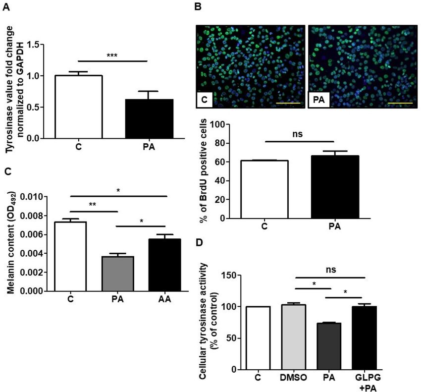
www.nature.com/scientificreports/
short-chain fatty acids (SCFAs) with chain lengths less than six carbons such as acetate, butyrate, and propionate,
the most potent agonist for FFAR220,21. Previously, we demonstrated that in vivo knockdown of FFAR2 in mouse
skin blocked butyric acid mediation of UV i rritation3.
C. acnes is abundant on the human skin surface accounting for > 60% of the bacteria2. Moreover, propionic
acid (PA), a fermentation metabolite from C. acnes, and its esterified derivatives, act as a potential antimicro-
bial agent against Staphylococcus aureu11,22 and poly(ethylene oxide) coatings are a promising method to avoid
infections23. Skin commensal probiotic bacteria can inhibit the growth of USA300 through fermentation of
poly(ethylene glycol) dimethacrylate as a selective fermentation initiator24. PF68, PEG-based polymer is a sta-
ble gel carrier for antimicrobial agents, increasing the surface solubility of the drug, and is usually used in the
treatment of infected wounds without any discernible side effects25. In this study, we determined that PF68 as
a polymer derived compound is a potentially safe and effective agent and the application of metabolites from
the fermentation of PF68 by the skin commensal C. acnes could inhibit UV-induced hyperpigmentation or
melanogenesis of skin.
Methods
Ethics statement. This study was carried out in strict with an approved Institutional Animal Care and Use
Committee (IACUC) protocol at National Central University (NCU), Taiwan (NCU-106-016) and in compli-
ance with the Arrive guidelines (https://arriveguidelines.org/). Institute Cancer Research (ICR) mice (8–9 weeks
old females; National Laboratory Animal Center, Taipei, Taiwan) were sacrificed using CO2 in a closed box. All
methods were performed in accordance with relevant guidelines and regulations.
Bacterial culture. C. acnes (ATCC 6919) was cultured on Reinforced Clostridium Medium (RCM, Oxford,
Hampshire, England) under anaerobic conditions using a Gas-Pak (BD, Sparks, MD, USA). Bacteria were cul-
tured at 37 °C until the logarithmic growth phase. Bacterial pellets were harvested by centrifugation at 5,000 × g
for 10 min, washed in phosphate-buffered saline (PBS), and then suspended in PBS or RCM for further experi-
ments.
Bacteria fermentation. C. acnes (105 colony-forming unit (CFU)/mL) was incubated in 10 mL TSB in
the presence or absence of 2% PF68 (BASF, NY, USA) under anaerobic conditions using Gas-Pak at 37 °C. PF68
alone in TSB was included as a control. The 0.002% (w/v) phenol red (Sigma) in TSB served as a fermentation
indicator. A color change from red–orange to yellow indicated the occurrence of bacterial fermentation, which
was detected by optical density at 560 nm (OD560).
Gas chromatography‑mass spectrometry (GC–MS) analysis. C. acnes (105 CFU/mL) was incubated
in TSB (10 mL) in the presence of 2% PF68. After 1 day fermented media was centrifuged at 5000 g for 10 min
and the supernatant was used for detection of SCFAs using previously published p rotocol26. The levels of acetic
acid (AA), PA, butyric acid (BA), and iso-butyric acid (I-BA) in the fermentation media were quantified by a
calibration curve made from six non-zero levels using the FFA Test Standard (Restek Corporation, Bellefonte,
PA, USA) which is diluted to 500-, 1,000-, 2,000-, 5,000- and 10,000-fold.
B16F10 melanoma cell culture. B16F10 melanoma cell line was kindly provided by Dr. Richard Gallo,
University of California San Diego, USA, and Dr. Cheng Ching-Yi, Chang Gung University of Science and Tech-
nology, Taiwan. The cells were cultured in Dulbecco’s Modified Eagle’s Medium (DMEM, Life Technologies,
Carlsbad, CA, USA) supplemented with 10% fetal bovine serum (FBS, Life Technologies) and 1% Penicillin–
streptomycin (Life Technologies) at 37 °C and 5% CO2. The cells were grown until 70–80% confluence and then
subcultured with Trypsin-Ethylenediaminetetraacetic acid (EDTA, Life Technologies).
Reverse transcription‑quantitative polymerase chain reaction (RT‑qPCR). RT-qPCR was
employed to study the tyrosinase gene expression in B16F10 melanoma cells treated with 4 mM PA for 48 h.
Total cellular RNA was extracted using a Quick-RNA MiniPrep Kit (Zymo research, CA, USA), followed by
reverse transcription to cDNA using iScript cDNA synthesis kit (Bio-Rad, Hercules, CA, USA) and amplified by
RT-qPCR in an ABI 7300 system (Applied Biosystems, Waltham, MA, USA). The comparative cycle threshold
(ΔΔCT) was used to determine the quantification of gene expression. The gene-level of glyceraldehyde 3-phos-
phate dehydrogenase (GAPDH) was used for the normalization of tyrosine kinase gene. Primer for tyrosinase
and GAPDH are 5′-TGACAAAGCCAAAACCCCCA-3′ (forward); 5′-TTGTTCAAAAATACTTCCAGTGTG
T-3′ (reverse) and 5′-TGTGTCCGTCGRGGATCTGA-3′ (forward); 5′-GATGCCTGCTTCACCACCTT3′
(reverse).
Cellular tyrosinase activity following FFAR2 gene knockdown. B16F10 melanoma cells were
seeded at a density of 5 × 10⁴ cells/well in 24-well plates. Further, the cells were treated with FFAR2 selective
antagonist GLPG0974 (0.1 µM, GLPG) and PA (4 mM) and incubated at 37 °C in 5% C O2 for 24 h. Cells were
trypsinized and lysed with RIPA buffer (Thermo Fisher Scientific, NJ, USA). The lysed cells were frozen at -80 °C
and thawed twice followed by centrifugation at 12,000 rpm for 10 min. The supernatant was mixed with 1 μg/
mL of Levodopa (L-DOPA, Sigma) in 2:1 ratio in a 96-well plate, incubated at room temperature (RT) for 1 h
and OD at 475 nm was detected27.
Scientific Reports | (2021) 11:11980 | https://doi.org/10.1038/s41598-021-91386-x 2
Vol:.(1234567890)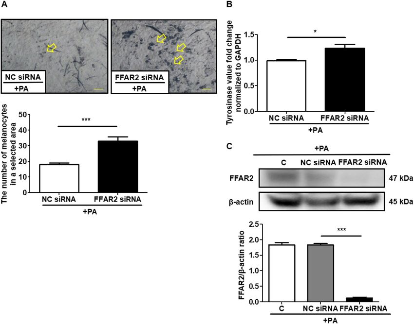
www.nature.com/scientificreports/
BrdU labeling. B16F10 melanoma cells were grown on 8-well chamber slides (Thermo Fisher Scientific) in
DMEM with 10% FBS, penicillin and streptomycin till 70% confluency achieved. Bromodeoxyuridine (10 μM,
BrdU, ACROS, New Jersey, USA) was added to the culture media along with 4 mM PA. BrdU added with PBS in
culture media was included as a control. After 24 h, cells were fixed with 4% perfluoroalkoxy (PFA, Sigma) and
then permeabilized with 0.2% triton X-100 (Sigma) for 10 min. Next, cells were treated with 2 N HCl at 37 °C
for 25 min, neutralized with HCl with 0.1 M Borate buffer (pH 8.5) for 10 min, and blocked with blocking buffer
before incubation anti-BrdU antibody (Abcam, MA, USA) overnight and donkey anti-goat Alexa Fluor 568 IgG
(H + L) (Life technologies) as second antibodies for 1 h at 4 °C. Nuclei were counterstained with 4,6-diamidino-
2-phe-nylindole (DAPI, Sigma). Images were acquired with cellSens software (https://www.olympus-lifescience.
com.cn/en/software/cellsens/; Version 1.2.1) connected to an Olympus BX63 microscope (Olympus, Tokyo,
Japan).
The mouse models created by UV light exposure. Mouse models have been instrumental in advanc-
ing the understanding of the many roles of U VR28. UVR increases DOPA-positive melanocytes in the skin,
specifically at the site of exposure29. The protocol for the treatment of C. acnes injection to the mice ears has
been described in our previous s tudy3 (This reference does not talk about C. acnes injection). The ears of ICR
mice were injected intradermally with C. acnes (107 CFU) with and without 2% PF68 followed by ultraviolet B
(UVB) exposure of 312 nm wavelength at a dose of 200 mJ/cm2 using an UV lamp (Model EB-280C, Spectronics
Corp., Westbury, NY, USA) for 2 min every day for 3 days. Mouse ears injected with PBS or PF68 followed by
UVB exposure were included as a control. In another group of experiment mice, ears were applied with 4 mM
PA followed by UV exposure, with PBS as control.
siRNA‑Mediated gene silencing of FFAR2. In order to silence FFAR2 gene, we used the chemically-
modified siRNA that targets FFAR2 receptor (FFAR2 siRNA), the siRNA negative control (NC siRNA) and
the control without injection (C), which were obtained from GenePharma Co. (Shanghai, China). Their oligo-
nucleotide sequences are siFFAR2: sense strand, 5′-CCGGUGCAGUACAAGUUAUTT-3′; anti-sense strand,
5′-AUAACUUGUACUGCACCGGTT-3′. SiControl: sense strand, 5′-UUCUCCGAACGUGUCACGUTT-3′;
anti-sense strand, 5′-ACGUGACACGUUCGGAGAATT-3′. These chemically-modified siRNAs were delivered
every day for 3 days by intradermal injection in the ears of mice using a microneedle (2 mg/kg of mice weight).
From day two, apply with 2% PA and UV exposure for 3 days. The pretreatment for injecting siRNA to the mouse
as described in reference30. Then, mice ears were excised and stained on the fourth day.
Western blotting. Mice ears were injected with chemically-modified FFAR2 and control siRNAs followed
by application with 2% PA and UVB exposure for 3 days. On third day mice ears were cut, homogenized and
then lysed with RIPA buffer (Thermo Fisher Scientific). Cell lysates (30 µg) were subjected to 10% SDS-PAGE
gel, which were then transferred to a poly(vinylidene fluoride) (PVDF) membrane (Sigma) and blocked with 5%
(w/v) nonfat milk before incubation overnight with primary antibodies to FFAR2 Rabbit PolyAb (Proteintech,
Rosemont, IL, USA) at 4 °C or β-actin (1:1,000; Cusabio Technology, Houston, TX, USA). This was followed by
treatment with horseradish peroxidase-conjugated goat anti-rabbit secondary antibody (1:5000) (Thermo Fisher
Scientific) for 1 h. Protein bands were detected with a chemiluminescent detection reagent (Thermo Fisher Sci-
entific) and Omega Lum C Imaging System (Gel Co., San Francisco, CA, USA). Protein bands were conducted
using ImageJ software (https://imagej.nih.gov/ij/; Version 1.53e).
Melanocyte counting. The cartilages from mouse were removed manually and the skin tissues were soaked
in ammonium thiocyanate (Sigma) solution at 37 °C for 20 min. The epidermal and basal layers were exfoliated
from the rest of the skin tissue, and melanocytes were stained by immersing in 0.1 M PBS (pH 7.2) containing
0.14% L-DOPA at RT for 3 h and the melanocyte count in skin tissues was determined microscopically accord-
ing to a previously published protocol29.
Statistical analyses. Data analysis was performed by unpaired t-test using Prism software (https://www.
graphpad.com/; Version 5.01, GraphPad Software, La Jolla, CA, USA). The levels of statistical significance were
indicated as the following: *P < 0.05, **P < 0.01, ***P < 0.001 and ns = non-significant. The mean ± standard devi-
ation (SD) for at least three independent experiments except two independent western blotting analysis for
Fig. 4C was displayed. Animal experiments were performed with at least three animals per treatment group.
Results
C. acnes induces fermentation of PF68. Previous studies have demonstrated the fermentation of skin
probiotics for induction of SCFA production in the presence of compounds as a carbon s ource26. To investigate
if C. acnes could ferment PF68, C. acnes was incubated with PF68 in TSB media for 1 day with phenol red as an
indicator to the monitor the bacterial fermentation. In TSB media incubated with bacteria alone, the phenol red
changed from red to orange due to bacterial replication, whereas in the TSB media containing bacteria and PF68,
the phenol red color changed to pale yellow with a decrease in pH indicating the use of PF68 as a carbon source
for fermentation (Fig. 1A). Furthermore, the OD560 of phenol red showed a significant decrease in pH value in
the TSB media containing bacteria and PF68 (Fig. 1B) compared to bacteria or PF68 alone. The SCFAs contained
in the C. acnes ferment cultured with PF68 were identified by GC–MS analysis (Fig. 1C) and found to be AA, PA,
BA, and I-BA acid, with PA11 having the highest concentration (Fig. 1D).
Scientific Reports | (2021) 11:11980 | https://doi.org/10.1038/s41598-021-91386-x 3
Vol.:(0123456789)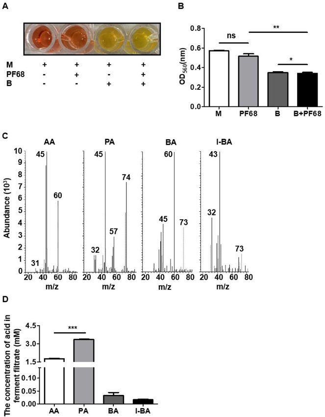
www.nature.com/scientificreports/
Figure 1. The bacterial fermentation with polymer was accompanied by the decrease in intracellular pH. (A)
The color change of phenol red from red to yellow when C. acnes was incubated with/without PF68 in TSB. (B)
The prevalence of fermentation was quantified by measuring the optical density of phenol red at O D560. The
pH of media with PF68 and C. acnes was significantly lower than the other groups. (C) The ion chromatogram
and mass spectrum showing the SCFAs present in the C. acnes ferment cultured with PF68. (D) Quantification
of SCFAs in the C. acnes ferment filtrate. A high amount of PA was produced by PF68 fermentation of C.
acnes. Medium (M), C. acnes (B), acetic acid (AA), propionic acid (PA), butyric acid (BA), and iso-butyric acid
(I-BA). Data are the mean ± SD from three separate experiments. *P < 0.05, **P < 0.01, ***P < 0.001 and ns = non-
significant (two-tailed t-test).
In vitro effect of PA on melanin production via PA‑FFAR2. Fatty acids modulate the degradation of
tyrosinase, a crucial enzyme involved in melanin biosynthesis in melanocytes and melanoma c ells31. Therefore,
to detect the in vitro effect of PA on melanin synthesis, B16F10 melanoma cells were treated with 4 mM PA for
48 h, with the decreased expression of the tyrosinase in PA treated melanoma cells compared to the control
(Fig. 2A). The immunostaining of PA treated melanoma cells after BrdU labeling showed that PA did not alter
the proliferation of melanoma cells (Fig. 2B). Furthermore, the effect of PA and AA, two highly expressed SCFAs
in the fermentation filtrate (Fig. 1D)32, on melanin production was assessed in melanoma cells, with PA causing
Scientific Reports | (2021) 11:11980 | https://doi.org/10.1038/s41598-021-91386-x 4
Vol:.(1234567890)www.nature.com/scientificreports/
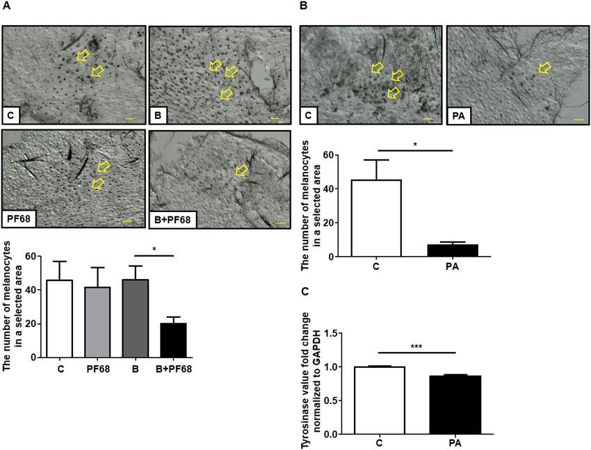
Figure 2. The effects of tyrosinase gene expression and cell proliferation in melanoma cells after PA treatment.
(A) Tyrosinase gene expression decreased in PA treated B16F10 melanoma cells quantified by RT-qPCR. (B)
The effects of PA on B16F10 melanoma cells evaluated by BrdU labeling. PA treatment did not alter the cell
proliferation. (C) Effects of PA and AA in melanocytes. The relatively higher efficacy of PA in attenuating
melanin content. (D) Blocking the signaling of FFar2 using GLPG prevents the PA mediated decreased cellular
tyrosinase activity in B16F10 cells. Control (C), propionic acid (PA), acetic acid (AA), dimethyl sulfoxide
(DMSO), and GLPG0974 (GLPG). Data are the mean ± SD from three separate experiments. *P < 0.05, **P < 0.01
and ns = non-significant (two-tailed t-test). Scale bar = 120 µm.
a > twofold decrease in melanin production compared to AA (Fig. 2C). SCFAs, like PA and AA, regulate their
functions via interaction with FFAR2 and FFAR3, with PA being among the most potent SCFA for both these
receptors33. Considering PA regulation via its interaction with FFAR2, blocking the signaling of this receptor
using a selective inhibitor could be a useful approach to evaluate the role of PA in the regulation of melanogen-
esis. The cellular tyrosinase activity was unchanged by blocking FFAR2 using the FFAR2 selective antagonist
GLPG before treatment with PA and decreased without GLPG in contrast to the control group (Fig. 2D). These
results demonstrated the effective role of PA-FFAR2 in the attenuation of the melanin content by inhibiting
tyrosinase kinase activity in melanoma cells with unaltered cell proliferation.
The mixture of C. acnes and PF68 inhibited UVB‑induced functioning melanocyte in mouse
ear. The topical application of fermentation extracts or fatty acids has been shown to decrease UV-induced
skin hyperpigmentation by regulating tyrosinase degradation34,35. Previous studies demonstrated that melano-
ermis36. Hav-
genesis after UV exposure is due to the activation in functioning melanocytes in the epidermis or d
ing established that PA as a major SCFA from C. acnes fermentation of PF68 could effectively reduce melanin
production in vitro, we then examined the effect of C. acnes plus PF68 on it in vivo. The ears of ICR mice were
exposed to UVB every other day for 3 days concurrent with the injection of C. acnes and PF68. Injection with
PBS or C. acnes or PF68 alone were included as controls. The epidermal and basal layers were exfoliated from the
skin tissue in the mouse ear and the melanocytes were stained with L-DOPA. There was a significant increase
of the melanocytes number after UVB exposure, with no change after injection with C. acnes or PF68 alone.
However, a significant reduction in number of DOPA-positive melanocytes was detected in the UVB exposure
groups after treatment with a mixture of C. acnes and PF68 (Fig. 3A).
Scientific Reports | (2021) 11:11980 | https://doi.org/10.1038/s41598-021-91386-x 5
Vol.:(0123456789)www.nature.com/scientificreports/
Figure 3. Induction of melanocytes by UVB and inhibition by different treatments. (A) The ears of mice
were applied PF68 + C. acnes. The same amount of PBS, PF68 only, and C. acnes only were applied as control.
Specimens were examined microscopically for the effects of PF68 + C. acnes treatment on the DOPA-positive
melanocytes proliferation in the epidermis. (B) The ears of mice were applied PA. The same amount of PBS
was applied as control. Specimens were examined microscopically for the effects of PA treatment on the
DOPA-positive melanocytes proliferation in the epidermis. (C) Tyrosinase gene expression after PA treatment
in vivo was quantified by RT-qPCR. Control (C), C. acnes (B), propionic acid (PA). The arrowhead in A and B
shows the staining of DOPA-positive melanocytes that was counted in the same millimeter squared. Data are
the mean ± SD from three separate experiments was replayed. *P < 0.05, ***P < 0.001 (two-tailed t-test). Scale
bar = 60 μm.
Amelioration of UVB‑induced functioning melanocyte in mouse ear by PA, a C. acnes fermen‑
tation metabolite. The effect of the direct application of PA on UV-induced melanogenesis was assessed.
UV-induced hyperpigmentation and tyrosinase expression in mouse ear skin in the control group was signifi-
cantly lowered after the topical application of PA for 3 days (Fig. 3B). Thus, PA, a metabolite from PF68 fer-
mentation of C. acnes inhibits functioning melanocytes production with melanin synthesis by UVB-induced.
Tyrosinase expression was noticeably decreased in PA treated cells compared to the control (Fig. 3C).
PA inhibits UVB‑induced functioning melanocytes via FFAR2 in vivo. To determine if the reduc-
tion in exogenous melanogenesis occurred via PA interaction with its cognate receptor, FFAR2 was knocked
down in the skin melanocytes of the mouse ear, followed by UVB exposure and PA treatment. UVB-induced
an increase in the proliferation of melanocytes and tyrosinase activity was significantly reduced in the mouse
ear epidermis injected with NC siRNA after application of PA (Fig. 4A,B). However, the number of melanocytes
and tyrosinase activity remains unchanged even after PA application in UVB irradiated mouse ear epidermis
knocked down with FFAR2. We confirmed the FFAR2 gene knockdown by measuring the protein expression
level of FFAR2 by Western blot analysis (Fig. 4C). Thus, PA interferes with melanogenesis by suppressing the
activity of tyrosinase, in which PA-FFAR2 plays a potential role in melanin production.
Discussion
Melanin, a critical factor of skin defense against UV-irradiation, is synthesized in the melanosomes of melano-
cytes, transferred to neighboring keratinocytes via the dendritic tips, and eventually distributed throughout the
skin epidermis37. Bacteria fermentation metabolites are increasingly used in skin therapy due to their beneficial
effect on UV-induced skin inflammation and infectious d isorders3. In this study, we demonstrated that PA, the
Scientific Reports | (2021) 11:11980 | https://doi.org/10.1038/s41598-021-91386-x 6
Vol:.(1234567890)www.nature.com/scientificreports/
Figure 4. The siRNA knockdown efficiency of the FFAR2 gene, along with the topical application of PA and
UV exposure to ICR mice ears. (A) Specimens were examined microscopically for the effects of PA treatment
after FFAR2 gene knockdown on the DOPA-positive melanocytes proliferation in the epidermis. (B) Tyrosinase
gene expression in vivo quantified by RT-qPCR. (C) Western blotting to confirm FFAR2 gene knockdown
and the expression of FFAR2 and β-actin in mice ears. Full-length gels images are available in Supplemental
Information. Control (C), propionic acid (PA), negative control (NC). The arrowhead in A and B shows the
staining of DOPA-positive melanocytes that was counted in the same millimeter squared. Data A and B are
the mean ± SD from three separate experiments except two independent western blotting analysis for C was
replayed. *P < 0.05, ***P < 0.001 (two-tailed t-test). Scale bar = 60 μm.
major metabolite from PF68 fermentation of C. acnes, reduced UVB-induced functioning melanocyte levels by
inhibiting tyrosinase activity in vitro and in vivo via FFAR2.
Previous studies demonstrated the inhibition of melanogenesis through tyrosinase inhibition by fermentation
metabolites from probiotic LA and Lactobacillus paracasei bacteria24,38,39. Also, the high molecular weight PEG-
based polymer (~ 20,000) was completely degraded through fermentation by gram-negative bacteria producing
acetate and p ropionate40. In this study, PA (~ 4 mM) was the most abundant SCFA in the filtrate from PF68 fer-
mentation of C. acnes and reduced the melanin content in melanocytes more effectively than AA. Furthermore,
treatment with 4 mM PA significantly inhibited cellular tyrosinase activity in melanocytes proving that inhibition
of melanogenesis by PA occurred via the reduction of tyrosinase gene expression. People have the same number
of melanocytes that is not the reason to affect skin pigmentation but the activation in functioning melanocytes
by UV-irradiation36,41. Here, 4 mM PA treatment did not alter melanocyte proliferation, indicating that it is an
effective treatment option, causing no cellular damage. The reduced melanocytes number and tyrosinase activity
were also observed in mice ear skin tissue injected with a mixture of C. acnes and PF68, demonstrating that the
inhibition of melanogenesis is likely to be mediated through fermentation metabolites from C. acnes fermentation
using PF68 as a carbon source. Additionally, our study validated PA as an excellent antimicrobial agent, with no
alteration in the growth of its parent bacteria C. acnes, showing PA as a potent metabolite that does not disrupt
the balance of the skin microbiome11.
Melanin production is initiated and regulated by several signaling systems, whereby tyrosinase catalyzes the
conversion of tyrosine to DOPA, and into dopaquinone, leading to the formation of melanocytic p igments42,43.
In most cases, skin lightening reagents act at various levels of melanin production via FFAR2 to affect tyrosi-
nase activity or directly targeting tyrosinase to inhibit melanogenesis in the s kin44–46. SCFAs such as PA and
AA exert their effects through binding to their selective cognate receptors like FFAR2 or FFAR3, with PA being
among the most potent SCFA for both receptors47. GLPG, a selective FFAR2 antagonist, and siRNA-mediated
gene silencing of FFAR2 to support the blocking of F FAR23,48. In the current study, the increased melanocytes
Scientific Reports | (2021) 11:11980 | https://doi.org/10.1038/s41598-021-91386-x 7
Vol.:(0123456789)www.nature.com/scientificreports/
number and tyrosinase gene expression in mice ear skin tissue did not changed in response to UVB radiation
even after PA application to FFAR2 knockdown in mice (Fig. 4A). Furthermore, tyrosinase gene expression
was decreased after PA treatment in vitro, however, blocking the FFAR2 gene with GLPG, a selective FFAR2
antagonist, and ameliorated the effect of PA to attenuate tyrosinase gene expression. After blocking FFAR2 as
loss of FFAR2 results in enhanced expression of tyrosinase activity after PA treatment. Although both PA and
AA, the fermentation metabolites from C. acnes, have a similar affinity for FFAR2 binding20, the relatively lower
efficacy of AA in attenuating tyrosinase activity in melanocytes validates the therapeutic potential of PA as a
postbiotic against melanogenesis.
The photo damaging effects caused by UVR on the skin have attracted extensive attention. Sun screen-
ing and anti-hyperpigmentation products are widely used but their safety, skin penetration, and therapeutic
efficacy are still in question49. Although avobenzone is a common component in sunscreens owing to its high
efficacy against UV, it is photo unstable and therefore not safe, and has been detected in blood after chronic
applications50,51. Development of effective skin whitening through blocking tyrosinase expression by fermentation
metabolites from probiotic bacteria or polymers as a carbon source is an effective and natural way to suppress
melanogenesis38,39. As the use of live probiotics for cosmetics is strictly regulated, the promotion of their beneficial
effects through fermentated metabolites from live probiotic bacteria has become a feasible application. Besides,
PF68 as a prebiotic can boost the production of SCFAs from C. acnes fermentation and has been used to enhance
the biological stability and assist various therapeutic agents such as 6-mercaptopurine loaded microspheres in
their interaction with the human b ody52. Furthermore, PF68 can be incorporated into cell membranes and trans-
locate into cells and has been used as a stable gel carrier for antimicrobial agents, increasing the surface solubility
of the drug in skin treatment12,23. Hence, PF68 is considered a safe and effective options for the development
of pharmaceuticals and cosmetics. In addition to the function of PF68 as a fermentation initiator to augment
the fermentation activity of C. acnes, it also has the potential to be an adjuvant to reduce the effective doses of
medicine reagents and improve the solubility of poor-water soluble drugs.
Overall, these results demonstrate that the PA-FFAR2 interaction may be a central mechanism in the effect
of probiotics or postbiotics on hyperpigmentation caused by UVR. PA treatment did not affect melanocyte pro-
liferation. Compared with chemical therapies, the metabolites of probiotics are milder and n atural53. Although
the exact mechanism by which the metabolites of PF68 fermentation of C. acnes inhibit melanogenesis is unclear,
the evidence from the present study suggests the involvement of the SCFAs-FFAR2-tyrosinase pathway. These
results are beneficial for the future clinical treatment of pigmentation disorders and to develop cosmetics that
increases the range of applications of whitening products. Lastly, the diversity of probiotics offers personalized
treatments for hyperpigmentation and the development of dedicated products with higher performance.
Received: 21 February 2021; Accepted: 18 May 2021
References
1. Patra, V., Byrne, S. N. & Wolf, P. The skin microbiome: is it affected by uv-induced immune suppression?. Front Microbiol. 7, 1235.
https://doi.org/10.3389/fmicb.2016.01235 (2016).
2. Wang, Y. et al. The response of human skin commensal bacteria as a reflection of UV radiation: UV-B decreases porphyrin produc-
tion. PLoS ONE. https://doi.org/10.1371/journal.pone.0047798 (2012).
3. Keshari, S. et al. Butyric acid from probiotic staphylococcus epidermidis in the skin microbiome down-regulates the ultraviolet-
induced pro-inflammatory IL-6 cytokine via short-chain fatty acid receptor. Int. J. Mol. Sci. https://doi.org/10.3390/ijms201844
77 (2019).
4. Hawryluk, E. B., Oztan, A. & Fisher, D. E. Effects of ultraviolet exposure behaviors on skin pigmentation and melanoma. J. Pigm.
Disord. https://doi.org/10.4172/jpd.1000113 (2014).
5. Tu, C. X. et al. Curcumin inhibits melanogenesis in human melanocytes. Phytother Res 26, 174–179. https://doi.org/10.1002/ptr.
3517 (2012).
6. Roth, R. R. & James, W. D. Microbial ecology of the skin. Annual Rev. Microbiol. 42, 441–464 (1988).
7. Grice, E. A. & Segre, J. A. The skin microbiome. Nat Rev Microbiol 9, 244–253. https://doi.org/10.1038/nrmicro2537 (2011).
8. Ji, G., Beavis, R. & Novick, R. P. Bacterial interference caused by autoinducing peptide variants. Science 276, 2027–2030. https://
doi.org/10.1126/science.276.5321.2027 (1997).
9. Kober, M. M. & Bowe, W. P. The effect of probiotics on immune regulation, acne, and photoaging. Int. J. Womens Dermatol. 1,
85–89. https://doi.org/10.1016/j.ijwd.2015.02.001 (2015).
10. Baur, M., Breton, L., Couzy, F. & Gueniche, A. Use of probiotic lactic acid bacteria for preventing ultraviolet radiation induced
inflammatory or allergic reaction or immunosuppression in the skin. French patent (2002).
11. Shu, M. et al. Fermentation of Propionibacterium acnes, a commensal bacterium in the human skin microbiome, as skin probiotics
against methicillin-resistant Staphylococcus aureus. PLoS ONE. https://doi.org/10.1371/journal.pone.0055380 (2013).
12. Futsaether, C. M., Kjeldstad, B. & Johnsson, A. Intracellular pH changes induced in Propionibacterium acnes by UVA radiation
and blue light. J. Photochem. Photobiol., B 31, 125–131 (1995).
13. Kjeldstad, B. Photoinactivation of propionibacterium acnes by near-ultraviolet light. Z Naturforsch C. 39, 300–302 (1984).
14. Woolridge, J. Galactomyces ferment filtrate reduces melanin synthesis and oxidative stress in normal human melanocytes. J. Am.
Acad. Dermatol. https://doi.org/10.1016/j.jaad.2014.01.527 (2014).
15. Ando, H., Funasaka, Y. & Oka, M. Possible involvement of proteolytic degradation of tyrosinase in the regulatory effect of fatty
acids on melanogenesis. J. Lipid Res. 40, 1312–1316 (1990).
16. D, D. Z. Skin lightening preparations and the hydroquinone controversy. Dermatol. Therapy 20, 308–313 (2007).
17. R., K. Fairness via formulations: A review of cosmetic skin-lightening ingredients. J. Cosmet. Sci. 63, 43–54 (2012).
18. Read, J. E. et al. Dynamic changes in gene expression and signalling during trophoblast development in the horse. Reproduction
https://doi.org/10.1530/rep-18-0270 (2018).
19. Pan, P. et al. Loss of FFAR2 promotes colon cancer by epigenetic dysregulation of inflammation suppressors. Int J Cancer 143,
886–896. https://doi.org/10.1002/ijc.31366 (2018).
20. Hong, Y. H. et al. Acetate and propionate short chain fatty acids stimulate adipogenesis via GPCR43. Endocrinology 146, 5092–5099.
https://doi.org/10.1210/en.2005-0545 (2005).
Scientific Reports | (2021) 11:11980 | https://doi.org/10.1038/s41598-021-91386-x 8
Vol:.(1234567890)www.nature.com/scientificreports/
21. Le Poul, E. et al. Functional characterization of human receptors for short chain fatty acids and their role in polymorphonuclear
cell activation. J. Biol. Chem. 278, 25481–25489. https://doi.org/10.1074/jbc.M301403200 (2003).
22. Wang, Y. et al. Propionic acid and its esterified derivative suppress the growth of methicillin-resistant Staphylococcus aureus
USA300. Benef Microbes 5, 161–168. https://doi.org/10.3920/BM2013.0031 (2014).
23. Bridgett, M. J. & Davies, M. Control of staphylococcal adhesion to polystyrene surfaces by polymer surface modification with
surfactants. Biomaterials 13, 411–416 (1992).
24. Kao, M. S. et al. Microbiome precision editing: Using PEG as a selective fermentation initiator against methicillin-resistant Staphy-
lococcus aureus. Biotechnol J. https://doi.org/10.1002/biot.201600399 (2017).
25. Treter, J. et al. Washing-resistant surfactant coated surface is able to inhibit pathogenic bacteria adhesion. Appl. Surf. Sci. 303,
147–154. https://doi.org/10.1016/j.apsusc.2014.02.123 (2014).
26. Keshari, S. et al. Skin cutibacterium acnes mediates fermentation to suppress the calcium phosphate-induced itching: a butyric
acid derivative with potential for uremic pruritus. J. Clin. Med. https://doi.org/10.3390/jcm9020312 (2020).
27. Ko, H. H. et al. Eupafolin, a skin whitening flavonoid isolated from Phyla nodiflora, downregulated melanogenesis: Role of MAPK
and Akt pathways. J. Ethnopharmacol. 151, 386–393. https://doi.org/10.1016/j.jep.2013.10.054 (2014).
28. Day, C. P., Marchalik, R., Merlino, G. & Michael, H. Mouse models of UV-induced melanoma: genetics, pathology, and clinical
relevance. Lab Invest. 97, 698–705. https://doi.org/10.1038/labinvest.2016.155 (2017).
29. Hiramoto, K., Yanagihara, N., Sato, E. F. & Inoue, M. Ultraviolet B irradiation of the eye activates a nitric oxide-dependent hypo-
thalamopituitary proopiomelanocortin pathway and modulates functions of alpha-melanocyte-stimulating hormone-responsive
cells. J Invest. Dermatol. 120, 123–127. https://doi.org/10.1046/j.1523-1747.2003.12004.x (2003).
30. Pan, J. et al. Intradermal delivery of STAT3 siRNA to treat melanoma via dissolving microneedles. Sci. Rep. 8, 1117. https://doi.
org/10.1038/s41598-018-19463-2 (2018).
31. Ando, H. et al. Fatty acids regulate pigmentation via proteasomal degradation of tyrosinase: A new aspect of ubiquitin-proteasome
function. J. Biol. Chem. 279, 15427–15433. https://doi.org/10.1074/jbc.M313701200 (2004).
32. LeBlanc, J. G. et al. Beneficial effects on host energy metabolism of short-chain fatty acids and vitamins produced by commensal
and probiotic bacteria. Microb. Cell Fact 16, 79. https://doi.org/10.1186/s12934-017-0691-z (2017).
33. Ulven, T. Short-chain free fatty acid receptors FFA2/GPR43 and FFA3/GPR41 as new potential therapeutic targets. Front Endocrinol
(Lausanne) 3, 111. https://doi.org/10.3389/fendo.2012.00111 (2012).
34. Ahn, H. Y., Choo, Y. M. & Cho, Y. S. Anti-pigmentation effects of eight phellinus linteus-fermented traditional crude herbal extracts
on brown guinea pigs of ultraviolet b-induced hyperpigmentation. J Microbiol Biotechnol 28, 375–380. https://doi.org/10.4014/
jmb.1711.11043 (2018).
35. Shigeta, Y. et al. Skin whitening effect of linoleic acid is enhanced by liposomal formulations. Biol. Pharm. Bull. 27, 591–594 (2004).
36. Jimbow, K. & Uesugi, T. New melanogenesis and photobiological processes in activation and proliferation of precursor melano-
cytes after UV-exposure: Ultrastructural differentiation of precursor melanocytes from Langerhans cells. J. Invest. Dermatol. 78,
108–115. https://doi.org/10.1111/1523-1747.ep12505758 (1982).
37. Schallreuter, K. U., Kothari, S., Chavan, B. & Spencer, J. D. Regulation of melanogenesis–controversies and new concepts. Exp.
Dermatol. 17, 395–404. https://doi.org/10.1111/j.1600-0625.2007.00675.x (2008).
38. Chen, Y. M., Shih, T. W., Chiu, C. P., Pan, T.-M. & Tsai, T.-Y. Effects of lactic acid bacteria-fermented soy milk on melanogenesis
in B16F0 melanocytes. J. Funct. Foods 5, 395–405. https://doi.org/10.1016/j.jff.2012.11.012 (2013).
39. Bs, N. Y. K., Ms, H. S. K. & Lee, H. Y. Effect of inhibition on tyrosinase and melanogenesis of Agastache. J. Cosmetic Dermatol. 16,
407–415. https://doi.org/10.1111/jocd.12264 (2017).
40. Bernhard, S. & Marion, S. Fermentative degradation of polyethylene glycol by a strictly anaerobic, gram-negative, nonsporeforming
bacterium, Pelobacter venetianus sp. nov. Appl. Environ. Microbiol. 45, 1905–1913 (1983).
41. Kittles, R. Nature, origin, and variation of human pigmentation. J. Black Stud. (1995).
42. D’Mello, S. A., Finlay, G. J., Baguley, B. C. & Askarian-Amiri, M. E. Signaling Pathways in Melanogenesis. Int. J. Mol. Sci. https://
doi.org/10.3390/ijms17071144 (2016).
43. Seim, I., Ma, S. & Gladyshev, V. N. Tyrosinase gene expression in human tissues. Pigment Cell Res. 6, 400–405. https://doi.org/10.
1038/npjamd.2016.14 (1993).
44. Kumar, K. J. et al. In vitro and in vivo studies disclosed the depigmenting effects of gallic acid: A novel skin lightening agent for
hyperpigmentary skin diseases. BioFactors 39, 259–270. https://doi.org/10.1002/biof.1064 (2013).
45. Truong, X. T. et al. Protocatechuic acid from pear inhibits melanogenesis in melanoma cells. Int. J. Mol. Sci. https://doi.org/10.
3390/ijms18081809 (2017).
46. Oliveira, K. B., Palú, É., Weffort-Santos, A. M. & Oliveira, B. H. Influence of rosmarinic acid and Salvia officinalis extracts on
melanogenesis of B16F10 cells. Rev. Bras 23, 249–258. https://doi.org/10.1590/s0102-695x2012005000135 (2013).
47. Sawzdargo, M. et al. A cluster of four novel human G protein-coupled receptor genes occurring in close proximity to CD22 gene
on chromosome 19q13.1. Biochem Biophys Res Commun 239, 543–547. https://doi.org/10.1006/bbrc.1997.7513 (1997).
48. Negari, I. P., Keshari, S. & Huang, C. M. Probiotic activity of Staphylococcus epidermidis induces collagen type I production
through FFaR2/p-ERK Signaling. Int J Mol Sci 22. https://doi.org/10.3390/ijms22031414 (2021).
49. Rai R, Shanmuga SC & C., S. Update on photoprotection.Update on photoprotection. Indian J Dermatol. 57, 335–342 (2012).
50. Kullavanijaya P & HW., L. Photoprotection. J Am Acad Dermatol. 52, 937–958 (2005).
51. Matta, M. K., Zusterzeel, R. & Pilli, N. R. Effect of sunscreen application under maximal use conditions on plasma concentration
of sunscreen active ingredients. JAMA 321, 2082–2091 (2019).
52. Praveen B. Kajjari, Lata S. Manjeshwar & Aminabhavi, T. M. Novel Blend Microspheres of Poly(3-hydroxybutyrate) and Pluronic
F68/127 for Controlled Release of 6-Mercaptopurine. J. APPL. POLYM. SCI. 131 (2014).
53. Shenderov, B. A. Metabiotics: novel idea or natural development of probiotic conception. Microb Ecol Health Dis 24, doi:https://
doi.org/10.3402/mehd.v24i0.20399 (2013).
Acknowledgements
The authors thank Dr. Cheng Ching-Yi for the technical support of this project. We would also like to thank Dr.
Lan Tian for providing valuable comments and proofreading.
Author contributions
C.-M.H.: Conceptualization; H.-J.K., and Y.-H.W.: methodology; S.K.: validation; H.-J.K., S.K., and Y.-H.W.:
formal analysis; H.-J.K.: investigation; C.-M.H.: resources; H.-J.K., and C.-M.H.: data curation; H.-J.K.: writing-
original draft preparation; S.K., J.J.Y., S.S., C.-C.C., and C.-M.H.: writing-review and editing; C.-M.H.: supervi-
sion, project administration, funding acquisition. All authors have read and agreed to the published version of
the manuscript.
Scientific Reports | (2021) 11:11980 | https://doi.org/10.1038/s41598-021-91386-x 9
Vol.:(0123456789)www.nature.com/scientificreports/
Funding
This work was funded by 106/107/108-Landseed Hospital-NCU joint grants and Ministry of Science and Tech-
nology (MOST) Grants 108-2622-B-008-001-CC1; 108-2314-B-008-003-MY3, and 107-2923-B-008-001-MY3.
Competing interests
The authors declare no conflict of interests.
Additional information
Supplementary Information The online version contains supplementary material available at https://doi.org/
10.1038/s41598-021-91386-x.
Correspondence and requests for materials should be addressed to C.-M.H.
Reprints and permissions information is available at www.nature.com/reprints.
Publisher’s note Springer Nature remains neutral with regard to jurisdictional claims in published maps and
institutional affiliations.
Open Access This article is licensed under a Creative Commons Attribution 4.0 International
License, which permits use, sharing, adaptation, distribution and reproduction in any medium or
format, as long as you give appropriate credit to the original author(s) and the source, provide a link to the
Creative Commons licence, and indicate if changes were made. The images or other third party material in this
article are included in the article’s Creative Commons licence, unless indicated otherwise in a credit line to the
material. If material is not included in the article’s Creative Commons licence and your intended use is not
permitted by statutory regulation or exceeds the permitted use, you will need to obtain permission directly from
the copyright holder. To view a copy of this licence, visit http://creativecommons.org/licenses/by/4.0/.
© The Author(s) 2021
Scientific Reports | (2021) 11:11980 | https://doi.org/10.1038/s41598-021-91386-x 10
Vol:.(1234567890)You can also read