Precise base editing for the in vivo study of developmental signaling and human pathologies in zebrafish - eLife
←
→
Page content transcription
If your browser does not render page correctly, please read the page content below
TOOLS AND RESOURCES

                                      Precise base editing for the in vivo study
                                      of developmental signaling and human
                                      pathologies in zebrafish
                                      Marion Rosello1,2, Juliette Vougny2, François Czarny1, Marina C Mione3,
                                      Jean-Paul Concordet4, Shahad Albadri1*, Filippo Del Bene1,2*
                                      1
                                        Sorbonne Université, INSERM, CNRS, Institut de la Vision, Paris, France; 2Institut
                                      Curie, PSL Research University, Inserm U934, CNRS UMR3215, Paris, France;
                                      3
                                        Department of Cellular, Computational and Integrative Biology – CIBIO, University
                                      of Trento, Trento, Italy; 4Museum National d’Histoire Naturelle, INSERM U1154,
                                      CNRS UMR 7196, Paris, France

                                      Abstract While zebrafish is emerging as a new model system to study human diseases, an
                                      efficient methodology to generate precise point mutations at high efficiency is still lacking. Here we
                                      show that base editors can generate C-to-T point mutations with high efficiencies without other
                                      unwanted on-target mutations. In addition, we established a new editor variant recognizing an
                                      NAA protospacer adjacent motif, expanding the base editing possibilities in zebrafish. Using these
                                      approaches, we first generated a base change in the ctnnb1 gene, mimicking oncogenic an
                                      mutation of the human gene known to result in constitutive activation of endogenous Wnt
                                      signaling. Additionally, we precisely targeted several cancer-associated genes including cbl. With
                                      this last target, we created a new zebrafish dwarfism model. Together our findings expand the
                                      potential of zebrafish as a model system allowing new approaches for the endogenous modulation
                                      of cell signaling pathways and the generation of precise models of human genetic disease-
                                      associated mutations.
*For correspondence:
shahad.albadri@inserm.fr (SA);
filippo.del-bene@inserm.fr (FDB)
                                      Introduction
Competing interests: The
                                      With the recent technological advances in precise gene editing, the use of zebrafish in genetic engi-
authors declare that no
                                      neering studies has drastically increased in the last 5 years (Patton and Tobin, 2019;
competing interests exist.
                                      Santoriello and Zon, 2012). The CRISPR (clustered regularly interspaced short palindromic repeats)/
Funding: See page 12                  Cas9 system is indeed a remarkably powerful gene-editing tool (Sander and Joung, 2014) that ena-
Received: 07 December 2020            bles the rapid and efficient generation of loss-of-function mutations in this animal model. This system
Accepted: 10 February 2021            relies on the specific binding of a sgRNA-Cas9 complex that initially interacts with DNA 20 base pair
Published: 12 February 2021           (bp) upstream of a NGG protospacer adjacent motif (PAM) sequence that triggers the Cas9 protein
                                      to introduce a double-strand break (DSB). This technique is nowadays widely used in zebrafish nota-
Reviewing editor: Tanya T
Whitfield, University of Sheffield,
                                      bly to produce knock-out alleles (Hwang et al., 2013), and more recently, it has also been demon-
United Kingdom                        strated that CRISPR/Cas9-mediated homology-directed repair (HDR) can be used to introduce
                                      exogenous DNA and single-nucleotide polymorphisms (Prykhozhij et al., 2018; Tessadori et al.,
   Copyright Rosello et al. This
                                      2018; Wierson et al., 2020).
article is distributed under the
                                          Recently, a CRISPR/Cas9-based technology has been developed to precisely edit single bases of
terms of the Creative Commons
Attribution License, which            DNA without introducing DSBs in human cells (Koblan et al., 2018; Komor et al., 2016;
permits unrestricted use and          Komor et al., 2017). The method is based on the fusion of a Cas9-D10A nickase with a cytidine
redistribution provided that the      deaminase giving rise to a cytidine base editor (CBE). CBE converts C-to-T bases in a restricted win-
original author and source are        dow of 13–19 nucleotides (nt) upstream of the PAM sequence (Figure 1A). In zebrafish, a CBE was
credited.                             shown to work but with limited efficiencies, inducing less than 29% of gene editing and, in most

Rosello et al. eLife 2021;10:e65552. DOI: https://doi.org/10.7554/eLife.65552                                                         1 of 15
Tools and resources Developmental Biology Genetics and Genomics
A Cytidine Base Editing technology B BE4-gam mRNA BE4-gam mRNA C BE4-gam, ctnnb1 (S33L)
+ control sgRNA + ctnnb1 sgRNA
A
N-ter APOBEC Cas9 (D10A) UGI UGI C-ter C
T
Tg(7xTCF-Xla.Siam:GFP), 1dpf
19 to 13 bases G
APOBEC
(Cytidine deaminase)
U C PAM
5’ 3’
3’ 5’
(rev) 5’ 3’
G
Cas9 (D10A)
5’ (Nickase)
UGI sgRNA TCA (S33) 27 26
3’
(Uracil glycosylase
inhibitor) to TTA (L33)
73 100 74
-13 -15
D BE4-gam, tek (Q94*) E BE4-gam, bap1 (Q273*) F BE4-gam, tp53 (Q21*) G BE4-gam, tp53 (Q170*)
A A A A
C C C C
T T T T
G G G G
(rev) 5’ 3’ (rev) 5’ 3’ 5’ 3’ 5’ 3’
73
86
86
14 14
33
-13 -14 -16 -14 -16
TGG (Q94) to TAG (*94) CAG (Q273) to TAG (*273) CAG (Q21) to TAG (*21) CAG (Q170) to TAG (*170)
TAA (*94), TGA (*94)
Figure 1. Efficient endogenous activation of Wnt signaling pathway and tumor suppressor genes targeting using BE4-gam in zebrafish. (A) Schematic
representation of the cytidine base editor technology. (B) Activation of Wnt signaling via S33L mutation in b-catenin. 1 dpf Tg(7xTCF-Xla.Siam:GFP)
representative embryos injected with BE4-gam mRNA and ctnnb1 (S33L) sgRNA or control scrambled sequence. The upper panel shows an overall
increase of GFP-positive cells in the head/anterior region upon the injection of the BE4-gam mRNA and ctnnb1 (S33L) sgRNA compared to the control
situation. The lower panel shows maximal z-projection of lateral view of the injected embryos where ectopic GFP signal in retinal progenitor cells (white
stars) can be detected, whereas control embryos do not show any fluorescence in the retina at this stage. (C–G) DNA sequencing chromatogram of
targeted loci with the BE4-gam and obtained C-to-T conversion efficiencies. The chromatograms correspond to the highest efficiency reported for the
single embryos analyzed as detailed in Table 2. (C) S33L mutation in b-catenin upon C-to-T conversion in ctnnb1 reached 73% of gene-editing
efficiency. The other edited C led to a silent mutation GAC (D) to GAT (D). (D) Q94* mutation in Tek upon C-to-T conversion in tek reached 18% of
gene-editing efficiency. (E) Q273* mutation in Bap1 upon C-to-T conversion in bap1 reached 14% of gene-editing efficiency. (F) Q21* mutation in p53
upon C-to-T conversion in tp53 reached 73% of gene-editing efficiency. (G) Q170* mutation in p53 upon C-to-T conversion in tp53 reached 86% of
gene-editing efficiency. For (C) and (E), the reverse complement of the sgRNA sequence is shown. Scale bars: (B) 50 mm. (D–G) Numbers in the boxes
represent the percentage of each base at that sequence position. In red are highlighted the base substitutions introduced by base editing, while the
original bases are in blue. The color code of the chromatogram is indicated in the upper left corner (Adenine green, Cytosine blue, Thymine red,
Guanine black). The distance from the PAM sequence of the targeted C base is indicated below each chromatogram. It is considered that the
quantifications under 5% are due to the background signal from Sanger sequencing and are thus non-significant (Kluesner et al., 2018).
The online version of this article includes the following figure supplement(s) for figure 1:
Figure supplement 1. List of targeted loci.
cases, at least 5% of unwanted INDEL (insertion or deletion) mutations were also detected
(Carrington et al., 2020; Zhang et al., 2017). For these reasons, this editing strategy has not been
so far favored by the zebrafish community. However, since this first generation of CBEs, several stud-
ies in cell culture have optimized and engineered new base editor variants with increased gene-edit-
ing efficiency reaching up to 90% without creating undesired INDEL mutations (Koblan et al.,
2018). A recent study reported the use of a second-generation CBE to generate a zebrafish model
of human ablepharon macrostomia syndrome (Zhao et al., 2020).
Rosello et al. eLife 2021;10:e65552. DOI: https://doi.org/10.7554/eLife.65552 2 of 15
Tools and resources                                                                Developmental Biology Genetics and Genomics

                                        Recent progress has also been made on the generation of CBEs able to recognize other PAM
                                    sequences, allowing to broaden gene-editing possibilities (Jakimo et al., 2018; Koblan et al.,
                                    2018). Thus, base editing offers a complementary and powerful approach in zebrafish to introduce
                                    specific single-nucleotide variants into the zebrafish genome. Here based on these technological
                                    advances, we optimized these second-generation gene-editing tools in zebrafish. As reported in ex
                                    vivo studies (Koblan et al., 2018), we tested different CBE variants and obtained highly efficient
                                    C-to-T conversion, reaching up to 91% efficiency without unwanted mutations and expanded base
                                    editing possibilities using a CBE variant recognizing the NAA PAM. Furthermore, compared to previ-
                                    ous studies, here we used these tools to target Wnt signaling, thus proving that endogenous path-
                                    ways can be modulated in their natural context. Finally, we demonstrated the power of this
                                    technology for introducing precise mutations in human cancer-associated genes with high efficiency
                                    in zebrafish and created a new fish model for dwarfism.

                                    Results and discussion
                                    BE4-gam base editing for the endogenous activation of Wnt signaling
                                    pathway
                                    To date, the main strategies used in zebrafish to study the constitutive activation of signaling path-
                                    ways and to dissect their role during embryonic development or tumorigenesis were based on over-
                                    expressing mutated genes. To gain further insights and to complement these studies, an important
                                    requirement is to have the ability to maintain the endogenous genetic and regulatory contexts by
                                    generating mutations of endogenous genes in vivo.
                                        To address this challenge, we decided to introduce an activating mutation in the ctnnb1 gene
                                    coding for the key effector b-catenin of canonical Wnt signaling, a major signaling pathway during
                                    embryonic development which is activated in many cancers (Steinhart and Angers, 2018). It was
                                    previously shown that the mutation of the Serine33 of the human b-catenin protein into a Leucine
                                    prevents its degradation by the ubiquitin-proteasome system, leading to its stabilization and thereby
                                    to the constitutive activation of Wnt signaling pathway (Hart et al., 1999; Liu et al., 1999).
                                        We first aimed at introducing this mutation in the genome of the zebrafish by using the Base Edi-
                                    tor 4 fused to the gam domain (BE4-gam) (Figure 1A). This CBE was indeed one of the first variants
                                    of CBEs to show high efficiency of gene editing and fewer INDELs formation in cultured cells
                                    (Komor et al., 2017). We injected the BE4-gam mRNA and synthetic ctnnb1 S33L sgRNA into one-
                                    cell stage Tg(7xTCF-Xla.Siam:GFP) zebrafish embryos to directly monitor the effect of the introduced
                                    mutation on the activity of the canonical Wnt signaling (Moro et al., 2012). Upon ctnnb1 S33L
                                    sgRNA injection, we observed an increase of GFP-positive cells at 1 dpf (n = 39/50 embryos) com-
                                    pared to the control embryos (n = 27 embryos) resulting from three independent experiments. By
                                    confocal imaging we quantified ectopic activation of the pathway in retinal progenitor cells and
                                    observed an average of 12 GFP-positive clones per retina, while GFP-positive cells were never
                                    detected in the retina of control injected Tg(7xTCF-Xla.Siam:GFP) embryos (n = 4 each) (Figure 1B).
                                    Using this strategy, we observed base editing in five of eight randomly chosen embryos and were
                                    able to reach up to 73% of editing efficiency in single embryo analysis (Table 1, Table 2). In addition,
                                    we also observed the conversion of another cytidine within the PAM [ 19, –13 bp] window leading
                                    to a silent mutation (GAC-to-GAT (D)) in four of the eight analyzed embryos with up to 74% effi-
                                    ciency (Figure 1C, Table 1, Table 2, Figure 1—figure supplement 1).
                                        With these results, we demonstrated that it is now possible to constitutively and efficiently acti-
                                    vate important developmental signaling pathways in their endogenous context, as we show here for
                                    Wnt signaling. Furthermore, several studies have implicated the S33L b-catenin mutation in tumori-
                                    genesis, making it possible to study the role of this oncogenic mutation in cancer development in
                                    zebrafish. In order to test the potential of CBE targeting in cancer modeling, we next decided to use
                                    it to target a series of tumor suppressor genes and oncogenes using the same editing strategy
                                    applied to endogenous b-catenin.

                                    Base-editing strategies for the generation of human cancer mutations
                                    Zebrafish is a powerful model system to study cancer genetics in vivo (Cagan et al., 2019;
                                    Cayuela et al., 2018). However, a robust method for modeling cancer-associated mutations in

Rosello et al. eLife 2021;10:e65552. DOI: https://doi.org/10.7554/eLife.65552                                                        3 of 15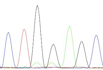
Tools and resources                                                                                              Developmental Biology Genetics and Genomics

Table 1. Base-editing efficiency using different CBE variants.
Number of edited embryos randomly chosen after injection of CBE mRNA and sgRNA. The efficiency varies between non-detected (n.
d.) and 91% depending on the targeted locus, the sgRNA, and the CBE used. Editing efficiency was quantified by editR analysis
(Kluesner et al., 2018), which does not detect editing efficiency below 5%.
                                                                                                                                                              nras             tp53
Targeted gene                     ctnnb1        tp53           cbl                 kras            Kras            dmd       dmd           rb1                (G13S)           (Q170*)
CBE used                          (S33L)        (Q170*)        (W577*)             (E62K)          (E62K)          (Q8*)     (Q8*)         (W63*)             spymac           spymac-
induced mutation                   BE4-gam      BE4-gam         BE4-gam             BE4-gam        ancBE4max       BE4-gam   ancBE4max     ancBE4max          -ancBE4max       ancBE4max

Number of edited embryos          5/8           7/8            8/10                0/8             4/7             0/8       2/4           8/8                2/4              1/4

Highest obtained efficiency       73%           86%            C16    C15          n.d.            19%             n.d.      14%           C17      C16       19%              16%
                                                               35%    50%                                                                  91%      65%

Table 2. Editing efficiency quantification.
Editing quantification of up to 10 single embryos randomly chosen after injection of indicated CBE mRNA and sgRNA. The efficiency
varies between non-detected (n.d.) to 91% in a single embryo depending on the targeted locus, the sgRNA, and the CBE used. Edit-
ing efficiency was quantified by editR analysis (Kluesner et al., 2018), which does not detect editing efficiency below 5%.
                      Number
Targeted gene         of edited                                                                                                                                            Emb.      Emb.
CBE used              embryos            Emb. 1           Emb. 2            Emb. 3            Emb. 4           Emb. 5        Emb. 6        Emb. 7         Emb. 8           9         10

ctnnb1 (S33L)         5/8                C15 C13          C15 C13           C15 C13           C15 C13          C15 C13       n.d.          n.d.           n.d.             –         –
 BE4-gam                                 74% 73%          n.d. 40%          44% 25%           7% 16%           n.d. 11%

tek (Q94*)            5/8                C14 C13          C14 C13           C14 C13           C14 C13          C14 C13       n.d.          n.d.           n.d.             –         –
 BE4-gam                                 18% 8%           10% n.d.          8% n.d.           6% n.d.          8% 9%

Bap1 (Q273*)          4/8                14%              12%               9%                8%               n.d.          n.d.          n.d.           n.d.             –         –
 BE4-gam

tp53 (Q21*)           6/8                63%              33%               37%               58%              8%            50%           n.d.           n.d.             –         –
 BE4-gam

tp53 (Q170*)          7/8                86%              46%               51%               62%              45%           20%           33%            n.d.             –         –
BE4-gam

cbl (W577*)           8/10               C16 C15          C16 C15           C16 C15           C16 C15          C16 C15       C16 C15       C16 C15        C16 C15          n.d.      n.d.
 BE4-gam                                 35% 50%          19% 31%           22% 38%           25% 41%          20% 35%       7% 9%         7% 12%         10% 17%

kras                  0/8                n.d.             n.d.              n.d.              n.d.             n.d.          n.d.          n.d.           n.d.             –         –
(E62K)
 BE4-gam

kras                  4/7                C17 C16          C17 C16           C17 C16           C17 C16          n.d.          n.d.          n.d.           –                –         –
(E62K) ancBE4max                         19% 21%          8% 11%            6% 8%             9% 10%

dmd                   0/8                n.d.             n.d.              n.d.              n.d.             n.d.          n.d.          n.d.           n.d.             –         –
(Q8*)
BE4-gam

dmd                   2/4                14%              6%                n.d.              n.d.             –             –             –              –                –         –
(Q8*) ancBE4max

sod2 (Q145*)          8/8                64%              45%               21%               54%              52%           24%           26%            33%              –         –
ancBE4max

rb1                   8/8                C19 C17 C16 C19 C17 C16 C19 C17 C16 C19 C17                     C16 C19 C17 C16 C19 C17 C16 C19 C17 C16 C19 C17 C16 –                       –
(W63*) ancBE4max                         n.d. 91% 65% 21% 79% 75% n.d. 27% 18% 13% 81%                   60% 8% 48% 33% 13% 76% 64% 13% 78% 69% 21% 77% 63%

nras                  2/4                19%              18%               n.d.              n.d.             –             –             –              –                –         –
(G13S) spymac-
ancBE4max

tp53 (Q170*)          1/4                16%              n.d.              n.d.              n.d.             –             –             –              –                –         –
spymac-ancBE4max

Rosello et al. eLife 2021;10:e65552. DOI: https://doi.org/10.7554/eLife.65552                                                                                                     4 of 15
Tools and resources Developmental Biology Genetics and Genomics
zebrafish is still lacking to date. We decided to create predictable premature stop codons in tumor
suppressor genes and to generate activating mutations in oncogenes of the RAS family (Li et al.,
2018) in order to test the ability of CBEs to induce cancer-related mutations in zebrafish.
We first developed an automated script to rapidly detect codons allowing to generate nonsense
mutations after a C-to-T conversion within the restricted PAM [ 19, –13] bp editing window
(Source code 1). Using this script, we designed a series of sgRNAs targeting in a selection of tumor
suppressor genes. We induced the Q94* mutation in Tek in five of eight randomly chosen embryos,
Q273* mutation in Bap1 in four of eight randomly chosen embryos and Q21* mutation in p53 in six
of eight randomly chosen embryos as well as Q170* in p53 in seven of eight randomly chosen
embryos by C-to-T conversions (Figure 1D–G, Table 1, Table 2). Among the different targeted
mutations, the highest efficiency was achieved with the tp53 tumor suppressor gene, for which we
reached up to 86% of C-to-T conversion for the introduction of the Q170* mutation (Figure 1G,
Table 1, Table 2). To assess the presence of INDELs or unwanted mutations upon BE4-gam injec-
tions in our targets, we amplified, cloned, and sequenced all targeted loci. For tek 6 of 20, bap1 2
of 12, tp53 (Q21*) 12 of 24, and lastly tp53 (Q170*) 21 of 24 colonies showed precise C-to-T conver-
sions, whereas all the other analyzed sequences were wild type, without any error or INDEL forma-
tion. Together these results show that, using BE4-gam, we efficiently targeted several genes
implicated in tumorigenesis in zebrafish without generating any unwanted INDELs, unlike what was
previously reported with the BE3 variant.
More recently, a new CBE variant, the ancBE4max, has been engineered and optimized in cell cul-
ture with increased efficiency compared to the classical BE4-gam, reaching up to 90% efficiency and
very low rates of INDELs (Koblan et al., 2018). We therefore decided to use this new CBE variant to
target the oncogenic mutation E62K in Kras and induce the creation of a Q8* stop codon in the
Dmd tumor suppressor for which we did not obtain any C-to-T conversions or other unwanted
changes using the BE4-gam in randomly chosen eight embryos for each condition (Table 1, Table 2).
By co-injection of ancBE4max mRNA with the kras E62K sgRNA, we were able to introduce the
E62K mutation in four of seven randomly chosen embryos and we were able to reach up to 19% of
editing efficiency in single embryo analysis. Another cytidine in the editing window was also con-
verted and led to the generation of a silent mutation (CAG-to-CAA (Q)) (Figure 2A, Table 1,
Table 2). Similar to what we observed in the case of kras editing, we were able to obtain a Q8*
mutation in the Dmd tumor suppressor in two of four randomly chosen embryos with up to 14% of
editing efficiency (Figure 2B, Table 1, Table 2). Thus, with this new ancBE4max variant, we are able
to introduce mutations that could not be achieved with BE4-gam using the same sgRNAs. Remark-
able editing efficiency was also observed using this CBE for two additional targets: the tumor sup-
pressor genes sod2 and rb1, for which, respectively, up to 64% and 91% of editing were reached
and 100% of the sequenced embryos were precisely mutated (from single embryo analysis of n = 8
randomly analyzed embryos) (Figure 2C,D, Table 1, Table 2; Bravard et al., 1992; Dyson, 2016).
It is interesting to note that in general all the cytidine bases present in the PAM [ 19, –13] bp
window can be edited by the CBE, with a higher efficiency for the cytidine bases located in the mid-
dle of this window while editing was below detection levels for cytidines located only 12 bp
upstream (Figure 2D). With the use of ancBE4max CBE, these results highlight the importance of
the cytidine distance from the PAM for efficient editing in zebrafish as shown previously in cell cul-
ture assays (Gaudelli et al., 2017).
Expanding gene-editing possibilities in zebrafish using a CBE
recognizing NAA PAM
Due to the PAM-dependent restriction of the editing window, many mutations could not be gener-
ated so far. We therefore decided to expand the editing possibilities in zebrafish by associating Spy-
macCas9 recognizing NAA PAMs with the efficient conversion capacity of the ancBE4max. To this
end, we replaced the PAM-interacting motif (PIM) domain of the SpCas9 with the one of the Spy-
macCas9 in the ancBE4max (Jakimo et al., 2018). The inserted PIM domain was codon optimized
for zebrafish. Using this newly generated ancBE4max-SpymacCas9, we were able to reproduce the
human G13S mutation in Nras oncogene in zebrafish in 2 out of 4 randomly analyzed embryo with
up to 19% of efficiency reached in single embryo analysis (Figure 2E, Table 1, Table 2). We also
introduced a stop codon by a C-to-T conversion in the tp53 gene in 1 out of 4 randomly analyzed
embryo with 16% efficiency (Figure 2F, Table 1, Table 2). These results demonstrate that in addition
Rosello et al. eLife 2021;10:e65552. DOI: https://doi.org/10.7554/eLife.65552 5 of 15Tools and resources Developmental Biology Genetics and Genomics Figure 2. Tumor suppressor genes and oncogenes targeting by the highly efficient ancBE4max and the ancBE4max-SpymacCas9 recognizing NAA PAM. (A–F) DNA sequencing chromatogram of targeted loci with the ancBE4max (in A–D) or ancBE4max-SpymacCas9 (in E,F) and obtained C-to-T conversion efficiencies. (A) E62K mutation in Kras upon C-to-T conversion in kras reached 19% gene-editing efficiency. The other edited C led to a silent mutation CAG (Q) to CAA (Q). (B) Q8* mutation in Dmd upon C-to-T conversion in dmd reached 14% of gene-editing efficiency. (C) Q145* mutation in Sod2 upon C-to-T conversion in sod2 reached 64% of gene-editing efficiency. (D) W63* mutation in Rb1 upon C-to-T conversion in rb1 Figure 2 continued on next page Rosello et al. eLife 2021;10:e65552. DOI: https://doi.org/10.7554/eLife.65552 6 of 15
Tools and resources                                                                       Developmental Biology Genetics and Genomics

Figure 2 continued
reached 21% for the C19 base, 79% for C17, and 75% for the C16 of gene-editing efficiency. (E) G13S mutation in Nras upon C-to-T conversion in nras
reached 19% of gene-editing efficiency. (F) Q170* mutation in p53 upon C-to-T conversion in tp53 reached 16% of gene-editing efficiency. For (A, D–
F), the reverse complement of the sgRNA sequence is shown. (A–F) The chromatograms correspond to the efficiency reported for the single embryos
provided in the first column of Table 2. The numbers in the boxes represent the percentage of each base at that sequence position. In red are
highlighted the base substitutions introduced by base editing, while the original sequence is in blue. The color code of the chromatogram is indicated
in the upper left corner (Adenine green, Cytosine blue, Thymine red, and Guanine black). The distance from the PAM sequence of the targeted C base
is indicated below each chromatogram. It is considered that the quantifications under 5% are due to the background signal from Sanger sequencing
and are thus non-significant (Kluesner et al., 2018).

                                    to the classical NGG PAM it is now also possible to target NAA PAMs in zebrafish, thereby signifi-
                                    cantly expanding the range of cytidine bases that can be converted. For these new CBEs, we added
                                    a function in our script to choose the PAM recognized by the Cas9-D10A of the chosen CBE to gen-
                                    erate the desired base editing (Source code 1). Together with the use of the ancBE4max and
                                    ancBE4max-SpymacCas9 CBE variants, we were now able to target mutations that could not be gen-
                                    erated with the BE4-gam base editor and reproduce a wider range of human cancer mutations in
                                    zebrafish.
                                        Genetic alterations that lead to oncogene activation and/or tumor suppressor inactivation are
                                    responsible for tumorigenesis. It is indeed well-established that in cancer patients, a series of genetic
                                    mutations in tumor suppressor genes and/or oncogenes are combined to all together lead to the
                                    appearance of the disease (Dash et al., 2019). With these efficient genetic tools that are now estab-
                                    lished in zebrafish, we have the possibility to rapidly test precise combinations of mutations identi-
                                    fied in cancer patients.

                                    Precise gene editing in the cbl tumor suppressor gene for the
                                    generation of human disease phenotypes in zebrafish
                                    With the technological advances in CRISPR/Cas9 gene editing, zebrafish has become an even more
                                    attractive system for modeling human genetic diseases. Among the chosen loci to test the efficiency
                                    of the BE4-gam, we targeted the tumor suppressor gene encoding for Cbl, an E3 ubiquitin ligase,
                                    that is found mutated in Noonan syndrome patients presenting short stature and other bone malfor-
                                    mations among several other phenotypes (Martinelli et al., 2010). In human, activating mutations in
                                    the fibroblast growth factor receptor 3 (FGFR3) gene are a leading cause of dwarfism achondropla-
                                    sia and related dwarf conditions. Indeed, FGFR3 hyperactivation triggers intracellular signaling
                                    within the chondrocytes of the growth plate which terminates its proliferation and bone growth
                                    (Harada et al., 2009). Interestingly, another study based on in vitro systems reported that some of
                                    these activating mutations in FGFR3 disrupt c-Cbl-mediated ubiquitination that serves as a targeting
                                    signal for lysosomal degradation and termination of receptor signaling (Cho et al., 2004). Using the
                                    CBE BE4-gam as previously described, we obtained up to 50% of gene-editing efficiency
                                    (Figure 3A, Table 1, Table 2), with 80% of the analyzed embryos showing the expected editing
                                    (n = 10 randomly analyzed embryos). Four of 15 adults carried the Cbl W577* mutation in germ cells
                                    and one of these carriers transmitted it to 28% of its F1 offspring (44 of 153 analyzed fish carried the
                                    mutation). The target sequence was analyzed in the F1 embryos and no INDELs were found
                                    (Figure 3B). The zygotic homozygous mutant fish (cbl / ) deriving from the incross of two heterozy-
                                    gous parents (cbl+/ ) did not develop any obvious phenotype and could be grown to adulthood.
                                    This could be due to the fact that in zebrafish, maternal factors stored as mRNAs and proteins in the
                                    egg can compensate for zygotic loss of function during embryonic stages. In order to obtain mater-
                                    nal-zygotic mutants (MZ cbl / ) that lacked wild-type cbl mRNAs and proteins provided by the
                                    mother, cbl / mutant parents were incrossed. As controls, the cbl+/+ siblings of the cbl / mutant
                                    fish were incrossed in parallel. Interestingly, 24% of the MZ cbl / mutants displayed a significantly
                                    reduced overall growth and size by 3 months post-fertilization, while 100% of the progeny of the
                                    cbl+/+ sibling fish showed a normal body size (means: 2.7 cm for the wild-type controls and 1.96 cm
                                    for the dwarf MZ cbl / , Figure 3C,D). Furthermore, this dwarf phenotype was never observed in
                                    any of the fish derived from the incrosses of the wild-type stocks used to generate this mutant line,
                                    while it was observed in the progeny of two other crosses of the cbl / line. Although we cannot for-
                                    mally exclude the presence of a distinct maternal zygotic mutation linked to the cblW577* allele, our

Rosello et al. eLife 2021;10:e65552. DOI: https://doi.org/10.7554/eLife.65552                                                                  7 of 15
Tools and resources Developmental Biology Genetics and Genomics
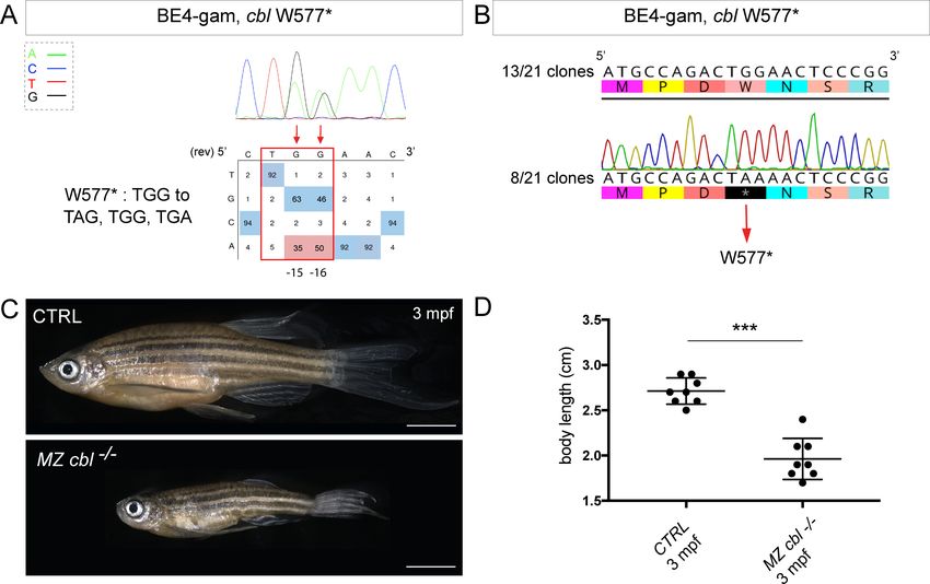
Figure 3. BE4-gam generated cbl maternal zygotic mutant fish show a reduced growth phenotype. (A) DNA sequencing chromatogram of targeted cbl
gene with the BE4-gam. W577* mutation in Cbl upon C-to-T conversion in cbl reached 50% for the C16 base and 35% for the C15 base of gene-editing
efficiency. The chromatogram refers to the efficiency reported for the embryo provided in the first column of Table 2. The numbers in the boxes
represent the percentage of each base at that sequence position. In red are highlighted the base substitutions introduced by base editing, while the
original sequence is in blue. The color code of the chromatogram is indicated in the upper left corner (Adenine green, Cytosine blue, Thymine red,
and Guanine black). The distance from the PAM sequence of the targeted C base is indicated below the chromatogram. It is considered that the
quantifications under 5% are due to the background signal from Sanger sequencing and are thus non-significant (Kluesner et al., 2018). (B)
Sequencing of individual clones of a pool of F1 embryos from a founder carrying the W577* mutation in Cbl. TGG-to-TAA precise mutation was found
in 8 of 21 clones. No editing or INDELs were detected in all other clones. (C) Three months post-fertilization (mpf) cbl wild type derived from the
incross of wild-type siblings (upper panel) and dwarf maternal zygotic (MZ) mutant fish found in 24% of the progeny (lower panel). (D) Quantification of
the body length of the cbl+/+ controls and of the dwarf MZ cbl / . The dwarf fish show a significant reduced size at three mpf compared to the wild-
type controls. n = 8 for each group. Mann–Whitney test, p=0,0002. Scale bars: (C) 5 mm.
data strongly support the role of Cbl W577* in the observed phenotype. Four germline mutations
located in the RING domain of Cbl (Q367P, K382E, D390Y, and R420Q) have been previously identi-
fied and associated to Noonan syndrome and related phenotypes (Martinelli et al., 2010). Our
results are in line with the growth defect phenotypes observed in these patients and directly impli-
cate Cbl loss-of-function as a cause of bone malformations in an animal model. In addition, a point
mutation in zebrafish Cbl (H382Y) has been implicated in myeloproliferative disorders. Unlike our
mutant, cblH382Y mutant fish do not survive to adulthood, suggesting that the CblW577* premature
stop reported here may have different consequences on the multiple functions of Cbl (Peng et al.,
2015). Although not lethal, it would be of interest to assess whether any hematopoietic defects are
present in our MZ cbl / mutants or whether this phenotype is only linked to the CblH328Y
Rosello et al. eLife 2021;10:e65552. DOI: https://doi.org/10.7554/eLife.65552 8 of 15Tools and resources Developmental Biology Genetics and Genomics
substitution found in the LDD731 zebrafish mutant (Peng et al., 2015). Our model represents a pow-
erful in vivo system to dissect the role of Cbl in bone morphogenesis and to explain the human phe-
notypes related to bone malformations.
Conclusions
In our work, we took advantage of base editors to generate C-to-T point mutations at unprece-
dented high efficiencies (up to 91%) without detecting any unwanted mutations that were often
problematic when using CBEs in zebrafish. In comparison, previous work has reported an efficiency
reaching a maximum of 29% using the BE3 (Zhang et al., 2017). Another more recent study
employed the ancBE4max variant in zebrafish with a slight difference of efficiency that might be due
to the choice of the specific locus targeted, the synthesis of the sgRNA (homemade vs commercially
synthetized) and the injection mode (yolk vs cell) (Carrington et al., 2020). More recently,
Zhao et al., 2020 have shown similar efficiencies as we obtained in our study. To expand the gene-
editing possibilities in this animal model, we established in addition a new editor variant recognizing
the NAA PAM. Using these approaches, we first performed the endogenous and constitutive activa-
tion of Wnt signaling by introducing the S33L mutation in b-catenin. In addition, we demonstrated
using these strategies that we were able to precisely target several cancer-associated genes for
which so far only transgenic over-expressions or imprecise deletions were used to elucidate their
functions. Among our targets, the introduced mutation in the cbl gene allowed us to generate a
new zebrafish model for dwarfism.
Together our work provides a panel of examples whereby, using gene-editing approaches, some
of which we established here in zebrafish for the first time, it is now possible to manipulate endoge-
nous signaling pathways, to generate models for human genetic disorders and to mimic precise can-
cer-associated mutations in zebrafish. While a recent study reported the use of ancBE4max in
zebrafish (Zhao et al., 2020), in our work we provide a direct comparison of BE4-gam, ancBE4max,
and Spymac-ancBE4max. Our study highlights the power and the need for these approaches to
increase the efficiency and the targeting flexibility in order to model pathological human mutations
in zebrafish.
Finally, the high efficiencies of CBEs obtained in this study should encourage future applications
where they could be implemented with mosaic mutation induction technologies such as the MAZER-
ATI (Modeling Approach in Zebrafish for Rapid Tumor Initiation) system (Ablain et al., 2018). This
will allow to rapidly model and study in vivo combinations of endogenous mutations occurring in
specific cancer patients or in genetic disorders caused by somatic mosaicism. Our approach could
thus be applied in zebrafish for the precise modeling of complex combinations of cancer-causing
mutations in adult animal models as currently possible by transgenic overexpression or somatic gene
inactivation (Callahan et al., 2018).
Materials and methods
Key resources table
Reagent type
(species) or Additional
resource Designation Source or reference Identifiers information
Genetic reagent Tg(7xTCF-Xla.Siam:GFP) ZIRC ZFIN ID:
(Danio rerio) ZBD-ALT-110113–1
Recombinant pCMV_BE4-gam Addgene Addgene:#100806
DNA reagent (plasmid) RRID:Addgene_100806
Recombinant pCMV_ancBE4max Addgene Addgene:#112094
DNA reagent (plasmid) RRID:Addgene_112094
Recombinant pCS2+_ancBE4max- This paper See Materials and methods
DNA reagent SpymacCas9
(plasmid)
Commercial NEBuilder HiFi DNA New England Biolabs Catalog# E5520S
assay or kit Assembly Cloning Kit
Continued on next page
Rosello et al. eLife 2021;10:e65552. DOI: https://doi.org/10.7554/eLife.65552 9 of 15Tools and resources Developmental Biology Genetics and Genomics
Continued
Reagent type
(species) or Additional
resource Designation Source or reference Identifiers information
Commercial mMESSAGE mMACHINE Ambion Catalog# AM1345
assay or kit T7 Ultra kit
Commercial mMESSAGE mMACHINE Ambion Catalog# AM1340
assay or kit Sp6 kit
Commercial PCR clean-up gel extraction kit Macherey-Nagel Catalog# 740609.50
assay or kit
Peptide, Phusion high-fidelity ThermoFisher Catalog# F-530XL
recombinant DNA polymerase
protein
Software, SequenceParser.py This paper See Source code 1
algorithm
Fish lines and husbandry
Zebrafish (Danio rerio) were maintained at 28˚C on a 14 hr light/10 hr dark cycle. Fish were housed
in the animal facility of our laboratory which was built according to the respective local animal wel-
fare standards. All animal procedures were performed in accordance with French and European
Union animal welfare guidelines. Animal handling and experimental procedures were approved by
the Committee on ethics of animal experimentation. The Tg(7xTCF-Xla.Siam:GFP) line was kindly
provided by Sophie Vriz (Moro et al., 2012).
Molecular cloning
To generate the pCS2+_ancBE4max-SpymacCas9 plasmid, the SpymacCas9 PIM domain sequence
has been codon optimized for expression in zebrafish using online software from IDT and synthe-
sized with the first UGI sequence as G-block from IDT. Then, three fragments have been inserted
into pCS2+ plasmid linearized with Xho1 using the NEBuilder HiFi DNA Assembly Cloning Kit (New
England Biolabs # E5520S): a first fragment of 4161 bp of the ancBE4max to the PIM domain (ampli-
fied using the primers F-50 -CGATTCGAATTCAAGGCCTCATGAAACGGACAGCCGAC-30 and R-50 -
CGGTCTGGATCTCGGTCTTTTTCACGATATTC-30 ), the Gblock fragment of 803 bp (amplified using
the primers F-50 -AAAGACCGAGATCCAGACCGTGGGACAG-30 and R-50 -TCCCGCCGCTATCC
TCGCCGATCTTGGAC-30 ), and a third fragment of 654 bp of the rest of the ancBE4max from the
PIM domain (amplified using the primers F-50 -CGGCGAGGATAGCGGCGGGAGCGGCGGG-30 and
R-50 -CTCACTATAGTTCTAGAGGCTTAGACTTTCCTCTTCTTCTTGGGCTCGAATTCGCTGCCGTCG-
30 ). pCMV_BE4-gam (a gift from David Liu, Addgene plasmid # 100806; Anzalone et al., 2019) has
been used to generate BE4-gam mRNAin vitro. This plasmid was linearized with Pme1 restriction
enzyme and mRNAs were synthesized by in vitro transcription with 1 ml of GTP from the kit added to
the mix, followed by Poly(A) tailing procedure and lithium chloride precipitation (using the mMES-
SAGE mMACHINE T7 Ultra kit #AM1345, Ambion). pCMV_ancBE4max (pCMV_AncBE4max was a
gift from David Liu [Addgene plasmid # 112094]) has been linearized using AvrII restriction enzyme;
mRNAs were synthesized by in vitro transcription with 1 ml of GTP from the kit added to the mix and
lithium chloride precipitation (using the mMESSAGE mMACHINE T7 Ultra kit #AM1345, Ambion).
The pCS2+_ancBE4max-SpymacCas9 has been linearized using KpnI restriction enzyme; mRNAs
were synthetized by in vitro transcription with 1 ml of GTP added to the mix and lithium chloride pre-
cipitation (using the mMESSAGE mMACHINE Sp6 kit #AM1340, Ambion).
sgRNA design
A sequenceParser.py python script was developed and used to design sgRNAs for the creation of a
stop codon. The first function of the script is to ask which PAM will be used to then execute the
rapid detection of codons that are in the right editing windows from this predefined PAM to gener-
ate a STOP in frame after C-to-T conversion. The ORF sequence file extension is .txt and the letters
in lower cases. The script can be executed from the command line interface (such as the terminal or
PowerShell console) using Python version 3.
Rosello et al. eLife 2021;10:e65552. DOI: https://doi.org/10.7554/eLife.65552 10 of 15Tools and resources Developmental Biology Genetics and Genomics
Efficiencies of sgRNAs were validated using CRISPOR online tool (Haeussler et al., 2016). All the
synthetic sgRNAs were synthesized by IDT as Alt-R CRISPR-Cas9 crRNA and Alt-R CRISPR-Cas9
tracrRNA.
List of the crRNAs used in this study and the targeted C bases for each targeted locus. Sequences
are oriented from 50 to 30 :
crRNA sequence used for base editing (5’ 3’)
ctnnb1 (S33L) CTGGACTCAGGAATACACTC
tek (Q94*) GGAGCTCCAGGTGACGGTAG
bap1 (Q273*) GACTCAGCAAGAATCAGGCC
tp53 (Q21*) AGTATTCAGCCCCCAGGTGG
tp53 (Q170*) CAATCAGCGAGCAAATTACA
kras (E62K) CCTCCTGACCTGCAGTGTCC
dmd (Q8*) CCACAGGACCAATGGGAGGA
sod2 (Q145*) GCTGTTCAGGGCTCAGGCTG
rb1 (W63*) TCTCCATGCATGATCACAGA
nras NAA (G13S) AACACCTCCTGCTCCCACAA
tp53 NAA (Q170*) ATCAGCGAGCAAATTACAGG
cbl (W577*) AGTTCCAGTCTGGCATGTTG
Micro-injection
Prior injections, a mix of 2 mL of the Alt-R CRISPR-Cas9 crRNA (100 pmol/mL) and 2 mL of Alt-R
CRISPR-Cas9 tracrRNA (100 pmol/mL) from IDT was incubated at 95˚C for 5 min, cooled down at
room temperature, and then kept on ice to form the synthetic sgRNA complex. One nanoliter of
another mix containing CBE mRNA (600 ng/mL) and the synthetic sgRNA complex (43 pmol/mL) was
then injected into the cell at one-cell stage zebrafish embryos.
Genotyping
To genotype the cbl mutant line, a PCR was performed with primers Fwd-50 -GTACGCCTGGA-
GACCCATCTC-30 and Rev-50 -CTTTTGGACTGTCATAATCCGATGC-30 . The PCR product was
digested with the restriction enzyme BsrI, which cut only on the WT allele. The WT allele resulted in
two fragments (300 bp and 69 bp) and the mutant allele only one fragment (369 bp).
Whole-embryo DNA sequencing
A series between 4 and 10 single embryos randomly chosen was analyzed for each target sequence,
and the embryo with the highest efficiency is shown. Generally, between 25% and 100% embryos
were positive for gene editing, that is showed >16% expected sequence modification. For genomic
DNA extraction, each single embryo was digested for 1 hr at 55˚C in 0.5 mL lysis buffer (10 mM Tris,
pH 8.0, 10 mM NaCl, 10 mM EDTA, and 2% SDS) with proteinase K (0.17 mg/mL, Roche Diagnos-
tics) and inactivated 10 min at 95˚C. To sequence and check for frequency of mutations, each target
genomic locus was PCR-amplified using Phusion High-Fidelity DNA polymerase (ThermoFisher Scien-
tific, # F-530XL). PCR products have been extracted from an agarose gel and purified (using the PCR
clean-up gel extraction kit #740609.50, Macherey-Nagel), and Sanger sequencing was performed by
Eurofins. Sequence analyses were achieved using ApE software and quantifications of the mutation
rate done using editR online software (Kluesner et al., 2018). For the verification of cbl mutant F1
embryos, tek, bap1 and tp53 mutations, PCR fragments were subsequently cloned into the pCR-
bluntII-TOPO vector (Invitrogen). Plasmid DNA was isolated from single colonies and sent for
sequencing. Mutant alleles were identified by comparison with the wild-type sequence using ApE
and Geneious softwares.
Primer sequences used to amplify the targeted loci:
Rosello et al. eLife 2021;10:e65552. DOI: https://doi.org/10.7554/eLife.65552 11 of 15Tools and resources Developmental Biology Genetics and Genomics
tek
(Q94*) ATCTCAGACGTGACTCTGGTGAAC TTCCTGTAGCATCTTGTAGGTGTAG
bap1 TTGTTTATTTTTCAGGACCATGGGG CACCTGAAGGTATTGGTGTTTCTTG
(Q273*)
tp53 CTTTGCATAAGAAACAACATCCCGA GTTCAACACTGAAAACCAAAAGAGG
(Q21*)
tp53 ATATCTTGTCTGTTTTCTCCCTGCT GTCCTACAAAAAGGCTGTGACATAC
(Q170*)
kras CGTTCCACTATGTCCACACATTTAG AACAGTACATTTTCTGCATACTCGC
(E62K)
dmd AGGGCTCCTTCCTTTTTCTGTTTAT TGATCGAGTTTTGATGATTTCTCCG
(Q8*)
sod2 GCATATGGCTGGAAATGATGAACC GCACTTTATGTGCATTCACTGAGG
(Q145*)
rb1 TCTGTCAACTGTTGTTTTTCCAGAC TTCAATATCTGCCACACATACCTCA
(W63*)
nras CCTTTTCTCTCTTTTTGTCTGGGTG CGCAATCTCACGTTAATTGTAGTGT
(G13S)
cbl GTACGCCTGGAGACCCATCTC CTTTTGGACTGTCATAATCCGATGC
(W577*)
Imaging
Embryos were oriented in low-melting agarose 0.6% with an anesthetic (Tricaine 0.013%) diluted in
egg solution. The inverted laser scanning confocal microscope Zeiss CLSM-LSM780 was used for
high-resolution microscopy, employing a 40 water immersion objective. Z-stacks were acquired
every 1–2 mm. Leica MZ10F was used to image the whole embryos the cbl mutant adult fish. Image
analyses were performed with ImageJ software.
Body size quantifications
Eight control wild-type siblings and eight dwarf MZcbl–/– in total were used to measure the body
size using a millimetric ruler. The length measured was from mouth to trunk. A non-parametric t-test
with the Mann–Whitney correction was applied to determine significance in growth. The software
used was Prism 7 (GraphPad).
Acknowledgements
We thank Sophie Vriz for sharing the Tg(7xTCF-Xla.Siam:GFP) transgenic line and the members of
the fish-facility in Institut Curie. We also thank Céline Revenu and Viviana Anelli for early contribu-
tion. MR was supported by the Fondation pour la Recherche Medicale (FRM grant number
ECO20170637481) and la Ligue Nationale Contre le Cancer. Work in the Del Bene laboratory was
supported by ANR-18-CE16 ‘iReelAx’, UNADEV in partnership with ITMO NNP/AVIESAN (National
Alliance for Life Sciences and Health) in the framework of research on vision and IHU FOReSIGHT
[ANR-18-IAHU-0001] supported by French state funds managed by the Agence Nationale de la
Recherche within the Investissements d’Avenir program. MCM was supported by World Wide Can-
cer Research, grant no. 0624, and by LILT –Trento, Program five per mille (year 2014).
Additional information
Funding
Funder Grant reference number Author
Agence Nationale de la Re- ANR-18-CE16 "iReelAx" Filippo Del Bene
cherche
Agence Nationale de la Re- [ANR-18-IAHU-0001 Filippo Del Bene
cherche
Rosello et al. eLife 2021;10:e65552. DOI: https://doi.org/10.7554/eLife.65552 12 of 15Tools and resources Developmental Biology Genetics and Genomics
Fondation pour la Recherche ECO20170637481 Marion Rosello
Médicale
Ligue Contre le Cancer Marion Rosello
UNADEV/AVIESAN Filippo Del Bene
Worldwide Cancer Research grant no. 0624 Marina C Mione
LILT -Trento Program 5 per mille Marina C Mione
The funders had no role in study design, data collection and interpretation, or the
decision to submit the work for publication.
Author contributions
Marion Rosello, Conceptualization, Formal analysis, Validation, Investigation, Visualization, Method-
ology, Writing - original draft, Writing - review and editing; Juliette Vougny, Validation, Investiga-
tion, Methodology; François Czarny, Software; Marina C Mione, Conceptualization, Writing - review
and editing; Jean-Paul Concordet, Conceptualization, Supervision, Methodology, Writing - original
draft, Writing - review and editing; Shahad Albadri, Conceptualization, Formal analysis, Validation,
Visualization, Writing - original draft, Writing - review and editing; Filippo Del Bene, Conceptualiza-
tion, Supervision, Funding acquisition, Methodology, Writing - original draft, Writing - review and
editing
Author ORCIDs
Marion Rosello https://orcid.org/0000-0003-3935-6971
Juliette Vougny http://orcid.org/0000-0002-7361-8405
Marina C Mione http://orcid.org/0000-0002-9040-3705
Shahad Albadri https://orcid.org/0000-0002-3243-7018
Filippo Del Bene https://orcid.org/0000-0001-8551-2846
Ethics
Animal experimentation: All procedures were performed on zebrafish embryos in accordance with
the European Communities Council Directive (2010/63/EU) and French law (87/848) and approved
by the Sorbonne Université ethic committee (Charles Darwin) and the French Ministry for research
(APAFIS agreement #21323 2019062416186982) and by the Institut Curie ethic committee and the
French Ministry for research (APAFIS agreement #6031 2016070822342309).
Decision letter and Author response
Decision letter https://doi.org/10.7554/eLife.65552.sa1
Author response https://doi.org/10.7554/eLife.65552.sa2
Additional files
Supplementary files
. Source code 1. SequenceParser.py STOP codon design source code. This python code highlights
in capital the codons that can converted as STOP codon by C-to-T conversion with the chosen PAM
sequence at the correct distance (PAM [ 19, –13] bp window).
. Transparent reporting form
Data availability
All data generated or analysed during this study are included in the manuscript and supporting files.
Rosello et al. eLife 2021;10:e65552. DOI: https://doi.org/10.7554/eLife.65552 13 of 15Tools and resources Developmental Biology Genetics and Genomics
References
Ablain J, Xu M, Rothschild H, Jordan RC, Mito JK, Daniels BH, Bell CF, Joseph NM, Wu H, Bastian BC, Zon LI,
Yeh I. 2018. Human tumor genomics and zebrafish modeling identify SPRED1 loss as a driver of mucosal
melanoma. Science 362:1055–1060. DOI: https://doi.org/10.1126/science.aau6509, PMID: 30385465
Anzalone AV, Randolph PB, Davis JR, Sousa AA, Koblan LW, Levy JM, Chen PJ, Wilson C, Newby GA, Raguram
A, Liu DR. 2019. Search-and-replace genome editing without double-strand breaks or donor DNA. Nature 576:
149–157. DOI: https://doi.org/10.1038/s41586-019-1711-4, PMID: 31634902
Bravard A, Sabatier L, Hoffschir F, Ricoul M, Luccioni C, Dutrillaux B. 1992. SOD2: a new type of tumor-
suppressor gene? International Journal of Cancer 51:476–480. DOI: https://doi.org/10.1002/ijc.2910510323,
PMID: 1592538
Cagan RL, Zon LI, White RM. 2019. Modeling Cancer with flies and fish. Developmental Cell 49:317–324.
DOI: https://doi.org/10.1016/j.devcel.2019.04.013, PMID: 31063751
Callahan SJ, Tepan S, Zhang YM, Lindsay H, Burger A, Campbell NR, Kim IS, Hollmann TJ, Studer L, Mosimann
C, White RM. 2018. Cancer modeling by transgene electroporation in adult zebrafish (TEAZ). Disease Models &
Mechanisms 11:dmm034561. DOI: https://doi.org/10.1242/dmm.034561, PMID: 30061297
Carrington B, Weinstein RN, Sood R. 2020. BE4max and AncBE4max are efficient in germline conversion of C:g
to T:a base pairs in zebrafish. Cells 9:1690. DOI: https://doi.org/10.3390/cells9071690
Cayuela ML, Claes KBM, Ferreira MG, Henriques CM, van Eeden F, Varga M, Vierstraete J, Mione MC. 2018. The
zebrafish as an emerging model to study DNA damage in aging, Cancer and other diseases. Frontiers in Cell
and Developmental Biology 6:178. DOI: https://doi.org/10.3389/fcell.2018.00178, PMID: 30687705
Cho JY, Guo C, Torello M, Lunstrum GP, Iwata T, Deng C, Horton WA. 2004. Defective lysosomal targeting of
activated fibroblast growth factor receptor 3 in achondroplasia. PNAS 101:609–614. DOI: https://doi.org/10.
1073/pnas.2237184100, PMID: 14699054
Dash S, Kinney NA, Varghese RT, Garner HR, Feng WC, Anandakrishnan R. 2019. Differentiating between Cancer
and normal tissue samples using multi-hit combinations of genetic mutations. Scientific Reports 9:1005.
DOI: https://doi.org/10.1038/s41598-018-37835-6, PMID: 30700767
Dyson NJ. 2016. RB1: a prototype tumor suppressor and an enigma. Genes & Development 30:1492–1502.
DOI: https://doi.org/10.1101/gad.282145.116, PMID: 27401552
Gaudelli NM, Komor AC, Rees HA, Packer MS, Badran AH, Bryson DI, Liu DR. 2017. Programmable base editing
of A.T to G.C in genomic DNA without DNA cleavage. Nature 551:464–471. DOI: https://doi.org/10.1038/
nature24644, PMID: 29160308
Haeussler M, Schönig K, Eckert H, Eschstruth A, Mianné J, Renaud JB, Schneider-Maunoury S, Shkumatava A,
Teboul L, Kent J, Joly JS, Concordet JP. 2016. Evaluation of off-target and on-target scoring algorithms and
integration into the guide RNA selection tool CRISPOR. Genome Biology 17:148. DOI: https://doi.org/10.
1186/s13059-016-1012-2, PMID: 27380939
Harada D, Yamanaka Y, Ueda K, Tanaka H, Seino Y. 2009. FGFR3-related dwarfism and cell signaling. Journal of
Bone and Mineral Metabolism 27:9–15. DOI: https://doi.org/10.1007/s00774-008-0009-7, PMID: 19066716
Hart M, Concordet JP, Lassot I, Albert I, del los Santos R, Durand H, Perret C, Rubinfeld B, Margottin F,
Benarous R, Polakis P. 1999. The F-box protein beta-TrCP associates with phosphorylated beta-catenin and
regulates its activity in the cell. Current Biology 9:207–211. DOI: https://doi.org/10.1016/S0960-9822(99)80091-
8, PMID: 10074433
Hwang WY, Fu Y, Reyon D, Maeder ML, Tsai SQ, Sander JD, Peterson RT, Yeh JR, Joung JK. 2013. Efficient
genome editing in zebrafish using a CRISPR-Cas system. Nature Biotechnology 31:227–229. DOI: https://doi.
org/10.1038/nbt.2501, PMID: 23360964
Jakimo N, Chatterjee P, Nip L, Jacobson JM. 2018. A Cas9 with complete PAM recognition for Adenine
dinucleotides. bioRxiv. DOI: https://doi.org/10.1101/429654
Kluesner MG, Nedveck DA, Lahr WS, Garbe JR, Abrahante JE, Webber BR, Moriarity BS. 2018. EditR: a method
to quantify base editing from Sanger sequencing. The CRISPR Journal 1:239–250. DOI: https://doi.org/10.
1089/crispr.2018.0014, PMID: 31021262
Koblan LW, Doman JL, Wilson C, Levy JM, Tay T, Newby GA, Maianti JP, Raguram A, Liu DR. 2018. Improving
cytidine and Adenine base editors by expression optimization and ancestral reconstruction. Nature
Biotechnology 36:843–846. DOI: https://doi.org/10.1038/nbt.4172, PMID: 29813047
Komor AC, Kim YB, Packer MS, Zuris JA, Liu DR. 2016. Programmable editing of a target base in genomic DNA
without double-stranded DNA cleavage. Nature 533:420–424. DOI: https://doi.org/10.1038/nature17946,
PMID: 27096365
Komor AC, Zhao KT, Packer MS, Gaudelli NM, Waterbury AL, Koblan LW, Kim YB, Badran AH, Liu DR. 2017.
Improved base excision repair inhibition and bacteriophage mu gam protein yields C:g-to-t:a base editors with
higher efficiency and product purity. Science Advances 3:eaao4774. DOI: https://doi.org/10.1126/sciadv.
aao4774, PMID: 28875174
Li S, Balmain A, Counter CM. 2018. A model for RAS mutation patterns in cancers: finding the sweet spot.
Nature Reviews Cancer 18:767–777. DOI: https://doi.org/10.1038/s41568-018-0076-6, PMID: 30420765
Liu C, Kato Y, Zhang Z, Do VM, Yankner BA, He X. 1999. beta-Trcp couples beta-catenin phosphorylation-
degradation and regulates Xenopus Axis formation. PNAS 96:6273–6278. DOI: https://doi.org/10.1073/pnas.
96.11.6273, PMID: 10339577
Martinelli S, De Luca A, Stellacci E, Rossi C, Checquolo S, Lepri F, Caputo V, Silvano M, Buscherini F, Consoli F,
Ferrara G, Digilio MC, Cavaliere ML, van Hagen JM, Zampino G, van der Burgt I, Ferrero GB, Mazzanti L,
Rosello et al. eLife 2021;10:e65552. DOI: https://doi.org/10.7554/eLife.65552 14 of 15Tools and resources Developmental Biology Genetics and Genomics
Screpanti I, Yntema HG, et al. 2010. Heterozygous germline mutations in the CBL tumor-suppressor gene cause
a noonan syndrome-like phenotype. The American Journal of Human Genetics 87:250–257. DOI: https://doi.
org/10.1016/j.ajhg.2010.06.015, PMID: 20619386
Moro E, Ozhan-Kizil G, Mongera A, Beis D, Wierzbicki C, Young RM, Bournele D, Domenichini A, Valdivia LE,
Lum L, Chen C, Amatruda JF, Tiso N, Weidinger G, Argenton F. 2012. In vivo wnt signaling tracing through a
transgenic biosensor fish reveals novel activity domains. Developmental Biology 366:327–340. DOI: https://doi.
org/10.1016/j.ydbio.2012.03.023, PMID: 22546689
Patton EE, Tobin DM. 2019. Spotlight on zebrafish: the next wave of translational research. Disease Models &
Mechanisms 12:dmm039370. DOI: https://doi.org/10.1242/dmm.039370, PMID: 30858282
Peng X, Dong M, Ma L, Jia XE, Mao J, Jin C, Chen Y, Gao L, Liu X, Ma K, Wang L, Du T, Jin Y, Huang Q, Li K,
Zon LI, Liu T, Deng M, Zhou Y, Xi X, et al. 2015. A point mutation of zebrafish c-cbl gene in the ring finger
domain produces a phenotype mimicking human myeloproliferative disease. Leukemia 29:2355–2365.
DOI: https://doi.org/10.1038/leu.2015.154, PMID: 26104663
Prykhozhij SV, Fuller C, Steele SL, Veinotte CJ, Razaghi B, Robitaille JM, McMaster CR, Shlien A, Malkin D,
Berman JN. 2018. Optimized knock-in of point mutations in zebrafish using CRISPR/Cas9. Nucleic Acids
Research 46:e102. DOI: https://doi.org/10.1093/nar/gky512, PMID: 29905858
Sander JD, Joung JK. 2014. CRISPR-Cas systems for editing, regulating and targeting genomes. Nature
Biotechnology 32:347–355. DOI: https://doi.org/10.1038/nbt.2842, PMID: 24584096
Santoriello C, Zon LI. 2012. Hooked! modeling human disease in zebrafish. Journal of Clinical Investigation 122:
2337–2343. DOI: https://doi.org/10.1172/JCI60434
Steinhart Z, Angers S. 2018. Wnt signaling in development and tissue homeostasis. Development 145:
dev146589. DOI: https://doi.org/10.1242/dev.146589, PMID: 29884654
Tessadori F, Roessler HI, Savelberg SMC, Chocron S, Kamel SM, Duran KJ, van Haelst MM, van Haaften G,
Bakkers J. 2018. Effective CRISPR/Cas9-based nucleotide editing in zebrafish to model human genetic
cardiovascular disorders. Disease Models & Mechanisms 11:dmm035469. DOI: https://doi.org/10.1242/dmm.
035469, PMID: 30355756
Wierson WA, Welker JM, Almeida MP, Mann CM, Webster DA, Torrie ME, Weiss TJ, Kambakam S, Vollbrecht
MK, Lan M, McKeighan KC, Levey J, Ming Z, Wehmeier A, Mikelson CS, Haltom JA, Kwan KM, Chien CB,
Balciunas D, Ekker SC, et al. 2020. Efficient targeted integration directed by short homology in zebrafish and
mammalian cells. eLife 9:e53968. DOI: https://doi.org/10.7554/eLife.53968, PMID: 32412410
Zhang Y, Qin W, Lu X, Xu J, Huang H, Bai H, Li S, Lin S. 2017. Programmable base editing of zebrafish genome
using a modified CRISPR-Cas9 system. Nature Communications 8:118. DOI: https://doi.org/10.1038/s41467-
017-00175-6, PMID: 28740134
Zhao Y, Shang D, Ying R, Cheng H, Zhou R. 2020. An optimized base editor with efficient C-to-T base editing in
zebrafish. BMC Biology 18:190. DOI: https://doi.org/10.1186/s12915-020-00923-z, PMID: 33272268
Rosello et al. eLife 2021;10:e65552. DOI: https://doi.org/10.7554/eLife.65552 15 of 15You can also read