A neurostructural biomarker of dissociative amnesia: a hippocampal study in dissociative identity disorder
←
→
Page content transcription
If your browser does not render page correctly, please read the page content below
Psychological Medicine                                 A neurostructural biomarker of dissociative
        cambridge.org/psm
                                                               amnesia: a hippocampal study in dissociative
                                                               identity disorder
                                                               Lora I. Dimitrova1,2, *            , Sophie L. Dean3, *, Yolanda R. Schlumpf4,5, †,
        Original Article
                                                               Eline M. Vissia6, †          , Ellert R. S. Nijenhuis5, Vasiliki Chatzi7, Lutz Jäncke4,8,
        *These authors contributed equally to this
        work                                                   Dick J. Veltman2, Sima Chalavi9 and Antje A. T. S. Reinders1
        †These authors contributed equally to this             1
                                                                 Department of Psychological Medicine, Institute of Psychiatry, Psychology & Neuroscience, King’s College
        work                                                   London, London, UK; 2Department of Psychiatry, Amsterdam UMC, Location VUmc, VU University Amsterdam,
                                                               Amsterdam, The Netherlands; 3Department of Psychosis Studies, Institute of Psychiatry, King’s College London,
        Cite this article: Dimitrova LI et al (2021). A
        neurostructural biomarker of dissociative              London, UK; 4Division of Neuropsychology, Department of Psychology, University of Zurich, Zurich, Switzerland;
                                                               5
        amnesia: a hippocampal study in dissociative             Clienia Littenheid AG, Private Clinic for Psychiatry and Psychotherapy, Littenheid, Switzerland; 6Heelzorg, Centre
        identity disorder. Psychological Medicine 1–9.         for Psychotrauma, Zwolle, The Netherlands; 7Department of Biomedical Engineering, King’s College London,
        https://doi.org/10.1017/S0033291721002154              London, UK; 8Research Unit for Plasticity and Learning of the Healthy Aging Brain, University of Zurich, Zurich,
                                                               Switzerland and 9Movement Control and Neuroplasticity Research Group, Department of Movement Sciences, KU
        Received: 14 September 2020                            Leuven, Leuven, Belgium
        Revised: 12 February 2021
        Accepted: 11 May 2021
                                                                 Abstract
        Key words:
                                                                 Background. Little is known about the neural correlates of dissociative amnesia, a transdiag-
        Dissociative experience scale; DES; childhood
        trauma; CA1; Freesurfer; dissociation                    nostic symptom mostly present in the dissociative disorders and core characteristic of dis-
                                                                 sociative identity disorder (DID). Given the vital role of the hippocampus in memory, a
        Author for correspondence:                               prime candidate for investigation is whether total and/or subfield hippocampal volume can
        Antje A. T. S. Reinders,                                 serve as biological markers of dissociative amnesia.
        E-mail: a.a.t.s.reinders@kcl.ac.uk;
        a.a.t.s.reinders@gmail.com                               Methods. A total of 75 women, 32 with DID and 43 matched healthy controls (HC), under-
                                                                 went structural magnetic resonance imaging (MRI). Using Freesurfer (version 6.0), volumes
                                                                 were extracted for bilateral global hippocampus, cornu ammonis (CA) 1–4, the granule cell
                                                                 molecular layer of the dentate gyrus (GC-ML-DG), fimbria, hippocampal−amygdaloid tran-
                                                                 sition area (HATA), parasubiculum, presubiculum and subiculum. Analyses of covariance
                                                                 showed volumetric differences between DID and HC. Partial correlations exhibited relation-
                                                                 ships between the three factors of the dissociative experience scale scores (dissociative
                                                                 amnesia, absorption, depersonalisation/derealisation) and traumatisation measures with hip-
                                                                 pocampal global and subfield volumes.
                                                                 Results. Hippocampal volumes were found to be smaller in DID as compared with HC in
                                                                 bilateral global hippocampus and bilateral CA1, right CA4, right GC-ML-DG, and left presu-
                                                                 biculum. Dissociative amnesia was the only dissociative symptom that correlated uniquely and
                                                                 significantly with reduced bilateral hippocampal CA1 subfield volumes. Regarding traumatisa-
                                                                 tion, only emotional neglect correlated negatively with bilateral global hippocampus, bilateral
                                                                 CA1, CA4 and GC-ML-DG, and right CA3.
                                                                 Conclusion. We propose decreased CA1 volume as a biomarker for dissociative amnesia. We
                                                                 also propose that traumatisation, specifically emotional neglect, is interlinked with dissociative
                                                                 amnesia in having a detrimental effect on hippocampal volume.

                                                               Introduction
                                                               Dissociative amnesia is a dissociative symptom common in the dissociative disorders and is
                                                               characterised by recurrent gaps in recalling everyday events and/or of important personal
                                                               (trauma-related) information, distinct from ordinary forgetting. The most severe of the dis-
                                                               sociative disorders is dissociative identity disorder (DID). DID is a debilitating psychiatric con-
        © The Author(s), 2021. Published by                    dition and is related to, among others, alternating states of consciousness and distinct
        Cambridge University Press. This is an Open            personality states with changing access to autobiographical information (American
        Access article, distributed under the terms of
                                                               Psychiatric Association, 2013). Although dissociative amnesia is a core symptom of DID
        the Creative Commons Attribution licence
        (http://creativecommons.org/licenses/by/4.0),          and of other dissociative disorders little is known about its neurobiological foundations.
        which permits unrestricted re-use, distribution            A few studies have found a negative correlation between dissociative symptoms and hippo-
        and reproduction, provided the original article        campal volume. Ehling, Nijenhuis, and Krikke (2008) found bilateral global hippocampal vol-
        is properly cited.                                     ume reductions in individuals with DID, which negatively correlated with dissociative
                                                               symptoms. Similarly, Chalavi et al. (2015b) found evidence for hippocampal global and sub-
                                                               field volume reductions in relation to dissociative symptoms and/or traumatisation. Left hip-
                                                               pocampal volume reduction has further been associated with dissociative symptoms in
                                                               individuals who suffered childhood sexual abuse as compared with healthy controls (HC)

Downloaded from https://www.cambridge.org/core. IP address: 46.4.80.155, on 11 Jul 2021 at 05:51:11, subject to the Cambridge Core terms of use, available at
https://www.cambridge.org/core/terms. https://doi.org/10.1017/S0033291721002154
2                                                                                                                                                   Lora I. Dimitrova et al.

       (Stein, Koverola, Hanna, Torchia, & McClarty, 1997) indicating                             absorption and depersonalisation/derealisation are separate
       that early traumatisation is a potential mediator of dissociative                          unique dissociative components that need to be examined as sep-
       symptoms. Adverse childhood experiences further increase in                                arate entities in research (Lyssenko et al., 2018; Soffer-Dudek,
       the likelihood of childhood autobiographical memory deficits                               Lassri, Soffer-Dudek, & Shahar, 2015). For instance, unlike
       (Brown et al., 2007). Amnesia has also been linked to the presence                         amnesia and depersonalisation/derealisation, absorption has
       of psychological stress and/or traumatisation (Markowitsch &                               been found to be normally distributed among healthy populations
       Staniloiu, 2012; Staniloiu & Markowitsch, 2012). On the other                              (Soffer-Dudek et al., 2015) and it is suggested that absorption
       hand, there is also information to suggest absence of hippocampal                          involves an alternation of consciousness that is not dissociative
       volume reductions in individuals with a dissociative disorder                              (Nijenhuis, Van der Hart, & Steele, 2002b). Therefore, this raises
       (Weniger, Lange, Sachsse, & Irle, 2008) and a study by Mutluer                             concerns as to whether absorption could diminish statistical
       et al. (2018) did not show significant correlations between hippo-                         effects when pathological dissociation is examined as a cumulative
       campal volume and dissociative symptoms in a group of PTSD                                 overall phenomenon. Consequently, it is important to assess indi-
       patients. Thus, although most studies reported decreased hippo-                            vidually the core diagnostic features of DID and their neural cor-
       campal volume, evidence for a negative correlation between hip-                            relates as well as to investigate the three dissociative constructs in
       pocampal volume and dissociative symptoms is not fully                                     relation to hippocampal morphology, which could provide a com-
       consistent. Variations of findings across these studies may be dri-                        prehensive understanding of the specificity of structural
       ven by the low numbers of participants and/or inconsistent meas-                           alterations.
       urement of the dissociative symptoms. Therefore, there is a need                               The aims of the current study are to extend previous research
       for further research into the role of the hippocampus in dissocia-                         by Chalavi et al. (2015b) by increasing the sample size and to
       tive amnesia in DID.                                                                       investigate the distinct contributions of the three separate DES
           The hippocampus plays an integral role in consolidating long-                          factors, that is amnesia, absorption and depersonalisation/dereal-
       term memories and in learning (Preston & Eichenbaum, 2014).                                isation, to decreases in hippocampal global and subfield volumes
       As such, it is relevant to consider the hippocampus in relation                            in a large sample of individuals with DID. In addition, due to the
       to dissociative amnesia. The hippocampus consists of different                             inter-linked nature of trauma and dissociation, we aim to investi-
       anatomical subfields, namely the cornu ammonis 1, 2, 3 and 4                               gate the global volume of the hippocampus and of its composite
       (CA1–4), the parasubiculum, presubiculum, subiculum and the                                smaller regions in relation to self-report measures of traumatisa-
       hippocampal tail, as well as other regions, namely the granule                             tion related to emotional abuse, emotional neglect, physical abuse,
       cell molecular layer of the dentate gyrus (GC-ML-DG), the hippo-                           sexual abuse and sexual harassment. We hypothesise that hippo-
       campal–amygdaloid transition area (HATA), the fimbria, the hip-                            campal global and subfield volumes will be smaller in individuals
       pocampal fissure and the molecular layer of the hippocampus                                with DID as compared to HC and that within the DID group the
       (Amaral & Lavenex, 2006). Research has shown that excessive                                global and subfield hippocampal volumes will negatively correlate
       stress from traumatic experiences, particularly childhood trau-                            with higher severity of dissociative amnesia as well as with greater
       matic experiences, contributes to the dysregulation of the hypo-                           traumatisation.
       thalamic pituitary adrenal axis (Kuhlman, Vargas, Geiss, &
       Lopez-Duran, 2017; Shea, Walsh, MacMillan, & Steiner, 2004),
       which may damage hippocampal function and structure                                        Methods
       (Bidzan, 2017; Teicher, Anderson, & Polcari, 2012; Woon,
                                                                                                  Design and participants
       Sood, & Hedges, 2010). Certain hippocampal regions have been
       identified as more important in memory and amnesia including                               Data of a total of 75 women (only female participants with DID
       the CA1 (Bartsch, Döhring, Rohr, Jansen, & Deuschl, 2011;                                  volunteered) were included in the current study which follows a
       Spiegel et al., 2017). CA1 is a subfield known to be critically                            between-group research design: 32 female volunteers with DID
       involved in the process of memory consolidation and could be                               and 43 HC matched for age, gender, years of education and eth-
       involved in dissociative amnesia (Spiegel et al., 2017).                                   nicity. Data were collected in the Netherlands in the University
       Vulnerability and susceptibility of the CA1 to the detrimental                             Medical Centre in Groningen (UMCG) and the Amsterdam
       effects of stress could therefore lead to dissociative amnesia. To                         Medical Centre (AMC), and in Switzerland at the University
       date, only one study has investigated the global volume of the                             Hospital in Zurich. Participant information has been detailed pre-
       hippocampus and of its composite smaller regions in relation to                            viously (Chalavi et al., 2015a, 2015b; Reinders et al., 2018, 2019;
       dissociative symptoms in DID participants and showed a negative                            Schlumpf et al., 2013, 2014). The DID participants were recruited
       correlation between total dissociation scores and hippocampal                              from psychiatric departments, outpatient psychotherapists and
       regions (Chalavi et al., 2015b).                                                           psychiatrists. Initial diagnosis fulfilled DSM-IV criteria and was
           Dissociative symptomatology is of a complex nature. Many dif-                          confirmed by trained clinicians with the Structural Clinical
       ferent theoretical and definitional frameworks have endeavoured                            Interview for DSM-IV Dissociative Disorders (SCID-D)
       to conceptualise it (Dell & O’Neil, 2009; Dorahy et al., 2014;                             (Steinberg, 1993). Twenty-nine volunteers with DID had a
       Holmes et al., 2005; Nijenhuis, 2015; Reinders & Veltman,                                  co-morbid diagnosis of PTSD and three had PTSD in remission.
       2020; Şar, Dorahy, & Krüger, 2017). Most research into patho-                              Additional co-morbidity was confirmed by participants and their
       logical dissociation has used total scores from the dissociative                           personal therapists based on DSM-IV classification (American
       experience scale (DES) (Bernstein & Putnam, 1986) to assess psy-                           Psychiatric Association, 1994), see for details Reinders et al.
       choform dissociative symptoms in relation to, for example, cogni-                          (2018, 2019) and online Supplementary Table S1. The HC
       tive functioning or neurobiology (Roydeva & Reinders, 2021).                               group was recruited through local newspaper advertisements.
       However, the DES is considered to have a three-factor structure,                           Exclusion criteria for all participants included age outside the
       the factors being dissociative amnesia, absorption and deperson-                           range of 18–65, pregnancy, systemic or neurological illness, claus-
       alisation/derealisation. Increasingly evidence shows that amnesia,                         trophobia, metal implants in the body and substance abuse.

Downloaded from https://www.cambridge.org/core. IP address: 46.4.80.155, on 11 Jul 2021 at 05:51:11, subject to the Cambridge Core terms of use, available at https://www.cambridge.org/core/terms.
https://doi.org/10.1017/S0033291721002154
Psychological Medicine 3
Additional exclusion criteria for the HC group included the pres- Fischl, Sereno, Tootell, & Dale, 1999b; Fischl et al., 2002, 2004a,
ence of dissociative symptoms and a history of trauma, past or 2004b; Han et al., 2006; Jovicich et al., 2006; Segonne et al.,
current psychiatric disorders and medication use (see for details 2004; Yeo et al., 2010a, 2010b – for full details, see Fischl, 2012;
Chalavi et al. 2015a, 2015b; Reinders et al. 2018; Schlumpf et al. Fischl et al., 2002), volumetric measures for the global hippocam-
2013, 2014). HC were required to score below 25 on the DES. pus, CA1, CA3, CA4, fimbria, HATA, subiculum, GC-DG, para-
The DES is a 28-item self-report screening tool for dissociative subiculum and presubiculum, for each hemisphere (Fig. 1), in
disorders which is often used in clinical research and as a basis addition to total intercranial volume (TIV), were extracted. For
for guiding diagnosis in the clinical interview (Carlson & one participant from the HC group FreeSurfer was not able to
Putnam, 1993). The DES scale has received meta-analytic valid- complete the hippocampal segmentation, and therefore this par-
ation (Van Ijzendoorn & Schuengel, 1996). The HC were also ticipant was excluded from subsequent statistical analyses.
required to have a score below and 29 on the Somatoform
Dissociation Questionnaire (Nijenhuis, Spinhoven, Van Dyck,
Dissociative amnesia
Van der Hart, & Vanderlinden, 1996). Traumatic experiences
were measured with the traumatic experience checklist (TEC), a Dissociative amnesia was measured as part of the DES. In add-
self-report measure of potentially traumatising events ition to total dissociation the three core dissociative symptoms,
(Nijenhuis, Van der Hart, & Kruger, 2002a). TEC total scores namely absorption, amnesia and depersonalisation/derealisation
were previously calculated for each category of abuse, namely were calculated.
emotional neglect, emotional abuse, physical abuse, sexual abuse
and sexual harassment (Reinders et al., 2018). As expected and
Statistical analysis
previously reported (Reinders et al., 2018, 2019) dissociative
symptoms and traumatic experiences were significantly higher All analyses were performed using SPSS (v25) (IBM Corp., 2017).
in the DID group (all data obtained in the predominant person- To confirm the findings of Chalavi et al. (2015b) ‘that hippocam-
ality state) than in the HC group (all p-values4 Lora I. Dimitrova et al.
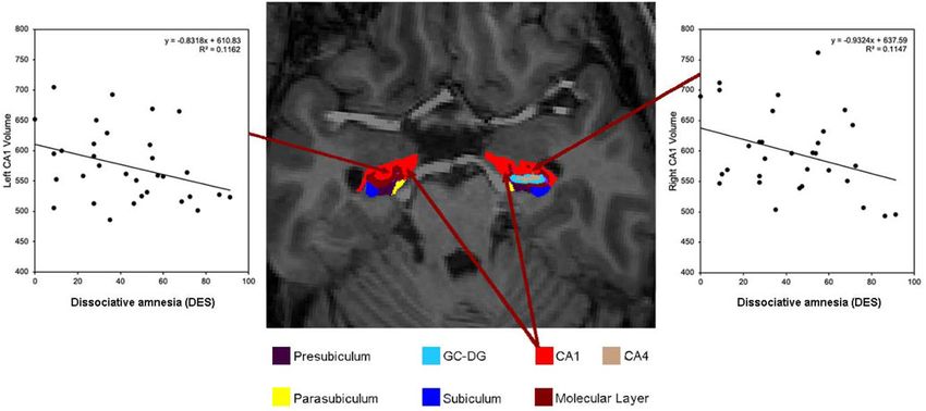
Fig. 1. Axial slice of the hippocampus from a DID participant with scatter plots showing the relationship between increased dissociative amnesia scores as assessed
by the dissociative experiences scale (DES) and decreased bilateral CA1 volume.
(see for discussion Chalavi et al. 2015a, 2015b; Dimitrova et al. 4.187, p = 0.045, ηp 2 = 0.061, d = 0.50), right GC-ML-DG
2020; Preacher, Rucker, MacCallum, & Nicewander, 2005). (F(1,65) = 4.130, p = 0.046, ηp 2 = 0.060, d = 0.50) and left presubi-
Partial correlations were conducted between volumetric measures culum (F(1,65) = 5.663, p = 0.020 ηp 2 = 0.080, d = 0.58). Details
and dissociation total and subscale measures, controlling for age are provided in online Supplementary Table S2.
and TIV. The Kolmogorov–Smirnov test detected no deviations
from normality in the dissociation measures within the DID
group. Within the DID group, the associations of total TEC scores Hippocampal volumes and dissociation
and subscale scores of abuse categories with hippocampal Dissociative amnesia, absorption, depersonalisation/derealisation
volumes for each hemisphere were explored with partial correla- scores as well as total DES scores correlated with hippocampal
tions, controlling for age and TIV. Due to the ordinal nature of global and subfield volumes. However, only dissociative amnesia
the TEC data and lack of normality as assessed by the and total DES scores correlated significantly, that is, between dis-
Kolmogorov–Smirnov test (all p < 0.05), Spearman’s partial corre- sociative amnesia and reduced bilateral hippocampal CA1 sub-
lations were conducted using a script in the SPSS syntax editor. field volumes (left: r = −0.396, p = 0.030; right: r = −0.363, p =
Results were controlled for co-morbidity by repeating the par- 0.049) and between total DES scores and left CA1 subfield (r =
tial correlations and adding the co-morbid diagnosis as a third −0.369, p = 0.045). Results are illustrated in Figs 1, 2 and online
variable to the analyses, in a similar manner as Chalavi et al. Supplementary Fig. S1. There were no significant correlations
(2015a). This meant that the correlations between dissociation between symptoms of absorption or depersonalisation/derealisa-
measures and hippocampal volumes were controlled for age, tion scores and hippocampal volumes. Details are provided in
TIV and co-morbidity. Additionally, we created a ‘total’ covariate online Supplementary Table S3. Online Supplementary Table S4
variable, data coded with 1 if any co-morbidity was present, and a contains the results corrected for co-morbidity using covariate
0 if none was present. PTSD was not included as a co-morbidity analyses.
because all individuals with DID also had a diagnosis of PTSD (in
remission). Adding PTSD as a covariate would therefore invali-
date the analyses. Hippocampal volumes and traumatisation
Measures of traumatic experiences correlated with hippocampal
Results volumes. Negative correlations were found between emotional
neglect and bilateral global hippocampus (left: r = −0.442, p =
Hippocampal volumes
0.021; right: r = −0.431, p = 0.025), bilateral CA1 (left: r =
Bilateral hippocampal global volume was significantly smaller in −0.408, p = 0.035; right: r = −0.392, p = 0.043), right CA3 (r =
the DID group compared with the HC group (left: F(1,69) = −0.411, p = 0.033), bilateral CA4 (left: r = −0.446, p = 0.020;
6.183, p = 0.015, ηp 2 = 0.087, d = 0.61; right: F(1,66) = 5.425, p = right: r = −0.462, p = 0.017) and bilateral GC-ML-DG (left: r =
0.023, ηp 2 = 0.076, d = 0.57). Regarding hippocampal subfield −0.460, p = 0.016; right: r = −0.469, p = 0.016). There were no sig-
and region volumes, smaller volumes for the DID group com- nificant negative correlations between hippocampal volumes and
pared with the HC group were found for bilateral CA1 (left: emotional, physical and sexual abuse subscales, sexual harassment
F(1,66) = 4.785, p = 0.032, ηp 2 = 0.068, d = 0.53; right: F(1,66) = and total traumatisation scores. Details are provided in online
5.812, p = 0.019, ηp 2 = 0.081, d = 0.59), right CA4 (F(1,65) = Supplementary Table S5.
Downloaded from https://www.cambridge.org/core. IP address: 46.4.80.155, on 11 Jul 2021 at 05:51:11, subject to the Cambridge Core terms of use, available at https://www.cambridge.org/core/terms.
https://doi.org/10.1017/S0033291721002154Psychological Medicine 5
Fig. 2. Scatter plot showing the relationship between dissociative amnesia as assessed by the DES and bilateral CA1 volume. The correlations indicate a reduction
in volume for both hemispheres with higher severity of dissociative amnesia.
Discussion
to a conditioned stimulus) in individuals presenting trauma,
The current study is the first to investigate decreased hippocampal implicating accurate context evaluation of non-threatening set-
global and subfield volumes in relation to dissociative amnesia, tings (Chen et al., 2018). Consequently, damage to or lack of mat-
absorption and depersonalisation/derealisation symptoms in the uration of CA1 regions may be related to memory disturbances.
largest cohort of individuals with DID to date. Our most import- The CA1 projects to the medial prefrontal cortex and the orbito-
ant finding is that in the DID group only dissociative amnesia and frontal frontal cortex (Zhong, Yukie, & Rockland, 2006) and it
total dissociation symptom scores and not absorption or deper- could be speculated that damage to the CA1 may contribute to
sonalisation/derealisation correlated significantly and negatively dissociation mechanisms and the formation of dissociative per-
with hippocampal volume. Our second most important finding sonality states (Forrest, 2001).
is that these negative correlations were only found for the CA1 We also found a link between the severity of childhood trau-
hippocampal subfield. matisation, specifically emotional neglect and reductions in hip-
We propose that the volume of CA1 can serve as a biomarker for pocampal volume including CA1. In support of our finding
dissociative amnesia because only dissociative amnesia and not research has demonstrated an association between emotional neg-
absorption or depersonalisation/derealisation correlated with smal- lect and dissociation severity accentuating the relationship
ler CA1 hippocampal subfield volume in individuals with DID. between traumatisation, dissociation and hippocampal volume
Research has previously shown that the total DES score correlates reductions (Şar, 2011; Şar, Akyüz, & Doğan, 2007; Schalinski
negatively with hippocampal volume in patients with DID and et al., 2016; Schimmenti, 2016). CA1 impairment in relation to
PTSD (Chalavi et al., 2015b; Ehling et al., 2008; Mutluer et al., childhood traumatisation can possibly lead to fragmentation of
2018; Stein et al., 1997), but our results indicate that this effect the mind and a scattered sense of self (Brown et al., 2007).
might be driven by dissociative amnesia. Further to this, the associ- Interestingly, Huntjens, Dorahy, and van Wees-Cieraad (2013)
ation of CA1 with dissociative amnesia remained significant even present lack of self-referential processing as a possible mechanism
after controlling for co-morbidity. This indicates that CA1 volume to explain the link between dissociation and fragmentation of the
reduction is primarily driven by dissociative amnesia in DID, not mind. Furthermore, research has shown memory source misattri-
other disorders, and presents a specific dissociative disorder effect. bution as a specific cognitive characteristic of dissociation, par-
Our novel findings can be linked to amnesia and memory ticularly for dissociative amnesia (Chiu et al., 2016, 2019). The
because damage to hippocampal subfield CA1 has been shown association between dissociative symptoms and misattributing
to lead to memory impairments (Bartsch et al., 2015, 2011; self-generated representations as an external doing suggests an
Ocampo, Squire, & Clark, 2017). Furthermore, CA1 has been amnestic barrier regarding access to self-relevant information.
found to be of particular importance for autobiographical mem- Chiu et al. (2016) state that this misattribution error could not
ories (Bartsch et al., 2011), which constitute building blocks of be fully explained by intellectual function and general psycho-
a person’s identity. Moreover, CA1 impairments have been linked pathology, which suggests that this cognitive blockage of informa-
to damage in extinction learning (the gradual decrease in response tion is a specific cognitive characteristic of dissociation. They also
Downloaded from https://www.cambridge.org/core. IP address: 46.4.80.155, on 11 Jul 2021 at 05:51:11, subject to the Cambridge Core terms of use, available at https://www.cambridge.org/core/terms.
https://doi.org/10.1017/S00332917210021546 Lora I. Dimitrova et al.
found that source misattribution of self-generated representations prefrontal cortex, posterior cingulate cortex, midline thalamus
correlated significantly with DES assessed dissociative amnesia and periventricular hypothalamus (Abdallah et al., 2017; Blessing,
(Chiu et al., 2016). Furthermore, it has been found that there is Beissner, Schumann, Brünner, & Bär, 2016). Future studies could
a link between dissociation proneness and reduced self-reference also formulate hypotheses about and include a wider range of mea-
ability, particularly for individuals with high dissociation prone- sures of dissociative symptomatology such as positive and negative
ness and high childhood relational trauma (Chiu et al., 2019). dissociative symptoms (Spiegel et al., 2013), detachment and com-
The inability to establish a self-referential perspective during trau- partmentalisation (Cardeña & Carlson, 2011) and trait and state
matisation prevents processing the experience as own and self- dissociation (Roydeva & Reinders, 2021).
relevant and inhibits assimilating those memories into the auto- Our study presents the following limitations. Firstly, our study
biographical memory base (Huntjens et al., 2013). This supports only included participants with DID and future research should
the altered sense of individuality prevalent in DID and the amnes- assess dissociative amnesia across other psychiatric disorders to
tic blockage of information as evident with dissociative amnesia. confirm our proposal of CA1 as a biomarker for dissociative
Furthermore, there are consistent reports on associations between amnesia. Furthermore, smaller hippocampal volume has been
abuse and dissociation severity across a range of clinical presenta- found in various mental disorders with and without dissociative
tions including PTSD, borderline personality disorder (BPD) and symptoms (Belli, 2014; Chalavi et al., 2015b; Luoni, Agosti,
psychosis (Schalinski & Teicher, 2015; Schalinski et al., 2016). A Crugnola, Rossi, & Termine, 2018; Roydeva & Reinders, 2021;
multitude of factors interplay with dissociation severity including van Huijstee & Vermetten, 2017). Due to the polysymptomatic
the relationship to the perpetrator (Plattner et al., 2003), attach- presentation of symptoms in DID it is important that future
ment style (Kong, Kang, Oh, & Kim, 2018) and genetics research controls for comorbidity findings. Secondly, our study
(Dackis, Rogosch, Oshri, & Cicchetti, 2012; Savitz et al., 2008; is limited by the exclusively female sample. Therefore our results
Wolf et al., 2014). may not extend to men with DID. Nevertheless, by focusing on
Our study is, to the best of our knowledge, the first to show that women exclusively, our study design is optimised to minimise
decreased hippocampal subfield volume is related to dissociative the neuroanatomical and clinical heterogeneity that could have
amnesia and is interlinked with emotional neglect. These findings been introduced by analysing data across gender categories
need to be confirmed in future research because emotional neglect (Reinders et al., 2018). It can be argued that seeing a correlation
is the only nonviolent traumatisation measure, while past research of hippocampal volume and (self-report) severity of amnesia in
has emphasised the importance of physical and sexual abuse in dis- the DID group is evidence that this neural substrate is relevant
sociative disorders (Mutluer et al., 2018; Stein et al., 1997; Twaite & to the severity of amnesia in DID, rather than the presence of dis-
Rodriguez-Srednicki, 2004). For instance, there are reports correlat- sociative amnesia because amnesia is a defining symptom of DID
ing dissociative symptoms with various adverse events, including and is therefore expected to be present in all participating indivi-
emotional neglect by family of origin, in addition to research duals with DID. Although this is indeed the case and symptom
suggesting emotional neglect (e.g. parental unavailability) during scores of absorption and depersonalisation/derealisation are 4–
childhood as a major predictor for developing a dissociative dis- 5% higher than for dissociative amnesia, our proposal of
order (Dutra, Bureau, Holmes, Lyubchik, & Lyons-Ruth, 2009; decreased CA1 volume as a biomarker for dissociative amnesia
Lyons-Ruth, Dutra, Schuder, & Bianchi, 2006; Nijenhuis, is also based on the absence of a correlation between hippocampal
Spinhoven, Van Dyck, Van der Hart, & Vanderlinden, 1998; (sub-)volumes and symptoms of absorption and depersonalisa-
Ogawa, Sroufe, Weinfield, Carlson, & Egeland, 1997). tion/derealisation instead of only the presence of dissociative
Our finding of the importance of CA1 in dissociative amnesia amnesia. Therefore, we propose that our findings are of interest
is of interest as it provides insight into further understanding bio- for neurobiological biomarker research.
markers of dissociative disorders. Furthermore, by showing the In conclusion, our study proposes decreased CA1 volume as a
variation of significance of the three subscales of the DES, that biomarker for dissociative amnesia. We also propose that emo-
is amnesia, absorption and depersonalisation/derealisation, we tional neglect is interlinked with dissociative amnesia in having
emphasise with others (Lyssenko et al., 2018; Soffer-Dudek a detrimental effect on hippocampal subfield volume.
et al., 2015) the importance of examining the subscales as separate
Supplementary material. The supplementary material for this article can
entities and not as a total cumulative unit. By identifying a region
be found at https://doi.org/10.1017/S0033291721002154
of interest that is linked to dissociative amnesia we provide evi-
dence of implications in the clinical realm (Reinders & Acknowledgements. The authors thank all the participants and their thera-
Veltman, 2020). Our study has possible clinical relevance because pists. We thank Dr Nel Draijer, Mechteld Giesen, Ekaterina Weder and Eva
there is some evidence to suggest hippocampal volume increase/ Zimmermann for arranging participant inclusion and scanning as well as
recovery after medication (Vermetten, Vythilingam, Southwick, for the assessment of diagnostic interviews and their support as research
Charney, & Bremner, 2003) and with phase-oriented psychother- clinicians.
apy (Ehling et al., 2008), but not with brief eclectic psychotherapy
Financial support. This article represents an independent research part-
(Lindauer et al., 2005). However, to date, there is little evidence funded by the National Institute for Health Research (NIHR) Biomedical
regarding treatment outcomes for DID and future research Research Centre at South London and Maudsley NHS Foundation Trust and
could investigate the role of CA1 hippocampal subfield volume King’s College London. The views expressed are those of the author(s) and not
during treatment of and recovery from dissociative disorders. necessarily those of the NHS, the NIHR or the Department of Health. A.A.T.S.
We also recommend that future studies provide more precise Reinders was supported by the Netherlands Organisation for Scientific
results in terms of hippocampal head, body and tail and their Research (www.nwo.nl), NWO-VENI grant no. 451-07-009. S. Chalavi was sup-
respective connectivity in a larger sample of DID participants. ported by David Caul graduate research grant from the International Society for
This is of interest because the anterior hippocampus has been the Study of Trauma and Dissociation (ISSTD) (www.isst-d.org/about/awards.
associated with trauma-related memories and intrinsic functional htm). Y. Schlumpf was supported by the Forschungskredit UZH (www.research-
ers.uzh.ch/de/funding/phd/fkcandoc.html).
connectivity with the amygdala, nucleus accumbens, medial
Downloaded from https://www.cambridge.org/core. IP address: 46.4.80.155, on 11 Jul 2021 at 05:51:11, subject to the Cambridge Core terms of use, available at https://www.cambridge.org/core/terms.
https://doi.org/10.1017/S0033291721002154Psychological Medicine 7
Conflict of interest. None. Chiu, C.-D., Tollenaar, M. S., Yang, C.-T., Elzinga, B. M., Zhang, T.-Y., & Ho,
H. L. (2019). The loss of the self in memory: Self-referential memory, child-
hood relational trauma, and dissociation. Clinical Psychological Science, 7
(2), 265–282. https://doi.org/10.1177/2167702618804794.
References
Chiu, C., Tseng, M., Chien, Y., Liao, S., Liu, C., Yeh, Y., … Hwu, H. (2016).
Abdallah, C., Wrocklage, K., Averill, C., Akiki, T., Schweinsburg, B., Roy, A., … Misattributing the source of self-generated representations related to dis-
Scott, J. (2017). Anterior hippocampal dysconnectivity in posttraumatic sociative and psychotic symptoms. Frontiers in Psychology, 7, 541. https://
stress disorder: A dimensional and multimodal approach. Translational doi.org/10.3389/fpsyg.2016.00541.
Psychiatry, 7(2), e1045. https://doi.org/10.1038/tp.2017.12. Cohen, J. (1988). Statistical Power Analysis for the Behavioral Sciences (2nd
Amaral, D., & Lavenex, P. (2006). Hippocampal neuroanatomy. In P. Ed.) New York: Lawrence Erlbaum Associates.
Andersen, R. Morris, D. Amaral, T. Bliss & J. O’Keefe (Eds.), The hippocam- Dackis, M. N., Rogosch, F. A., Oshri, A., & Cicchetti, D. (2012). The role of
pus book (pp. 37–115). Oxford: Oxford University Press. limbic system irritability in linking history of childhood maltreatment
American Psychiatric Association (1994). Diagnostic and statistical manual of and psychiatric outcomes in low-income, high-risk women: Moderation
mental disorders: DSM-IV. Washington, DC: American Psychiatric Association. by FK506 binding protein 5 haplotype. Development and Psychopathology,
American Psychiatric Association (2013). Diagnostic and statistical manual of 24(4), 1237–1252. https://doi.org/https://doi.org/10.1017/S09545794120006
mental disorders: DSM-5 (5th Ed.). Washington, DC: American Psychiatric 73.
Publishing. Dale, A., Fischl, B., & Sereno, M. I. (1999). Cortical surface-based analysis I:
Bartsch, T., Döhring, J., Reuter, S., Finke, C., Rohr, A., Brauer, H., … Jansen, Segmentation and surface reconstruction. NeuroImage, 9, 179–194.
O. (2015). Selective neuronal vulnerability of human hippocampal CA1 https://doi.org/10.1006/nimg.1998.0395.
neurons: Lesion evolution, temporal course, and pattern of hippocampal Dale, A. M., & Sereno, M. I. (1993). Improved localization of cortical activity
damage in diffusion-weighted MR imaging. Journal of Cerebral Blood by combining EEG and MEG with MRI cortical surface reconstruction: A
Flow & Metabolism, 35(11), 1836–1845. https://doi.org/https://doi.org/10. linear approach. Journal of Cognitive Neuroscience, 5, 162–176. https://doi.
1038/jcbfm.2015.137. org/10.1162/jocn.1993.5.2.162.
Bartsch, T., Döhring, J., Rohr, A., Jansen, O., & Deuschl, G. (2011). CA1 neu- Dell, P. F., & O’Neil, J. A. (2009). Dissociation and the dissociative disorders:
rons in the human hippocampus are critical for autobiographical memory, DSM-V and beyond. New York: Routledge.
mental time travel, and autonoetic consciousness. Proceedings of the Dimitrova, L., Fernando, V., Vissia, E. M., Nijenhuis, E. R. S., Draijer, N., &
National Academy of Sciences of the United States of America, 108(42), Reinders, A. A. T. S. (2020). Sleep, trauma, fantasy and cognition in dis-
17562–17567. https://doi.org/https://doi.org/10.1073/pnas.1110266108. sociative identity disorder, post-traumatic stress disorder and healthy con-
Belli, H. (2014). Dissociative symptoms and dissociative disorders comorbidity trols: A replication and extension study. European Journal of
in obsessive compulsive disorder: Symptom screening, diagnostic tools and Psychotraumatology, 11(1), 1705599. https://doi.org/10.1080/20008198.
reflections on treatment. World Journal of Clinical Cases, 2(8), 327–331. 2019.1705599.
https://doi.org/10.12998/wjcc.v2.i8.327. Dorahy, M. J., Brand, B. L., Şar, V., Krüger, C., Stavropoulos, P.,
Bernstein, E. M., & Putnam, F. W. (1986). Development, reliability, and valid- Martínez-Taboas, A., … Middleton, W. (2014). Dissociative identity dis-
ity of a dissociation scale. Journal of Nervous and Mental Disease, 174(12), order: An empirical overview. Australian and New Zealand Journal of
727–735. http://doi.org/10.1097%2F00005053-198612000-00004. Psychiatry, 48(5), 402–417. https://doi.org/10.1177/0004867414527523.
Bidzan, M. (2017). Biological bases of dissociative amnesia. Acta Dutra, L., Bureau, J.-F., Holmes, B., Lyubchik, A., & Lyons-Ruth, K. (2009).
Neuropsychologica, 15(1), 1–11. https://doi.org/10.5604/12321966.1233199. Quality of early care and childhood trauma: A prospective study of develop-
Blessing, E. M., Beissner, F., Schumann, A., Brünner, F., & Bär, K.-J. (2016). A mental pathways to dissociation. Journal of Nervous and Mental Disease,
data-driven approach to mapping cortical and subcortical intrinsic func- 197(6), 383–390. https://doi.org/doi:10.1097/NMD.0b013e3181a653b7.
tional connectivity along the longitudinal hippocampal axis. Human Ehling, T., Nijenhuis, E. R. S., & Krikke, A. P.. (2008). Volume of discrete
Brain Mapping, 37(2), 462–476. https://doi.org/10.1002/hbm.23042. brain structures in complex dissociative disorders: Preliminary findings.
Brown, D. W., Anda, R. F., Edwards, V. J., Felitti, V. J., Dube, S. R., & Giles, W. Progress in Brain Research, 167, 307–310. https://doi.org/10.1016/S0079-
H. (2007). Adverse childhood experiences and childhood autobiographical 6123(07)67029-0.
memory disturbance. Child Abuse and Neglect, 31(9), 961–969. https://doi. Fischl, B. (2012). FreeSurfer. NeuroImage, 62(2), 774–781. https://doi.org/10.
org/10.1016/j.chiabu.2007.02.011. 1016/j.neuroimage.2012.01.021.
Cardeña, E., & Carlson, E. (2011). Acute stress disorder revisited. Annual Fischl, B., & Dale, A. M. (2000). Measuring the thickness of the human cere-
Review of Clinical Psychology, 7(1), 245–267. https://doi.org/10.1146/ bral cortex from magnetic resonance images. Proceedings of the National
annurev-clinpsy-032210-104502. Academy of Sciences of the United States of America, 97, 11050–11055.
Carlson, E. B., & Putnam, F. W. (1993). An update of the dissociative experi- https://doi.org/10.1073/pnas.200033797.
ence scale. Dissociation, 7, 16–27. Fischl, B., Liu, A., & Dale, A. M. (2001). Automated manifold surgery:
Chalavi, S., Simmons, A., Dijkstra, H., Barker, G. J., & Reinders, A. S. (2012). Constructing geometrically accurate and topologically correct models of
Quantitative and qualitative assessment of structural magnetic resonance the human cerebral cortex. IEEE Transactions on Medical Imaging, 20,
imaging data in a two-center study. BMC Medical Imaging, 12. https:// 70–80. https://doi.org/10.1109/42.906426.
doi.org/10.1186/1471-2342-12-27. Fischl, B., Salat, D. H., Busa, E., Albert, M., Dieterich, M., Haselgrove, C., …
Chalavi, S., Vissia, E. M., Giesen, M. E., Nijenhuis, E. R. S., Draijer, N., Barker, G. J., Dale, A. M. (2002). Whole brain segmentation: Automated labeling of
… Reinders, A. A. T. S. (2015a). Similar cortical but not subcortical gray neuroanatomical structures in the human brain. Neuron, 33(3), 341–355.
matter abnormalities in women with posttraumatic stress disorder with versus https://doi.org/10.1016/S0896-6273(02)00569-X.
without dissociative identity disorder. Psychiatry Research – Neuroimaging, Fischl, B., Salat, D. H., van der Kouwe, A. J., Makris, N., Segonne, F., Quinn, B.
231(3), 308–319. https://doi.org/10.1016/j.pscychresns.2015.01.014. T., & Dale, A. M. (2004a). Sequence-independent segmentation of magnetic
Chalavi, S., Vissia, E. M., Giesen, M. E., Nijenhuis, E. R. S., Draijer, N., Cole, J. resonance images. NeuroImage, 23, S69–S84. https://doi.org/10.1016/j.neu-
H., … Reinders, A. A. T. S. (2015b). Abnormal hippocampal morphology in roimage.2004.07.016.
dissociative identity disorder and post-traumatic stress disorder correlates Fischl, B., Sereno, M. I., & Dale, A. M. (1999a). Cortical surface-based analysis.
with childhood trauma and dissociative symptoms. Human Brain II: Inflation, flattening, and a surface-based coordinate system. NeuroImage,
Mapping, 36(5), 1692–1704. https://doi.org/10.1002/hbm.22730. 9, 195–207. https://doi.org/10.1006/nimg.1998.0396.
Chen, L. W., Sun, D., Davis, S. L., Haswell, C. C., Dennis, E. L., Swanson, C. A., Fischl, B., Sereno, M. I., Tootell, R. B., & Dale, A. M. (1999b). High-resolution
… Morey, R. A. (2018). Smaller hippocampal CA1 subfield volume in post- intersubject averaging and a coordinate system for the cortical surface.
traumatic stress disorder. Depression and Anxiety, 35(11), 1018–1029. Human Brain Mapping, 8, 272–284. https://doi.org/10.1002/(SICI)1097-
https://doi.org/10.1002/da.22833. 0193(1999)8:43.0.CO;2-4.
Downloaded from https://www.cambridge.org/core. IP address: 46.4.80.155, on 11 Jul 2021 at 05:51:11, subject to the Cambridge Core terms of use, available at https://www.cambridge.org/core/terms.
https://doi.org/10.1017/S00332917210021548 Lora I. Dimitrova et al.
Fischl, B., van der Kouwe, A., Destrieux, C., Halgren, E., Segonne, F., Salat, D. Nijenhuis, E. R. S., Spinhoven, P., Van Dyck, R., Van der Hart, O., &
H., … Dale, A. M. (2004b). Automatically parcellating the human cerebral Vanderlinden, J. (1998). Degree of somatoform and psychological dissoci-
cortex. Cerebral Cortex, 14, 11–22. https://doi.org/10.1093/cercor/bhg087. ation in dissociative disorder is correlated with reported trauma. Journal of
Forrest, K. A. (2001). Toward an etiology of dissociative identity disorder: A Traumatic Stress, 11(4), 711–730. https://doi.org/10.1023/A:1024493332751.
neurodevelopmental approach. Consciousness and Cognition, 10(3), 259– Nijenhuis, E. R. S., Van der Hart, O., & Kruger, K. (2002a). The psychometric
293. https://doi.org/https://doi.org/10.1006/ccog.2001.0493. characteristics of the traumatic experiences checklist (TEC): First findings
Han, X., Jovicich, J., Salat, D., van der Kouwe, A., Quinn, B., Czanner, S., … among psychiatric outpatients. Clinical Psychology and Psychotherapy, 9
Fischl, B. (2006). Reliability of MRI-derived measurements of human cere- (3), 200–210. https://doi.org/10.1002/cpp.332.
bral cortical thickness: The effects of field strength, scanner upgrade and Nijenhuis, E. R. S., Van der Hart, O., & Steele, K. (2002b). The emerging psy-
manufacturer. NeuroImage, 32, 180–194. https://doi.org/10.1016/j.neuro- chobiology of trauma-related dissociation and dissociative disorders. In
image.2006.02.051. H. A. H. D’Heanen, J. A. den Boer & P. Willner (Eds.), Biological psychiatry
Holmes, E. A., Brown, R. J., Mansell, W., Fearon, R. P., Hunter, E. C. M., (Vol. 2, pp. 1079–1098). West Sussex: Wiley & Sons.
Frasquilho, F., … Oakley, D. A. (2005). Are there two qualitatively distinct O’Brien, L. M., Ziegler, D. A., Deutsch, C. K., Frazier, J. A., Herbert, M. R., &
forms of dissociation? A review and some clinical implications. Clinical Locascio, J. J. (2012). Statistical adjustments for brain size in volumetric neuroi-
Psychology Review, 25(1), 1–23. https://doi.org/10.1016/j.cpr.2004.08.006. maging studies: Some practical implications in methods. Psychiatry Research,
Howell, D. C. (2010). Statistical methods for psychology (7th Ed.). Australia: 193(2), 113–122. https://doi.org/10.1016%2Fj.pscychresns.2011.01.007.
Belmont, CA: Thomson Wadsworth. Ocampo, A. C., Squire, L. R., & Clark, R. E. (2017). Hippocampal area CA1
Huntjens, R. J. C., Dorahy, M. J., & van Wees-Cieraad, R. (2013). Dissociation and remote memory in rats. Learning & Memory, 24(11), 563–568.
and memory fragmentation. In D. P. F. Kennedy & H. Kennerley (Eds.), https://doi.org/10.1101/lm.045781.117.
Cognitive behavioural approaches to the understanding and treatment of dis- Ogawa, J. R., Sroufe, L. A., Weinfield, N. S., Carlson, E. A., & Egeland, B. (1997).
sociation (pp. 92–103). London: Routledge. Development and the fragmented self: Longitudinal study of dissociative
IBM Corp. (2017). IBM SPSS statistics for Macintosh, Version 25.0. Armonk, symptomatology in a nonclinical sample. Development and Psychopathology,
NY: IBM Corp. 9(4), 855–879. https://doi.org/10.1017/s0954579497001478.
Jovicich, J., Czanner, S., Greve, D., Haley, E., van der Kouwe, A., Gollub, R., … Plattner, B., Silvermann, M. A., Redlich, A. D., Carrion, V. G., Feucht, M.,
Dale, A. (2006). Reliability in multi-site structural MRI studies: Effects of gra- Friedrich, M. H., & Steiner, H. (2003). Pathways to dissociation:
dient non-linearity correction on phantom and human data. NeuroImage, 30, Intrafamilial versus extrafamilial trauma in juvenile delinquents. The
436–443. https://doi.org/10.1016/j.neuroimage.2005.09.046. Journal of Nervous and Mental Disease, 191(12), 781–788.
Kong, S. S., Kang, D. R., Oh, M. J., & Kim, N. H. (2018). Attachment insecurity Preacher, K. J., Rucker, D. D., Maccallum, R. C., & Nicewander, W. A. (2005).
as a mediator of the relationship between childhood trauma and adult dis- Use of the Extreme Groups Approach: A Critical Reexamination and New
sociation. Journal of Trauma & Dissociation, 19(2), 214–231. https://doi. Recommendations. Psychological Methods, 10(2), 178–192.
org/10.1080/15299732.2017.1329772. Preston, A. R., & Eichenbaum, H. (2014). Interplay of hippocampus and
Kuhlman, K. R., Vargas, I., Geiss, E. G., & Lopez-Duran, N. L. (2017). Age of prefrontal cortex in memory. Current Biology, 23(17), R764–R773. https://
trauma onset and HPA axis dysregulation among trauma-exposed youth. doi.org/10.1016/j.cub.2013.05.041.
Journal of Traumatic Stress, 28(6), 572–579. https://doi.org/10.1002/jts.22054. Reinders, A. A. T. S., Chalavi, S., Schlumpf, Y. R., Vissia, E. M., Nijenhuis, E. R. S.,
Lindauer, R. J. L., Vlieger, E.-J., Jalink, M., Olff, M., Carlier, I. V. E., Majoie, Jäncke, L., … Ecker, C. (2018). Neurodevelopmental origins of abnormal cor-
C. B. L. M., … Gersons, B. P. R. (2005). Effects of psychotherapy on hippo- tical morphology in dissociative identity disorder. Acta Psychiatrica
campal volume in out-patients with post-traumatic stress disorder: A MRI Scandinavica, 137(2), 157–170. https://doi.org/10.1111/acps.12839.
investigation. Psychological Medicine, 35(10), 1421–1431. https://doi.org/10. Reinders, A. A. T. S., Marquand, A. F., Schlumpf, Y. R., Chalavi, S., Vissia, E. M.,
1017/S0033291705005246. Nijenhuis, E. R. S., … Veltman, D. J. (2019). Aiding the diagnosis of dissocia-
Luoni, C., Agosti, M., Crugnola, S., Rossi, G., & Termine, C. (2018). tive identity disorder: Pattern recognition study of brain biomarkers. British
Psychopathology, dissociation and somatic symptoms in adolescents who Journal of Psychiatry, 215(3), 536–544. https://doi.org/10.1192/bjp.2018.255.
were exposed to traumatic experiences. Frontiers in Psychology, 9(Dec), 1– Reinders, A. A. T. S., & Veltman, D. J. (2020). Dissociative identity disorder:
9. https://doi.org/10.3389/fpsyg.2018.02390. Out of the shadows at last? British Journal of Psychiatry, 1–2.
Lyons-Ruth, K., Dutra, L., Schuder, M., & Bianchi, I. (2006). From infant Roydeva, M. I., & Reinders, A. A. T. S. (2021). Biomarkers of pathological dis-
attachment disorganization to adult dissociation: Relational adaptations or sociation: A systematic review. Neuroscience and Biobehavioral Reviews, 123,
traumatic experiences? Psychiatric Clinics of North America, 29(1), 63– 120–202. https://doi.org/j.neubiorev.2020.11.019.
viii. https://doi.org/doi:10.1016/j.psc.2005.10.011. Şar, V. (2011). Epidemiology of dissociative disorders: An overview. Epidemiology
Lyssenko, L., Schmahl, C., Bockhacker, L., Vonderlin, R., Bohus, M., & Research International, 1, 1–8. https://doi.org/10.1155/2011/404538.
Kleindienst, N. (2018). Dissociation in psychiatric disorders: A meta-analysis Şar, V., Akyüz, G., & Doğan, O. (2007). Prevalence of dissociative disorders
of studies using the dissociative experiences scale. American Journal of among women in the general population. Psychiatry Research, 149(1–3),
Psychiatry, 175(1), 37–46. https://doi.org/10.1176/appi.ajp.2017.17010025. 169–176. https://doi.org/10.1016/j.psychres.2006.01.005.
Markowitsch, H. J., & Staniloiu, A. (2012). Amnesic disorders. Lancet Şar, V., Dorahy, M. J., & Krüger, C. (2017). Revisiting the etiological aspects of
(London, England), 380(9851), 1429–1440. https://doi.org/10.1016/S0140- dissociative identity disorder: A biopsychosocial perspective. Psychology
6736(11)61304-4. Research and Behavior Management, 10, 137–146. https://doi.org/10.2147/
Mutluer, T., Şar, V., Kose-Demiray, Ç., Arslan, H., Tamer, S., Inal, S., & Kaçar, PRBM.S113743.
A. Ş. (2018). Lateralization of neurobiological response in adolescents with Savitz, J. B., van der Merwe, L., Newman, T. K., Solms, M., Stein, D. J., &
post-traumatic stress disorder related to severe childhood sexual abuse: The Ramesar, R. S. (2008). The relationship between childhood abuse and dis-
tri-modal reaction (T-MR) model of protection. Journal of Trauma & sociation. Is it influenced by catechol-O-methyltransferase (COMT) activ-
Dissociation, 19(1), 108–125. https://doi.org/10.1080/15299732.2017.1304489. ity? The International Journal of Neuropsychopharmacology, 11(02), 1240–
Nijenhuis, E. R. S. (2015). The trinity of trauma: Ignorance, fragility, and con- 1243. https://doi.org/10.1017/S1461145707007900.
trol: The evolving concept of trauma/The concept and facts of dissociation in Schalinski, I., & Teicher, M. H. (2015). Type and timing of childhood maltreat-
trauma. Göttingen: Vandenhoeck & Ruprecht. ment and severity of shutdown dissociation in patients with schizophrenia
Nijenhuis, E. R. S., Spinhoven, P., Van Dyck, R., Van der Hart, O., & spectrum disorder. PLoS ONE, 10(5), e0127151. https://doi.org/10.1371/
Vanderlinden, J. (1996). The development and psychometric characteristics journal.pone.0127151.
of the somatoform dissociation questionnaire (SDQ-20). The Journal of Schalinski, I., Teicher, M. H., Nischk, D., Hinderer, E., Müller, O., &
Nervous and Mental Disease, 184(11), 688–694. https://doi.org/10.1097/ Rockstroh, B. (2016). Type and timing of adverse childhood experiences dif-
00005053-199611000-00006. ferentially affect severity of PTSD, dissociative and depressive symptoms in
Downloaded from https://www.cambridge.org/core. IP address: 46.4.80.155, on 11 Jul 2021 at 05:51:11, subject to the Cambridge Core terms of use, available at https://www.cambridge.org/core/terms.
https://doi.org/10.1017/S0033291721002154Psychological Medicine 9
adult inpatients. BMC Psychiatry, 16(1), 295. https://doi.org/https://doi.org/ Twaite, J. A., & Rodriguez-Srednicki, O. (2004). Childhood sexual and physical
10.1186/s12888-016-1004-5. abuse and adult vulnerability to PTSD: The mediating effects of attachment
Schimmenti, A. (2016). The developmental roots of dissociation: A multiple and dissociation. Journal of Child Sexual Abuse, 13(1), 17–38. https://doi.
mediation analysis. Psychoanalytic Psychology, 34, 96–105. https://doi.org/ org/https://doi.org/10.1300/J070v13n01_02.
https://doi.org/10.1037/pap0000084. van Huijstee, J., & Vermetten, E. (2017). The dissociative subtype of post-
Schlumpf, Y. R., Nijenhuis, E. R. S., Chalavi, S., Weder, E. V., Zimmermann, E., traumatic stress disorder: Research update on clinical and neurobiological
Luechinger, R., … Jäncke, L. (2013). Dissociative part-dependant biopsy- features. In Behavioral neurobiology of PTSD (Vol. 38, pp. 229–248).
chosocial reactions to backward masked angry and neutral faces: An Springer. https://doi.org/10.1007/7854_2017_33.
fMRI study of dissociative identity disorder. NeuroImage: Clinical, 3(July), Van Ijzendoorn, M., & Schuengel, C. (1996). The measurement of dissociation
54–64. https://doi.org/10.1016/j.nicl.2013.07.002. in normal and clinical populations: Meta-analytic validation of the dissocia-
Schlumpf, Y. R., Reinders, A. A. T. S., Nijenhuis, E. R. S., Luechinger, R., Van tive experiences scale (DES). Clinical Psychology Review, 16(5), 365–382.
Osch, M. J. P., & Jäncke, L. (2014). Dissociative part-dependant resting-state https://doi.org/10.1016/0272-7358(96)00006-2.
activity in dissociative identity disorder: A controlled fMRI perfusion study. Vermetten, E., Vythilingam, M., Southwick, S. M., Charney, D. S., & Bremner,
PLoS ONE, 9(6), 1–15. https://doi.org/10.1371/journal.pone.0098795. J. D. (2003). Long-term treatment with paroxetine increases verbal declara-
Segonne, F., Dale, A. M., Busa, E., Glessner, M., Salat, D., Hahn, H. K., & Fischl, B. tive memory and hippocampal volume in posttraumatic stress disorder.
(2004). A hybrid approach to the skull stripping problem in MRI. NeuroImage, Biological Psychiatry, 54(7), 693–702. https://doi.org/https://doi.org/10.
22, 1060–1075. https://doi.org/10.1016/j.neuroimage.2004.03.032. 1016/S0006-3223(03)00634-6.
Shea, A., Walsh, C., MacMillan, H., & Steiner, M. (2004). Child maltreatment Weniger, G., Lange, C., Sachsse, U., & Irle, E. (2008). Amygdala and hippo-
and HPA axis dysregulation: Relationship to major depressive disorder and campal volumes and cognition in adult survivors of childhood abuse with
posttraumatic stress disorder in females. Psychoneuroendocrinology, 30, dissociative disorders. Acta Psychiatrica Scandinavica, 118(4), 281–290.
162–178. https://doi.org/https://doi.org/10.1016/j.psyneuen.2004.07.001. https://doi.org/10.1111/j.1600-0447.2008.01246.x.
Soffer-Dudek, N., Lassri, D., Soffer-Dudek, N., & Shahar, G. (2015). Wolf, E. J., Rasmusson, A. M., Mitchell, K. S., Logue, M. W., Baldwin, C. T., &
Dissociative absorption: An empirically unique, clinically relevant, dissocia- Miller, M. W. (2014). A genome-wide association study of clinical symp-
tive factor. Consciousness and Cognition, 36, 338–351. https://doi.org/http:// toms of dissociation in a trauma-exposed sample. Depression and
dx.doi.org/10.1016/j.concog.2015.07.013. Anxiety, 31(4), 352–360. https://doi.org/10.1002/da.22260.
Spiegel, D., Lewis-Fernández, R., Lanius, R., Vermetten, E., Simeon, D., & Woon, F., Sood, S., & Hedges, D. (2010). Hippocampal volume deficits asso-
Friedman, M. (2013). Dissociative disorders in DSM-5. Annual Review of ciated with exposure to psychological trauma and posttraumatic stress dis-
Clinical Psychology, 9(1), 299–326. https://doi.org/10.1146/annurev- order in adults: A meta-analysis. Progress in Neuro-Psychopharmacology
clinpsy-050212-185531. and Biological Psychiatry, 34(7), 1181–1188. https://doi.org/https://doi.org/
Spiegel, D. R., Smith, J., Wade, R. R., Cherukuru, N., Ursani, A., Dobruskina, 10.1016/j.pnpbp.2010.06.016.
Y., … Dreyer, N. (2017). Transient global amnesia: Current perspectives. World Medical Association (2013). World Medical Association. World
Neuropsychiatric Disease and Treatment, 13, 2691–2703. https://doi.org/ Medical Association Declaration of Helsinki ethical principles for medical
10.2147/NDT.S130710. research involving human subjects. Journal International de Bioéthique,
Staniloiu, A., & Markowitsch, H. J. (2012). The remains of the day in dissocia- 15(1), 124. https://doi.org/10.3917/jib.151.0124.
tive amnesia. Brain Sciences, 2, 101–129. https://doi.org/doi:10.3390/ Yeo, B., Sabuncu, M., Vercauteren, T., Ayache, N., Fischl, B., & Golland, P.
brainsci2020101. (2010a). Spherical demons: Fast diffeomorphic landmark-free surface regis-
Stein, M. B., Koverola, C., Hanna, C., Torchia, M. G., & McClarty, B. (1997). tration. IEEE Transactions on Medical Imaging, 39, 650–668. https://dx.doi.
Hippocampal volume in women victimized by childhood sexual abuse. org/10.1109%2FTMI.2009.2030797.
Psychological Medicine, 27(4), 951–959. https://doi.org/10.1017/ Yeo, B., Sabuncu, M., Vercauteren, T., Holt, D., Amunts, K., Zilles, K., &
S0033291797005242. Golland, P. B. F. (2010b). Learning task-optimal registration cost functions
Steinberg, M. (1993). Structured clinical interview for DSM-IV dissociative dis- for localizing cytoarchitecture and function in the cerebral cortex. IEEE
orders (SCID-D). Washington DC: American Psychiatric Press. Transactions on Medical Imaging, 29, 1424–1441. https://dx.doi.org/10.
Teicher, M. H., Anderson, C. M., & Polcari, A. (2012). Childhood maltreat- 1109%2FTMI.2010.2049497.
ment is associated with reduced volume in the hippocampal subfields Zhong, Y., Yukie, M., & Rockland, K. S. (2006). Distinctive morphology of hip-
CA3, dentate gyrus, and subiculum. Proceedings of the National pocampal CA1 terminations in orbital and medial frontal cortex in
Academy of Sciences, 109(9), E563–E572. https://doi.org/10.1073/pnas. macaque monkeys. Experimental Brain Research, 169(4), 549–553. https://
1115396109. doi.org/https://doi.org/10.1007/s00221-005-0187-7.
Downloaded from https://www.cambridge.org/core. IP address: 46.4.80.155, on 11 Jul 2021 at 05:51:11, subject to the Cambridge Core terms of use, available at https://www.cambridge.org/core/terms.
https://doi.org/10.1017/S0033291721002154You can also read