Applicability of hiPSC-derived neuronal co-cultures and rodent primary cortical cultures for in vitro seizure liability assessment
←
→
Page content transcription
If your browser does not render page correctly, please read the page content below
Applicability of hiPSC-derived neuronal co-cultures and rodent primary cortical cultures
for in vitro seizure liability assessment

Anke M. Tukker, Fiona M.J. Wijnolts, Aart de Groot and Remco H. S. WesterinkA

Neurotoxicology Research Group, Toxicology Division, Institute for Risk Assessment Sciences

                                                                                                              Downloaded from https://academic.oup.com/toxsci/advance-article/doi/10.1093/toxsci/kfaa136/5899742 by guest on 07 September 2020
(IRAS), Faculty of Veterinary Medicine, Utrecht University, P.O. Box 80.177, NL-3508 TD
Utrecht, The Netherlands.

A   Corresponding author:
Dr. R.H.S. Westerink,
Neurotoxicology Research Group
Toxicology Division
Institute for Risk Assessment Sciences (IRAS)
Faculty of Veterinary Medicine, Utrecht University
P.O. Box 80.177
3508 TD Utrecht, The Netherlands
Phone: +31 30 2533487
Email: R.Westerink@uu.nl

    © The Author(s) 2020. Published by Oxford University Press on behalf of the Society of Toxicology.
    This is an Open Access article distributed under the terms of the Creative Commons Attribution Non-
    Commercial License (http://creativecommons.org/licenses/by-nc/4.0/), which permits non-commercial
    re-use, distribution, and reproduction in any medium, provided the original work is properly cited. For
    commercial re-use, please contact journals.permissions@oup.com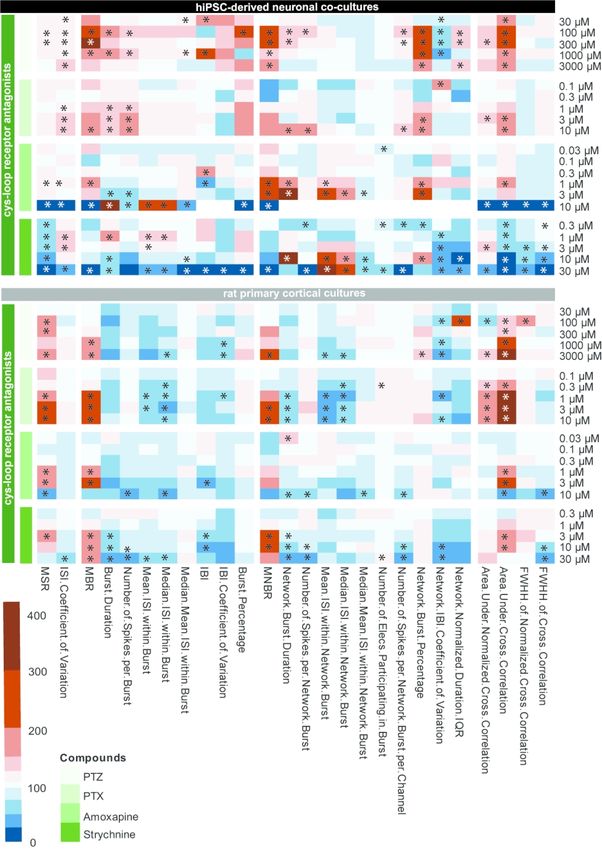
Abstract
Seizures are life-threatening adverse drug reactions which are investigated late in drug
development using rodent models. Consequently, if seizures are detected, a lot of time, money
and animals have been used. Thus, there is a need for in vitro screening models using human
cells to circumvent inter-species translation. We assessed the suitability of co-cultures of human
induced pluripotent stem cell (hiPSC)-derived neurons and astrocytes compared to rodent
Downloaded from https://academic.oup.com/toxsci/advance-article/doi/10.1093/toxsci/kfaa136/5899742 by guest on 07 September 2020
primary cortical cultures for in vitro seizure liability assessment using micro-electrode arrays
(MEA).
HiPSC-derived and rodent primary cortical neuronal co-cultures were exposed to nine
known (non-)seizurogenic compounds (pentylenetetrazole, amoxapine, enoxacin, amoxicillin,
linopirdine, pilocarpine, chlorpromazine, phenytoin and acetaminophen) to assess effects on
neuronal network activity using MEA recordings. All compounds affect activity in hiPSC-
derived co-cultures. In rodent primary cultures all compounds, except amoxicillin changed
activity. Changes in activity patterns for both cell models differ for different classes of
compounds. Both models had a comparable sensitivity for exposure to amoxapine (LOEC 0.03
M), linopirdine (LOEC 1 M) and pilocarpine (LOEC 0.3 M). However, hiPSC-derived
cultures were about 3 times more sensitive for exposure to pentylenetetrazole (LOEC 30 M)
than rodent primary cortical cultures (LOEC 100 M). Sensitivity of hiPSC-derived cultures
for chlorpromazine, phenytoin and enoxacin was 10-30 times higher (LOECs 0.1, 0.3 and 0.1
M respectively) than in rodent cultures (LOECs 10, 3 and 3 M respectively).
Our data indicate that hiPSC-derived neuronal co-cultures may outperform rodent
primary cortical cultures with respect to detecting seizures, thereby paving the way towards
animal-free seizure assessment.
Keywords: alternatives to animal testing, human induced pluripotent stem cell (hiPSC)-derived
neuronal models, micro-electrode array (MEA), rodent primary cortical cultures, seizure
liability assessment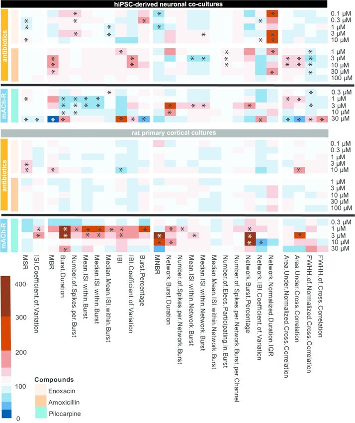
1. Introduction
       Even though many test strategies are in place to detect adverse drug reactions, drug
attrition still occurs frequently. Often this is due to central nervous system (CNS) related safety
issues (Arrowsmith and Miller 2013; Onakpoya et al. 2016). A quarter of the failures occurring
during clinical development, is due to CNS-related issues (Cook et al. 2014). These issues were
not detected before clinical trials started, thus emphasizing the need for continued improvement

                                                                                                      Downloaded from https://academic.oup.com/toxsci/advance-article/doi/10.1093/toxsci/kfaa136/5899742 by guest on 07 September 2020
of pre-clinical test strategies. Seizures and convulsions, defined as periods of abnormal hyper-
excitability of neurons that fire in a highly synchronized way (Easter et al. 2009; Jiruska et al.
2013), are a commonly encountered life threatening problem during pre-clinical drug
development (Authier et al. 2016). Especially drugs that target the CNS give a higher risk of
seizures due to higher brain penetration and higher affinity for CNS targets (Gao et al. 2016).
However, the potential of drugs to induce seizures is not investigated until late in the drug
development process during in vivo studies. If seizures are detected, the need to develop
alternative compounds arises, causing delays in the drug developmental process. Also, animal
experiments could have been avoided if seizure liability was detected earlier in in vitro studies.
There is thus a clear need for better CNS safety testing prior to clinical studies. Hence, it would
be beneficial if a reliable high throughput in vitro system becomes available for seizure liability
assessment. This system would preferably use human cells to avoid inter-species translation
and ethically debated animal experiments.
       Currently, the acute rat hippocampal slice assay is one of the most used methods for in
vitro seizure liability assessment (Authier et al. 2016), although brain slices have also been
isolated from dogs, minipigs and non-human primates (Accardi et al. 2018). Brain organization
is largely maintained in this assay (Grainger et al. 2018), with active and intact neuronal
networks. However, this ex vivo system is not suitable for high-throughput screens as slices
have a short life-span (Buskila et al. 2015). Recently developed organotypic slice cultures
already have a longer life-span and it has been shown that these slices can develop epileptiform
activity making them a potential tool for drug screening (Magalhães et al. 2018). However, both
acute and organotypic slices recordings are labor intensive and require specific expertise and
equipment (Grainger et al. 2018). Also, both brain slice models are still of non-human origin.
       Some of these drawbacks can be resolved by multiwell micro-electrode array
(mwMEA) recordings. This non-invasive tool makes it possible to record electrical activity of
neuronal networks without affecting the integrity of the cells (for review see: Johnstone et al.,
2010). All cell types that exhibit spontaneous activity can be cultured on mwMEAs, but rodent
primary cortical cultures are the current gold standard (Authier et al. 2016). Primary cortical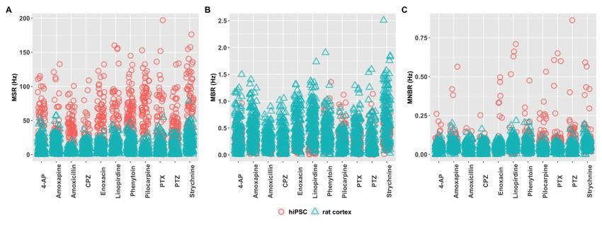
neurons grown on mwMEAs have many characteristics of in vivo neurons, including
(spontaneous network) burst activity (Cotterill et al. 2016). The cortical cultures can be
modulated with neurotransmitters and pharmacological and toxicological agents (Hogberg et
al. 2011; McConnell et al. 2012; Nicolas et al. 2014; Hondebrink et al. 2016). It has been shown
that rodent primary neuronal networks can be successfully used for seizure liability assessment
(Bradley et al. 2018; Kreir et al. 2018; Fan et al. 2019; Tukker et al. 2020).

                                                                                                     Downloaded from https://academic.oup.com/toxsci/advance-article/doi/10.1093/toxsci/kfaa136/5899742 by guest on 07 September 2020
       In vitro seizures can present themselves in different ways. According to Bradley et al.
(2018), two patterns of in vitro seizurogenicity can be distinguished. Distinctive for pattern one
is an overall increase in activity, but more important is the higher synchronicity and
organization of bursts. This increased network organization is reflected in the higher number of
spikes that occur within a burst (burst percentage) and the increased inter-spike-interval
coefficient of variation (ISI CoV). In the case of GABAA receptor antagonists also an increase
in burst duration can be observed. Picrotoxin (PTX), PTZ and 4-aminopyridine (4-AP) are
examples of compounds that match this pattern (Bradley et al. 2018). Characteristics of pattern
two are a decrease in activity and a deterioration of organization and synchronicity reflected in
a decreased ISI CoV, shorter bursts and longer inter-burst-intervals (IBIs) (Bradley et al. 2018).
However, there is often only a narrow concentration window in which this pattern is visible,
because of the rapid decrease in firing rate, ultimately resulting in a silent neuronal network.
Examples of compounds that match this pattern include strychnine, linopirdine and amoxapine
(Bradley et al. 2018).
       The recent introduction of human induced pluripotent stem cell (hiPSC)-derived
neurons offers an interesting opportunity to circumvent the use of animals for neurotoxicity
testing. These cells lack the ethical concerns of embryonic stem cells and in vivo experiments.
Human iPSC-derived neurons can be cultured on mwMEAs and develop spontaneous network
activity and (network) bursting comparable to mature neurons (Odawara et al. 2016;
Paavilainen et al. 2018; Sasaki et al. 2019 Apr 4). Also, these hiPSC-derived neuronal networks
can be modulated with neurotransmitters and known neurotoxicants (Tukker et al. 2016;
Odawara et al. 2018). Over the last years, an increasing number of hiPSC-derived neurons
became commercially available. The use of commercial iPSCs significantly reduces the time
these cells need to be in culture until they develop spontaneous activity from months (Kuijlaars
et al. 2016; Odawara et al. 2016) to just a few weeks (Meneghello et al. 2015; Tukker et al.
2020). This short culture duration enhances the time- and cost-efficiency of in vitro drug
screening methods. The commercial availability also reduces the potential variability between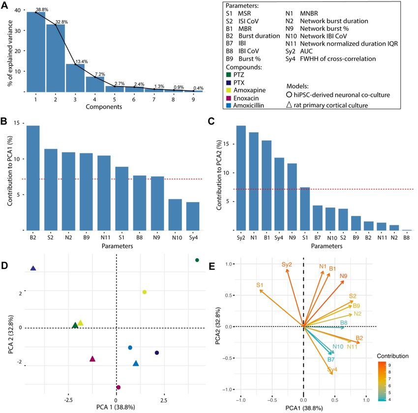
self-cultured batches as they can be purchased in large, quality controlled quantities (Anson et
al. 2011; Little et al. 2019).
        For the current research, we hypothesized that hiPSC-derived neuronal co-cultures are
equally suitable for in vitro seizure liability assessment as rodent primary cortical cultures.
Therefore, in this study, we investigated the potential of hiPSC-derived neuronal co-cultures
compared to rodent primary cortical cultures for seizure liability assessment using MEA

                                                                                                   Downloaded from https://academic.oup.com/toxsci/advance-article/doi/10.1093/toxsci/kfaa136/5899742 by guest on 07 September 2020
recordings.

2. Materials and Methods
2.1 Chemicals
        Neurobasal®-A Medium, L-glutamine, fetal bovine serum (FBS), B27 supplement
(without vitamin A), N2 supplement and penicillin – streptomycin (5000 U/mL – 5000 µg/mL
for rat primary cortical culture media and 10.000 U/mL – 10.000 µg/mL for hiPSC medium)
were obtained from Life Technologies (Bleiswijk, The Netherlands). iCell® Neural Supplement
B and Nervous System Supplement were obtained from Cellular Dynamics International
(Madison, WI, USA). BrainPhys™ neuronal medium was obtained from StemCell
Technologies (Cologne, Germany). Ethanol (EtOH) was obtained from VWR Chemicals
(Amsterdam, The Netherlands). Laminin (L2020), 50% polyethyleneimine (PEI) solution,
sodium borate, boric acid and all other chemicals (unless described otherwise) were obtained
from Sigma-Aldrich (Zwijndrecht, The Netherlands).
        A set of nine compounds was chosen as reference set (Table 1). Test concentrations
were chosen based on the NC3R crack-it Neuratect project and the closely related work from
the HESI-MEA sub-team. All reference compounds were obtained from Sigma-Aldrich
(Zwijndrecht, The Netherlands). An additional set of three compounds (PTX, strychnine and 4-
AP; Table 1) of which data were published previously (Tukker et al. 2020) has been added to
the heatmaps for more extensive mode of action comparisons. Stock solutions of compounds
dissolved in dimethyl sulfoxide (DMSO) were stored in the freezer till further use. Solutions of
compounds dissolved in medium or EtOH were freshly prepared on the day of the experiment.

2.2 Cell cultures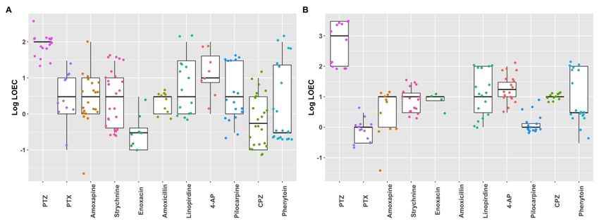
Both rat primary cortical cells and hiPSC-derived neuronal co-cultures were cultured at
37°C in a humidified 5% CO2 incubator. All cell culture surfaces were pre-coated with 0.1%
PEI solution diluted in borate buffer (24 mM sodium borate/50 mM boric acid in Milli-Q
adjusted to pH 8.4).
       For each plating round, primary rat cortical cells were isolated from post-natal day
(PND) 1 Wistar rat pups (Envigo, Horst, The Netherlands) in accordance with Dutch law and

                                                                                                   Downloaded from https://academic.oup.com/toxsci/advance-article/doi/10.1093/toxsci/kfaa136/5899742 by guest on 07 September 2020
approved by the Ethical Committee for Animal Experimentation of Utrecht University. Cells
obtained from the Wistar rat cortices were pooled, meaning that the resulting cultures consist
of cells derived from male and female pups. Animals were treated humanely and all efforts
were made to alleviate suffering. Cortical cultures were prepared as described previously
(Dingemans et al. 2016; Tukker et al. 2016). Briefly, PND1 pups were decapitated and cortices
were rapidly dissected on ice and kept in dissection medium (Neurobasal®-A supplemented
with 25g/L sucrose, 450 µM L-glutamine, 30 µM glutamate, 1% penicillin/streptomycin and
10% FBS, pH 7.4) during the entire procedure. Cortices were dissociated to a single-cell
suspension by mincing, trituration and filtering through a 100 µm mesh (EASYstrainer,
Greiner). The cell suspension was diluted to 2 x 10⁶ cells/mL. Then, a 50 µL droplet was placed
on the electrode field of a pre-coated 48-well MEA plate (Axion BioSystems Inc., Atlanta, GA,
USA). Cells were left for ~2 hr to adhere before adding 450 µL dissection medium. To prevent
glial overgrowth, 90% of the dissection medium was replaced by glutamate medium
(Neurobasal®-A supplemented with 25 g/L sucrose, 450 µM L-glutamine, 30 µM glutamate,
1% penicillin/streptomycin and 2% B-27 supplement, pH 7.4) at day in vitro (DIV) 1 resulting
in a culture with ± 46% astrocytes (Tukker et al. 2016). At DIV4, 90% of the glutamate medium
was replaced with FBS medium (Neurobasal®-A supplemented with 25 g/L sucrose, 450 µM
L-glutamine,   1% penicillin/streptomycin and 10% FBS, pH 7.4).
       For each plating round, iCell® Glutaneurons (Lot# 103288, male donor, containing ±
70% glutamatergic neurons and 30% GABAergic neurons; Cellular Dynamics International,
Madison, WI, USA) and iCell® Astrocytes (Lot# 103956 or 11493, female donors; Cellular
Dynamics International, Madison, WI, USA) were thawed and cultured according to
manufacturer’s protocol as described previously (Tukker et al. 2020). Briefly, cells were thawed
separately in BrainPhys™ medium supplemented with 2% iCell® Neural Supplement B, 1%
Nervous System Supplement, 1% N2, 1% penicillin – streptomycin (10.000 U/mL – 10.000
µg/mL) and 0.1% laminin. The cell pellet was dissolved in dotting medium (i.e. supplemented
BrainPhys™ medium with 10% laminin). Before plating, iCell® Glutaneurons and iCell®
Astrocytes were premixed into a co-culture containing 15 x 103 iCell® Glutaneurons/µL and
6.7 x 10³ iCell® Astrocytes/µL so that each well contains 120 x 103 iCell® Glutaneurons and 20
x 103 iCell® Astrocytes. Cells were plated in 11 µL droplets (140 x 103 cells/droplet with 85%
iCell® Glutaneurons and 15% iCell® Astrocytes) over the electrode field of 48-well MEA
plates. Following plating, cells were allowed to adhere for ~1hr after which 300 µL of room
temperature (RT) supplemented BrainPhys™ medium was added. Hereafter, 50% medium
changes with RT supplemented BrainPhys™ medium took place at DIV1, 2, 4, 6, 8, 10, 12 and
Downloaded from https://academic.oup.com/toxsci/advance-article/doi/10.1093/toxsci/kfaa136/5899742 by guest on 07 September 2020
14.
2.3 MEA recordings
Each well of a 48-well MEA plate contains 16 nanotextured gold micro-electrodes (~40-
50 µm diameter; 350 µm spacing) with 4 integrated ground electrodes. This yields a total of
768 channels that can be recorded simultaneously (for review see: Johnstone et al., 2010).
Spontaneous electrical activity was recorded as described previously (Nicolas et al. 2014;
Tukker et al. 2019). Briefly, signals were recorded, in the absence of extra CO2, at the day of
experiments (DIV9-11 for rat primary cortical cultures or DIV14 for hiPSC-derived co-
cultures) using a Maestro 768-channel amplifier with integrated heating system and temperature
controller and a data acquisition interface (Axion BioSystems Inc., Atlanta, GA, USA). Data
acquisition was managed with Axion’s Integrated Studio (AxIS 2.4.2.13) and recorded as
.RAW files. All channels were sampled simultaneously with a gain of 1200x and a sampling
frequency of 12.5kHz/channel, using a 200-5000 Hz band-pass filter. Prior to the recording,
MEA plates were allowed to equilibrate for 5-10 min in the Maestro.
In order to determine the effects of reference compounds on spontaneous neuronal
activity (spiking and (network) bursting behavior) on the two types of cell cultures, a 30 min
baseline recording was made. Following this recording, wells were exposed (10 x dilution for
primary rat cortical cultures, 30 x dilution for hiPSC-derived co-cultures) to the reference
compounds or the appropriate solvent control. Test concentrations were determined by the
NC3R’s CRACK-it team, who based their choice on the list of the HESI NeuTox MEA Sub-
team. Then activity was recorded for another 30 min. Stock solutions of compounds dissolved
in DMSO or EtOH were diluted in culture medium to obtain desired concentrations. In all
experiments, the solvent concentration never exceeded 0.1% v/v. In order to prevent receptor
(de)sensitization, each well was exposed to a single concentration. For each experimental
condition, MEA plates from at least two different plating rounds were used.2.4 Data analysis and statistics
To determine (modulation of) spontaneous activity, .RAW data files were re-recorded
to obtain Alpha Map files for further data analysis. In this re-recording, spikes were detected
using the AxIS spike detector (Adaptive threshold crossing, Ada BandFIt v2) with a variable
threshold spike detector set at 7x (primary cortical culture) or 5.5x (hiPSC culture) standard
Downloaded from https://academic.oup.com/toxsci/advance-article/doi/10.1093/toxsci/kfaa136/5899742 by guest on 07 September 2020
deviation (SD) of internal noise level (rms) on each electrode. This difference in spike detection
threshold does not significantly affect effect size (see Fig. S3). Post/pre-spike duration was set
to 3.6/2.4 ms. For further analysis, spike files were loaded in NeuralMetric Tool (version 2.2.4,
Axion BioSystems). Only active electrodes (MSR ≥ 6 spikes/min) in active wells (≥ 1 active
electrode) were included in the data analysis. Mean number of active electrodes per well for
hiPSC-derived neuronal co-cultures was 12.6 and for rat primary rodent cultures 9.8 (see Fig.
S4). The (network) bursting behavior was analyzed as it is crucial for (in vivo) transmission of
information (Izhikevich et al. 2003) using the Poisson Surprise method (Legéndy and Salcman
1985) with a minimal surprise of 10 and a minimum bursting frequency of 0.3 bursts/min.
Network bursts were extracted with the adaptive threshold algorithm.
Effects of reference compounds on the spontaneous activity pattern were determined in
exposure experiments where baseline activity prior to exposure was compared with activity
following exposure. To prevent inclusion of exposure artefacts, windows for data analysis were
corrected for the time it took to expose the whole plate. In other words, if exposure took 2 min,
the first 2 min were not included in data analysis or when a window of 20-30 min post exposure
was analyzed, analysis started at 20 + 2 min. Optimum effect windows were determined
separately for all reference compounds. For all reference compounds the window of 20-30 min
post exposure was used for analysis of effects.
A custom-made MS Excel macro was used to calculate treatment ratios (TR) per well
for the different metric parameters (Table 2) by: (parameterexposure/parameterbaseline) x 100%.
Hereafter, TRs were normalized to appropriate vehicle control. Outliers were identified well-
based and defined as not within average ± 2 x SD were removed (3.4% for hiPSC-derived
neuronal data and 4.8% for rat primary cortical data). To test for biological relevant
concentration-dependent effects, the variability around control values was defined. SD values
as a percentage of the mean value per parameter of the control were calculated. This SD was
added and subtracted from 100% to create effect threshold levels on that parameter. Response
values above or below this threshold were marked as a ‘hit’. This was done for every single
parameter. Heatmaps were created in R (version 3.6.0) using the pheatmap package (by Kolde,2019, version 1.0.12). Data are presented as mean ± SEM from the number of wells (n)
indicated, derived from at least 2 independent plating rounds (i.e. independent rodent culture
isolations or thawing of vials for hiPSC-derived co-cultures). As not all spiking wells show
burst, and not all bursting wells show network burst, the number of wells (n) used for analysis
of the different parameters may differ, in particular for treatments that lower or eliminate
(network) burst activity (see Supplemental tables S1-S11 for exact number of wells used for
Downloaded from https://academic.oup.com/toxsci/advance-article/doi/10.1093/toxsci/kfaa136/5899742 by guest on 07 September 2020
calculations). Lowest observed effect concentrations (LOECs) are defined as lowest ‘hit’
concentrations at one of the activity parameters.
2.5 Principal component analysis
Principal component analysis (PCA) can be used to visualize the level of variation
between different culture models following exposure to compounds with the same mode of
action. Therefore, a PCA was performed to segregate MEA results of hiPSC-derived co-cultures
and rat primary cortical cultures following exposure to GABAA antagonists (PTZ, PTX,
amoxapine, enoxacin and amoxicillin) and visualize a (potential) inter-species difference. The
class of GABAA antagonists was chosen, because they form the majority of compounds in this
study. The PCA was conducted in R using the packages FactoMineR (Lê et al., 2008) and
FactoExtra (by Mundt, 2017; version 1.0.5). To prevent empty cells in the matrix, average
values per parameter of the middle test concentration were calculated. Next, highly correlated
parameters were removed from the data set. Correlation was determined based on visual
inspection of the correlation matrix and the heatmap, resulting in removal of 10 parameters.
The resulting 14 parameters included in the analysis are depicted in italic in Table 2, including
MSR, MBR, MNBR, parameters related to (network) burst duration and percentage and
synchronicity parameters. A scree plot was created to visualize the percentage of variation
explained by each component and eigen-values were calculated. The loading of each parameter
to a component was calculated. Parameters contributing more than the expected contribution of
7% (expected value = (1/ number of variables x 100%)) were considered important for a
component. The 14-dimensional vector was projected on the plane created by the first 2
principal components that explained most of the variation to visualize the level of (potential)
inter-species variation between the two models.3. Results
3.1 Baseline activity of the two models
In a previous study we have shown that hiPSC-derived neuronal co-cultures and rat
primary cortical cultures both develop spontaneous activity with (network) bursting behavior,
Downloaded from https://academic.oup.com/toxsci/advance-article/doi/10.1093/toxsci/kfaa136/5899742 by guest on 07 September 2020
although with different activity patterns (Tukker et al. 2020). The average mean spike rate
(MSR) is 47.5 ± 1.13 Hz for hiPSC-derived neuronal co-cultures, whereas the average is much
lower for rat primary cortical cultures with a MSR of 10.5 ± 0.22 Hz (Fig. 1A). The mean burst
rate (MBR) is more comparable between the two models (Fig. 1B). The average rate is 0.25 ±
0.007 Hz for the hiPSC-derived neuronal culture and 0.40 ± 0.073 for rat primary cortical
cultures. The mean network burst rate (MNBR) is 0.082± 0.003 Hz and 0.050 ± 0.001 Hz for
hiPSC-derived neuronal co-cultures and rat primary cortical cultures, respectively (Fig. 1C).
3.2 Effects on spontaneous network activity
To assess and compare the suitability of hiPSC-derived neuronal co-cultures and rat
primary cortical cultures for seizure liability assessment, both models were exposed to
compounds with different modes of action (Table 1). Observed neurotoxic effects were not
caused by cytotoxicity (see supplemental data Fig. S1).
As a negative control, cells were exposed to acetaminophen. Exposure to this compound
did not result in changes in neuronal activity in rat primary cortical cultures (Zwartsen et al.
2019) or hiPSC-derived neuronal co-cultures (supplemental data Fig. S2).
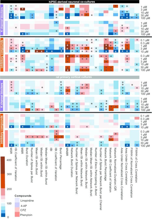
3.2.1 Cys-loop receptor antagonists
In the hiPSC-derived neuronal co-culture, PTZ increases MSR, MBR and MNBR
particularly at intermediate concentrations (100-300 μM; Fig. 2A). PTZ at intermediate
concentration also increases the MSR in rat primary cortical cultures. At high concentrations,
PTZ also increases MBR and MNBR in rat primary cortical cultures (Fig. 2A).
Amoxapine increases the MSR in both cultures (1-3 μM), but at the highest
concentration tested (10 μM), a strong decrease in activity occurs (Fig. 2B; left). The same trend
occurs in the MBR (Fig. 2B; middle) and MNBR (Fig. 2C; right). Overall, exposure toamoxapine increases activity in both models, but activity decreases following exposure to 10
μM.
3.2.2 Antibiotics
MSR of hiPSC-derived neuronal co-cultures is only slightly affected following
Downloaded from https://academic.oup.com/toxsci/advance-article/doi/10.1093/toxsci/kfaa136/5899742 by guest on 07 September 2020
exposure to enoxacin (Fig. 3A; left), although biologically relevant decreases occur following
exposure to 0.3, 1 and 10 μM. Contrary, an increase can be observed when rat primary cortical
cultures are exposed to enoxacin (3-10 μM). Enoxacin has little effect on MBR of hiPSC-
derived co-cultures, but increases MBR at the highest test concentration (10 μM) in rat primary
cortical cultures (Fig. 3A; middle). At low test concentrations (0.1, 1 μM), enoxacin decreases
MNBR in hiPSC-derived co-cultures, but enoxacin has little effect on MNBR in rat primary
cortical cultures (Fig. 3A; right).
Amoxicillin has no effect on MSR or MNBR in either of the models (Fig. 3B; left/right
respectively), but at intermediate concentrations (3-30 μM) increases MBR in hiPSC-derived
co-cultures (Fig. 3B; middle), whereas there is no change in the rat primary cortical culture.
3.2.3 Muscarinic acetylcholine receptor agonist
Pilocarpine increases MSR in hiPSC-derived neuronal co-cultures following exposure
to 1 μM, but decreases MSR and MBR following exposure to 30 μM (Fig. 4A-B). In rat primary
cortical cultures, pilocarpine has little effect on MSR and MBR. Pilocarpine has little effect on
MNBR of hiPSC-derived co-cultures. In rat primary cortical cultures, pilocarpine strongly
increases network bursting following exposure to 3 and 10 μM, but not at 30 μM (Fig. 4C).
3.2.4 Potassium channel blocker
Linopirdine concentration-dependently decreases MSR in hiPSC-derived co-cultures,
in contrast to rat primary cortical cultures where a biologically relevant increase is observed
(Fig. 5A). MBR increases significantly following exposure to 3 μM linopirdine in hiPSC-
derived co-cultures, followed by a decrease at higher test concentrations (Fig. 5B). This trend
is not observed in rat primary cortical cultures where MBR is not affected by linopirdine. At
high concentrations (30-100 μM), linopirdine decreases MNBR of hiPSC-derived co-cultures(Fig. 5C). In rat primary cortical cultures, linopirdine increases MNBR but only at high
concentrations (10-100 μM).
3.2.5. Inhibitory compounds
CPZ (0.1-3 μM) increases MSR in hiPSC-derived co-cultures followed by a decrease at
Downloaded from https://academic.oup.com/toxsci/advance-article/doi/10.1093/toxsci/kfaa136/5899742 by guest on 07 September 2020
the highest test concentration (10 μM; Fig. 6A; left). CPZ has little effect on MSR in rat primary
cortical cultures, except for a decrease at the highest test concentration. A comparable trend is
observed for MBR: in hiPSC-derived co-cultures CPZ increases MBR, whereas MBR decreases
at high concentrations. CPZ has little effect on MBR in rat primary cortical cultures, except for
a decrease at the highest test concentration (Fig. 6A; middle). At low concentrations, MNBR
increases in hiPSC-derived co-cultures following exposure to CPZ, which is followed by a
decrease at the highest test concentration. CPZ has little effect on MNBR in rat primary cortical
cultures, except for a decrease at the highest test concentration (Fig. 6A; right).
Phenytoin decreases MSR in both models at the highest test concentration in a
biologically relevant way (Fig. 6B; left), although MSR is increased in hiPSC-derived co-
cultures at intermediate concentrations (1-3 µM). At low test concentrations, MBR increases in
hiPSC-derived co-cultures, whereas MBR is not affected in rat primary cortical cultures (Fig.
6B; middle). MNBR increases following phenytoin exposure in hiPSC-derived neuronal co-
cultures (Fig. 6B; right). Phenytoin has little effect on MNBR in rat primary cortical cultures,
except for a decrease at 100 µM.
3.3 Finger prints of the different compound classes
Heatmaps of the concentration-response curve of all tested compounds on hiPSC-
derived neuronal co-cultures and rodent primary cortical cultures were created to further
illustrate the effect on different parameters. These heatmaps also visualize that compounds with
the same mode of action have a comparable chemical fingerprint (Fig 7-9). Because these
heatmaps show more parameters than just the classical activity parameters as depicted in Fig.
2-6, they create a more complete overview of how a network is affected by exposure. For
example, when looking at the heatmaps, it is immediately clear that parameters such as
(network) burst percentage, mean and median ISI within (network) burst and area under cross
correlation are strongly affected by exposure to these (non-)seizurogenic compounds. Theseheatmaps thus provide a valuable tool for visualizing changes in neuronal networks following
modulation, including potential seizurogenicity patterns as described by Bradley et al., 2018.
When the hiPSC-derived neuronal co-cultures are exposed to PTZ or PTX, both
GABAA-R antagonists, a comparable pattern occurs and strongest effects can be seen on burst
related parameters (Fig 7; left). However, effects are more profound following exposure to PTZ
compared to PTX. Exposure of hiPSC-derived co-cultures to amoxapine, also a GABAA-R
Downloaded from https://academic.oup.com/toxsci/advance-article/doi/10.1093/toxsci/kfaa136/5899742 by guest on 07 September 2020
antagonist, increases MSR, MBR, MNBR, network burst duration and the area under cross
correlation, just like PTZ and PTX. However, the strong increasing effect on burst related
parameters is not so clear following exposure to amoxapine. Following exposure to PTZ and
amoxapine, the intermediate concentrations have the strongest effect on burst and network burst
rate. The fourth compound in the cys-loop receptor antagonist category, strychnine, shows a
different pattern with a clear decrease in activity in hiPSC-derived co-cultures. This is also the
only compound with a different target, the glycine receptor. Exposure of rat primary cortical
cultures to the same set of cys-loop receptor antagonists yields a pattern different from the
pattern of hiPSC-derived neuronal co-cultures (Fig. 7; right). Overall, activity decreases
following exposure of rat primary cortical cultures. The pattern for all four compounds is
comparable, with increases in MSR, MBR, MNBR and the area under cross correlation.
The next group consists of two antibiotics, both GABAA-receptor antagonists (Fig. 8;
orange: enoxacin and amoxicillin). When looking at the pattern resulting from exposure of
hiPSC-derived co-cultures it can be seen that the magnitude of change of effects is less than
with the other GABAA-receptor antagonists PTZ and PTX. Exposure of rat primary cortical
cultures results in even milder effects (Fig. 8; right). In the case of exposure to amoxicillin, no
effects can be observed in rat primary cortical cultures.
Exposure of both models to the mACh receptor agonist pilocarpine (Fig. 8; blue,
bottom) results in a pattern that indicates this different mode of action as it has few similarities
with other compounds. For both hiPSC-derived neuronal co-cultures and rat primary cortical
co-cultures it can be seen that exposure to the intermediate concentrations results in the biggest
effect. Other than that, there are few similarities between the pattern in hiPSC-derived neuronal
co-cultures and rat primary cortical co-cultures.
Two potassium channel blockers were tested (Fig. 9; purple, linopirdine and 4-AP). For
neither cell model it is clear from the heatmap that the compounds have the same mode of
action. In both cell models, linopirdine affects network burst parameters strongest, whereas 4-
AP has more effect on burst related parameters.The last group consists of two inhibitory compounds (Fig. 9; pink, CPZ and phenytoin).
CPZ has the D2-receptor as a target and strongly increases MBR, MNBR and network burst
percentage and decreases the IBI in hiPSC-derived neuronal networks. This same increase can
be observed when hiPSC-cells are exposed to phenytoin, a sodium channel blocker. Both
compounds have relatively mild effects on spike parameters. When rat primary cortical cultures
are exposed to CPZ or phenytoin, spike parameters decrease. Also, bursting and network
Downloaded from https://academic.oup.com/toxsci/advance-article/doi/10.1093/toxsci/kfaa136/5899742 by guest on 07 September 2020
bursting decrease with increasing concentration. Overall, effects on rat primary cortical cultures
are milder than on hiPSC-derived co-cultures.
3.4 Principal component analysis to distinguish between human and rodent model
In order to visualize the potential inter-species differences following exposure to
GABAA receptor antagonists (PTZ, PTX, amoxapine, enoxacin, amoxicillin) between the
hiPSC-derived neuronal co-culture and rat primary cortical culture, a PCA was performed. PCA
efficiently clusters MEA data based on the activity profile of the networks following exposure.
Parameters for the PCA were selected based on visual inspection of a correlation matrix
(Supplemental Table 12) and the heatmaps (Figs. 7-9). A PCA was performed on 10 items (5
compounds per model) and 14 parameters. Four principal components have an eigenvalue
greater than 1. Together, these components explain 92.2% of the variability as becomes clear
from the scree plot (Fig. 10A). In total, 8 parameters score higher than the expected 7%
contribution to component 1 (Fig. 10B) and 5 parameters for component 2 (Fig. 10C). Notably,
the most important parameters contributing to PCA1 relate to duration of (network) bursts and
the percentage of spikes that occur in (network) bursts. This is in contrast to PCA2 where
classical activity parameters (mean burst rate and mean network burst rate) as well as
parameters related to synchronicity are more important. What becomes apparent from the plot
(Fig. 10D) is that there is a clear segregation between data points from hiPSC-derived neuronal
co-cultures and rat primary cortical neurons. With the exception of amoxicillin, all rat primary
cortical culture data is presented in left quadrants of the graph, whereas the hiPSC-derived
neuronal co-culture data is presented in the right quadrants. Contribution of the different
parameters to the two components is depicted in Fig. 10E. Interestingly, the mean spike rate
has a strong negative contribution to PCA1 (Fig. 10E).
3.5 Comparison of seizure liability between the two modelsIn order to compare the suitability and sensitivity for seizure liability assessment
between the two models, for all compounds LOECs (defined as lowest hit concentration) were
calculated for all 26 metric parameters (Fig. 11). LOECs were compared between the two
models. The model with the lowest LOEC is defined as the most sensitive model. For most
tested compounds, the hiPSC-derived neuronal culture is more sensitive (Table 3). Sensitivity
is equal between the two cultures for exposure to amoxapine, linopirdine and pilocarpine. No
Downloaded from https://academic.oup.com/toxsci/advance-article/doi/10.1093/toxsci/kfaa136/5899742 by guest on 07 September 2020
LOECs could be determined following exposure of rat primary cortical cultures to amoxicillin.
4. Discussion
The aim of this study was to assess the suitability of hiPSC-derived neuronal co-cultures
for seizure liability assessment using MEA-recordings. We compared data obtained with human
neuronal co-cultures with data obtained in parallel from rat primary cortical cultures.
Our MEA recordings (Fig. 2-6) show that this type of experiment is suitable for the
detection of changes in network activity following modulation by different drugs. Parameters
that are important for in vitro seizure liability assessment include parameters that describe the
activity and the organization of the activity (Bradley et al. 2018). Examples of these parameters
are spike, and (network) burst rate, but also the percentage of spikes that is incorporated in
(network) bursts as well as parameters related to synchronicity. Importance of these parameters
becomes clear from the heatmaps (Fig. 7-9) and PCA (Fig. 10). If in vitro seizures are defined
by changes in these aforementioned parameters, then our data confirm that both models despite
the differences, are able to detect seizure liability in vitro. No changes were detected following
exposure to amoxicillin in the rodent culture. However, sensitivity for the different test
compounds differs between the human and rodent model as becomes clear when comparing
LOECs (Fig. 11; Table 3). Our heatmaps indicate that it is possible to create fingerprints and
group compounds with comparable mechanisms of action (Fig. 7-8). From these heatmaps it
becomes clear that burst and network burst parameters are more sensitive for excitatory changes
in hiPSC-derived co-cultures than in rat primary cortical cultures. Spike parameters are more
affected in rodent primary cultures than in hiPSC-derived co-cultures. Our PCA analysis shows
that it is possible to differentiate within a subset of compounds with a similar mode of action
between two cell models (Fig. 10).
Looking at just PTZ, a compound used to induce seizures in laboratory rodents
(Klioueva et al. 2001; Singh et al. 2019) and thus a known seizurogenic compound, activityincreased in both the rodent and hiPSC-derived neuronal network model at concentrations
comparable to concentrations reported in literature (Fig 2A; Bradley et al., 2018; Kreir et al.,
2018).. The observed trend of increased bursting after PTZ exposure on hiPSC-derived co-
cultures, followed by a decrease at higher concentrations, is comparable with previous studies
(Odawara et al. 2018). The cyclic anti-depressant amoxapine is known to cause seizures as an
adverse drug reaction (Litovitz and Troutman 1983; Kumlien and Lundberg 2010). Not
Downloaded from https://academic.oup.com/toxsci/advance-article/doi/10.1093/toxsci/kfaa136/5899742 by guest on 07 September 2020
surprisingly, amoxapine increased activity in both models (Fig. 2B). Increase in network
activity was only observed in the hiPSC-derived neuronal co-culture. However, in both models,
activity decreased to a minimum at the highest concentration tested. A previous study reported
no significant change in spike rate following exposure of rat primary cortical cultures to
amoxapine (Kreir et al. 2018), nor did this research find seizures in vivo when rats were dosed
intravenously, indicating a potential inter-species difference in amoxapine sensitivity.
The fluoroquinolone antibiotic enoxacin is known to cause seizures as adverse drug
reaction (Simpson and Brodie 1985). Decreases in activity were found in the hiPSC-derived
model following enoxacin exposure (Fig. 3A), whereas in the rodent cell culture model, spike
and burst rate did increase. However, in rat hippocampal slices, seizures were found at 30 M
(Gao et al. 2017). This is above our highest test concentration. Exposure to the penicillin
antibiotic amoxicillin had a more profound effect on hiPSC-derived co-cultures as compared to
rat primary cortical cultures (Fig. 3B), with an increase in burst rate. Lack of effect on rat
primary cortical cultures following amoxicillin exposure is in line with previous reports
(McConnell et al. 2012). However, amoxicillin has been reported to cause seizures as an
adverse drug reaction in humans (Raposo et al. 2016). This thus matches our hiPSC-derived
neuronal data.
When looking at the GABAA receptor antagonists (PTZ, PTX, amoxapine, enoxacin
and amoxicillin) as a group in the heatmap (Fig. 7-8), it can be observed that all increased the
overall activity in both models, with the exception of enoxacin on the hiPSC-derived neuronal
co-culture. However, burst duration did not increase following exposure of rat primary cortical
cultures. This is in contrast to hiPSC-derived co-cultures, where an increase in burst duration
was frequently observed. The hiPSC-derived co-cultures thus follow pattern one. Bradley et al.
(2018) named amoxapine as a compound following pattern two. However, that was based on
an exposure of 5 M. Our data indicate that activity and network synchronicity decreased
between 3 and 10 M on both models following amoxapine exposure. At this point,
seizurogenic pattern two can be observed (Fig. 7), indicating that at low test concentrationsamoxapine follows pattern one and then at higher concentrations pattern two. Both antibiotics,
enoxacin and amoxicillin, are GABAA receptor antagonists and follow pattern one in both
models. However, in both models and with both compounds, the observed effect is not as strong
as with the other GABAA receptor antagonists. Overall, the increased network organization and
synchronicity are more pronounced following exposure of hiPSC-derived co-cultures as
compared to rat primary cortical cultures. The differences between the human and rodent model
Downloaded from https://academic.oup.com/toxsci/advance-article/doi/10.1093/toxsci/kfaa136/5899742 by guest on 07 September 2020
following exposure to GABAA receptor antagonists were confirmed by PCA (Fig. 10). This
analysis efficiently identified differences between the two models and thus emphasizes inter-
species differences. In this study, PCA has been performed on one set of compounds with a
similar mode of action. However, this type of analysis holds great potential for further
identifying differences and similarities between models following exposure to groups of
compounds with other modes of action. Strychnine has been shown to follow pattern two. In
our data, exposure of hiPSC-derived co-cultures to strychnine clearly results in pattern two (Fig.
7). In the case of rodent primary cortical cultures activity increases. However, network
organization decreases in line with pattern two, as reflected in the decreased ISI CoV, burst
duration and burst percentage.
Pilocarpine is used to model epilepsy in vivo in rodents (Marchi et al. 2007). At low test
concentrations pilocarpine increased activity of hiPSC-derived neuronal co-cultures, but at the
highest test concentration a decrease was observed (Fig. 3A-B). This biphasic effect with a
strong decrease in activity following exposure to high concentrations has been reported for
other compounds such as endosulfan, lindane and fipronil (Wallace et al. 2015; Dingemans et
al. 2016). It could also be the result of receptor desensitization or of a non-specific effect on ion
channels, such as inhibition of voltage-gated calcium channels (Heusinkveld et al. 2010). Rat
primary cortical cultures showed a strong increase in network burst activity, in line with other
studies that reported seizurogenic effects on rodent primary cultures and hippocampal slices
following exposure to comparable concentrations (Nagao et al. 1996; Kreir et al. 2018). The
data presented here do not allow for a clear indication that pilocarpine follows either
seizurogenicity pattern. This problem was also mentioned by Bradley et al. (2018).
In both tested models, linopirdine decreased activity, except network burst activity in
the rodent model, (Fig. 5) at concentrations in line with previous reports (Bradley et al. 2018).
Exposure of rat primary cortical cultures to linopridine (60 M) has been shown to result in
seizurogenicity pattern two (Bradley et al. 2018). Our data showed that in both models exposure
to linopirdine at a comparable concentration resembles pattern two (Fig. 8). The otherpotassium channel blocker shown (Fig. 8), 4-AP, should resemble pattern one (Bradley et al.
2018). However, this is not clear from our data.
CPZ increased spiking activity in hiPSC-derived neuronal co-cultures (Fig. 6A). At
higher test concentrations activity decreased. Bursting and network bursting decreased
following exposure to this compound. These findings are in line with literature (Kreir et al.
2018). In rat hippocampal slices, CPZ has been shown to induce seizures at 1 – 10 M (Easter
Downloaded from https://academic.oup.com/toxsci/advance-article/doi/10.1093/toxsci/kfaa136/5899742 by guest on 07 September 2020
et al. 2007), in line with our hiPSC-derived data. Exposure of hiPSC-derived co-cultures to CPZ
results in pattern one. However, this pattern cannot be observed in rat primary cortical cultures.
Phenytoin is designed as an antiepileptic drug and therefore expected to decrease
network activity. Phenytoin (Fig. 6B) decreases activity at the highest test concentration in
hiPSC-derived co-cultures, at similar concentrations as others found with hiPSC-cultures
(Odawara et al. 2018). Pattern one can be observed when hiPSC-derived co-cultures are
exposed to phenytoin, whereas this is not clear in rat primary cortical cultures (Fig. 8).
There are several factors that could contribute to the observed differences between the
two models. It must be kept in mind that the medium of the rodent cortical culture contains
serum, whereas the medium of the hiPSC-derived co-cultures is serum free. Therefore, the
bioavailability of test compounds might be slightly lower in de rodent culture due to serum-
compound interactions. Also, there is a difference between the two cultures in the percentage
of astrocytes present. The rat primary cortical culture contains about 45% astrocytes (Görtz et
al. 2004; Tukker et al. 2016). The percentage of astrocytes in the hiPSC-derived neuronal co-
culture can easily be increased. However, using higher percentages of astrocytes in the iPSC-
derived cultures hampers spontaneous neuronal activity and (network) bursting, likely due to
profound cell clustering. On the data analysis side, it is important to note that defining ‘hits’ in
this way may make the method less sensitive for decreases in activity. Therefore, a reduction
in activity will less likely be a hit than an increase. However, this should not be a major issue
for seizure liability assessment.
We have shown that hiPSC-derived co-cultures can be used already as an initial screen
for seizurogenic activity in vitro. Our data indicates differences between the human and rodent
model, highlighting the need to move towards human iPSC-derived neuronal co-culture models
for more predictive in vitro seizure liability assessment. This need is emphasized by the fact
that the hiPSC-derived model seems to be more sensitive than the primary rodent cortical
culture model. However, these hiPSC-derived neuronal cells must be further characterized.
Also, this approach must be thoroughly validated using a larger set of test compounds includingcompounds that are known to cause seizures in vivo and negative controls before they can
replace the current gold standard of rodent primary cortical cultures.
Conflict of interest
The authors declare no conflict of interest.
Downloaded from https://academic.oup.com/toxsci/advance-article/doi/10.1093/toxsci/kfaa136/5899742 by guest on 07 September 2020
Funding
This work was funded by a grant from the National Centre for the Replacement, Refinement
and Reduction of Animals in Research (NC3Rs; project number 50308-372160) and by the
Faculty of Veterinary Medicine (Utrecht University, The Netherlands).
Acknowledgements
We gratefully acknowledge members of the Neurotoxicology Research Group for helpful
discussions.References
Accardi M V., Huang H, Authier S. 2018. Seizure liability assessments using the hippocampal
tissue slice: Comparison of non-clinical species. J Pharmacol Toxicol Methods. 93:59–68.
doi:10.1016/j.vascn.2017.11.003.
Alachkar A, Łażewska D, Latacz G, Frank A, Siwek A, Lubelska A, Honkisz-Orzechowska
E, Handzlik J, Stark H, Kieć-Kononowicz K, et al. 2018. Studies on Anticonvulsant Effects of
Downloaded from https://academic.oup.com/toxsci/advance-article/doi/10.1093/toxsci/kfaa136/5899742 by guest on 07 September 2020
Novel Histamine H3R Antagonists in Electrically and Chemically Induced Seizures in Rats.
Int J Mol Sci. 19(11):3386. doi:10.3390/ijms19113386. [accessed 2019 Sep 17].
http://www.mdpi.com/1422-0067/19/11/3386.
Aminoshariae A, Khan A. 2015. Acetaminophen: Old Drug, New Issues. J Endod. 41:588–
593. doi:10.1016/j.joen.2015.01.024. [accessed 2018 Dec 20].
http://dx.doi.org/10.1016/j.joen.2015.01.024.
Anson BD, Kolaja KL, Kamp TJ. 2011. Opportunities for use of human iPS cells in predictive
toxicology. Clin Pharmacol Ther. 89(5):754–758. doi:10.1038/clpt.2011.9.
Arrowsmith J, Miller P. 2013. Phase II and Phase III attrition rates 2011–2012. Nat Rev Drug
Discov. 12(8):569–569. doi:10.1038/nrd4090. [accessed 2019 Sep 4].
http://www.nature.com/articles/nrd4090.
Authier S, Arezzo J, Delatte MS, Kallman M-J, Markgraf C, Paquette D, Pugsley MK,
Ratcliffe S, Redfern WS, Stevens J, et al. 2016. Safety pharmacology investigations on the
nervous system: An industry survey. J Pharmacol Toxicol Methods. 81:37–46.
doi:10.1016/J.VASCN.2016.06.001. [accessed 2019 Jan 16].
https://www.sciencedirect.com/science/article/pii/S1056871916300570?via%3Dihub.
Boyd-Kimball D, Gonczy K, Lewis B, Mason T, Siliko N, Wolfe J. 2018 Jul 16. Classics in
Chemical Neuroscience: Chlorpromazine. ACS Chem Neurosci.:acschemneuro.8b00258.
doi:10.1021/acschemneuro.8b00258. [accessed 2018 Dec 20].
http://pubs.acs.org/doi/10.1021/acschemneuro.8b00258.
Bradley JA, Luithardt HH, Metea MR, Strock CJ. 2018. In Vitro Screening for Seizure
Liability Using Microelectrode Array Technology. Toxicol Sci. 163(1):240–253.
doi:10.1093/toxsci/kfy029. [accessed 2018 Aug 28].
https://academic.oup.com/toxsci/article/163/1/240/4844105.
Burn DJ, Tomson CRV, Seviour J, Dale G. 1989. Strychnine poisoning as an unusual cause of
convulsions. Postgrad Med J. 65(766):563–564. doi:10.1136/pgmj.65.766.563.
Buskila Y, Breen PP, Tapson J, van Schaik A, Barton M, Morley JW. 2015. Extending the
viability of acute brain slices. Sci Rep. 4(1):5309. doi:10.1038/srep05309. [accessed 2019May 6]. http://www.ncbi.nlm.nih.gov/pubmed/24930889.
Cook D, Brown D, Alexander R, March R, Morgan P, Satterthwaite G, Pangalos MN. 2014.
Lessons learned from the fate of AstraZeneca’s drug pipeline: A five-dimensional framework.
Nat Rev Drug Discov. 13(6):419–431. doi:10.1038/nrd4309.
Cotterill E, Charlesworth P, Thomas CW, Paulsen O, Eglen SJ. 2016. A comparison of
computational methods for detecting bursts in neuronal spike trains and their application to
Downloaded from https://academic.oup.com/toxsci/advance-article/doi/10.1093/toxsci/kfaa136/5899742 by guest on 07 September 2020
human stem cell-derived neuronal networks. J Neurophysiol. 116(2):306–321.
doi:10.1152/jn.00093.2016. [accessed 2018 Dec 20].
http://www.ncbi.nlm.nih.gov/pubmed/27098024.
Dhir A. 2012. Pentylenetetrazol (PTZ) Kindling Model of Epilepsy. In: Current Protocols in
Neuroscience. Vol. Chapter 9. Hoboken, NJ, USA: John Wiley & Sons, Inc. p. Unit9.37.
[accessed 2018 Dec 20]. http://www.ncbi.nlm.nih.gov/pubmed/23042503.
Dingemans MML, Schütte MG, Wiersma DMM, de Groot A, van Kleef RGDM, Wijnolts
FMJ, Westerink RHS. 2016. Chronic 14-day exposure to insecticides or methylmercury
modulates neuronal activity in primary rat cortical cultures. Neurotoxicology. 57:194–202.
doi:10.1016/j.neuro.2016.10.002.
Easter A, Bell ME, Damewood JR, Redfern WS, Valentin J-P, Winter MJ, Fonck C, Bialecki
RA. 2009. Approaches to seizure risk assessment in preclinical drug discovery. Drug Discov
Today. 14(17–18):876–84. doi:10.1016/j.drudis.2009.06.003. [accessed 2019 Apr 28].
https://linkinghub.elsevier.com/retrieve/pii/S1359644609001895.
Easter A, Sharp TH, Valentin JP, Pollard CE. 2007. Pharmacological validation of a semi-
automated in vitro hippocampal brain slice assay for assessment of seizure liability. J
Pharmacol Toxicol Methods. 56(2):223–233. doi:10.1016/j.vascn.2007.04.008.
Fan J, Thalody G, Kwagh J, Burnett E, Shi H, Lewen G, Chen S-J, Levesque P. 2019.
Assessing seizure liability using multi-electrode arrays (MEA). Toxicol In Vitro. 55:93–100.
doi:10.1016/j.tiv.2018.12.001. [accessed 2019 May 6].
https://linkinghub.elsevier.com/retrieve/pii/S0887233318303631.
Gao M, Igata H, Takeuchi A, Sato K, Ikegaya Y. 2017. Machine learning-based prediction of
adverse drug effects: An example of seizure-inducing compounds. J Pharmacol Sci.
133(2):70–78. doi:10.1016/j.jphs.2017.01.003.
Gao Z, Chen Y, Cai X, Xu R. 2016. Predict drug permeability to blood–brain-barrier from
clinical phenotypes: drug side effects and drug indications. Bioinformatics. 33(6):btw713.
doi:10.1093/bioinformatics/btw713. [accessed 2019 Sep 5].
https://academic.oup.com/bioinformatics/article-lookup/doi/10.1093/bioinformatics/btw713.Görtz P, Fleischer W, Rosenbaum C, Otto F, Siebler M. 2004. Neuronal network properties of
human teratocarcinoma cell line-derived neurons. Brain Res. 1018(1):18–25.
doi:10.1016/j.brainres.2004.05.076.
Grainger AI, King MC, Nagel DA, Parri HR, Coleman MD, Hill EJ. 2018. In vitro Models for
Seizure-Liability Testing Using Induced Pluripotent Stem Cells. Front Neurosci. 12:590.
doi:10.3389/fnins.2018.00590. [accessed 2019 May 6].
Downloaded from https://academic.oup.com/toxsci/advance-article/doi/10.1093/toxsci/kfaa136/5899742 by guest on 07 September 2020
https://www.frontiersin.org/article/10.3389/fnins.2018.00590/full.
Heusinkveld HJ, Thomas GO, Lamot I, van den Berg M, Kroese ABA, Westerink RHS. 2010.
Dual actions of lindane (γ-hexachlorocyclohexane) on calcium homeostasis and exocytosis in
rat PC12 cells. Toxicol Appl Pharmacol. 248(1):12–19. doi:10.1016/j.taap.2010.06.013.
[accessed 2018 Sep 11]. http://www.ncbi.nlm.nih.gov/pubmed/20600211.
Hogberg HT, Sobanski T, Novellino A, Whelan M, Weiss DG, Bal-Price AK. 2011.
Application of micro-electrode arrays (MEAs) as an emerging technology for developmental
neurotoxicity: Evaluation of domoic acid-induced effects in primary cultures of rat cortical
neurons. Neurotoxicology. 32(1):158–168. doi:10.1016/j.neuro.2010.10.007.
Hondebrink L, Verboven AHA, Drega WS, Schmeink S, De Groot MWGDM, Van Kleef
RGDM, Wijnolts FMJ, De Groot A, Meulenbelt J, Westerink RHS. 2016. Neurotoxicity
screening of (illicit) drugs using novel methods for analysis of microelectrode array (MEA)
recordings. Neurotoxicology. 55:1–9. doi:10.1016/j.neuro.2016.04.020.
http://dx.doi.org/10.1016/j.neuro.2016.04.020.
Izhikevich EM, Desai NS, Walcott EC, Hoppensteadt FC. 2003. Bursts as a unit of neural
information: selective communication via resonance. Trends Neurosci. 26(3):161–167.
doi:10.1016/S0166-2236(03)00034-1. [accessed 2018 Dec 21].
https://www.sciencedirect.com/science/article/pii/S0166223603000341.
Jiruska P, de Curtis M, Jefferys JGR, Schevon CA, Schiff SJ, Schindler K. 2013.
Synchronization and desynchronization in epilepsy: controversies and hypotheses. J Physiol.
591(4):787–797. doi:10.1113/jphysiol.2012.239590. [accessed 2019 Apr 28].
http://www.ncbi.nlm.nih.gov/pubmed/23184516.
Johnstone Andrew F.M., Gross GW, Weiss DG, Schroeder OH-U, Gramowski A, Shafer TJ.
2010. Microelectrode arrays: A physiologically based neurotoxicity testing platform for the
21st century☆. Neurotoxicology. 31(4):331–350. doi:10.1016/j.neuro.2010.04.001. [accessed
2019 Sep 5]. http://www.ncbi.nlm.nih.gov/pubmed/20399226.
Johnstone Andrew F M, Gross GW, Weiss DG, Schroeder OHU, Gramowski A, Shafer TJ.
2010. Microelectrode arrays: A physiologically based neurotoxicity testing platform for theYou can also read