Post synthesis nanostructuration of BSA Capsaicin nanoparticles generated by sucrose excipient - Nature
←
→
Page content transcription
If your browser does not render page correctly, please read the page content below
www.nature.com/scientificreports
OPEN Post‑synthesis nanostructuration
of BSA‑Capsaicin nanoparticles
generated by sucrose excipient
Ramón Carriles1, Laura E. Zavala‑García2, Sofía Nava‑Coronel3, Alejandro Sánchez‑Arreguín2,
Mercedes G. López4 & Lino Sánchez‑Segura2*
In the pharmaceutical industry nano-hydrocolloid systems frequently coalesce or present nanoparticle
aggregation after a long storage periods. Besides, the lyophilization process used to dry nanoparticles
(NPs) produces loss of their original properties after dispersion. In this work we evaluated the effect on
morphology and physicochemical properties of different protective excipients during drying of bovine
serum albumin (BSA) NPs loaded with different concentrations of capsaicin. Capsaicin concentrations
of 0, 812, 1625, 2437, and 3250 µg mL−1 were used; subsequently, NPs were dried with deionized
water (DW), NaCl (DN), sucrose (DS), and not dried (ND). We found that ND, DW, and DN treatments
showed a negative effect on the NPs properties; while, DS reduced the aggregation and produced the
formation of isolated nanoparticles at higher concentrations of capsaicin (3250 µg mL−1), improving
their circular shape, morphometrical parameters, and ζ-potential. The stability of the BSA-capsaicin
NPs was associated to complex capsaicin/amino acid/water, in which GLY/GLN, ALA/HIS, ARG, THR,
TYR, and Iso/CYS amino acids are involved in the restructuration of capsaicin molecules into the
surface of nanoparticles during the drying process. The secondary nanostructuration in the post-
synthesis stage can improve the molecular stability of the particles and the capacity of entrapping
hydrophobic drugs, like capsaicin.
Nanotechnology has been used with great impact in the pharmaceutical industry. The encapsulation of the active
formula into nanocarriers produces an increment of drug bioavailability in the target cells and tissues and reduces
adverse effects1. Among these technologies, special attention has been dedicated to the study of bovine (BSA) and
human (HSA) serum albumins nanoparticle (NP) production. These biopolymers have been used to encapsulate
several drugs; for example salicylic acid, pranoprofen, antineoplastic agents (curcumin, vincristine, vinorelbine
(Navelbine IV) and vinblastine), and chemotherapy drugs such as 5-fluorouracil (5-FU), paclitaxel (Abraxane)2–7.
In agricultural science, albumin has been used to potentiate antimicrobial effects, load extra cellular chitinase,
and to encapsulate hydrophobic secondary metabolites of plants to inhibit phytopathogens growth8–10.
The mechanism of albumin transformation into nanoparticles has been greatly s tudied11. It has been found
that the main parameters that affect the synthesis are initial protein concentration, temperature, pH, glutaralde-
hyde concentration, agitation speed, rate of addition of crosslink/desolvation agent, and drug load10,12–14. Only
few studies have focused on the description of post-synthesis stages, i.e. hydrocolloid stability, storage, effect of
the excipient during dried freeze, and changes of physicochemical properties after redispersion.
Nano-hydrocolloidal systems suffer from NP aggregation after long storage periods, limiting their use for
drug delivery1,4; shelf life can be improved by removing the water contained in them. Lyophilization is commonly
employed to stabilize different types of N Ps1,15,16; however, after lyophilization and dispersion, most nanoparticles
do not maintain their original properties. Anhorn et al.1 found that the sucrose excipient at 3% in freeze-drying of
HSA nanoparticles improves long-term storage stability with respect to the particle diameter and polydispersity
after reconstitution; while, Kim et al.2 found that the freeze-drying with water maintained a stable aspect of the
1
División de Fotónica, Centro de Investigaciones en Óptica, A.C, Loma del Bosque 115, León, Guanajuato 37150,
México. 2Departamento de Ingeniería Genética, Centro de Investigación y de Estudios Avanzados del Instituto
Politécnico Nacional, Unidad Irapuato, Km. 9.6 Libramiento Norte, Carretera Irapuato‑León, Guanajuato 36824,
México. 3Departamento de Ingeniería en Nanotecnología, Instituto Tecnológico Superior de Ciudad Hidalgo,
Av. Ing. Carlos Rojas Gutiérrez 2120, Ciudad Hidalgo, Michoacán 61100, México. 4Departamento de Bioquímica
y Biotecnología, Centro de Investigación y de Estudios Avanzados del Instituto Politécnico Nacional,
Unidad Irapuato, Km. 9.6, Libramiento Norte, Carretera Irapuato‑León, Guanajuato 36824, México. *email:
lino.sanchez@cinvestav.mx
Scientific Reports | (2021) 11:7549 | https://doi.org/10.1038/s41598-021-87241-8 1
Vol.:(0123456789)www.nature.com/scientificreports/
Figure 1. Effect of capsaicin concentration in the quantified BSA in nanoparticles subjected to drying
treatments: not dried (ND), dried with water (DW), dried with sucrose (DS), and dried with NaCl (DN).
All treatments showed a linear tendency for low concentrations. Values are means of three experimental
replicates ± standard deviations.
lyophilized cake. Interestingly, both studies theorize about the negative influence of lyophilization in NP stabil-
ity; however, in neither study the morphology or any physicochemical parameters of the NPs were evaluated.
Capsaicin [(E)-N-(4-hydroxy-3-methoxyphenyl)methyl-8-methylnon-6-enamide], the metabolite found
in pungent chili peppers (Capsicum spp.), has been used in novel biotechnological applications entrapped in
BSA nanoparticles10,14,17. Nonetheless, the effects of drying conditions in BSA structured NPs and of different
concentrations of capsaicin in nano-hydrocolloid stabilization have not been studied. In this work, we evalu-
ated the influence of protective excipients during the drying process with deionized water (DW), NaCl (DN),
and sucrose (DS) solutions and not dried (ND) treatment of bovine serum albumin nanoparticles loaded with
different capsaicin concentrations, and the effect in the morphological and physicochemical parameters after
redispersion of the NPs.
Results and discussion
Effect of drying on yield and efficiency of BSA‑capsaicin nanoparticles. The ND treatment
showed a positive correlation (R = 0.8376) between the transformation of native BSA into nanoparticles and
drug concentration during the coacervation process (Fig. 1, full circles); a similar linear tendency was reported
by Sánchez-Segura et al.14 and Sánchez-Arreguin et al.10 while the nanoparticle yield showed the highest values
at 1625 and 2438 µg mL−1 of capsaicin (91.8% and 91.7%, respectively), the yield decreased for 3250 µg mL−1
(Table 1 and red dashed ellipse in Fig. 1). This reduction is due to saturation of the amino acids responsible for
entrapping the capsaicin. This result confirms previous observations that the increment of capsaicin concentra-
tion affects the transformation of native BSA into n anoparticles10. The interaction of BSA molecules with an
hydrophobic drug, improved the yield of nanoparticles respect to simple nanostructuration of BSA in which the
yield reached between 68 and 70%7. On the other hand, drying treatments with different excipients showed a
slightly higher correlation between BSA transformed into NPs and the increment in the concentration of cap-
saicin. No additional BSA or capsaicin were supplemented during the drying process. The correlation factors for
different treatments were R = 0.8786 for DW (Fig. 1, empty squares), R = 0.8675 for DN (Fig. 1, empty circles)
and R = 0.8858 for DS (Fig. 1, full triangles). On the other hand, the DS treatment showed more affinity of the
BSA to capsaicin molecules at a low concentration of capsaicin (812 and 1625 µg mL−1, see Table 1). The maxi-
mal values in the ratio BSA/capsaicin at higher concentrations (2437 and 3250 µg mL−1, see Table 1) were found
in ND and DW, respectively. However, the DS treatment maintains a high molecular affinity (see Table 1). The
yield values for all drying treatments showed a slight decrement as compared to the ND treatment; however, the
DS treatment showed a lower loss of BSA during the drying process. The nanoparticle yield of the DS treatment
showed its highest values, 89.9% ± 0.1 and 90.2% ± 0.4, at 1625 and 2438 µg mL−1 of capsaicin concentration,
respectively; but at 3250 µg mL−1 the yield decreased (Table 1).
The post-nanostructuration effect triggered by the excipients and drying process has not been described yet
for BSA nanoparticles; however, biopolymer reorganization by temperature and chemical stimulus was studied
previously on nanoparticles. Yang et al.18 found that nanoparticles formed by blocks of bis(pyrene)-Lys-Leu-Val-
Phe-Phe-Gly-polyethylene glycol (BP-KLVFFG-PEG, BKP) and hydrophilic polyethylene glycol (PEG) showed
a spontaneous reorganization of structure. Probably this change was due to the strong hydrophobic interactions
when the BKP self-assemblies in water. A similar effect was observed by Costa et al.19; they found that microcap-
sules of Chitosan/ELRs (biomimetic elastin-like recombiner) showed stimuli-responsive effect caused by different
solvent temperatures. The synergy of these stimuli produces significant changes probably as a result of a layer
rearrangement of chitosan that allowed improving the control of the permeability of these multilayer systems.
Scientific Reports | (2021) 11:7549 | https://doi.org/10.1038/s41598-021-87241-8 2
Vol:.(1234567890)www.nature.com/scientificreports/
BSA nanoparticles yield (%)a
Capsaicin concentration (µg mL )
−1
ND DW DS DN
0 62.9 ± 1.1 60.0 ± 0.6 60.8 ± 1.2 60.0 ± 3.0
812 81.7 ± 2.1 71.7 ± 1.9 76.8 ± 2.5 73.0 ± 4.2
1625 91.8 ± 2.0 82.2 ± 1.2 89.9 ± 0.1 84.4 ± 5.3
2437 91.7 ± 1.4 82.0 ± 0.6 90.2 ± 0.4 83.7 ± 3.0
3250 87.3 ± 2.3 70.0 ± 0.6 82.9 ± 1.2 71.3 ± 2.4
Encapsulated efficiency (%)b
Capsaicin concentration (µg mL )
−1
ND DW DS DN
0 00.0 ± 0.0 00.0 ± 0.0 00.0 ± 0.0 00.0 ± 0.0
812 49.0 ± 0.3 44.3 ± 0.3 49.1 ± 0.8 46.3 ± 0.3
1625 59.0 ± 2.8 46.0 ± 0.5 58.5 ± 1.9 46.3 ± 3.7
2437 75.6 ± 3.3 60.0 ± 1.6 72.7 ± 2.2 64.4 ± 4.7
3250 55.8 ± 0.8 46.2 ± 0.6 55.4 ± 1.5 48.8 ± 3.3
Ratio BSA/Capsaicin
Capsaicin concentration (µg mL−1) ND DW DS DN
0 0.0 0.0 0.0 0.0
812 4.7 4.9 5.0 4.8
1625 10.0 8.6 10.1 8.7
2437 19.2 17.9 18.8 17.1
3250 19.9 21.3 20.8 20.5
Table 1. Nanoparticle yield, entrapment efficiency and ratio BSA/Capsaicin. a,b n = 3 ± SD.
Figure 2. Encapsulated capsaicin in nanoparticles as a function of initial concentration. The quantified
capsaicin showed a linear tendency followed by a plateau for all drying treatments. Values are means of three
experimental replicates ± standard deviations.
On the other hand, the encapsulated capsaicin recovered from ND treated samples showed a linear cor-
relation (R = 0.9404) with respect to capsaicin concentration, 0, 812, 1625, 2437, and 3250 μg mL−1 (Fig. 2, full
circles). During the coacervation process, it was also possible to observe the effect of BSA saturation (Fig. 2, red
ellipse). Drying treatments with water, sucrose, and NaCl excipients showed similar linear trends with R = 0.9547,
R = 0.9523, and R = 0.9457, respectively (Fig. 2). The correlation showed slight changes, probably due to the
amino acid-capsaicin interaction in the nanoparticles being affected by the drying process, it is possible that
low quantities of capsaicin were delivered from the NP surface to the supernatant during reconstitution of the
nanoparticles. De Freitas et al.20 reported that BSA-capsaicin nanoparticles stored at freezing conditions (− 4 °C)
showed a lower drug release reaching 7% of capsaicin entrapped, compared to NPs stored at room temperature
that reached 31% and NPs stored under refrigeration (4 °C) showed 18% of released capsaicin after 3 months.
Regarding the Encapsulated Efficiency percentage (EE%), it showed large changes between different dry-
ing treatments (Table 1); actually, the EE% was more affected by the drying process than any other variables
reported in this work. A capsaicin concentration of 2437 μg mL−1 produced the highest EE% for all four dry-
ing treatments; 75.6 ± 3.3% for ND, 60.0 ± 1.6% for DW, 64.4 ± 4.7% for DN, and 72.7 ± 2.2% for DS (Table 1).
Scientific Reports | (2021) 11:7549 | https://doi.org/10.1038/s41598-021-87241-8 3
Vol.:(0123456789)www.nature.com/scientificreports/
Therefore, the highest capsaicin loss occurred when deionized water or NaCl were used as protective excipients
in the drying process. It has been reported that albumin NPs suffer loss of loaded drug impacting negatively the
pharmacokinetics, drug delivery, and therapeutic e fficacy21. This has been partially resolved by using mannitol,
sucrose, and trehalose excipients during the freeze-drying of BSA nanoparticles, allowing a reduced loss of drug
load of up to 1%1.
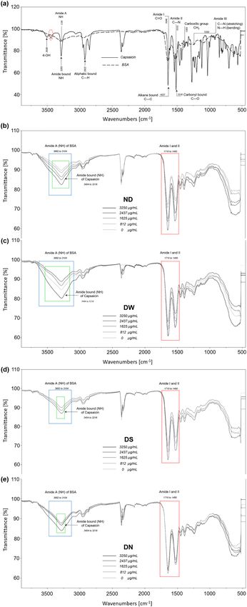
Structural changes of BSA‑capsaicin nanoparticles after drying treatments. Reported Fourier
Transform Infra Red (FTIR) spectra from pure capsaicin and native B SA10,22,23 were compared against the spec-
tra from our samples. The solid line in Fig. 3a shows the FTIR spectrum from our native capsaicin; the phenolic
4-OH group was assigned to the peak at 3506 cm−1, this resonance showed slight changes with respect to previ-
ous reports10,24. In this work we found a peak at 3443 cm−1 probably due to photochemical oxidation of capsaicin
during analysis (Fig. 3a, red ellipse). The 4-OH of capsaicin, show high reactivity and decomposition during
analysis of the chemical structure by NMR, HPLC, and FTIR25. A third peak was found at 3283 cm−1 and cor-
responds to the amide stretching bond N–H of the capsaicin molecule. The peak at 2922 cm−1 was associated to
the aliphatic bond C–H stretching vibration. Finally, the resonances at 1637 and 1516 cm−1 were assigned to the
C–C and C–O stretching vibrations, according to Leela et al.26.
The dashed line in Fig. 3a, shows the spectrum for native BSA; similar profiles have been observed by Bronze-
Uhle et al.3 and Sánchez-Arreguin et al.10. The N–H functional group associated to amide A showed a broad
stretching with a maximal peak at 3288 cm−1. The second maximal peak, found at 1646 cm−1, corresponds to a
stretching of the carbonyl group (C=O) of amide I. The subsequent regions showed some intense peaks associ-
ated to functional groups with double molecular behavior. C–N stretching and N–H bending of amide II was
identified at 1515 cm−1. The C H2 bending groups were associated to the peak at 1393 cm−1, while the vibrance
at 1250 cm−1 was correlated to C–N stretching and N–H bending in amide III.
All treatments (ND, DW, DN, and DS) and formulations of capsaicin (0, 812, 1625, 2437, and 3250 μg mL−1)
showed deformations of the peaks on the region from 3682 to 3104 cm−1; this spectral region corresponds to the
N–H amide A of the BSA, and overlaps with the 4-OH group and NH amide bond of capsaicin (see blue rectan-
gles in Fig. 3b–e). For all treatments with capsaicin concentration of 3250 μg mL−1 the NH amide bond signal
of pure capsaicin (N–H stretching) in the region from 3404 to 3216 cm−1 increased as shown by the black line
in the green rectangle of Fig. 3b–e. The spectral analysis of each treatment serves to identify possible structural
changes in the nanoparticles after the drying procedure. The DW and DN treatments at 3250 μg mL−1 showed an
intense peak deformation in the 3404 to 3216 cm−1 region as displayed by the black lines in the blue rectangles
in Fig. 3c,e, respectively. These perturbations were probably due to the instability of the capsaicin during the
drying process; deionized water and NaCl excipients showed no protection capacity against changes generated
in the interaction of the amino acid of albumin/capsaicin/water.
As Fig. 3d shows (see blue and green rectangles) the spectral resonances of the overlapping peaks of N–H
amide and A-/-NH amide bonds of capsaicin (3404–3216 cm−1) for the DS treatment did not show perturba-
tions in intensity and functional groups for all concentrations. This observation suggests the low deformation
of the peak and the low distance between bands of transmittance were probably due to the ordered interchange
of the amino acid of BSA in the microstructure and the subsequent migration of capsaicin from the core to the
surface of the n anoparticles10,14. This leads to an homogeneous distribution of capsaicin patches on the surface
of NPs dried with sucrose.
The DS treatment showed the best protective effect on the functional groups of amino acids of BSA NPs;
a similar effect was observed by Lee and T imasheff27 they found that sucrose does not perturb the spectral
fingerprint of several proteins during the drying process. The stabilization of proteins by sucrose excipient has
been proposed by the formation of polyhydric alcohols that induce a conformational change in some functional
groups of proteins, producing milder changes. In this study, during the drying process with sucrose, the capsaicin
experienced an equilibrium of repulsive forces between capsaicin and hydrophobic amino acid of BSA, into the
nanoparticles. During this process, the restructuration of capsaicin is carried out, and the capsaicin passed from
the core to the surface of the nanoparticles; we found this restructuration is less violent when NPs are dried with
the sucrose excipient.
The efficiency of excipients in the drying process of NPs has been evaluated through the presence of free
H2O or molecular water (O–H) content in the FTIR spectrum at 1644 cm−1 wavelength28. In this work, this
region corresponds to the overlapping of amide I and II bands of BSA with the hydrophobic side chain of the
capsaicin (1710–1480 cm−1), see the red rectangle in Fig. 3b–e. The presence of molecular water was observed
in nanoparticles dried with and without water, while in the DS and DN treatments the presence of O–H was not
observed. Moreover, a protein–sugar interaction was not observed for the DS treatment; this peak was reported
at 1580 cm−1, which is ascribed to the H-bond interaction with the carboxylate groups29.
Morphology of nanoparticles. Regarding the NPs size, they increased as the concentration of capsaicin
increased; this trend has been observed before10,14,17. Beside size changes, the TEM images showed morphologi-
cal changes in the nanoparticles depending on the drying treatment. As shown in Fig. 4a (for more images see
Fig. S2 of Supporting Information), the ND treatment showed a transitional change of shape in nanoparticles
from circular shape, for 0 µg mL−1 of capsaicin concentration, to elliptical shape for 1625 µg mL−1. It can also
be observed that at higher capsaicin concentrations NP coalescence is more extensive and small aggregates are
produced.
The treatment DW produced large particles with an elliptical shape and secondary particles branching from
the structure and forming columnar aggregates as seen in Fig. 4b (for more images see Fig. S2 of Supporting
Information). It is also noticeable that the surface of the NPs was slightly rougher; a similar effect was observed
Scientific Reports | (2021) 11:7549 | https://doi.org/10.1038/s41598-021-87241-8 4
Vol:.(1234567890)www.nature.com/scientificreports/
Figure 3. Molecular changes of the BSA-capsaicin nanoparticles after drying treatments with several excipients and the effect
of the increment of capsaicin in the nanostructuration. (a) FTIR spectrum of native BSA (dotted line) and pure capsaicin
(solid line); the scanning spectral range was between 4500 and 400 c m−1. (b) FTIR spectral characterization of not dried
treatment (ND) of BSA-capsaicin nanoparticles at 0, 812, 1625, 2437 and 3250 µg m L−1, rectangles show the main change
in amide A and bound amide of capsaicin. (c) NPs dried with water (DW), rectangles show changes in alkane and carbonyl
bonds, functional groups associated with molecular water. (d) NPs dried with sucrose (DS), spectra did not show deformation.
(e) NPs dried with NaCl (DN), spectra showed a low deformation in treatments from 0 to 2437 μg m L−1 of capsaicin
concentration, at 3250 μg m L−1 an increase of transmitted signal of the capsaicin was observed.
Scientific Reports | (2021) 11:7549 | https://doi.org/10.1038/s41598-021-87241-8 5
Vol.:(0123456789)www.nature.com/scientificreports/
Figure 4. Morphology of BSA-capsaicin nanoparticles. (a) Not dried (ND), (b) dried with water (DW), (c)
dried with NaCl (DN), and (d) dried with sucrose (DS). Electron micrographs showed a synergy effect between
loaded capsaicin and drying procedure. The isolated nanoparticles showed a change of shape, size and, in some
cases, showed coalescence. TEM micrographs showed several magnifications (upper right margin) due to the
increased NPs size.
in BSA-capsaicin nanoparticles reported by De Freitas et al.20. It is probable that the dual effect of freeze drying
and high capsaicin concentrations produced columnar aggregates with quasi-spherical shape.
Among the four treatments, DN resulted in the most negative effects regarding NPs shape. In this drying
procedure even the small nanoparticles with 0 µg mL−1 of capsaicin fused between them as can be appreciated in
Fig. 4c (for more images see Fig. S2 of Supporting Information). Similar coalescence was observed for the other
capsaicin concentrations; also it is worth noting that as the drug load increased, the structural complexity of the
NPs decreased, probably due to amino acids disassembling and producing an amorphous coagulated protein.
Interestingly, the DS treatment resulted in better morphology even than the ND case. We found that the
sucrose at 1 mmol, allowed to protect the circular shape of nanoparticles observed at 0 µg mL−1 of capsaicin,
see Fig. 4d (for more images see Fig. S2 of Supporting Information) and improved the morphology of the NPs
loaded with capsaicin; this was probably because the removal of water molecules increased sucrose concentra-
tion leading to the formation of a sol, the residual water molecules produced nanofluidic drag (microcirculation
phenomena)30. This drag induced the separation between NPs due to shear forces or strong capillary forces27.
Physical separation of NPs produced an improvement in the morphology of the dried nanoparticles with sucrose.
Morphometric analysis of the nanoparticles. Several studies have reported that the morphometry of
nanoparticles changes as a function of the increment of capsaicin during the synthesis of NPs10,14,17. In this study,
as aforementioned in the morphology analysis section, we observe that the shape of the NPs was affected by the
drying process. In order to evaluate this change, TEM images of isolated nanoparticles were analyzed by digital
image analysis (DIA). As seen in Fig. 5a all the treatments showed an increment of effective diameter (Ed) with
increasing drug load. The Ed for the ND treatment changed by a factor of 5.0 from 0 to 3250 µg mL−1 of capsai-
cin concentration; for the other treatments the factors were 3.4, 2.9, and 3.3 for DS, DW, and DN procedures,
respectively. While three of the treatments show similar behavior, DS deviates for high concentrations reaching a
plateau, see dashed red rectangle in Fig. 5a. This means the sucrose excipient reduces the negative effect over dry-
ing NPs and their coalescence when the drug load is augmented. In the case of the DN treatment, we propose the
Na+ and Cl− ions altered the pH and the net charge on the protein surface through amino acid hydrogens inter-
acting with different ions in solution, resulting in an acidic pH change. At pH 4.9 there is a lack of electrostatic
repulsion and thus amorphous aggregates are readily formed through nonspecific interactions3. Additionally,
the increase in capsaicin concentration generates a more hydrophobic environment, which increases the forma-
tion of aggregates. Future work may be directed towards establishing possible relations between formulation and
drying processes.
Scientific Reports | (2021) 11:7549 | https://doi.org/10.1038/s41598-021-87241-8 6
Vol:.(1234567890)www.nature.com/scientificreports/
Figure 5. (a) Effective diameter parameter of isolated BSA-capsaicin nanoparticles versus capsaicin
concentration for different drying procedures. The DS treatment showed a protective effect that allowed to
reduce the size of nanoparticles at a high concentration of capsaicin. (b) The isolated nanoparticles showed
a gradual loss of shape compared with NPs without drying (ND) treatment as a function of initial capsaicin
concentration. DW and DN treatments did not show protective effect in any formulation. DS showed a
protective effect and increase of the circular shape (Sf = 1). The equation of shape factor (Sf) is also displayed. (c)
Aspect ratio of BSA-nanoparticles versus capsaicin concentration. ND treatment resulted in a gradual transition
from circular to ellipsoidal shape. DW and DN did not show a protective effect in all formulations. The NS
treatment showed a protective effect that reduced the change to elliptical shape at a high concentration of
capsaicin (3250 μg mL−1). Values are means of three experimental replicates ± standard deviations.
Scientific Reports | (2021) 11:7549 | https://doi.org/10.1038/s41598-021-87241-8 7
Vol.:(0123456789)www.nature.com/scientificreports/
Figure 6. (a) The ζ-potential of BSA-capsaicin nanoparticles showed an increase in the electronegativity
associated with drying treatments. ND, DW, and DN treatments showed a negative effect, at 3250 µg m L−1 the
saturation of capsaicin produces a significant increment of the ζ-potential. While the DS treatment showed a low
increase of electronegativity due to the controlled reorganization of the amino acids and capsaicin molecules on
the surface of nanoparticles. (b) The size of the aggregates of BSA-capsaicin nanoparticles showed an oscillatory
behavior associated with drying treatments. Not drying (ND), dried with water (DW) and dried with NaCl
(DN) treatments showed a negative effect. In all treatments 812 µg m L−1 of capsaicin produces a significant
increment of the size of aggregates. The treatment dried with sucrose (DS) showed a protective effect and reduce
the increment of size. Values are means of three experimental replicates ± standard deviations.
In contrast to the Ed parameter, the shape factor (Sf, related to circularity) and aspect ratio (Ar, related to
ellipticity) showed only small changes against capsaicin concentration for DW, DN, and DS treatments, see
empty squares, empty circles and full triangles in Fig. 5b,c. On the other hand, samples with no drying treat-
ment, ND, showed affectations with the concentration as can be seen in Fig. 5b,c, full circles; thus, the change of
shape was related to the drying process. This means that the particles not dried show gradual changes of Sf and
Ar associated to an increment of capsaicin concentration. Also, interestingly, the treatment with sucrose (DS)
resulted in the shape factor closest to 1, i.e. rounder particles, and the lowest aspect ratio, i.e. less ellipticity as
can be seen in the full triangles of Fig. 5b,c. This is related to less aggregation and branching as a consequence
of reduction in the coalescence.
A possible explanation for our results with the sucrose excipient is that when it is incorporated into the
colloidal system, it could exert pressure to reduce the surface of contact between the BSA molecules of near
nanoparticles due to decrease radius of gyration (spatial expansion), thus inhibiting the unfolding of the BSA
anoparticles26, and consequently improving the hydrodynamic shape of the NPs. In contrast, the loss of
in the n
circular shape for the ND treatment is attributed to the increased coalescence due to higher content of capsaicin
crystals, affecting the coagulation of the BSA m olecules14. For the DW and DN procedures, the aggregation was
probably due to a change in the surface charges of amino acid, thus altering the electrostatic properties of BSA31.
The use of sucrose to dry nanoparticles improves the pharmaceutical formulations, in which, the circular shape
and elliptical shape of nanoparticles are important factors for the internalization into the c ell28,32.
ζ‑Potential and hydrodynamic diameter of aggregates. In a previous work, we observed an incre-
ment of the ζ-potential as the drug load increased from low to medium capsaicin concentrations10. In this study,
we found that the electric charge is affected by the drying process. In particular, while we observed somewhat
similar values of the ζ-potential for the ND, DW, and DN treatments, a higher value of this parameter was found
for the NPs subjected to the DS procedure for all drug concentration procedures (Fig. 6a, compare full circles,
empty squares, and empty circles to full triangles). Also, we measured relatively large aggregates for the ND,
DW, and DN treatments, while the DS samples presented smaller aggregates for all capsaicin concentrations
(Fig. 6b, compare full circles, empty squares, and empty circles to full triangles). The increased electronegative
values at 3250 µg mL−1 of capsaicin concentration for the ND, DW, and DN drying treatments (Fig. 6a, full cir-
cle, empty square, and empty circle) were not observed in the previous report. They are attributed to exposition
of capsaicin molecules in the surface of NPs, and liberation of amino acids with negative charge on the Nernst
layer of the particle. Eisele et al.33 describe that the negative charges on the surface of BSA are generated from
deprotonation of the carboxyl end of acidic amino acids (glutamate and aspartate). According to Yang et al.18,
the reorganization in some biopolymer nanoparticles was affected by the surface properties and internal bonds
(H-bonds). In this study probably the molecular interaction between some amino acids and capsaicin affects the
stability and morphology of nanoparticles due to the changes in hydrophilic/lipophilic balance, thus affecting
the self-assembly process and even structures and morphologies of self-assembled materials. Capsaicin has the
capacity to form a stable protein−ligand complex with BSA that mostly involve hydrophobic and electrostatic
interactions34. However, this mechanism is not capable of binding great quantities of capsaicin, so probably there
exists an alternative mechanism related to drug sites I and II in the BSA molecule (commonly called hydropho-
bic cavities) that facilitates the incorporation of hydrophobic drugs into the structure of B SA3. The modification
of the drug sites I–II displays some effects such as the coalescence between nanoparticles, loss of circularity,
Scientific Reports | (2021) 11:7549 | https://doi.org/10.1038/s41598-021-87241-8 8
Vol:.(1234567890)www.nature.com/scientificreports/
Figure 7. Electrophoretic patterns of disassembled molecules of BSA after nanoparticle drying treatments
with several excipients and the effect of the increment of capsaicin in the nanostructuration. For all gels, lane 1
shows molecular weight MW from 10 to 170 kDa. Line 2 shows a positive control of native BSA (500 µg m L−1).
The formulations were run on lines 3, 4, 5, 6, and 7 (nanoparticles at 0, 812, 1625, 2437, and 3250 µg m L−1 of
capsaicin, respectively). (a) Treatment not drying (ND), (b) treatment dried with water (DW), (c) treatment
dried with NaCl (DN), and (d) treatment dried with sucrose (DS).
aggregation, aberrant morphology and changes in surface of nanoparticles (ζ-potential). In nanoencapsulation
of hydrophilic drugs (5-fluorouracil, vinorelbine tartrate and salicylic acid), the coalescence and aggregation of
particles were not observed3,5,6. NPs dried with sucrose showed low aggregated size (351.1 ± 30.9 nm; see Fig. 6b,
full triangles); this could be attributed to sucrose inhibiting an irreversible formation of a ggregates26 because
during the interaction of sucrose with the protein, the sucrose does not crystallize during vacuum d rying28. This
effect probably produces minor inter-particle contact and insulation of surface electrostatic charges allowed a
controlled reorganization of capsaicin molecules with the amino acids of the nanoparticles.
Stability of the nanoparticles after drying treatment. Electrophoresis was used to determine molec-
ular size and purity of proteins; moreover, it verified the homogeneity of the protein samples, as well as the
number and molecular size of subunits35. In SDS-PAGE the native BSA protein migrates in response to an
electrical field through pores of acrylamide gel. In this experiment, polyacrylamide gel (12% SDS-PAGE), at a
constant voltage of 70 V for stacking gel and 80 V for resolving gel, allowed to determine the purity of the BSA
and its molecular weight (MW). The molecular weight of native BSA was found to be 66 kDa (Fig. 7a–d, lane 2)
according to the MW marker (Fig. 7a–d, lane 1). The BSA-capsaicin nanoparticles did not show migration in
the acrylamide gel for any of the drying treatments, therefore, the nanoparticles were found in the loading well
(Fig. 7a–d). The ND treatment at 812 µg mL−1 capsaicin concentration (Fig. 7a, black arrow on line 4), and DW
at 1625 µg mL−1 (Fig. 7b, black arrow on line 5), showed a trace of free albumin at 66 kDa probably as a result
of the dissembling of some albumin molecules caused by the drying process. A similar effect was observed by
Wang et al.36, they found that the delivery of BSA from biohybrid nanoparticles increased after the exposition of
NPs to excipients with low pH. The SDS-PAGE showed an intensity dependent on incubation time. This post-
Scientific Reports | (2021) 11:7549 | https://doi.org/10.1038/s41598-021-87241-8 9
Vol.:(0123456789)www.nature.com/scientificreports/
Retention time (min)
Peak Amino acid ND DW DS DN
Group 1
1 ASP 2.618 ± 0.015 2.718 ± 0.046 2.745 ± 0.015 2.682 ± 0.056
2 GLU 2.922 ± 0.036 2.980 ± 0.177 3.121 ± 0.016 2.985 ± 0.156
3 SER –a 6.265 ± 0.111 6.356 ± 0.020 6.321 ± 0.044
4 PRO 7.277 ± 0.106 7.245 ± 0.028 7.323 ± 0.059 7.323 ± 0.016
5 VAL 10.413 ± 0.125 10.343 ± 0.052 10.366 ± 0.066 10.450 ± 0.061
6 MET 11.289 ± 0.115 10.862 ± 0.021 11.000 ± 0.020 11.100 ± 0.031
7 LEU 13.027 ± 0.032 13.019 ± 0.022 13.211 ± 0.024 13.364 ± 0.026
8 PHE 14.942 ± 0.065 14.289 ± –b 14.514 ± –b 14.726 ± –b
9 TRP 15.977 ± 0.036 15.731 ± 0.262 16.022 ± 0.191 16.064 ± 0.034
10 LYS 16.587 ± 0.047 16.509 ± 0.265 16.866 ± 0.031 17.023 ± 0.048
Group 2
11 ASN –a –a –a 5.807 ± –b
a
12 GLY/GLN 6.230 ± 0.028 – 6.636 ± 0.019 –a
b b
13 ALA/HIS 6.501 ± – 6.613 ± – 6.904 ± 0.065 6.852 ± –b
b a
14 ARG 6.767 ± – – 7.025 ± 0.095 –a
b a
15 THR 7.211 ± – – 7.179 ± 0.076 8.533 ± 2.223
16 TYR –a –a 10.057 ± 0.030 10.695 ± 1.229
17 ILE/CYS –a 12.82 ± –b –a 12.769 ± 0.148
18 Iso/CYS –a 12.780 ± 0.012 12.930 ± 0.064 12.878 ± 0.022
Table 2. Amino acids liberated during dried processes of BSA-Capsaicin nanoparticles. a Not peak was
registered. b One peak was registered.
nanostructuration effect is excipient-time–pH-dependent. On the contrary, the DN and DS treatments showed
no traces of free albumin.
Our results indicate that the BSA molecules showed high chemical stability into the structure of NPs and
maintained their assemblage during the drying process. In contrast, Tarhini et al.7 found the BSA nanoparticles
migrate in the gel of acrylamide and showed an intense band at 66 kDa of MW, this implied that the BSA NPs
showed instability and a high degree of disassembly. Since the BSA NPs are constituted by several BSA molecules,
a possible symptom of disassembly of the NPs is the presence of free albumin at 66 kDa. The orientation of the
capsaicin from the core towards the surface of the NPs implies the reorganization of the hydrophobic amino
acids and probably their loss during this process; in order to investigate this phenomenon, the quantification of
the free amino acid was carried out.
Quantification of free amino acids from dried nanoparticles with different concentrations of capsaicin allowed
to establish two groups of amino acids. Group 1 includes 10 amino acids with a relevant presence in the NPs
after ND, DW, DN, and DS treatments; while group 2 includes 8 amino acids with low presence, both groups
are detailed in Table 2 (presence of amino acids and retention time). Notably, in group 1 serine (SER) was not
observed in the ND treatment at any capsaicin concentration but was present for the other drying procedures and
phenylalanine (PHE) showed irregular presence on DW, DN, and DS treatment. Regarding group 2, the DS and
DN treatments showed a higher percentage of the total amino acids present in group 2, 12.27% and 6.75% respec-
tively (see Fig. 8). The two treatments presented a larger number of amino acids of group 2, with 6 amino acids;
in particular, DS had the lowest percentage of total amino acids of group 1 and the highest of group 2 (Fig. 8).
Overall, sucrose was the excipient that showed the best post-drying properties; probably, this was due to the
low loss of amino acids of group 1. The molecules of sucrose reduce the instability and dragging of those amino
acids that had strong water-binding into the BSA protein28. DS had the highest percentage of group 2 amino
acids, GLY/GLN, ALA/HIS, ARG, THR, TYR, and Iso/CYS, this observation could be related to the stability of
capsaicin during the reorganization into the surface of the NPs. Anand et al.34 describe 14 possible interactions
with amino acid of BSA, of which five are strong interactions (Tyr400–capsaicin, Asn401–capsaicin, Lys524–cap-
saicin, Phe506–capsaicin, and Phe550–capsaicin); it is possible that PHE, TYR, and LYS interact strongly with
capsaicin resulting in increased NP stability. Our results also suggest that the dynamical formation of complexes
of capsaicin-amino acid (BSA)-water affects the structural stability of the BSA nanoparticle and its capacity to
reorganize hydrophobic drugs like capsaicin. To our knowledge, this is the first report that supports the secondary
post-synthesis restructuration of nanoparticles and the interaction between amino acids with hydrophobic drugs.
Conclusions
In this study, NPs of BSA were loaded with increasing concentrations of capsaicin to evaluate the effect and
changes on properties of nanoparticles after drying with several excipients. We found that the nanoparticles dried
with sucrose 1 mmol reduced their aggregation and the formation of isolated NPs at the highest concentrations
of capsaicin (3250 µg mL−1) leading to an improvement of their circular shape, reducing the elongation of the
Scientific Reports | (2021) 11:7549 | https://doi.org/10.1038/s41598-021-87241-8 10
Vol:.(1234567890)www.nature.com/scientificreports/
Figure 8. Total percentage of free amino acids quantified in ND, DW, DS, and DN treatments. The DS
treatment showed the highest loss of amino acids of group 2 (GLY/GLN, ALA/HIS, ARG, THR, TYR, and Iso/
CYS) involved in the mechanism of restructuration of capsaicin.
aggregates and controlling their electrochemical stability. The sucrose excipient inhibited the disassembly of BSA
molecules of the nanoparticles. The instability of NPs subjected to ND, DW, and DN treatments was associated
to parallel extraction of molecular water with amino acids ASP, GLU, SER, PRO, VAL, MET, LEU, PHE, TRP
and LYS; in contrast, the sucrose excipient reduced the loss of these amino acids. On the other hand, the second
group of amino acids, GLY/GLN, ALA/HIS, ARG, THR, TYR, and Iso/CYS, are involved in the mechanism of
restructuration of the capsaicin molecules towards the surface of the NPs during the drying process. Accord-
ing to previous s tudies10,14,34 PHE, TYR, and LYS showed strong interaction in the relation capsaicin-amino
acid–water molecules during formation of BSA-capsaicin NPs. This mechanism of secondary post-synthesis nano
structuration can improve the molecular stability of the particles and their capacity of entrapping hydrophobic
drugs like capsaicin. The drying parameters presented in this work could be applied to several types of nanopar-
ticles elaborated with proteins that encapsulate hydrophobic drugs, this method of conservation improves the
properties of nanoparticles and enhances parameters important to pharmaceutical and agricultural industries.
Materials and methods
Chemicals. The biological reagents used in this study were BSA (lyophilized powder, 66 kD) (Equitech-
Bio, Kerrville, TX, USA) and pharmaceutical grade capsaicin (≥ 99%) from Capsicum spp. (Handim Chemical
Co., Ltd, Shanghai, China). The chemical reagents were glutaraldehyde (25%) (Electron Microscopy Science,
Hatfield, PA, USA), sodium chloride analytical grade (Merck, Darmstandt, Germany), acetonitrile (≥ 99.9%)
(Sigma-Aldrich, St Louis, MS, USA) and absolute ethanol (≥ 99.8%) (Merck, Darmstandt, Germany). Poly-
acrylamide gel electrophoresis components, which consisted of acrylamide (40%), tris electrophoresis purity
reagent, sodium dodecyl sulfate, ammonium persulfate, and tetramethylethylenediamine (TEMED) (Bio-Rad
Laboratories, Richmond, CA, USA), Coomassie brilliant blue G-250 (Sigma-Aldrich, St Louis, MS, USA) and
acetic acid glacial HPLC grade (Merck, Darmstandt, Germany) for Coomassie staining solution (CBB). For
derivatization of free amino acids methanol HPLC grade (Sigma-Aldrich, St Louis, MS, USA), triethylamine
(Sigma-Aldrich, St Louis, MS, USA), and phenyl isothiocyanate (Sigma-Aldrich, St Louis, MS, USA) were used.
Preparation of BSA‑capsaicin nanoparticles. Nanoparticles were prepared by the desolvation tech-
nique as described by Langer et al.11 modified by Sánchez-Segura et al.14 and readjusted from Sánchez-Arreguin
et al.10. Briefly, 200 mg of BSA powder were dissolved in 2 mL of 10 mmol NaCl solution, pH 9.4, and filtered
through a 0.22 μm syringe filter (Sartorius, Goettingen, Germany). The solution was maintained under agita-
tion at 200 rpm using a stirrer (Eurostar 20, IKA, Wilmington, NC, USA) for 30 min at room temperature. The
formulations of increasing concentration of capsaicin were generated by the addition of 4 mL of an ethanol-
capsaicin solution at 0, 812, 1625, 2437, and 3250 µg mL−1 for each drying treatment. The rate of addition was
1.0 mL min−1 at 200 rpm of stirring speed. The crosslinking process was carried out by the addition of 5 mL of
4% glutaraldehyde in a 10 mmol NaCl solution in agitation at 1000 rpm for 30 min in dark conditions.
Protective excipients of BSA‑capsaicin nanoparticles and vacuum drying. Nanoparticles of
albumin have been typically dried without water or deionized water. In order to compare the effect of excipients
in the drying treatment versus traditional drying with deionized water and without drying treatment (diluted in
deionized water), as a negative control, we used two protective excipients, NaCl (10 mmol, pH 9.4) and sucrose
1 mmol. The samples (1000 µL) were taken from stock of NPs and washed by three cycles of centrifugation at
12,485×g, 10 min, at room temperature (MC-12V, DuPont, Newtown, CONN, USA), and the dispersion of the
pellet was carried out in deionized water, during every cycle. After the final cycle, the first sample that was not
dried was maintained in deionized water (ND), the second sample was resuspended in 1000 µL of deionized
Scientific Reports | (2021) 11:7549 | https://doi.org/10.1038/s41598-021-87241-8 11
Vol.:(0123456789)www.nature.com/scientificreports/
water (DW), the third in 10 mmol NaCl solution (DN), pH 9.4, and the fourth treatment was resuspended in
a 1 mmol sucrose solution (DS). Samples were homogenized by shaking in a vortex mixer (Super Mixer, LAB
LINE, Melrose Park, ILL, USA) for 10 min. In DW, DN and DS treatments, the samples were dried with a
MAXI-dry vacuum centrifuge (Heto-Holten A/S, Alleroed, Denmark) at 30 °C, 1300 rpm. Finally, the powder
was stored at − 4 °C.
Quantification of BSA transformed in nanoparticles and encapsulated capsaicin. The quan-
tification of encapsulated capsaicin in NPs was done by extraction with acetonitrile as described by Sganzerla
et al.37 and modified by Sánchez-Segura et al.14 and Sánchez-Arreguin et al.10. The BSA nanoparticles yield were
calculated from recovered BSA after extraction of capsaicin. While, the encapsulated efficiency (EE%) was cal-
culated from capsaicin quantified by HPLC as described by Bhalekar et al.38. The entire procedure is described
in Supporting Information (see S1)10,14,37,38.
Fourier transform infra red spectroscopy (FTIR). Determination of Fourier transform infrared spec-
tra of pure capsaicin, pure BSA and BSA-capsaicin NPs dried were performed by method described by Sánchez-
Arreguin et al.10. These experiments are explained in Supporting Information (refer to S2)10.
Determination of ζ‑potential, and hydrodynamic diameter of aggregates. The ζ-potential and
hydrodynamic diameter of aggregates were determined by method previously reported by Sánchez-Segura
et al.14 and modified by Sánchez-Arreguin et al.10 as explained in Supporting Information (S3)10,14,39.
Transmission electron microscopy (TEM) and morphometric analysis of nanoparticles. The
morphology of the resuspended nanoparticles was examined by TEM. The experimental preparation of the sam-
ples and operating conditions of the microscope were similar to those previously reported by Sánchez-Segura
et al.14, Sánchez-Arreguin et al.10 and modified by Castro-González et al.40. On the other hand, the morphomet-
ric parameters were calculated with equations proposed by Syverud et al.41. Parakhonskiy et al.32 and Bouwman
et al.42 and modified for nanoparticle description by Sánchez-Segura et al.14 and Sánchez-Arreguin et al.10. The
procedure is detailed in the Supporting Information (see S4)10,14,32,40–42.
Quantification of disassembled BSA by polyacrylamide‑gel electrophoresis. Sodium dodecyl
sulphate polyacrylamide gel electrophoresis (SDS-PAGE) was performed using the Tris–glycine buffer system of
Laemmli43 with modifications. In addition, to estimate the protein molecular weight (MW) we used the EZ-Run
pre-stained Rec protein ladder with bands in the range of 10–170 kDa (Fisher Scientific, Waltham, MA, USA). To
reference the MW of native BSA, lyophilized BSA (66 kDa), was dissolved in deionized water to stock concentra-
tion [500 µg mL−1] (Equitech-Bio, Kerrville, TX, USA). From stock, the solution was adjusted to 20 µg per lane.
Finally, the BSA-capsaicin nanoparticles after drying procedures were dissolved and adjusted at a concentration
of 20 µg per lane. The samples were resolved on 12% SDS-PAGE at a constant voltage of 70 V for stacking and
80 V for resolving. Then, gels were washed three times with deionized water for 5 min each and were boiled for
1 min with CBB staining solution (0.025% Coomassie dye, G-250 in 10% acetic acid). Subsequently, gels were
washed again with deionized water for 5 min and clarified to visualize polypeptide bands. Gels were captured
(Documentation Systems Bio-Rad) at 600 dpi resolution in tagged image file (.tif) format with 1580 × 1489 pixels
in grey scale. In this format, 0 was assigned to black and 255 to white in the grey scale.
Extraction and derivatization of free amino acids from BSA‑capsaicin nanoparticles. The
identification and quantification of amino acids were determined according to Bidlingmeyer et al.44 with modi-
fications. The NPs (powder) were washed with 1000 µL of deionized water and were homogenized for 15 min
and sonicated for 10 min at 25 °C. The samples were centrifuged at 12,485×g, for 6 min, at room temperature,
the supernatant was discarded, and the pellets were dried during 20 min. The derivatization of the samples was
carried out by addition of 20 µL of methanol/water/triethylamine (2:2:1) solution; then, the samples were dried
for 30 min at 45 °C. The samples were resuspended with 20 µL of methanol/water/triethylamine/phenyl isothio-
cyanate (7:1:1:1) solution and incubated for 30 min at 25 °C, subsequently, the samples were dried. Finally, they
were dissolved with 200 µL of sodium acetate trihydrate 0.1 M, pH 6.5 and were stored at − 70 °C. The samples
were resolved with Shimadzu ultra-fast liquid chromatography (UFLC) prominence series system (Shimadzu,
Kyoto, Japan), equipped with a LC-20AD pump coupled to a DGU-20A degassing unit, a SPD-20A dual wave-
length detector, a CMB-20A system controller, a SIL-20A HT auto-sampler and a CTO-20A column oven. There
were employed as the components separation role. The system was controlled by LabSolution software ver. 5.87
SP1. Chromatographic separation was performed on a C18 column (Agilent Technologies Eclipse XDB-C18
4.6 × 150 mm, 5 µm). Separation conditions were mobile phase A: sodium acetate trihydrate 0.1 M, pH 6.5;
mobile phase B: acetonitrile/water (4:1), flow rate at 0.9 mL min−1; column temperature 40 °C, and UV detection
at 254 nm. Analytical standards were used to confirm the identity of the peaks. Calibration solution was based on
a solution of 21 amino acids standard reference material LAA21, (Sigma-Aldrich, St Louis, MS, USA) containing
l-alanine, l-arginine hydrochloride, l-asparagine, l-aspartic acid, l-cysteine hydrochloride, l-cystine, l-glu-
tamic acid, l-glutamine, glycine, l-histidine hydrochloride, trans-4-hydroxy-l-isoleucine, l-leucine, l-lysine
hydrochloride, l-methionine, l-phenylalanine, l-proline, l-serine, l-threonine, l-tryptophan, l-tyrosine, and
l-valine. The standard samples were prepared by injecting 60 μL in real triplicates.
Scientific Reports | (2021) 11:7549 | https://doi.org/10.1038/s41598-021-87241-8 12
Vol:.(1234567890)www.nature.com/scientificreports/
Received: 26 July 2020; Accepted: 25 March 2021
References
1. Anhorn, M. G., Mahler, H. C. & Langer, K. Freeze drying of human serum albumin (HSA) nanoparticles with different excipients.
Int. J. Pharm. 36, 162–169. https://doi.org/10.1016/j.ijpharm.2008.07.004 (2008).
2. Kim, T. H. et al. Preparation and characterization of water-soluble albumin-bound curcumin nanoparticles with improved anti-
tumor activity. Int. J. Pharm. 403, 285–291. https://doi.org/10.1016/j.ijpharm.2010.10.041 (2011).
3. Bronze-Uhle, E. S., Costa, B. C., Ximenes, V. F. & Lisboa-Filho, P. N. Synthetic nanoparticles of bovine serum albumin with
entrapped salicylic acid. Nanotechnol. Sci. Appl. 10, 11–21. https://doi.org/10.2147/NSA.S117018 (2017).
4. Abrego, G. et al. Design of nanosuspensions and freeze-dried PLGA nanoparticles as a novel approach for ophthalmic delivery of
pranoprofen. J. Pharm. Sci. 103, 3153–3164. https://doi.org/10.1002/jps.24101 (2014).
5. Maghsoudi, A., Shojaosadati, S. A. & Farahani, E. V. 5-Fluorouracil-loaded BSA nanoparticles: Formulation optimization and
in vitro release study. AAPS Pharm. Sci. Tech. 9, 1092–1096. https://doi.org/10.1208/s12249-008-9146-5 (2008).
6. Li, Y. et al. A novel active targeting preparation, vinorelbine tartrate (VLBT) encapsulated by folate-conjugated bovine serum
albumin (BSA) nanoparticles: Preparation, characterization and in vitro release study. Materials 5, 2403–2422. https://doi.org/10.
3390/ma5112403 (2012).
7. Tarhini, M. et al. Protein-based nanoparticle preparation via nanoprecipitation method. Materials 11, 394. https://d oi.o rg/1 0.3 390/
ma11030394 (2018).
8. Ghosh, D. K. et al. Antimicrobial nano-zinc oxide-2S albumin protein formulation significantly inhibits growth of “Candidatus
Liberibacter asiaticus” in planta. PLoS One 13, e0204702. https://doi.org/10.1371/journal.pone.0204702 (2018).
9. Narendrakumar, G. et al. Enhancement of biocontrol potential of biocompatible bovine serum albumin (BSA) based protein
nanoparticles loaded bacterial chitinase against major plant pathogenic fungi Alternaria alternata. Biocatal. Agric. Biotechnol. 15,
219–228. https://doi.org/10.1016/j.bcab.2018.05.015 (2018).
10. Sánchez-Arreguin, A., Carriles, R., Ochoa-Alejo, N., López, M. G. & Sánchez-Segura, L. Generation of BSA-capsaicin nanoparticles
and their hormesis effect on the Rhodotorula mucilaginosa yeast. Molecules 24, 2800. https://doi.org/10.3390/molecules24152800
(2019).
11. Elzoghby, A. O., Samy, W. M. & Elgindy, N. A. Albumin-based nanoparticles as potential controlled release drug delivery systems.
J. Control Release 157, 168–182. https://doi.org/10.1016/j.jconrel.2011.07.031 (2012).
12. Langer, K. et al. Optimization of the preparation process for human serum albumin (HSA) nanoparticles. Int. J. Pharm. 257,
169–180. https://doi.org/10.1016/S0378-5173(03)00134-0 (2003).
13. Rahimnejad, M., Najafpour, G. & Bakeri, G. Investigation and modeling effective parameters influencing the size of BSA protein
nanoparticles as colloidal carrier. Colloids. Surf. A. Physicochem. Eng. Asp. 412, 96–100. https://doi.org/10.1016/j.colsurfa.2012.
07.022 (2012).
14. Sánchez-Segura, L., Ochoa-Alejo, N., Carriles, R. & Zavala-García, L. E. Development of bovine serum albumin–capsaicin nano-
particles for biotechnological applications. Appl. Nanosci. 8, 1877–1886. https://doi.org/10.1007/s13204-018-0874-x (2018).
15. Saez, A., Guzman, M., Molpeceres, J. & Aberturas, M. R. Freeze-drying of polycaprolactone and poly(D, l-lactic-glycolic) nano-
particles induce minor particle size changes affecting the oral pharmacokinetics of loaded drugs. Eur. J. Pharm. Biopharm. 50,
379–387. https://doi.org/10.1016/S0939-6411(00)00125-9 (2000).
16. Abdelwahed, W., Degobert, G., Stainmesse, S. & Fessi, H. Freeze-drying of nanoparticles: Formulation, process and storage con-
siderations. Adv. Drug. Deliv. Rev. 58, 1688–1713. https://doi.org/10.1016/j.addr.2006.09.017 (2006).
17. Sánchez-Segura, L. Food nano- and microconjugated systems: The case of albumin–capsaicin. In Food Nanoscience and Nanotech-
nology (eds Hernández-Sánchez, H. & Gutiérrez-López, G. F.) 187–203 (Springer, 2015).
18. Yang, P. P., Zhao, X. X., Xu, A. P., Wang, L. & Wang, H. Reorganization of self-assembled supramolecular materials controlled by
hydrogen bonding and hydrophilic–lipophilic balance. J. Mater. Chem. B 4, 2662–2668. https://d oi.o
rg/1 0.1 039/c 6tb00 097e (2016).
19. Costa, R. R., Custódio, C. A., Arias, F. J., Rodríguez-Cabello, J. C. & Mano, J. F. Nanostructured and thermoresponsive recombinant
biopolymer-based microcapsules for the delivery of active molecules. Nanomedicine 9, 895–902. https://doi.org/10.1016/j.nano.
2013.01.013 (2013).
20. De Freitas, G. B. et al. Formulation, characterization, and in vitro/in vivo studies of capsaicin loaded albumin nanoparticles. Mater.
Sci. Eng. C. 93, 70–79. https://doi.org/10.1016/j.msec.2018.07.064 (2018).
21. Karimi, M. et al. Albumin nanostructures as advanced drug delivery systems. Expert. Opin. Drug. Deliv. 13, 1609–1623. https://
doi.org/10.1080/17425247.2016.1193149 (2016).
22. Jincheng, W., Xiaoyu, Z. & Sihao, C. Preparation and properties of nanocapsulated capsaicin by complex coacervation method.
Chem. Eng. Commun. 197, 919–933. https://doi.org/10.1080/00986440903249700 (2010).
23. Peng, W. et al. Oral delivery of capsaicin using MPEG-PCL nanoparticles. Acta Pharmacol. Sin. 36, 139–148. https://doi.org/10.
1038/aps.2014.113 (2015).
24. D’Souza, L., Devi, P., Shridhar, M. P. & Naik, C. G. Use of Fourier Transform Infrared (FTIR) spectroscopy to study cadmium-
induced changes in Padina tetrastromatica (Hauck). Anal. Chem. Insights. 3, 135–143 (2008).
25. Martínez-Juárez, V. M. et al. Specific synthesis of 5,5’-dicapsaicin by cell suspension cultures of Capsicum annuum. var annuum
(chili jalapeño chigol) and their soluble and NaCl-extracted cell wall protein fractions. J. Agric. Food Chem. 52, 972–979. https://
doi.org/10.1021/jf035214p (2004).
26. Leela, J. S. P. P., Hemamalini, R., Muthu, S. & Al-Saadi, A. A. Spectroscopic investigation (FTIR spectrum), NBO, HOMO–LUMO
energies, NLO and thermodynamic properties of 8-Methyl-N-vanillyl-6-nonenamide by DFT methods. Spectrochim. Acta A 146,
177–186. https://doi.org/10.1016/j.saa.2015.03.027 (2015).
27. Lee, J. C. & Timasheff, S. N. The stabilization of proteins by sucrose. J. Biol. Chem. 256, 7193–7201 (1981).
28. Rahman, I. A., Vejayakumaran, P., Sipaut, C. S., Ismail, J. & Chee, C. K. Effect of the drying techniques on the morphology of silica
nanoparticles synthesized via sol–gel process. Ceram. Int. 34, 2059–2066. https://doi.org/10.1016/j.ceramint.2007.08.014 (2008).
29. Wang, B., Tchessalov, S., Warne, N. W. & Pikal, M. J. Impact of sucrose level on storage stability of proteins in freeze-dried solids:
I. correlation of protein–sugar interaction with native structure preservation. J. Pharm. Sci. 98, 3131–3144. https://doi.org/10.
1002/jps.21621 (2009).
30. Iskandar, F., Gradon, L. & Okuyama, K. Control of the morphology of nanostructured particles prepared by the spray drying of a
nanoparticle sol. J. Colloid Interface Sci. 265, 296–303. https://doi.org/10.1016/S0021-9797(03)00519-8 (2003).
31. Ikeda, S. & Morris, V. J. Fine-stranded and particulate aggregates of heat-denatured Whey proteins visualized by atomic force
microscopy. Biomacromol 3, 382–389. https://doi.org/10.1021/bm0156429 (2002).
32. Parakhonskiy, B. et al. The influence of the size and aspect ratio of anisotropic, porous CaCO3 particles on their uptake by cells.
Dig. J. Nanobiotechnol. 13, 1–13. https://doi.org/10.1186/s12951-015-0111-7 (2015).
33. Eisele, K. et al. Fine-tuning DNA/albumin polyelectrolyte interactions to produce the efficient transfection agent cBSA-147.
Biomaterials 31, 8789–8801. https://doi.org/10.1016/j.biomaterials.2010.07.088 (2010).
34. Anand, B. G., Dubey, K., Shekhawat, D. S. & Kar, K. Capsaicin-coated silver nanoparticles inhibit amyloid fibril formation of serum
albumin. Biochemistry 55, 3345–3348. https://doi.org/10.1021/acs.biochem.6b00418 (2016).
Scientific Reports | (2021) 11:7549 | https://doi.org/10.1038/s41598-021-87241-8 13
Vol.:(0123456789)You can also read