The Expression of a Mitochondria-Localized Glutamic Acid-Rich Protein (MGARP/OSAP) Is Under the Regulation of the HPG Axis
←
→
Page content transcription
If your browser does not render page correctly, please read the page content below
NEUROENDOCRINOLOGY
The Expression of a Mitochondria-Localized Glutamic
Acid-Rich Protein (MGARP/OSAP) Is Under the
Regulation of the HPG Axis
Mingxue Zhou,* Yifeng Wang,* Shaoling Qi, Jian Wang, and Shuping Zhang
State Key Laboratory of Biomembrane and Membrane Biotechnology, School of Life Sciences, Tsinghua
University, Beijing 100084, China
The hypothalamic-pituitary-gonadal (HPG) axis exerts a profound effect on animal development,
reproduction, and response to stress, and new insights into its complicated functional activities are
continuously being made. In the present study, by using immunohistochemical studies and dif-
ferent mouse models (ovariectomy and ob/ob mice), we systemically analyzed the expression of a
novel mitochondria-localized glutamic acid-rich protein (MGARP)/ovary-specific acid protein and
demonstrated that MGARP is under the regulation of the HPG axis. MGARP is highly enriched in
steroidogenic tissues and the visual system. Interestingly, its expression increases as mice develop.
Early in development, MGARP is mainly detected in the retina and adrenal gland. At this early
developmental stage, its expression is not detectable in the gonads, but its expression in the gonads
dramatically increases during the first 2– 4 wk after birth. Importantly, MGARP levels correlate with
estrogen levels in the ovaries during the estrous cycle, and estrogen regulates the expression of
MGARP in a tissue-specific manner and through a feedback regulatory mechanism. Functional
inhibition of GnRH with an antagonist strongly reduces MGARP levels, and knockout of leptin
(ob/ob) significantly reduces the MGARP expression in follicular granular cells. We proposed a
model that elucidates the role MGARP plays in the HPG axis. Within the HPG axis loop, MGARP
participates in hormone biosynthesis while being under the regulation of the hormones derived
from the HPG axis. (Endocrinology 152: 2311–2320, 2011)
teroid hormones have a variety of metabolic activities, spermiogensis. Both LH and FSH participate in the regu-
S including neuromodulatory, neuroendocrine, and neu-
roprotective effects, and they play crucial roles in mamma-
lation of the estrous cycle, menstrual cycle, gonad devel-
opment, and reproduction. Furthermore, androgen and
lian reproduction, development, aging, and stress responses estrogen use a feedback mechanism to regulate GnRH,
by acting on the brain and other organs (1, 2). All of these LH, and FSH biosynthesis (7). The HPG axis is an essential
activities of steroids are critically regulated by the hypo- and very complicated system throughout life, and there are
thalamic-pituitary-gonadal (HPG) axis (1, 3, 4). In the still many questions remaining to be answered.
HPG axis, GnRH, a factor secreted by the hypothalamus, The mitochondria-localized glutamic acid-rich pro-
travels down the anterior portion of the pituitary via the tein (MGARP) is a novel mitochondrial protein identi-
hypophyseal portal system and binds to the receptors on fied by large-scale screenings for genes specifically ex-
the secretary cells of the adenohypophysis (5). Pulsatile pressed in the ovary, retina, cornea, and adrenal gland
release of GnRH stimulates the secretion of LH and FSH (8 –13). It was previously named as mouse ovary-spe-
in the adenohypophysis (6). LH regulates the synthesis of cific acidic protein and human corneal endothelium-
the gonadal hormones and ovulation, whereas FSH pro- specific protein (8, 13), but its function was not sub-
motes ovarian follicle maturation, estrogen release, and stantially defined. In our previous report, we proposed
ISSN Print 0013-7227 ISSN Online 1945-7170 * M.Z. and Y.W. contributed equally to this work.
Printed in U.S.A. Abbreviations: CNS, Central nervous system; E2, 17-Estradiol; E2-H, E2 high dose; E2-L,
Copyright © 2011 by The Endocrine Society E2 low dose; GST, glutathione transferase; HE, hematoxylin and eosin; HPG, hypothalamic-
doi: 10.1210/en.2011-0050 Received January 18, 2011. Accepted March 8, 2011. pituitary-gonadal; LGN, lateral geniculate nucleus; MGARP, mitochondria-localized glu-
First Published Online March 29, 2011 tamic acid-rich protein; opt, optic tract; OVX, ovariectomy.
Endocrinology, June 2011, 152(6):2311–2320 endo.endojournals.org 2311
The Endocrine Society. Downloaded from press.endocrine.org by [${individualUser.displayName}] on 14 May 2015. at 07:56 For personal use only. No other uses without permission. . All rights reserved.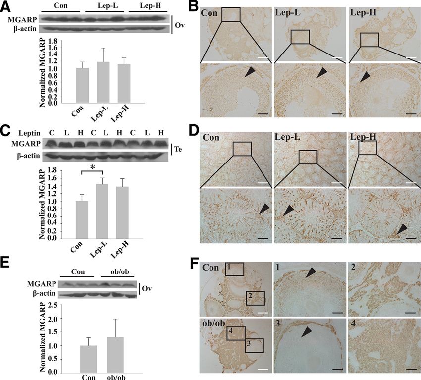
2312 Zhou et al. MGARP/OSAP Mediated by Hormones in the HPG Axis Endocrinology, June 2011, 152(6):2311–2320
an accordant and universal name, MGARP, for the pro- adjuvant and boostered weekly for 4 wk. The serum of the
tein in consideration of its enrichment with glutamic rabbit was collected 1 wk later, and the purity and specificity
acids and specific cellular localization (11). Our study of the antibody were analyzed by Western blotting and im-
munocytochemistry by using anti-GST, preimmunized serum
on MGARP in the retina showed robust levels of ex-
as control. The purified MGARP protein was used to do an-
pression in the inner segment of the photoreceptor, tigen preabsorption.
outer plexiform layer, and ganglion cell layer of the
retina (11). A reduction in MGARP expression results in Identification of the female mice estrous cycle
mitochondrial fragmentation and overexpression of A vaginal cast-off cell smear, hematoxylin and eosin (HE)
MGARP with a deletion of the N terminus causes severe staining method was used to identify the estrous cycle of fe-
mitochondrial aggregation (10, 11). In Y-1 cells, knock- male mice. A dipped wet cotton bud with 0.9% isotonic so-
down of MGARP significantly inhibits 8-bromoad- dium chloride was inserted into the vagina and rotated several
enosine-cAMP-induced progesterone production (10). times. Samples were smeared onto slices and fixed with meth-
anol. The vaginal cast-off cellular morphology was observed
The detailed regulatory mechanisms controlling MGARP
after HE staining.
expression, however, still remain to be elucidated. It is also
unclear how MGARP interacts with the different elements GnRH antagonist experiment
of the HPG axis, which includes the main components of ICR mice were divided into the following four groups: male
both the neurosecretory and steroidogenic systems. control group, male GnRH antagonist group, female control
In this study, we systemically analyzed the expression group, and female GnRH antagonist group (n ⫽ 4 – 6/group).
and the regulatory network of MGARP by Western blot- Mice in the GnRH antagonist groups were treated with daily sc
ting and immunohistochemical analysis using different injections of 0.5 mg/kg cetrorelix acetate (14) for 7 d, whereas the
control groups were treated with the same volume of saline. The
mouse models and found that MGARP is predominantly
phase of the estrous cycle for all of the female mice was identified
expressed in the steroidogenic tissues and the compart-
by the vaginal cast-off cell smear test before and after cetrorelix
ments of the visual system. We also found that MGARP treatment.
expression increases as the gonads develop. It can be reg-
ulated by estrogen, GnRH, and leptin, all of which are Leptin intervention
regulators or effectors of the HPG axis. Our results suggest ICR mice were divided into the following six groups: male
that the interaction between MGARP and elements of the control group, male leptin low-dose group, male leptin high-dose
HPG axis forms a functional loop, with steroid hormones group, female control group, female leptin low-dose group, and
as the mediators. female leptin high-dose group (n ⫽ 3/group). A dose of either 100
or 500 g/kg leptin was given daily for 5 d by ip injection. The
dose of 100 g/kg leptin corresponds to the physiological dose
(15), and 500 g/kg of leptin corresponds to a hyperphysiologi-
Materials and Methods cal dose. The control group was treated with the same volume of
saline. Euthanasia was performed on all mice (1% sodium pen-
Reagents and animals tobarbital). The phase of the estrous cycle for all the female mice
17-Estradiol (E2) (E8875) and leptin (L3772) were pur- was identified by the vaginal cast-off cell smear test before and
chased from Sigma (St. Louis, MO). Cetrorelix acetate was from
after leptin treatment.
Shanghai Taishi Biotechnology Co., Ltd. (Shanghai, China). ICR
mice were bred by the Animal Facility of Tsinghua University.
Ob/ob mice were obtained from The Jackson Laboratory (Bar
Immunohistochemistry
Harbor, ME). All animal experiments were performed in com- Tissues and organs, except for the eyeballs, from each adult
pliance with the relevant laws and institutional guidelines. The mouse were fixed in 10% formalin for 24 h and embedded in
animal care procedures were institutionally reviewed a by the paraffin. Eyeballs were fixed in a special fixative solution (glacial
Institutional Animal Care and Use Committee of Tsinghua Uni- acetic acid:formalin:0.9% sodium chloride:75% alcohol, 1:2:7:
versity. The euthanasia was performed by adopting 1% sodium 10). Two serial 5-m paraffin sections were used for immuno-
pentobarbital by ip injection. histochemical staining. Mouse MGARP antibody or GST anti-
body, used as a control, was added to the sections using a drop-
Antibody preparation wise technique.
The full-length MGARP cDNA sequence was inserted into After the mice were euthanized, the tissues from each mouse
pGEX-4T-1 vector and transformed into the BL21 expression were removed, fixed for 24 h, and embedded in paraffin. Then
strain. Isopropyl -D-1-thiogalactopyranoside (0.1 mM) was 5-m sections were cut and stained using an immunohistochem-
used to induce the expression of a glutathione transferase (GST)- ical method to observe and analyze MGARP expression. Addi-
MGARP fusion protein in Escherichia coli, and the protein was tionally, the same tissue types used for immunohistochemical
purified with an affinity column. A rabbit was inoculated with staining were also taken from the contralateral side of each
purified GST-MGARP fusion protein mixed with Freund’s mouse for Western blotting.
The Endocrine Society. Downloaded from press.endocrine.org by [${individualUser.displayName}] on 14 May 2015. at 07:56 For personal use only. No other uses without permission. . All rights reserved.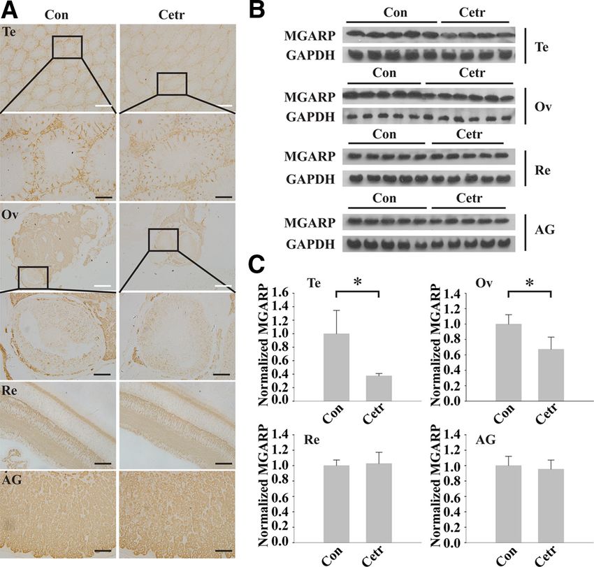
Endocrinology, June 2011, 152(6):2311–2320 endo.endojournals.org 2313
Group, Inc., Chicago, IL), followed by a sec-
ondary antibody (goat antimouse IgG horse-
radish peroxidase-conjugated antibody,
1:5000; Zhongshan Golden Bridge). Protein
expression was detected with an enhanced
chemiluminescence detection system (Vigor-
ous, Beijing, China).
Surgical procedures and estrogen
treatment
Mice were weighed and anesthetized with
1% sodium pentobarbital by ip injection. Sur-
gical castration was performed through the
backside to gain bilateral access to the ovary.
After the ovaries were removed, the skin was
sutured with 3– 0 vicryl. Mice in the sham
group only had fatty tissue near the ovaries
removed as a control. Ten days after the op-
erations, mice were divided into the following
four groups: control group with ovariectomy
(OVX) (n ⫽ 5), OVX ⫹ E2 low-dose (E2-L)
group (n ⫽ 6, 10 g/kg), OVX ⫹ E2 high-
dose (E2-H) group (n ⫽ 6, 100 g/kg), and
sham treatment group (n ⫽ 5). Estrogen (E2)
was dissolved in 10% alcohol and 90% pea-
nut oil (16) and given daily by sc injections for
1 wk. The dose of 10 g/kg of E2 corresponds
to the physiological dose, and 100 g/kg of E2
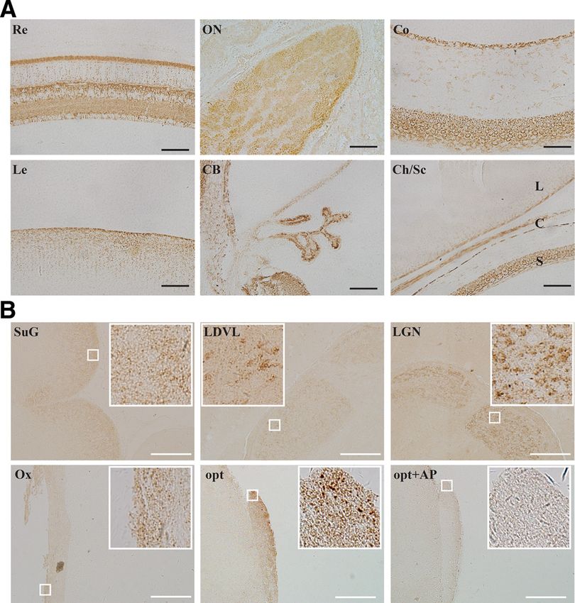
FIG. 1. Immunohistochemical staining shows the expression pattern of MGARP in the visual
corresponds to a hyperphysiological dose.
tissues of ICR mice. A, Immunostaining of different regions in the eyeballs: retina (Re), optic nerve
(ON), cornea (Co), lens (Le), ciliary body (CB), and choroid and sclera (Ch/Sc). B, MGARP expression Mice in the control group and sham group
in the visual system of the brain: superficial gray layer of the superior collicullus (SuG), laterodorsal were sc injected with the same dose of vehicle
thalamic nucleus ventrolateral part (LDVL), LGN, optic chiasma (Ox), and opt. Opt ⫹ AP means (90% peanut oil and 10% alcohol).
that anti-MGARP antibody was previously preabsorbed with antigen. AP, Antigen preabsorption.
Images in white squares are magnification of areas enclosed by small white squares. Antibody Statistical analysis
labeling appears brown. Black and white scale bars, 50 and 200 m, respectively.
Data are presented as the means ⫾ SE
and were analyzed by ANOVA, followed
Antigen preabsorption by Tukey’s test for multiple comparisons or Student’s t test.
Anti-MGARP antiserum was diluted (1:10,000) with purified Differences are considered significant when P ⬍ 0.05.
MGARP-GST fusion protein dissolved in PBS with 1% BSA. The
protein level of purified MGARP-GST fusion protein is 5.2
mg/ml (determined by bicinchoninic acid assay; Pierce, Rockford,
IL). Anti-MGARP antiserum diluted (1:10,000) with PBS supple-
Results
mented with 1% BSA was set as positive control. Both of which
MGARP is highly enriched in the steroidogenic and
described above were incubated at 37 C for 1 h and followed by
standard immunohistochemistry procedure. The tissue sections visual systems
were also preincubated with 1% BSA in room temperature for 2 h. The systemic study on the MGARP expression in adult
mouse by immunohistochemistry demonstrated that, in
Western blotting addition to the retina, MGARP is highly expressed in all
Total protein was isolated from mice tissues. Tissues were other parts of the eyeball, including cornea, lens, ciliary
collected and homogenized in protein extraction buffer [50 mM body, sclera, and choroid. There was robust expression in
Tris-HCl (pH 7.4), 0.25 M NaCl, 1% Nonidet P-40, 1 mM EDTA,
the corneal epithelial cells, scleral fibroblast cells, epithe-
and 1% protease inhibitor cocktail]. The lysate was centrifuged
at 12,000 ⫻ g for 10 min, and the supernatant was collected. The lial cells in the lens, and pigment epithelial layer of the
supernatant (30 g of protein) was resolved on a 10% SDS- ciliary body (Fig. 1A). The systemic expression profile also
PAGE gel and transferred onto nitrocellulose membranes. After showed that MGARP is highly expressed in the ovary,
being blocked with 5% nonfat milk, the membranes were probed testis, adrenal gland, eye, and brain, but not detectable in
with the primary mouse MGARP antibody (1:10,000) for 1 h,
heart, liver, spleen, lung, kidney, skeletal muscle, fat tis-
followed by a secondary antibody (goat antirabbit IgG horseradish
peroxidase-conjugated antibody, 1:5000; Zhongshan Golden sue, stomach, small intestine, uterus, pancreas, prostrate
Bridge, Beijing, China), or probed with the primary antibodies an- thymus, parathyroid gland, pituitary gland, and thyroid
tiactin (1:500; Sigma) and anti-GAPDH (1:4000; Proteintech gland by immunohistochemistry (Supplemental Fig. 1A,
The Endocrine Society. Downloaded from press.endocrine.org by [${individualUser.displayName}] on 14 May 2015. at 07:56 For personal use only. No other uses without permission. . All rights reserved.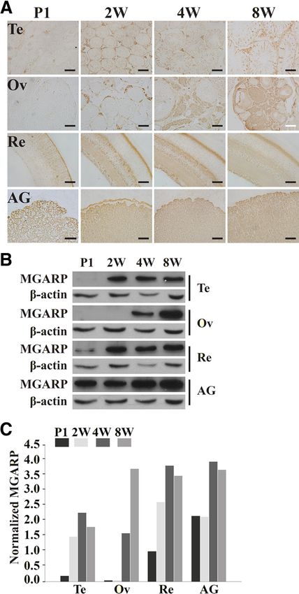
2314 Zhou et al. MGARP/OSAP Mediated by Hormones in the HPG Axis Endocrinology, June 2011, 152(6):2311–2320
published on The Endocrine Society’s Journals Online
web site at http://endo.endojournals.org). Using the anti-
gen-adsorbed MGARP antibody, the staining was com-
pletely eliminated (Supplemental Fig. 1B). Further analy-
sis indicated that MGARP was enriched in the lutein cells
of corpus luteum, theca cells, and granulosa cells of ovar-
ian follicles, Sertoli cells, and interstitial cells of mice testis
as well as the zona glomerulosa, zona fasciculata, and
zona reticularis of adrenal cortex. All these areas are made
up by steroidogenic cells (Supplemental Fig. 2). Most im-
portantly, our results showed that MGARP was detected
in different regions of the brain, including the optic chi-
asma, optic tract (opt), lateral geniculate nucleus (LGN),
laterodorsal thalamic nucleus ventrolateral part, and su-
perficial gray layer of the superior collicullus (SuG) (Fig.
1B). All of these comprise the main components of the
visual nervous system. Together, our observations indi-
cate that MGARP is enriched in steroidogenic tissues and
the visual system. Considering that the visual nervous sys-
tem, including the retina and related areas in the brain,
belongs to the central nervous system (CNS) as well as
responds to hormones, we hypothesized that MGARP is
potentially involved in the functional activities of the HPG
axis.
The expression of MGARP during mouse
development
To understand the role of MGARP in development that is
critically regulated by the HPG axis, we studied MGARP
expression during steroidogenic tissue development. As
shown in Fig. 2, at postnatal d 1, MGARP is readily detected
in the adrenal gland and only slightly detected in the retina. FIG. 2. MGARP protein expression is associated with the progression
It was not detectable, however, in the mouse ovaries and of mouse development. Black and white scale bars, 50 and 200 m,
respectively. A, Immunohistochemical staining of MGARP in the testes
testes at this stage. In the gonads, MGARP expression was (Te), ovaries (Ov), retinas (Re), and adrenal glands (AG) of mice at
clearly detected in pups 2– 4 wk after birth (Fig. 2, A–C). postnatal d 1 (P1), postnatal wk 2 (2W), 4W, and 8W. B, Western
In addition, we studied MGARP expression in more detail blotting of total proteins harvested from the Te, Ov, Re, and AG during
development. C, Column diagram indicates the quantitative analysis of
during the five phases of ovarian follicle development by
the Western blotting.
immunohistochemistry. The expression of MGARP in the
later phases was much higher than in the early phases
(Supplemental Fig. 3). To further determine the effects of sex hormones on
MGARP expression, we generated OVX mice to reduce
The effects of estrogen on MGARP expression the endogenous estrogen or performed sc injection into the
To study whether the expression of MGARP is regu- OVX mice with different doses of E2 to restore the estro-
lated by sex hormones and associated with steroidogenic gen level. The expression of MGARP was decreased in the
activity of the HPG axis, we examined MGARP expres- retina of the OVX mice compared with the sham group,
sion in female mice at different estrous cycle phases. We but the magnitude is not statistically significant, whereas
found that in ovaries, the expression of MGARP was sig- injection of E2 in OVX mice at a dose of 100 g/kg could
nificantly higher during estrus and diestrus than during increase the expression of MGARP by 25% compared
proestrus and metestrus (Fig. 3, A–C). However, there was with the OVX group (Fig. 3D). In contrast, the expression
no significant fluctuation in MGARP expression in the of MGARP in the adrenal gland was increased in the OVX
mice adrenal glands and retinas at different estrous cycle mice compared with the sham group. Furthermore, injec-
phases (Fig. 3C). tion of E2 at a dose of 100 g/kg could reduce the expres-
The Endocrine Society. Downloaded from press.endocrine.org by [${individualUser.displayName}] on 14 May 2015. at 07:56 For personal use only. No other uses without permission. . All rights reserved.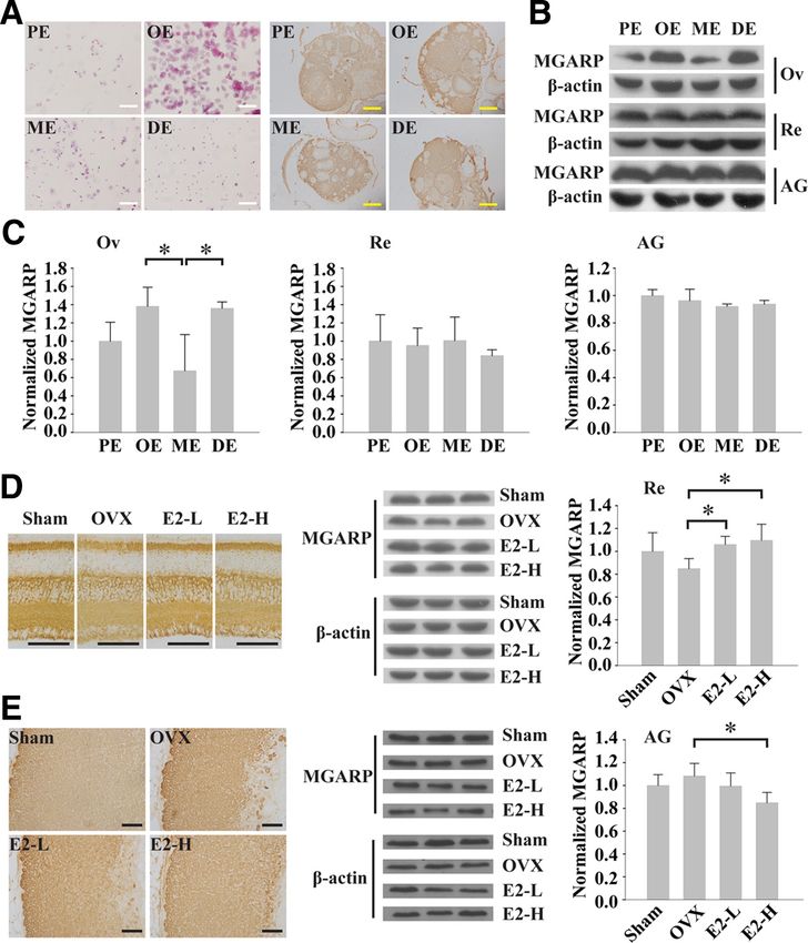
Endocrinology, June 2011, 152(6):2311–2320 endo.endojournals.org 2315
in follicular cells of ovary was also
reduced, but it is not as significant as
that occurring in treated testes (Fig.
4A). As shown by Western blotting,
cetrorelix treatment led to the reduc-
tion in MGARP expression by 62% in
testes and 33% in ovaries compared
with the control groups (Fig. 4, A–C).
However, no difference was observed
in the retina and adrenal gland.
The effects of leptin on MGARP
expression
To further examine a role of the HPG
axis in regulating MGARP expression,
we investigated the effects of leptin,
which has been reported to stimulate the
GnRH secretion through acting on the
hypothalamus (17). As shown in Fig. 5, A
and B, exogenous injection of leptin into
female mice did not induce an obvious
change in MGARP expression in the ova-
ries. In the follicular granular cells of the
sinusoid follicle, however, a low-dose
of leptin (100 g/kg) clearly increased
MGARP expression compared with the
FIG. 3. MGARP expression fluctuates during the estrous cycle in the ovary, and its expression control. To discount the effect of the es-
is estrogen independent in the retinas and adrenal glands. Black, white, and yellow scale bars, trous cycle on MGARP expression, we
50, 200, and 500 m, respectively. A, left panel, Proestrus (PE), estrus (OE), metestrus (ME), carried out similar studies by using male
and diestrus (DE) in the estrous cycle of female mice are identified by HE staining of vaginal
cast-off cells. Right panel, Mice ovaries at different estrous cycle phases are immunostained
mice. Injection of exogenous leptin at a
with MGARP antibody. B, Western blotting (WB) results of MGARP levels in mouse ovaries dose of 100 g/kg into adult male mice
(OV), retinas (Re), and adrenal glands (AG) at different estrous cycle phases. C, Quantitative also increased MGARP expression by
analysis (column diagram) of MGARP levels in mouse ovaries (OV), retinas (Re), and adrenal
45% in the testes compared with the con-
glands (AG) at different estrous cycle phases based on WB results (n ⫽ 5). D, left panel,
Retinas from sham, OVX, E2-L, and E2-H mouse groups are immunostained with anti-MGARP trol, with a particularly higher stimula-
body. Middle and right panels, Western blotting results and quantitative analysis (column tion in Leydig cells (Fig. 5, C and D).
diagram) of MGARP levels in the retinas from sham, OVX, E2-L, and E2-H mouse groups. E, Another model used in this study is the
left panel, Adrenal glands from sham, OVX, E2-L, and E2-H mouse groups are immunostained
with anti-MGARP body. Middle and right panels, Western blotting results and quantitative female leptin knockout (ob/ob) mice,
analysis (column diagram) of MGARP levels in the adrenal glands from sham, OVX, E2-L, and which have been demonstrated to be in-
E2-H mouse groups. fertile (18). We studied the expression of
MGARP in ovaries of ob/ob mice and
sion levels of MGARP by 21% in the adrenal gland com- wile-type C57 mice by immunohistochemistry. Consistently,
pared with the control of OVX group (Fig. 3E). the overall expression of MGARP in ovaries of the ob/ob
mice did not show significant differences, but its expression
The effects of functional inhibition of GnRH on in follicular granular cells was markedly reduced compared
MGARP expression with the wild-type control mice (Fig. 5, E and F).
Next, we studied MGARP expression by manipulation
of GnRH, a key factor in the HPG axis. Cetrorelix was
used in these tests, which is a GnRH antagonist that com- Discussion
petes the binding of GnRH to its receptors to inhibit go-
nadotropin level. The staining of MGARP in the cyto- We previously identified the MGARP gene by a microar-
plasm of Leydig cells and Sertoli cells of mice testes was ray from the mouse retina (11). Retina, a particularly ac-
reduced under the treatment of cetrorelix (GnRH antag- cessible part of the CNS, is critical for the capture of im-
onist) compared with the untreated control. Its expression ages and the response to light. These processes require the
The Endocrine Society. Downloaded from press.endocrine.org by [${individualUser.displayName}] on 14 May 2015. at 07:56 For personal use only. No other uses without permission. . All rights reserved.2316 Zhou et al. MGARP/OSAP Mediated by Hormones in the HPG Axis Endocrinology, June 2011, 152(6):2311–2320
mone (estrogen, progesterone, and an-
drogen) actions. Various physiological
conditions, such as age, menstrual cy-
cles, pregnancy, and menopause or an-
dropause can affect vision (24). Studies
have also shown the presence of sex ste-
roid hormone receptors in various oc-
ular tissues, such as the lens, retina,
choroid, cornea, and ciliary body, and
the response of the retina to sex steroid
hormone action is similar to that of the
CNS (24). With in situ hybridization
techniques, estrogen receptor mRNA
was detected in both the retina and
brain (25, 26). The retina was also able
to synthesize steroid hormones by the
progesterone pathway (27). Consider-
ing the CNS (brain and spinal cord) and
retina are steroidogenic tissues (27–
29), and steroid hormones are regu-
lated by the HPG axis, our findings sug-
gest a profound role of MGARP in the
FIG. 4. The GnRH antagonist cetrorelix acetate down-regulates MGARP levels in the testis
and ovary but not in the retinas and adrenal glands. Black and white scale bars, 50 and 200 regulatory loop of the HPG axis.
m, respectively. A, Immunostaining for MGARP in the mouse testes (Te), ovaries (Ov), retinas However, why is MGARP predom-
(Re), and adrenal glands (AG) without [control (con)] and with GnRH antagonist [cetrorelix inantly expressed within the visual sys-
acetate (Cetr), 0.5 mg/kg] treatment for 7 d. B, Total protein from comparable tissues
previously mentioned in A was harvested for Western blot analysis of MGARP. Each column
tem? This is a critical question that is
represents one mouse. GAPDH, Glyceraldehyde-3-phosphate dehydrogenase. C, Column worth of further study. Here, we pro-
diagrams indicating the quantitative analysis of the Western blotting. *, P ⬍ 0.05; n ⫽ 4, 5, pose several reasons. 1) The visual sys-
or 6.
tem has higher demand for both energy
and steroid hormones. 2) As part of the
collaboration of several different parts of the eyeball. To
CNS, the visual system is the major regions of the animal
understand a potential role of MGARP in the visual sys-
body to sense or receive the stimuli from the environment.
tem, we tested for MGARP expression in each part of the
The visual perception is not simply a translation of stimuli
eyeball by immunohistochemistry. The results demon-
and formation of the image on the retina and it should need
strated that, in addition to the retina, MGARP is also
the eye and the related visual tissues in the brain to work
highly expressed in all other parts of the eyeball. To gather
together in a very complicated way. This is a question
more information about MGARP, we conducted a sys-
scientists in this field have long struggled to explore. Our
temic analysis of MGARP expression by immunohisto-
chemical staining. The results showed that MGARP is observation that MGARP is highly expressed in visual
highly expressed in steroidogenic and nervous tissues, es- system implies that MGARP may play critical function
pecially in areas that make up the main components of the in transmitting the stimuli through responding to the
visual nervous system. The optic chiasma, opt, and LGN steroid hormones or through regulating hormone syn-
are the main components of the visual pathway in brain. thesis. 3) The expression difference in the brain reflects
These regions are responsible for transmitting visual in- that MGARP expression is under the regulation of dif-
formation from the eyeball to the optic center in the brain. ferent hormones in a direct or indirect manner, because
The superficial gray layer of the superior collicullus is re- different regions of the brain produce distinct hormones
sponsible for providing a powerful excitatory input to the and those hormones always function in different tissues
intermediate layer, which plays a critical role in initiating through circulation. It can be considered that HPG axis
rapid orienting movements of the eye (19 –23). Thus, most may steer the entire process through regulating the ex-
of the regions positive for MGARP expression are directly pression level of MGARP. Certainly, all these need more
or indirectly involved in visual processing. The visual sys- studies to be clearly addressed.
tem is a special system that has long been used as a model The HPG axis plays an active and essential role in mam-
for CNS developmental and functional studies of sex hor- malian development via modulating various hormones.
The Endocrine Society. Downloaded from press.endocrine.org by [${individualUser.displayName}] on 14 May 2015. at 07:56 For personal use only. No other uses without permission. . All rights reserved.Endocrinology, June 2011, 152(6):2311–2320 endo.endojournals.org 2317
and MGARP is less expressed in the
early phase of follicular development
than in other phases (31). Our results
of the MGARP expression during the
five phases of ovarian follicle develop-
ment indicate that MGARP expression
is gradually increased during follicle de-
velopment, which is most likely regu-
lated by gonadotropin.
One physiological function of the
HPG axis is to regulate the synthesis of
sex hormones. Conversely, the sex hor-
mones regulate the HPG axis through a
feedback regulatory mechanism. Estro-
gen is a sex hormone that not only af-
fects the sex organs, but it also affects
the structure and function of the ner-
vous system, because its receptors are
expressed in the brain regions that are
involved in sex differentiation and mat-
uration (1). To determine whether the
expression of MGARP is involved in
the steroidogenic activity of the HPG
FIG. 5. MGARP expression is stimulated by leptin (Lep) and reduced in the granulosa cells of
ob/ob mice. Black and white scale bars, 50 and 200 m, respectively. A, Western blot analysis
axis, we used a female mouse model,
of MGARP levels in mouse ovaries (Ov) without or with leptin treatment for 5 d. Column because they have a clear estrous cycle
diagram shows the quantitative analysis based on the Western blotting. *, P ⬍ 0.05; that is linked to the endogenous fluctu-
n ⫽ 3. Leptin-L, Leptin low dose (100 g/kg); Leptin-H, leptin high dose (500 g/kg). B,
ations of sex hormones. In estrous cy-
Immunostaining shows that leptin treatment increases MGARP expression in the follicular
granular cells of the antral follicle from ICR mice compared with controls. Black arrowhead cles, estrogen and progestin levels are
indicates follicular granular cells. C, Western blot analysis of MGARP levels in mouse testes dominant during estrous and diestrous
(Te) without or with leptin treatment for 5 d. Column diagram shows the quantitative analysis phase, thus, the MGARP expression
based on the Western blotting. *, P ⬍ 0.05; n ⫽ 3. D, Immunostaining shows that leptin
treatment increases MGARP expression in the Te of ICR mice, specifically in the Leydig cells change in ovaries at estrous cycle suggests
compared with controls. Black arrowhead indicates Leydig cells. E, Western blot analysis of that MGARP expression is correlated
MGARP expression in the Ov from normal C57 mice [control (con)] and ob/ob (leptin with the levels of estrogen and progesto-
knockout) mice. Column diagram shows the quantitative analysis based on the Western
blotting result (n ⫽ 3). F, Immunostaining shows that MGARP expression is reduced in the
gen in the ovary. It is reasonable that no
granulosa cells (1, 3) and interstitial cells (2, 4) of ob/ob mice ovaries compared with controls. significant change in MGARP expression
Black arrowheads indicate granulosa cells. C, Control; L, leptin low dose; H, leptin high dose. can be found in the adrenal gland and
retina during the estrous cycle, because
Observations of MGARP expression in mice retinas and
the estrous cycle mainly affects the fluctuation of sex hor-
adrenal glands during mouse development indicate that
mones in the gonads. Together, our findings indicate that
MGARP plays an early role in the development of the
the expression of MGARP is regulated by steroid hor-
organs. In the gonads, MGARP expression was clearly
mones in a tissue-specific manner.
detected in pups only 2– 4 wk after birth. We speculate that
OVX mice provide a model to monitor the effects of
as the levels of sex hormones increase in the gonads, they
stimulate the expression of MGARP. The increased ex- estrogen under a reduced background. Our results indi-
pression of MGARP, in turn, directs the biosynthesis of cate that estrogen in OVX mice exerts opposite effects on
distinct sex hormones in the gonads and facilitates the MGARP expression in the retina and the adrenal gland,
development of the sex organs. further suggesting a tissue-specific regulatory mechanism
During the development of mice ovaries, many ovar- that controls MGARP expression. The decreased expres-
ian follicles are involved in the process of maturation, sion of MGARP in the retina is likely due to a direct effect
whereas only a few of them named dominant follicles of estrogen reduction after removal of the ovaries. Con-
will eventually develop into mature ova (30). It has been versely, the increased expression of MGARP in the adrenal
reported that MGARP mRNA is up-regulated in dom- gland may be due to the negative feedback mechanism in
inant follicles compared with the subordinate follicles, the HPG axis that regulates estrogen levels (32). Simi-
The Endocrine Society. Downloaded from press.endocrine.org by [${individualUser.displayName}] on 14 May 2015. at 07:56 For personal use only. No other uses without permission. . All rights reserved.2318 Zhou et al. MGARP/OSAP Mediated by Hormones in the HPG Axis Endocrinology, June 2011, 152(6):2311–2320
sion by stimulating the secretion of GnRH via
the HPG axis.
Female leptin knockout (ob/ob) mice have
been demonstrated to be infertile, and leptin
treatment can restore the reproductive ability
of the ob/ob mice through reactivating the
HPG axis (36 –38). Consistently, our results
demonstrated that the overall expression of
MGARP in ovaries of the ob/ob mice did not
show significant difference, but its expression
in follicular granular cells was markedly re-
duced. Considering that estrogen is a major
FIG. 6. Model demonstrating the potential role of MGARP in the HPG axis. Solid steroid hormone secreted by follicular granular
arrows represent known pathways, whereas dashed arrows represent proposed cells, these results strongly support the belief
pathways. StAR, Steroidogenic acute regulatory protein; PKA, protein kinase A.
that MGARP is under the regulation of specific
hormones in the HPG axis. Based on these find-
larly, the sc injection of endogenous estrogen into OVX
ings, we would suggest a mechanism by which leptin reg-
mouse also exerts the opposite effect on the expression
ulates various reproductive events. We speculate that lep-
of MGARP in the retina and the adrenal gland. In mice,
tin stimulates MGARP expression and steroidogenesis in
the adrenal gland may function as one of the major
follicular granular cells, followed by the restoration of
organs that secrete sex hormones by increasing the ex-
estrogen levels, the ability of ob/ob mice to reproduce and
pression of MGARP in the adrenal cortex once the ova-
the ability of GnRH-deficient mice to ovulate (36).
ries have been removed.
Mitochondria are the initial site of steroid biosynthesis.
The hypothalamus is the center that controls the secre-
Steroidogenic acute regulatory protein promotes the trans-
tion of steroid hormones by releasing a major steroid stim-
portation of cholesterol from the mitochondrial inner mem-
ulator, GnRH, which is also a major regulator in the HPG
brane to the outer membrane and accelerates the synthesis of
axis (6). GnRH binds to receptors on secretary cells of the
steroid hormones (39, 40). Steroidogenic acute regulatory
adenohypophysis (5). Pulsatile release of GnRH can stim-
protein is regulated by cAMP, a major second messenger of
ulate the secretion of LH and FSH in the adenohypophysis
(6). LH regulates estrogen synthesis, whereas FSH pro- the gonadotropins (41). MGARP is a mitochondrial trans-
motes estrogen release. Cetrorelix, a GnRH antagonist, membrane protein that functions during steroidogenesis
can inhibit the level of gonadotropin by competing with (10). The properties of its localization and unfolded struc-
GnRH for the binding sites on its receptors (14, 33, 34). ture suggest that MGARP may play a critical role in trans-
From our results, we can clearly see that disruption of porting the precursors for synthesizing the steroid hor-
GnRH activity by cetrorelix decreases MGARP expres- mones into the mitochondria or helping the synthesized
sion. These results imply that functional inhibition of steroid hormones export out of the mitochondria. Given
GnRH by cetrorelix may interfere with the regulatory loop all of these points, we proposed a model that summarizes
in the HPG axis and lead to the suppression of gonado- and elucidates the role MGARP plays in the HPG axis (Fig.
tropin, LH, and FSH production. These events would then 6). Within the HPG axis loop, MGARP participates in
cause the down-regulation of MGARP expression. How- hormone biosynthesis while being under the regulation of
ever, no difference was observed in the retina and adrenal the hormones derived from the HPG axis.
gland, suggesting that MGARP expression in these tissues In summary, our study revealed a regulatory loop
is not affected as strongly and quickly as that in gonads, or mechanism that exists between MGARP expression and
MGARP expression is regulated by tissue-specific factors. steroidogenesis in the HPG axis. Additionally, we dem-
Leptin can stimulate GnRH secretion through acting on onstrated that MGARP is highly expressed both in the
the hypothalamus (35). Our results showed that exoge- visual nervous system and steroidogenesic tissues.
nous injection of leptin into female mice clearly increased MGARP is regulated in a development-dependent and tis-
the MGARP expression in follicular granular cells of an- sue-specific manner. This also implies that an animal’s
tral follicle, especially under the treatment of lower dose of body keeps the balance of hormone levels by combinatory
leptin (100 g/kg). Similarly, exogenous injection of leptin mechanisms that are both feedback regulatory and tissue
into male mice could also stimulate MGARP expression specific. The coordination between steroid hormones and
by 45% in the testis, much higher than in ovary. These neuronal control regulates important events within the
results suggest that leptin can induce the MGARP expres- animal body, including organogenesis, growth, aging, the
The Endocrine Society. Downloaded from press.endocrine.org by [${individualUser.displayName}] on 14 May 2015. at 07:56 For personal use only. No other uses without permission. . All rights reserved.Endocrinology, June 2011, 152(6):2311–2320 endo.endojournals.org 2319
stress response, and the clearing of most diseases. Thus, Teranishi T, Hattori Y, Furuya M, Higuchi T, Asai S, Kim SH,
Miyakoshi K, Yoshimura Y 2009 Expression of ovary-specific
this finding demonstrating the involvement of MGARP in
acidic protein in steroidogenic tissues: a possible role in steroido-
the HPG axis will be of great biological and clinical genesis. Endocrinology 150:3353–3359
significance. 11. Qi S, Wang Y, Zhou M, Ge Y, Yan Y, Wang J, Zhang SS, Zhang S 2010
A mitochondria-localized glutamic acid-rich protein (MGARP/OSAP)
is highly expressed in retina that exhibits a large area of intrinsic dis-
order. Mol Biol Rep 10.1007/s11033-010-9948-x
Acknowledgments 12. Bailey MJ, Coon SL, Carter DA, Humphries A, Kim JS, Shi Q,
Gaildrat P, Morin F, Ganguly S, Hogenesch JB, Weller JL, Rath MF,
We thank Prof. Bruce S. McEwen (Laboratory of Neuroendo- Møller M, Baler R, Sugden D, Rangel ZG, Munson PJ, Klein DC
crinology, Rockefeller University, New York, NY), Prof. Hon- 2009 Night/day changes in pineal expression of ⬎600 genes: central
role of adrenergic/cAMP signaling. J Biol Chem 284:7606 –7622
gjun Song (Departments of Neurology and Neuroscience, Johns 13. Kinouchi R, Kinouchi T, Hamamoto T, Saito T, Tavares A, Tsuru
Hopkins University School of Medicine, Baltimore, MD), and T, Yamagami S 2006 Distribution of CESP-1 protein in the corneal
Dr. Shaoyong Chen (Beth Israel Deaconess Medical Center, Har- endothelium and other tissues. Invest Ophthalmol Vis Sci 47:1397–
vard Medical School, Boston, MA) for constructive suggestions 1403
and reading our manuscript. We also thank Prof. Peng Li (School 14. Meirow D, Assad G, Dor J, Rabinovici J 2004 The GnRH antagonist
cetrorelix reduces cyclophosphamide-induced ovarian follicular de-
of Life Sciences, Tsinghua University) for the help with the ob/ob struction in mice. Hum Reprod 19:1294 –1299
mice. 15. Watanobe H, Suda T, Wikberg JE, Schiöth HB 1999 Evidence that
physiological levels of circulating leptin exert a stimulatory effect on
Address all correspondence and requests for reprints to: luteinizing hormone and prolactin surges in rats. Biochem Biophys
Shuping Zhang, School of Life Sciences, Tsinghua University, Res Commun 263:162–165
Beijing 100084, China. E-mail: bczhang@tsinghua.edu.cn. 16. Fujita T, Kawata T, Tokimasa C, Tanne K 2001 Influence of oes-
trogen and androgen on modelling of the mandibular condylar bone
This work was supported by funds from the State Key Lab-
in ovariectomized and orchiectomized growing mice. Arch Oral Biol
oratory of Biomembrane and Membrane Biotechnology (No. 46:57– 65
19881880001), the National Basic Research Program (also 17. Nagatani S, Guthikonda P, Thompson RC, Tsukamura H, Maeda
called the 973 Program) of China Grants 2006CB705700, KI, Foster DL 1998 Evidence for GnRH regulation by leptin: leptin
and National Natural Science Foundation of China Grants administration prevents reduced pulsatile LH secretion during fast-
ing. Neuroendocrinology 67:370 –376
30671036 and 30971513. 18. Chehab FF, Lim ME, Lu R 1996 Correction of the sterility defect in
Disclosure Summary: The authors have nothing to disclose. homozygous obese female mice by treatment with the human re-
combinant leptin. Nat Genet 12:318 –320
19. May PJ 2006 The mammalian superior colliculus: laminar structure
and connections. Prog Brain Res 151:321–378
References 20. Lee PH, Helms MC, Augustine GJ, Hall WC 1997 Role of intrinsic
synaptic circuitry in collicular sensorimotor integration. Proc Natl
1. Behl C 2002 Oestrogen as a neuroprotective hormone. Nat Rev Acad Sci USA 94:13299 –13304
Neurosci 3:433– 442 21. Isa T, Endo T, Saito Y 1998 The visuo-motor pathway in the local
2. McEwen BS, Milner TA 2007 Hippocampal formation: shedding circuit of the rat superior colliculus. J Neurosci 18:8496 – 8504
light on the influence of sex and stress on the brain. Brain Res Rev 22. Helms MC, Ozen G, Hall WC 2004 Organization of the interme-
55:343–355 diate gray layer of the superior colliculus. I. Intrinsic vertical con-
3. Craig MC, Murphy DG 2007 Estrogen: effects on normal brain nections. J Neurophysiol 91:1706 –1715
function and neuropsychiatric disorders. Climacteric 10(Suppl 2): 23. Ilg UJ, Hoffmann KP 1996 Responses of neurons of the nucleus of
97–104 the optic tract and the dorsal terminal nucleus of the accessory optic
4. Spencer JL, Waters EM, Romeo RD, Wood GE, Milner TA, McEwen tract in the awake monkey. Eur J Neurosci 8:92–105
BS 2008 Uncovering the mechanisms of estrogen effects on hippocam- 24. Gupta PD, Johar Sr K, Nagpal K, Vasavada AR 2005 Sex hormone
pal function. Front Neuroendocrinol 29:219 –237 receptors in the human eye. Surv Ophthalmol 50:274 –284
5. Charlton H 2008 Hypothalamic control of anterior pituitary func- 25. Kobayashi K, Kobayashi H, Ueda M, Honda Y 1998 Estrogen re-
tion: a history. J Neuroendocrinol 20:641– 646 ceptor expression in bovine and rat retinas. Invest Ophthalmol Vis
6. Vadakkadath Meethal S, Atwood CS 2005 The role of hupotha- Sci 39:2105–2110
lamic-pituiary-gonadal hormones in the normal structure and func- 26. Toran-Allerand CD, Miranda RC, Hochberg RB, MacLusky NJ
tioning of the brain. Cell Mol Life Sci 62:257–270 1992 Cellular variations in estrogen receptor mRNA translation in
7. Lu M, Tang Q, Olefsky JM, Mellon PL, Webster NJ 2008 Adi- the developing brain: evidence from combined [125I]estrogen au-
ponectin activates adenosine monophosphate-activated protein ki- toradiography and non-isotopic in situ hybridization histochemis-
nase and decreases luteinizing hormone secretion in LT2 gonado- try. Brain Res 576:25– 41
tropes. Mol Endocrinol 22:760 –771 27. Cascio C, Russo D, Drago G, Galizzi G, Passantino R, Guarneri R,
8. Hennebold JD, Tanaka M, Saito J, Hanson BR, Adashi EY 2000 Guarneri P 2007 17-Estradiol synthesis in the adult male rat retina.
Ovary-selective genes I: the generation and characterization of an Exp Eye Res 85:166 –172
ovary-selective complementary deoxyribonucleic acid library. En- 28. Mellon SH 2007 Neurosteroid regulation of central nervous system
docrinology 141:2725–2734 development. Pharmacol Ther 116:107–124
9. Sakai R, Kinouchi T, Kawamoto S, Dana MR, Hamamoto T, Tsuru 29. Mellon SH, Griffin LD 2002 Neurosteroids: biochemistry and clin-
T, Okubo K, Yamagami S 2002 Construction of human corneal ical significance. Trends Endocrinol Metab 13:35– 43
endothelial cDNA library and identification of novel active genes. 30. Ginther OJ, Wiltbank MC, Fricke PM, Gibbons JR, Kot K 1996
Invest Ophthalmol Vis Sci 43:1749 –1756 Selection of the dominant follicle in cattle. Biol Reprod 55:1187–
10. Matsumoto T, Minegishi K, Ishimoto H, Tanaka M, Hennebold JD, 1194
The Endocrine Society. Downloaded from press.endocrine.org by [${individualUser.displayName}] on 14 May 2015. at 07:56 For personal use only. No other uses without permission. . All rights reserved.2320 Zhou et al. MGARP/OSAP Mediated by Hormones in the HPG Axis Endocrinology, June 2011, 152(6):2311–2320
31. Liu Z, Youngquist RS, Garverick HA, Antoniou E 2009 Molecular reduced by fasting and [corrected] in ob/ob and db/db mice, but is
mechanisms regulating bovine ovarian follicular selection. Mol Re- stimulated by leptin. Diabetes 47:294 –297
prod Dev 76:351–366 36. Barkan D, Hurgin V, Dekel N, Amsterdam A, Rubinstein M 2005
32. Taniguchi F, Couse JF, Rodriguez KF, Emmen JM, Poirier D, Ko- Leptin induces ovulation in GnRH-deficient mice. FASEB J 19:133–
rach KS 2007 Estrogen receptor-␣ mediates an intraovarian negative 135
37. Cunningham MJ, Clifton DK, Steiner RA 1999 Leptin’s actions on
feedback loop on thecal cell steroidogenesis via modulation of
the reproductive axis: perspectives and mechanisms. Biol Reprod
Cyp17a1 (cytochrome P450, steroid 17␣-hydroxylase/17,20 lyase) 60:216 –222
expression. FASEB J 21:586 –595 38. Mounzih K, Lu R, Chehab FF 1997 Leptin treatment rescues the
33. Matikainen T, Ding YQ, Vergara M, Huhtaniemi I, Couzinet B, sterility of genetically obese ob/ob males. Endocrinology 138:1190 –
Schaison G 1992 Differing responses of plasma bioactive and im- 1193
munoreactive follicle-stimulating hormone and luteinizing hormone 39. Arakane F, Sugawara T, Nishino H, Liu Z, Holt JA, Pain D, Stocco
to gonadotropin-releasing hormone antagonist and agonist treat- DM, Miller WL, Strauss 3rd JF 1996 Steroidogenic acute regulatory
ments in postmenopausal women. J Clin Endocrinol Metab 75:820 – protein (StAR) retains activity in the absence of its mitochondrial
825 import sequence: implications for the mechanism of StAR action.
34. Rabinovici J, Rothman P, Monroe SE, Nerenberg C, Jaffe RB 1992 Proc Natl Acad Sci USA 93:13731–13736
40. Miller WL 2007 Steroidogenic acute regulatory protein (StAR), a
Endocrine effects and pharmacokinetic characteristics of a potent
novel mitochondrial cholesterol transporter. Biochim Biophys Acta
new gonadotropin-releasing hormone antagonist (Ganirelix) with 1771:663– 676
minimal histamine-releasing properties: studies in postmenopausal 41. Stocco DM, Wang X, Jo Y, Manna PR 2005 Multiple signaling
women. J Clin Endocrinol Metab 75:1220 –1225 pathways regulating steroidogenesis and steroidogenic acute regu-
35. Mizuno TM, Kleopoulos SP, Bergen HT, Roberts JL, Priest CA, latory protein expression: more complicated than we thought. Mol
Mobbs CV 1998 Hypothalamic pro-opiomelanocortin mRNA is Endocrinol 19:2647–2659
Subscribe Now to a Valuable New CME Resource
Translational Endocrinology & Metabolism
Integrating Basic Science and Clinical Practice.
www.endo-society.org
The Endocrine Society. Downloaded from press.endocrine.org by [${individualUser.displayName}] on 14 May 2015. at 07:56 For personal use only. No other uses without permission. . All rights reserved.You can also read