Type 2 Innate Lymphoid Cells Accumulate in the Brain After Hypoxia-Ischemia but Do Not Contribute to the Development of Preterm Brain Injury ...
←
→
Page content transcription
If your browser does not render page correctly, please read the page content below
ORIGINAL RESEARCH
                                                                                                                                             published: 07 August 2020
                                                                                                                                        doi: 10.3389/fncel.2020.00249

                                             Type 2 Innate Lymphoid Cells
                                             Accumulate in the Brain After
                                             Hypoxia-Ischemia but Do Not
                                             Contribute to the Development of
                                             Preterm Brain Injury
                                             Aura Zelco 1 , Eridan Rocha-Ferreira 2 , Arshed Nazmi 1 , Maryam Ardalan 1 ,
                                             Tetyana Chumak 1 , Gisela Nilsson 1 , Henrik Hagberg 2 , Carina Mallard 1 and
                                             Xiaoyang Wang 1,3*
                                             1
                                              Department of Physiology, Institute of Neuroscience and Physiology, Sahlgrenska Academy, University of Gothenburg,
                                             Gothenburg, Sweden, 2 Centre of Perinatal Medicine & Health, Institute of Clinical Sciences, Sahlgrenska Academy,
                                             University of Gothenburg, Gothenburg, Sweden, 3 Henan Key Laboratory of Child Brain Injury, Institute of Neuroscience
                                             and Third Affiliated Hospital of Zhengzhou University, Zhengzhou, China
                           Edited by:
                          Chao Chen,
          Children’s Hospital of Fudan       Background: The immune system of human and mouse neonates is relatively
                      University, China      immature. However, innate lymphoid cells (ILCs), commonly divided into the subsets
                      Reviewed by:           ILC1, ILC2, and ILC3, are already present in the placenta and other fetal compartments
           Hector Rosas-Hernandez,
     National Center for Toxicological       and exhibit higher activity than what is seen in adulthood. Recent reports have
      Research (FDA), United States          suggested the potential role of ILCs, especially ILC2s, in spontaneous preterm labor,
                     Josephine Herz,
                                             which is associated with brain damage and subsequent long-term neurodevelopmental
  Essen University Hospital, Germany
                                             deficits. Therefore, we hypothesized that ILCs, and especially ILC2s, play a role in
                 *Correspondence:
                     Xiaoyang Wang           preterm brain injury.
       xiaoyang.wang@fysiologi.gu.se
                                             Methods: C57Bl/6J mice at postnatal day 6 were subjected to hypoxia-ischemia
                   Specialty section:        (HI) insult induced by left carotid artery ligation and subsequent exposure to 10%
        This article was submitted to
                                             oxygen in nitrogen. The presence of ILCs and ILC2s in the brain was examined
             Cellular Neuropathology,
                a section of the journal     at different time points after HI. The contribution of ILC2s to HI-induced preterm
    Frontiers in Cellular Neuroscience       brain damage was explored using a conditionally targeted ILC2-deficient mouse strain
        Received: 19 February 2020           (Rorαfl/fl IL7rCre ), and gray and white-matter injury were evaluated at 7 days post-HI.
           Accepted: 17 July 2020
         Published: 07 August 2020
                                             The inflammatory response in the injured brain was assessed using immunoassays and
                            Citation:
                                             immunochemistry staining.
            Zelco A, Rocha-Ferreira E,
      Nazmi A, Ardalan M, Chumak T,
                                             Results: Significant increases in ILCs and ILC2s were observed at 24 h, 3 days, and
Nilsson G, Hagberg H, Mallard C and          7 days post-HI in the injured brain hemisphere compared with the uninjured hemisphere
         Wang X (2020) Type 2 Innate         in wild-type mice. ILC2s in the brain were predominantly located in the meninges of
          Lymphoid Cells Accumulate
  in the Brain After Hypoxia-Ischemia        the injured ipsilateral hemispheres after HI but not in the brain parenchyma. Overall, we
         but Do Not Contribute to the        did not observe changes in cytokine/chemokine levels in the brains of Rorαfl/fl IL7rCre
Development of Preterm Brain Injury.
        Front. Cell. Neurosci. 14:249.
                                             mice compared with wild type animals apart from IL-13. Gray and white-matter tissue
       doi: 10.3389/fncel.2020.00249         loss in the brain was not affected after HI in Rorαfl/fl IL7rCre mice. Correspondingly, we

Frontiers in Cellular Neuroscience | www.frontiersin.org                           1                                             August 2020 | Volume 14 | Article 249
Zelco et al.                                                                                                                     ILCs and Preterm Brain Injury

                                             did not find any differences in reactive microglia and astrocyte numbers in the brain in
                                             Rorαfl/fl IL7rCre mice compared with wild-type mice following HI insult.
                                             Conclusion: After HI, ILCs and ILC2s accumulate in the injured brain hemisphere.
                                             However, ILC2s do not contribute to the development of brain damage in this mouse
                                             model of preterm brain injury.
                                             Keywords: preterm brain injury, innate lymphoid cells, hypoxia-ischemia, innate immunity, newborns

INTRODUCTION                                                                       thymus, the liver, cord blood, and bone marrow (Forsberg
                                                                                   et al., 2014; Jones et al., 2018; Miller et al., 2018; Xu
Preterm infants are highly susceptible to brain damage, which                      et al., 2018) and therefore might be especially important for
may lead to long-term neurodevelopmental deficits such                             immune responses in early life. Recent studies have shown
as cerebral palsy and cognitive impairments. Therapeutic                           that ILC2s are involved in asthma-like responses after neonatal
hypothermia is used for term newborns, and other strategies                        hyperoxia (Cheon et al., 2018) and that together with ILC3s
such as erythropoietin are being tested; however, there are still                  they are increased when spontaneous preterm birth occurs
no effective treatments for brain damage in preterm newborns                       (Xu et al., 2018), suggesting the possible involvement of
(Shankaran et al., 2005; Zhu et al., 2009; Shankaran, 2015;                        these cells in injurious events around birth. Additionally,
Natalucci et al., 2016; Song et al., 2016; Juul et al., 2020). The                 ILCs, in particular ILC2s, have been found in the murine
mechanisms underlying perinatal brain damage are not fully                         central nervous system (Mair and Becher, 2014; Besnard et al.,
understood, but hypoxia-ischemia (HI) and maternal/neonatal                        2015; Russi et al., 2015; Fung et al., 2020), especially in the
inflammation have been suggested as major etiological factors.                     meninges (Gadani et al., 2017; Russi et al., 2018), and they
Apoptosis, mitochondrial dysfunction, excitotoxicity, and                          are involved in the neuroinflammatory response associated
impaired oligodendrocyte maturation are also implicated in the                     with experimental autoimmune encephalomyelitis (Mair and
injury process, as is the involvement of immune cells (Yang et al.,                Becher, 2014), aging (Fung et al., 2020), and cerebral malaria
2014; Hagberg et al., 2015; Zhang et al., 2017; Albertsson et al.,                 (Besnard et al., 2015). In a mouse model of spinal cord injury,
2018; Herz et al., 2018; Nazmi et al., 2018).                                      meningeal ILC2s are functionally activated, enter the injury site,
    Since their discovery about a decade ago, innate lymphoid cells                and produce type 2 cytokines that up-regulate inflammatory
(ILCs), which are part of the innate immune system, have been                      genes and improve recovery, thus suggesting that these cells
studied intensively. ILCs lack antigen recognition and therefore                   can also play a beneficial role in preventing CNS injury
can respond quickly to a variety of stimuli in their immediate                     (Gadani et al., 2017).
surroundings (Spits et al., 2013; Vivier et al., 2018). ILCs are                       Given the early development and functional maturity of ILCs
usually divided into subtypes based on the expression of the                       in ontogeny, the rapid response of ILCs to insults in various
transcription factors that regulate their development, cytokine                    tissues, and their potential role in cerebral pathologies, we
production, and function. These cells mirror the T-helper (Th)                     hypothesize that ILCs accumulate in the brain after HI insult
cells of the adaptive immune system in terms of their cytokine                     and are involved in the injury process in the mouse model of
production and functions (Almeida and Belz, 2016). Type 1 ILCs                     preterm brain injury.
(ILC1s), including natural killer cells, respond similarly to Th1
cells. ILC2s mirror Th2 cells with the production of cytokines
such as interleukin (IL)-4, IL-5, and IL-13, which are involved in                 MATERIALS AND METHODS
allergic inflammation. ILC3s are similar to Th17 cells and secrete
IL-17 and IL-22 upon activation (Spits et al., 2013; Vivier et al.,                Experimental Animals
2018). The immune response in neonates is biased toward the                        Mice were bred at our animal facility (Experimental Biomedicine,
Th2 type (Adkins et al., 2004; Debock and Flamand, 2014), but                      University of Gothenburg), and pups of both sexes were
after HI insult there is a Th1/Th17-type immune response in                        used. C57Bl/6J mice (Charles River, Germany) were used for
the neonatal mouse brain (Albertsson et al., 2014; Yang et al.,                    flow cytometry experiments and meningeal immunofluorescence
2014) and blockage of lymphocyte trafficking to the brain is                       staining. The transgenic mouse strain Rorαfl/fl IL7rCre , that were
neuroprotective (Yang et al., 2014). Further, the Th2 cytokines                    generated by crossing Rorαfl/fl mice and IL7-Cre mice, was a kind
IL-4 and IL-13 have been found to protect the mouse brain from                     gift from Professor Andrew McKenzie (Cambridge University,
injury (Walsh et al., 2015; Kolosowska et al., 2019).                              United Kingdom). The floxed gene was retinoid-related orphan
    ILCs have been shown to play an important role in the                          receptor alpha (RORα), a transcription factor that is critical for
immune response against viruses, allergens, and lung and                           ILC2 development. The Cre-recombinase enzyme was linked
intestinal inflammation (Nussbaum et al., 2013; Bernink et al.,                    to interleukin-7 receptor (IL7R), a transmembrane receptor
2014; Oliphant et al., 2014; Besnard et al., 2015). Furthermore,                   restricted to the lymphoid lineage. This combination leads to
ILCs develop early in ontogeny and are present already                             impairment of ILC2s, as described previously (Oliphant et al.,
in the placenta and in other fetal compartments like the                           2014). Rorαfl/fl IL7r+/Cre (ILC2-impaired) and Rorαfl/fl IL7r+/+

Frontiers in Cellular Neuroscience | www.frontiersin.org                       2                                         August 2020 | Volume 14 | Article 249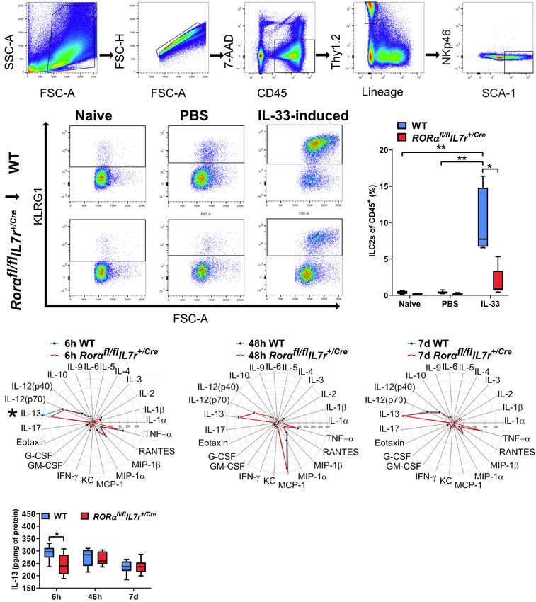
Zelco et al.                                                                                                        ILCs and Preterm Brain Injury

(wild-type controls) mouse littermates were used in the                  were incubated with an enzymatic solution composed of 0.01%
experiments. These mice were phenotypically indistinguishable            papain, 0.01% DNase I (Worthington, NJ, United States),
from commercially available C57Bl/6J mice. Genotyping of                 0.1% Dispase II (Roche, Sweden), and 12.4 mM MgSO4 in
Rorαfl/fl mice was undertaken using PCR primers (50 TGA                  Ca2+ /Mg2+ -free HBSS (Thermo Fisher Scientific, Sweden) at
GTG GTA ACA CCA CGG CAC GC 30 and 50 TGG AGC                             37◦ C for 20 min. The monocyte population was then separated
AGA ATC ATC CAG GAG GCC 30 ), giving a wild-type                         on a Percoll gradient (30/70%). After blocking non-specific
product of 573 bp and a targeted product of ∼650 bp.                     binding with Fc block (C16/32; clone 2.4G2; cat. 553142, BD
For genotyping of IL7-Cre mice, the PCR primers were                     Pharmingen), the following primary antibodies were used: anti-
50 CCT GAA AAC TTT GCC CCC TCC ATA 30 , 50                               lineage cocktail (FITC; CD3, CD5, B220, CD11b, CD11c, Gr-
CCA TAG AAT AGT GCA GCC TTG CCT C 30 , and                               1, TCRgd, and Ter-119; cat. 22-7770-72, eBioscience), anti-
50 AGC GAA AGC TCT ACC CAG AGC 30 , and these                            CD45 (APCcy7; clone 30-F77; cat. 103115, BioLegend) and anti-
generated a wild-type product of 584 bp and a targeted                   CD90 (Thy1.2, PEcy7; cat. ABIN477038, eBioscience). ILC2s
product of 680 bp.                                                       were identified by the addition of anti-SCA-1 (PE; clone D7;
   The animal facility had a 12-h light-dark cycle, and the mice         cat. 108107, BioLegend), anti-NKp46 (BV510; clone 29A1.4;
had free access to standard chow (B&K, Solna, Sweden) and                cat. 563455; BD Biosciences), anti-KLRG1 (APC; clone 2F1;
water. All experiments were performed with the approval of               cat. 561620, BD Biosciences), and anti-GATA3 PE (Catalog #
the Regional Animal Ethical Committee of Gothenburg (ethical             12-9966-42, eBiosciences) antibodies. For intracellular staining,
permit numbers 58/2016 and 2042/18). The experimental design             the permeabilization and fixation procedure was performed
and the number of animals used for each experiment are shown             according to manufacturer’s protocol. Briefly, after incubation
in Figure 1.                                                             with 250 µl of BD Cytofix/Cytoperm solution, the cells
                                                                         were washed in 1 × BD Perm/Wash buffer (BD Biosciences,
Hypoxic-Ischemic Model                                                   CA, United States) and resuspended in 100 µl of 1 × BD
The day of birth was defined as postnatal day (PND)0. At PND6,           Perm/Wash buffer before the GATA3 intracellular staining. The
HI surgery was performed as described previously (Albertsson             samples were run on a BD FACSCanto IITM flow cytometer.
et al., 2014). The pups were anesthetized with isoflurane (5% for        Fluorescence-minus-one controls for each antibody were run
induction and 3% for maintenance) and underwent unilateral               together with each experiment. Lung tissues in which ILC2s
left carotid artery ligation followed by xylocaine as the local          were enriched were included as positive controls for all
anesthetic. After recovering with their dam for 1 h, the pups were       brain tissue flow cytometry experiments, and the data were
transferred to a humidified chamber for exposure to 70 min of            analyzed with the FlowJo v10 software (Tree Star, Ashland,
hypoxia (10% O2 in N2 , 36.0 ± 0.5◦ C) (Rice et al., 1981). Before       OR, United States).
and after the hypoxia, the pups rested for 10 min at 36.0 ± 0.5◦ C          The lung single-cell suspensions were prepared following a
in the chamber. The pups were then returned to the dam’s cage            previously described method (Moro et al., 2015) with minor
until being sacrificed.                                                  modifications. Briefly, after perfusion the lungs were extracted,
                                                                         minced into small pieces, and then digested for 45 min at 37◦ C
IL-33 Stimulation of ILC2 Expansion                                      with the same enzyme mix used for the brain dissociation. The
To test the Cre efficiency in neonatal ILC2-impaired mice, we            supernatant was incubated with Red Blood Cell Lysing Buffer
challenged wild type and ILC2-impaired pups with IL-33, which            (R7757, Sigma-Aldrich, Sweden) at room temperature. Samples
promotes ILC2 expansion (Oliphant et al., 2014). Briefly, the            were then stained and analyzed as above.
pups received intraperitoneal (i.p.) injections of either mouse
recombinant IL-33 (0.04 µg/µl/g body weight in PBS, BioLegend)           Immunoassay for Cytokines and
or PBS alone (n = 6–9/group) every 24 h from PND3 to PND6
                                                                         Chemokines
for a total of four injections. The pups were sacrificed 24 h
                                                                         Samples were collected at 6 h, 48 h, and 7 days after HI from wild
after the last injection (PND7), and their lungs were collected
                                                                         type and ILC2-impaired mice (n = 7/group) for immunoassays of
for flow cytometry analysis to determine the number of IL-33–
                                                                         cytokine and chemokine protein expression. In summary, after
induced ILC2s.
                                                                         pentobarbital i.p. injection the animals underwent transcardial
                                                                         saline perfusion. The ipsilateral and contralateral hemispheres,
Flow Cytometry Experiments
                                                                         including the meninges, were collected separately and stored at
Flow cytometry was used to investigate the presence of all
                                                                         -80◦ C until processing. The Bio-Plex ProTM Mouse Cytokine 23-
ILCs in the brains of C57Bl/6J mice at 6 h, 24 h, 3 days,
                                                                         plex Assay (#m60009rdpd, Bio-Rad, Table 1) was used according
and 7 days after HI with naïve littermates as controls (n = 6–
                                                                         to the manufacturer’s instructions.
8/group). Transcardial saline perfusion was performed after i.p.
injection of pentobarbital (50 mg/ml, Abcur AB, Helsingborg,
Sweden). The brains were dissected out, the cerebellum was               Immunohistochemistry and
removed, and the ipsilateral and contralateral hemispheres,              Immunofluorescence Staining
including the meninges, were processed separately to obtain              At PND13 (7 days after HI), wild type and ILC2-impaired mice
single-cell suspensions as described previously (Zhang et al.,           were deeply anesthetized by i.p. injection with pentobarbital and
2017; Nazmi et al., 2018). In brief, brain homogenate samples            then perfused-fixed with 5% buffered formaldehyde (Histofix,

Frontiers in Cellular Neuroscience | www.frontiersin.org             3                                      August 2020 | Volume 14 | Article 249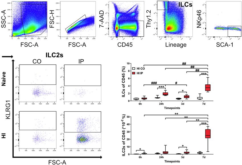
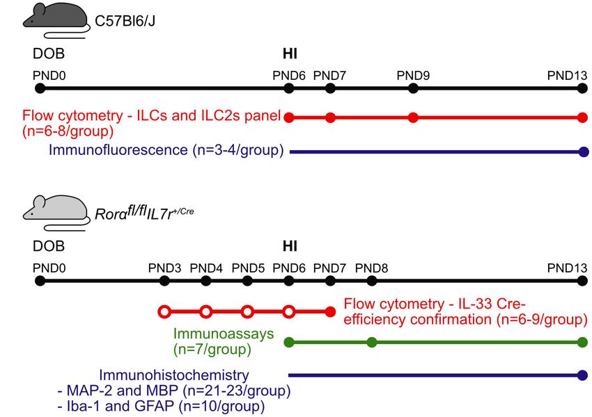
Zelco et al.                                                                                                                               ILCs and Preterm Brain Injury

  FIGURE 1 | Graphical abstract of the experiments. Representative scheme of the methods, numbers of animals, and time points used for experiments in (A) the
  C57Bl6/J mouse strain and (B) the Rorαfl/fl IL7rCre mouse strain. Colored dots correspond the data collection time points for the respective methods, while the
  empty dots correspond to the IL-33 injections. Abbreviations: DOB: date of birth, PND: postnatal day, IL-33: interleukin-33, HI: hypoxia-ischemia.

TABLE 1 | Cytokines and chemokines analyzed with the Bio-Plex ProTM Mouse Cytokine 23-plex Immunoassay.

Abbreviation                     Cytokine/Chemokine                      Abbreviation                    Cytokine/Chemokine

IL-1α                                 Interleukin 1α                         IL-17                       Interleukin 17
IL-1β                                 Interleukin 1β                        Eotaxin                      Eotaxin
IL-2                                  Interleukin 2                         G-CSF                        Granulocyte-colony stimulating factor
IL-3                                  Interleukin 3                        GM-CSF                        Granulocyte-macrophage colony-stimulating factor
IL-4                                  Interleukin 4                          IFN-γ                       Interferon γ
IL-5                                  Interleukin 5                           KC                         Chemokine (C-X-C motif) ligand 1
IL-6                                  Interleukin 6                         MCP-1                        Monocyte chemoattractant protein 1
IL-9                                  Interleukin 9                         MIP-1α                       Macrophage inflammatory protein 1α
IL-10                                 Interleukin 10                        MIP-1β                       Macrophage inflammatory protein 1β
IL-12(p40)                         Interleukin 12 p40                      RANTES                        Regulated on activation, normal T cell expressed and secreted
IL-12(p70)                         Interleukin 12 p70                        TNF-α                       Tumor necrosis factor α
IL-13                                 Interleukin 13

Histolab, Sweden). For Immunohistochemistry staining, the                                assessment, and microglial and astrocyte assessments, every
brains with meninges were collected and stored at 4◦ C in                                100th section was used (3 sections/animal). Briefly, the sections
Histofix before paraffin-embedding and sectioning. Coronal                               were de-paraffinized followed by antigen retrieval and 3%
sections of the forebrain were cut with a thickness of                                   H2 O2 in phosphate buffer. After blocking, sections were
7 µm. To obtain a representation of the areas of interest,                               incubated with mouse anti-microtubule-associated protein 2
the staining was performed for evaluating gray matter on                                 (MAP-2, 1:1,000 dilution; M4403, Sigma-Aldrich, United States),
every 50th section (6 sections/animal). For white-matter                                 mouse anti-myelin basic protein (MBP, 1:1,000 dilution;

Frontiers in Cellular Neuroscience | www.frontiersin.org                             4                                             August 2020 | Volume 14 | Article 249
Zelco et al.                                                                                                           ILCs and Preterm Brain Injury

SMI-94 Covance, United States), rabbit anti-ionized calcium                and ipsilateral hemispheres were measured separately in order
binding adaptor molecule 1 (Iba-1, 1:2000 dilution, 019-19741,             to calculate the tissue damage in the ipsilateral hemisphere
FUJIFILM Wako Chemicals, United States), and rabbit anti-                  against the internal control (the contralateral hemisphere). The
Glial fibrillary acidic protein (GFAP, 1:500, Z0334, Dako,                 total tissue loss was calculated with the following formula
Denmark) primary antibodies overnight at 4◦ C. The following               (Albertsson et al., 2014):
day, the sections were incubated with the corresponding
secondary antibodies for 1 h at room temperature. The                      (contralateral area − ipsilateral area)/contralateral area × 100%
sections were then incubated for 1 h in ABC Elite, and
                                                                           The total tissue loss volume was calculated as:
the immunoreactivity was visualized with 0.5 mg/ml 3.3-
diaminobenzidine in a buffer consisting of NiSO4 , β-D-                           Volume = sum of section area × section thickness
glucose, NH4 Cl, and β-D-glucose oxidase (all from Sigma-
Aldrich, Sweden).                                                                             × (1/sampling fraction)
    Immunofluorescence was performed to identify ILC2s at
7 days after HI (n = 3/group). After saline perfusion, the meninges        Quantification of Iba-1 Positive Brain
from each hemisphere were peeled off from the parenchyma                   Region and GFAP-Positive Astrocytes
and placed separately on a glass slide. Brains and lungs were              The border zone between the injured and non-injured brain
collected as well and stored in Histofix followed by sucrose               areas in the ipsilateral hemispheres, as well as the corresponding
30%. Both tissues were then frozen in isopentane and cut                   brain regions in the contralateral brain hemispheres were
at 30 µm of thickness. After fixation and blocking with 5%                 selected for analysis. Using ImageJ software (v1.52a, NIH,
donkey serum at room temperature for 1 h, the tissues were                 United States), GFAP-positive astrocytes were counted on
incubated with rat anti-mouse CD3 (1:500 dilution, cat. 100202,            captured images with an Olympus Optical microscope using
BioLegend) and rabbit anti-mouse ST2 (1:250 dilution, PA5-                 a 40× objective lens from selected areas in two regions of
20077, Invitrogen) primary antibodies overnight at 4◦ C. The               interest per section (in total 6 regions of interest per brain),
tissues were then incubated for 1 h at room temperature with               and expressed as cell density (cells/mm2 ). The Iba-1–positive
donkey anti-rat Alexa Fluor 488 (1:1,500 dilution, A21208,
                                             R
                                                                           stained brain regions were measured on the images which
Invitrogen) and donkey anti-rabbit Alexa Fluor 594 (1:1,500R
                                                                           were captured by a 20× objective lens from eight regions
dilution, A21207, Invitrogen) secondary antibodies. Lung tissue            of interest per brain. Analysis was performed on the images
was used as the positive control, and omitting the primary                 with final resolution of 1360 × 1024 pixel after adjusting
antibodies was used as the negative control (Supplementary                 threshold of positive stained areas on ImageJ. Data were
Figure 1). The slides were then stained with DAPI and afterward            expressed as the ratio between Iba-1 stained area and total
mounted with coverslips and ProLongTM gold antifade reagent                regions of interest.
(P36930, Invitrogen).
                                                                           Meningeal ILC2 Cell Counting
Quantification of Gray and White-Matter                                    A systematic set of Z-stacks of three regions of interest per
Tissue Loss                                                                hemisphere were acquired with a 20× objective lens on LSM
Gray-matter tissue loss was assessed by measuring MAP-2+ areas,            800 confocal microscope (Carl Zeiss, Germany). ILC2s (defined
either as a percentage of volume (mm3 ) or area (mm2 ) for                 as CD3− ST2+ ) were blindly counted in the height of the Z (z
each stained section at different levels as described previously           step = 1 µm), and volumes of each regions of interest were
(Zhang et al., 2017; Albertsson et al., 2018). Images of the stained       calculated. Estimation of number density was performed by
sections were acquired with an Olympus Optical microscope                  applying the following formula (McNaught and Wilkinson, 1997)
using a 1.25 × objective lens. MAP-2+ areas were delineated                of paired regions of interests from contralateral and ipsilateral
using ImageJ software (Rasband, W.S., US National Institutes of            meninges:
Health, United States1 ).                                                                            N = 6Q− /V
   To assess white-matter injury, a newly established automated
                                                                           N is the total number of cells per volume of brain region; 6Q− is
segmentation method, MyelinQ, was used to measure the
                                                                           the number of counted cells; V is the volume of regions of interest
size of the MBP+ areas (Mottahedin et al., 2018). Briefly,
                                                                           per sampling frame.
we captured images of each section using a 5 × objective
lens with the newCAST software (Visiopharm, Denmark) on
a modified Leica microscope (Leica DM6000 B, Germany)                      Statistical Analysis
equipped with a motorized stage (Ludl MAC 5000, United States)             All statistical analyses were performed with IBM SPSS Statistics
and a digital camera (Leica DFC 295, Germany). MyelinQ was                 25 (IBM Corp, Armonk, NY, United States). Data were
used for automatic detection of MBP+ areas in the whole                    tested for normal distribution through the generation of QQ
brain hemisphere.                                                          plots, and equality of variance was assessed by Leven’s test.
   For the quantification of gray and white-matter loss, the               If normally distributed, the data were analyzed with the
sizes of the MAP-2+ and MBP+ areas in the contralateral                    corresponding parametric test, while in the case when the
                                                                           data were non-normally distributed, appropriate non-parametric
1
    https://imagej.nih.gov/ij/, 1997–2016                                  tests were applied. All data are presented as boxplots (5th –

Frontiers in Cellular Neuroscience | www.frontiersin.org               5                                       August 2020 | Volume 14 | Article 249
Zelco et al. ILCs and Preterm Brain Injury
95th percentiles). Statistical significance was considered as of the ipsilateral hemisphere after HI (Figures 3C–E) and
p-values < 0.05. only few cells in the meninges contralaterally (Figures 3B,E,
p = 0.013), or in naïve mice (Figure 3A). In addition, we did not
observe ILC2s in the brain parenchyma in either the naïve mice
or after HI (data not shown). Because we observed this increase
RESULTS
of ILC2s in the injured brain, their role in brain injury after HI
was further investigated.
The Frequencies of ILCs and ILC2s
Increased in the Neonatal Mouse Brain ILC2-Impaired Neonatal Mice Did Not
After HI Injury Respond to IL-33 Stimulation
ILC2s are the subtype that are most frequently observed in the To explore the role of ILC2s in neonatal brain injury, we used
adult mouse brain compared to ILC1s and ILC3s (Russi et al., the ILC2-deficient mouse strain Rorαfl/fl IL7rCre (Oliphant et al.,
2015; Gadani et al., 2017). To determine if ILCs were present in 2014). To confirm the ILC2 deficiency in the newborn mice,
the neonatal mouse brain, we performed flow cytometry analysis which has not been investigated previously, we used IL-33, which
on single-cell suspensions isolated from brain homogenate is known to efficiently induce the expansion and activation of
(Figures 2A–C, Supplementary Figure 2), and measured the ILC2s (Oliphant et al., 2014; Van Dyken et al., 2014; Besnard et al.,
frequencies of both ILCs and ILC2s in naïve mice (n = 6/time 2015; Bartemes et al., 2017), including ILC2s in the CNS (Gadani
point) and in mice at 6 h (n = 8), 24 h (n = 7), 3 days et al., 2017). We performed IL-33 stimulation experiments to
(n = 8), and 7 days (n = 7) after HI. Because of the lack of compare the ILC2 response in wild type mice and Rorαfl/fl IL7rCre
single specific markers, we selected a panel of surface markers mice (Figures 4A,B, Supplementary Figure 4). Naïve mouse
in the FACS analysis for their identification, and ILCs were pups without any injection showed similar amount of ILC2s
defined as CD45+ Lin− Thy1.2+ (Pelly et al., 2016) and ILC2s between genotypes, and PBS injections did not evoke ILC2s
were defined as CD45+ Lin− Thy1.2+ SCA-1+ NKp46− KLRG1+ expansion, as shown by similar amount of ILC2s as in naïve mice
(Cardoso et al., 2017). (Figure 4B). In contrast, IL-33 triggered massive ILC2 expansion
For both ILCs and ILC2s, we did not detect any differences in wild type animals (n = 7), but failed to do so in Rorαfl/fl IL7rCre
between naive and contralateral brain hemispheres at any time mouse pups (n = 9, p = 0.006) (Figure 4B), thus confirming
points after HI; therefore, data from naïve mice are not shown. that the ILC2 expansion in response to IL-33 stimulation in
The frequencies of ILCs were significantly increased in the Rorαfl/fl IL7rCre mouse pups was significantly impaired.
brain hemisphere ipsilateral to the injury compared to the
uninjured contralateral hemisphere at 24 h (p < 0.001), 3 days ILC2 Impairment Attenuated the IL-13
(p = 0.010), and 7 days (p < 0.001) after HI, but not at Increase After HI
6 h after HI (Figure 2B). Quantification of ILC2s showed a To illustrate the inflammatory response in the brain after
significant increase in the ipsilateral hemisphere compared with HI, we performed a Bio-Plex ProTM Mouse Cytokine 23-
the uninjured contralateral hemisphere at 6 h (p = 0.011), 3 days plex Assay using brain homogenate from wild type and
(p = 0.020), and 7 days (p < 0.001) after HI (Figure 2C). Rorαfl/fl IL7rCre mice. This method allowed the study of 23
Furthermore, the frequencies of both ILCs (Figure 2B) and different cytokine/chemokines at the same time (Table 1) at
ILC2s (Figure 2C) in the ipsilateral hemisphere of HI mice 6 h, 48 h, and 7 days after HI (n = 7/group) (Figures 4C–
showed an increasing trend over time, with the highest at 7 days F). IL-13 protein levels in the brain were significantly lower at
after HI for both populations. We observed a first small peak for 6 h after HI in Rorαfl/fl IL7rCre mice compared with wild-type
ILCs at 24 h, which was higher than 6 h (p < 0.001) and the mice (p = 0.046) (Figures 4C,F), while no significant differences
subsequent 3 days (p = 0.031) time points. The ILCs showed a were observed for any other cytokine/chemokine between wild
second peak and were significantly greater at 7 days compared type and Rorαfl/fl IL7rCre mice (Figures 4C–E) in the ipsilateral
with 6 h (p < 0.001), 24 h (p = 0.009), and 3 days (p = 0.002) hemispheres at any of the time points analyzed.
after HI (Figure 2B). ILC2s instead increased drastically only
at 7 days compared with 6 h (p < 0,001), 24 h (p = 0.002), ILC2 Impairment Did Not Affect Tissue
and 3 days (p = 0.002) after HI (Figure 2C). The increased
ILC2s at 7 days after HI in the ipsilateral hemisphere were
Loss or the Neuroinflammatory
further confirmed using intracellular staining for GATA3, the Response in the Neonatal Mouse Brain
key transcription factor and master regulator that is critical for After HI
the development and maintenance of ILC2s (Yagi et al., 2014) Next, we investigated the involvement of ILC2s in HI-induced
(Supplementary Figure 3). preterm brain injury using the Rorαfl/fl IL7rCre mouse strain.
ILC2s are known to be enriched in the meninges compared Brain injury was evaluated at 7 days after HI for both the
with the brain parenchyma (Gadani et al., 2017; Russi et al., gray and white matter. ILC2 impairment did not impact the
2018). We examined the presence of ILC2s at 7 days after severity of gray-matter injury either in terms of total tissue
HI, when ILC2 accumulation in the brain peaked, using loss (Figure 5B) or at different brain levels (Figure 5C) as
immunofluorescent staining of the meninges compared to naïve evaluated by immunohistochemical staining of the neuronal
mice (Figures 3A–E). ILC2s were found mostly in the meninges marker MAP-2 (Figures 5A–C). To evaluate white-matter
Frontiers in Cellular Neuroscience | www.frontiersin.org 6 August 2020 | Volume 14 | Article 249Zelco et al. ILCs and Preterm Brain Injury FIGURE 2 | ILCs and ILC2s increased in the ipsilateral hemispheres in the neonatal mouse brain in a time-dependent fashion after HI injury. Single-cell suspensions generated from brain homogenates were analyzed using flow cytometry in mouse pups at different time points after HI, with naïve C57Bl6/J mouse littermates as controls (n = 6–8/group). (A) Representative flow cytometry plots showing the gating strategy for ILCs and ILC2s. Both are represented as percentages of the CD45+ population. ILCs (B) and ILC2s (C) in the ipsilateral and contralateral hemisphere at 24 h, 3 days, and 7 days after HI. A mixed model ANOVA with Games-Howell post-hoc were used. *#: p < 0.05, **##: p < 0.01, ***###: p < 0.001. in panels (B,C). Abbreviations: N: naïve, HI: hypoxia-ischemia, IP: ipsilateral hemisphere, CO: contralateral hemisphere. injury, we performed MBP immunohistochemical staining DISCUSSION (Figure 5D), and the MBP+ white-matter volumes in the whole brain hemisphere (Figure 5E) and areas at different ILCs and ILC2s have previously been found in the mouse brain levels (Figure 5F) were measured. Similarly, ILC2 impairment and have recently been studied in adult murine models of various did not affect the tissue loss in white matter in the whole brain pathologies (Mair and Becher, 2014; Besnard et al., 2015; brain hemisphere (Figures 5E,F) or in the subcortical white- Hatfield and Brown, 2015; Russi et al., 2015; Gadani et al., 2017; matter area (data not shown). Further, no sex difference was Russi et al., 2018; Romero-Suarez et al., 2019; Fung et al., 2020). noted regarding either the gray or white-matter injury after HI Here we found accumulation of these cells in the meninges after (data not shown). HI injury in neonatal mice, and to our knowledge this is the first To explore the neuroinflammatory response in Rorαfl/fl IL7rCre study to investigate their presence and function in a neonatal after HI insult, we examined the microglia and astrocyte reactivity mouse model of brain injury. using immunochemistry staining for the microglia marker Iba- Among ILCs, ILC2 is the most abundant subtype in the 1 and the astrocyte marker GFAP. At 7 days after HI, there healthy adult mouse brain (Gadani et al., 2017; Russi et al., were significantly increased staining for microglia (Figures 5G– 2018). ILC2s have been shown to be increased in tissues in K) and number of astrocytes (Figures 5L–P) in the ipsilateral different disease models such as spinal cord injury (Gadani et al., hemispheres in both the wild type (microglia: p = 0.001, 2017), experimental autoimmune encephalomyelitis (Russi et al., astrocytes: p = 0.015) and Rorαfl/fl IL7rCre mice (microglia: 2015; Russi et al., 2018), experimental cerebral malaria (Besnard p = 0.003, astrocytes: p < 0.001); however, no differences et al., 2015), and aging (Fung et al., 2020). In our study, ILCs were observed for microglia or astrocytes after HI between and ILC2s were present in the normal neonatal mouse brain, the wild type and Rorαfl/fl IL7rCre mice in either of the two and after HI insult ILCs and particularly ILC2s accumulated brain hemispheres. in the injured brain, which agrees with previous studies Frontiers in Cellular Neuroscience | www.frontiersin.org 7 August 2020 | Volume 14 | Article 249
Zelco et al.                                                                                                                               ILCs and Preterm Brain Injury

  FIGURE 3 | The presence of ILC2s in the meninges in neonatal mice after HI. Representative confocal images of immunofluorescent staining showing the presence
  of ILC2s (CD3- ST2+ , arrowheads) in the meninges taken from brain hemispheres in a naïve mouse (A) and from the contralateral hemisphere (B) and ipsilateral
  hemisphere (C) in a mouse at 7 days after HI. (D) High magnification orthogonal views of cells stained with ST2. The merged picture shows cytoplasm localization of
  ST2 surrounding DAPI+ nuclei. Scale bars: (A–C) 100 µm; (D) 10 µm. (E) The number density of ILC2-positive cells in the meninges in the ipsilateral hemisphere and
  contralateral hemispheres after HI. Paired t-test was used. *p < 0.05. Abbreviations: HI: hypoxia-ischemia, IP: ipsilateral hemisphere, CO: contralateral hemisphere.

(Mair and Becher, 2014; Hatfield and Brown, 2015; Romero-                              tissue (Oliphant et al., 2014; Van Dyken et al., 2014; Besnard
Suarez et al., 2019). This accumulation occurred in a time-                            et al., 2015; Bartemes et al., 2017) and the CNS (Gadani et al.,
dependent manner, and reached the highest level at 7 days                              2017). In the current study, we found that neonatal ILC2s in
after HI. This indicated that ILCs, and especially ILC2s, are                          the lung tissue in the wild type mice were also expanded by
stimulated and expanded by HI-induced tissue injury. ILC2s                             IL-33 stimulation, which did not occur in the ILC2-deficient
are recognized as tissue-resident cells (Moro et al., 2010; Neill                      mice. Together, this supports the hypothesis that ILC2s in
et al., 2010; Price et al., 2010; Molofsky et al., 2013) and                           both the peripheral nervous system and CNS are able to
are expanded upon IL-33 stimulation in both the peripheral                             respond and expand to either a stimulator like IL-33 and/or

Frontiers in Cellular Neuroscience | www.frontiersin.org                           8                                               August 2020 | Volume 14 | Article 249
Zelco et al.                                                                                                                                 ILCs and Preterm Brain Injury

  FIGURE 4 | ILC2s in Rorαfl/fl IL7rCre mice did not expand after IL-33 stimulation, but their cytokine/chemokine profile in the brain after HI was similar to wild-type
  mice. (A) Representative flow cytometry plots show the gating strategy for IL-33–stimulated ILC2 expansion in the lungs (n = 7–9/group). (B) IL-33–stimulated ILC2
  expansion in wild-type and Rorαfl/fl IL7rCre mouse pups. (C–E) Radar plots from immunoassays show the cytokine/chemokine changes in the ipsilateral hemisphere
  from the wild type and Rorαfl/fl IL7rCre mouse pups at 6 h (C), 48 h (D), and 7 days (E) after HI. (F) IL-13 in the ipsilateral hemispheres in the wild type and
  Rorαfl/fl IL7rCre mice at different time points after HI. *p < 0.05, **p < 0.01. Mann–Whitney U-tests were used in (B), and 2-Way ANOVA with Games-Howell
  post-hoc were used in (F). Abbreviations: HI: hypoxia-ischemia, IP: ipsilateral hemisphere, WT: wild type.

stress induced by tissue injury and that they are functional                            resident in the meninges and were seldom found in the
already in early life.                                                                  brain parenchyma under either normal conditions or
   To identify the localization of ILC2 in the neonatal                                 after HI-induced brain injury, thus suggesting a role for
mouse brain, we performed immunofluorescence staining                                   meningeal immune cells as sentinels for brain-derived
and found that – similar to previous findings (Gadani                                   alarmins as part of the immune response after brain
et al., 2017) – ILC2s in the neonatal mouse brain were                                  injury in neonates.

Frontiers in Cellular Neuroscience | www.frontiersin.org                            9                                               August 2020 | Volume 14 | Article 249
Zelco et al.                                                                                                                                     ILCs and Preterm Brain Injury

  FIGURE 5 | ILC2 impairment did not affect brain tissue loss 7 days after HI. (A,D) Representative pictures show immunochemistry staining for MAP-2 (A) and MBP
  (D). Gray matter (A–C) in Rorαfl/fl IL7rCre mice (n = 23) after HI compared to WT littermates (n = 21) in terms of brain tissue volume loss (B) or area loss per level (C).
  White matter (D–F) tissue loss in the whole hemisphere in terms of tissue volume loss (E) or area loss at each level (F) in the wild type and Rorαfl/fl IL7rCre mice.
  Representative images (G–J,L–O) and quantification (K,P) of Iba-1+ cells (G–K) and GFAP+ astrocytes (L–P) in the wild type and Rorαfl/fl IL7rCre mouse pups at
  7 days after HI. Paired t-test was used for comparisons between brain hemispheres, and independent t-test was used for comparisons between genotypes. Scale
  bars: 100 µm. Abbreviations: MAP-2: microtubule-associated protein-2, MBP: myelin basic protein; WT: wild type, IP: ipsilateral hemisphere. CO: contralateral
  hemisphere. *p < 0.05, **p < 0.01, ***p < 0.001.

   In the current study, among all the cytokine/chemokines                                 and an important modulator of peripheral allergic reactions. In
examined, we found a small yet significant decrease in expression                          the brain, IL-13 contributes to the death of activated microglia
of IL-13 in the ILC2-deficient mice compared with the wild                                 (Yang et al., 2002; Yang et al., 2006) and potentiates the
type mice. IL-13 is considered an anti-inflammatory cytokine                               effects of oxidative stress on neurons during neuroinflammation

Frontiers in Cellular Neuroscience | www.frontiersin.org                              10                                                August 2020 | Volume 14 | Article 249
Zelco et al.                                                                                                               ILCs and Preterm Brain Injury

(Park et al., 2009), and both neuroprotective and neurotoxic                   or not there are compensatory changes in ILC1 and ILC3 that
effects of IL-13 have been proposed. IL-13 has been previously                 contribute to the lack of an effect from ILC2 deficiency also needs
found to be increased both in the serum of term newborns                       to be explored in the future.
(Orrock et al., 2016; Al-Shargabi et al., 2017; Massaro et al.,                    Exploration of the neuroinflammatory response after HI by
2018) and in brain mRNA levels in mice (Albertsson et al.,                     immunohistochemistry revealed increased numbers of astrocytes
2014) early after HI injury. In newborns, increased serum IL-                  and staining for microglia in the ipsilateral hemisphere at 7 days
13 levels are associated with worse brain injury (Massaro et al.,              after HI in both ILC2-deficient and wild-type mice, and this
2018), reduced heart rate variability metrics (Al-Shargabi et al.,             agreed with previous findings (Doverhag et al., 2010; Bonestroo
2017), and adverse outcome after 24 h of therapeutic hypothermia               et al., 2013). However, there was no significant difference in
(Orrock et al., 2016). ILC2s are one of the major producers of IL-             astrocytes and microglia in either of the brain hemispheres
13 in response to insults in different tissues (Fallon et al., 2006;           between wild type and ILC2-deficient mice, and these results are
Price et al., 2010; Klein Wolterink et al., 2012; Kabata et al., 2018),        thus in line with our findings that no significant differences were
including in a model of spinal cord injury, where IL-13+ cells                 observed for cytokines/chemokines between wild type and ILC2-
were found to make up the majority ILC2s (Gadani et al., 2017).                deficient mice apart from IL-13 and that there were no differences
The reduction in IL-13 protein levels that we observed here might              in the severity of brain injury between the two mouse genotypes.
suggest that ILC2s have a detrimental role. However, in spite                      We found increased accumulation of ILC2s in the injured
of the observed significant increase in ILC2s in the brain after               brain after HI, but ILC2 deficiency did not affect the severity of
the HI insult, ILC2 impairment did not affect the HI-induced                   the injury. We found no differences in the neuroinflammatory
inflammatory responses in the brain or the extent of brain injury              response after HI between ILC2-deficient and wild-type mice
after HI in neonatal mice. Similarly, a previous study showed                  that might partly explain the lack of effect of ILC2 deficiency
that ILCs infiltrated the brain in an experimental autoimmune                  on brain injury. However, the in-depth molecular mechanisms
encephalomyelitis model but did not affect the severity of injury              behind these findings are not known. A limitation of the study
(Mair and Becher, 2014). Further, we did not observe any sex                   is that we only assessed effect of ILC2 deficiency on brain injury
differences regarding to the degree of injury in the brain of ILC2-            at 7 days after HI. We cannot exclude the possibility that ILC2
deficient mice compared with wild-type mice, although it has                   deficiency might have an effect at a later time point after HI.
been reported previously that the role of ILC2 is sex-dependent                    In conclusion, ILCs and ILC2s accumulate in the injured brain
under certain circumstances (Bartemes et al., 2017; Russi et al.,              after HI insult in the neonatal mouse brain. However, ILC2s did
2018). Even though ILCs were increased in the CNS after insult,                not affect the major inflammatory response in the brain and did
it is possible that there are still not enough ILCs to affect the brain        not contribute to the development of brain damage in this mouse
damage development in a substantial manner. Indeed, ILC2s                      model of preterm brain injury.
reside mainly in the meninges and choroid plexus, and not in the
parenchyma, and their frequencies are generally low in relation
to the whole leukocyte population (Hatfield and Brown, 2015;
Gadani et al., 2017; Russi et al., 2018; Fung et al., 2020).
                                                                               DATA AVAILABILITY STATEMENT
    ILC2s were the main cell type shown to be responsible                      All datasets generated for this study are included in the
for allergic response in a mouse model of lung inflammation                    article/Supplementary Material.
(Van Dyken et al., 2014). However, genetic ablation of ILC2s
triggered an increase in gamma delta T-cells, thus revealing
compensatory mechanisms among innate immune cells (Van
Dyken et al., 2014). Another example of cross talk between ILC2s               ETHICS STATEMENT
and other innate immune cells was found in a murine model of
experimental autoimmune encephalomyelitis where both mast                      The animal study was reviewed and approved by the Regional
cells and ILC2s were coordinated in the development of brain                   Animal Ethical Committee of Gothenburg (ethical permit
damage (Russi et al., 2018). We speculate that such cross talk                 numbers: 58/2016 and 2042/18).
and compensatory mechanisms between ILC2s and other innate
immune cells might be among the reasons why ILC2 impairment
did not impact HI-induced brain injury in neonatal mice.                       AUTHOR CONTRIBUTIONS
    In addition, the role of other ILCs such as ILC1 and ILC3
remains an important topic for future study. The lymphoid tissue               XW conceptualized the study. AZ, ER-F, and AN designed the
inducer cells comprise the ILC3 population. They present as early              experiments and performed the flow cytometry experiments. AZ
as the embryonic stage and play an important role in intestinal                and ER-F performed the HI model. AZ performed the protein
homeostasis after birth (Eberl, 2012), this might indicate the                 assays, immunohistochemistry, and immunofluorescence
importance of ILC3s in the immune response in the perinatal                    staining. AZ and TC performed fluorescent imaging and
period during development. In the current study, we did not                    analyzed the immunofluorescence data. AZ and MA performed
observe any obvious increase of ILC1 and ILC3 in the brain after               the statistical analysis. AZ and XW drafted the manuscript. All
HI (data not shown), which might be due to the time points at                  authors contributed to data interpretation and critical revision of
which ILC1 and ILC3 levels were assessed. Furthermore, whether                 the manuscript and approved the submitted version.

Frontiers in Cellular Neuroscience | www.frontiersin.org                  11                                       August 2020 | Volume 14 | Article 249
Zelco et al. ILCs and Preterm Brain Injury
FUNDING thank Anna Rehammar from Mathematical Sciences, Chalmers
University of Technology and University of Gothenburg, and
This work was supported by the Swedish Research Council Thomas Karlsson from Biostatistics, School of Public Health
(2018-02682 to XW), the Brain Foundation (FO2017-0102 to and Community Medicine, Institute of Medicine, University of
XW), grants from the Swedish state under the agreement Gothenburg for the statistic consultations.
between the Swedish Government and the county councils,
the ALF-agreement (ALFGBG-813291 to XW), the National
Natural of Science Foundation of China (81771418 to XW), SUPPLEMENTARY MATERIAL
H&K Jacobssons stiftelse (to AZ, 7/h18), Wilhelm & Martina
Lundgren’s Foundation (to AZ, 2017-1972), and the Frimurare The Supplementary Material for this article can be found
Barnhus Foundation (150511 to AN). online at: https://www.frontiersin.org/articles/10.3389/fncel.
2020.00249/full#supplementary-material
FIGURE S1 | Representative images show the positive controls (lung tissue, A),
ACKNOWLEDGMENTS and negative controls omitting the primary antibodies for the immunofluorescent
staining of ST2 and CD3 for lung tissue (B) and meninges (C). Arrow head: ST2+
We thank Anna-Lena Leverin (Department of Physiology, ILC2s, star: CD3+ cells. Scale bars: 50 µm.
Sahlgrenska Academy, University of Gothenburg) for help FIGURE S2 | Representative flow cytometry plots for fluorescence-minus-one
with the protein assays and Kajsa Groustra (Experimental controls (FMO) used in flow cytometry experiments.
Biomedicine, Sahlgrenska Academy, University of Gothenburg)
FIGURE S3 | (A) Representative flow cytometry plots for intracellular staining of
for assistance with animal maintenance and breeding. The GAGA-3. (B) The frequency of Lin− CD45+ GATA3+ ILC2s at 7 days post HI in the
Rorαfl/fl IL7rCre mouse strain was a kind gift from Dr. Andrew neonatal wild-type mice. Paired t-test was used in B for comparison between
McKenzie (Cambridge University, United Kingdom) with the hemispheres. ∗ p < 0.05.
kind permission from Hans-Reimer Rodewald (Deutsches FIGURE S4 | Representative flow cytometry plots for fluorescence-minus-one
Krebsforschungszentrum, Heidelberg, Germany) for the use of controls (FMO) used in flow cytometry experiments for the lung tissue
IL7rCre mice in the ILC2-impaired mouse strain. We also IL-33 experiments.
REFERENCES Cheon, I. S., Son, Y. M., Jiang, L., Goplen, N. P., Kaplan, M. H., Limper,
A. H., et al. (2018). Neonatal hyperoxia promotes asthma-like features through
Adkins, B., Leclerc, C., and Marshall-Clarke, S. (2004). Neonatal adaptive IL-33-dependent ILC2 responses. J. Allergy Clin. Immunol. 142, 1100–1112.
immunity comes of age. Nat. Rev. Immunol. 4, 553–564. doi: 10.1038/nri1394 doi: 10.1016/j.jaci.2017.11.025
Albertsson, A., Bi, D., Duan, L., Zhang, X., Leavenworth, J. W., Qiao, L., et al. Debock, I., and Flamand, V. (2014). Unbalanced neonatal CD4(+) T-cell
(2014). The immune response after hypoxia-ischemia in a mouse model of immunity. Front. Immunol. 5:393. doi: 10.3389/fimmu.2014.00393
preterm brain injury. J. Neuroinflamm. 11:153. Doverhag, C., Hedtjarn, M., Poirier, F., Mallard, C., Hagberg, H., Karlsson, A.,
Albertsson, A., Zhang, X., Vontell, R., Bi, D., Bronson, R. T., Supramaniam, V., et al. (2010). Galectin-3 contributes to neonatal hypoxic-ischemic brain injury.
et al. (2018). Gammadelta T cells contribute to injury in the developing brain. Neurobiol. Dis. 38, 36–46. doi: 10.1016/j.nbd.2009.12.024
Am. J. Pathol. 188, 757–767. Eberl, G. (2012). Development and evolution of RORgammat+ cells in a microbe’s
Almeida, F. F., and Belz, G. T. (2016). Innate lymphoid cells: models of plasticity world. Immunol. Rev. 245, 177–188. doi: 10.1111/j.1600-065x.2011.01
for immune homeostasis and rapid responsiveness in protection. Mucosal 071.x
Immunol. 9, 1103–1112. doi: 10.1038/mi.2016.64 Fallon, P. G., Ballantyne, S. J., Mangan, N. E., Barlow, J. L., Dasvarma, A.,
Al-Shargabi, T., Govindan, R. B., Dave, R., Metzler, M., Wang, Y., Du Plessis, A., Hewett, D. R., et al. (2006). Identification of an interleukin (IL)-25-dependent
et al. (2017). Inflammatory cytokine response and reduced heart rate variability cell population that provides IL-4, IL-5, and IL-13 at the onset of helminth
in newborns with hypoxic-ischemic encephalopathy. J. Perinatol. 37, 668–672. expulsion. J. Exp. Med. 203, 1105–1116.
doi: 10.1038/jp.2017.15 Forsberg, A., Bengtsson, M., Eringfalt, A., Ernerudh, J., Mjosberg, J., and Jenmalm,
Bartemes, K., Chen, C. C., Iijima, K., Drake, L., and Kita, H. (2017). IL-33- M. C. (2014). GATA binding protein 3(+) group 2 innate lymphoid cells
responsive group 2 innate lymphoid cells are regulated by female sex hormones are present in cord blood and in higher proportions in male than in female
in the uterus. J. Immunol. 200, 229–236. doi: 10.4049/jimmunol.1602085 neonates. J. Allergy Clin. Immunol. 134, 228–230.
Bernink, J. H., Germar, K., and Spits, H. (2014). The role of ILC2 in pathology of Fung, I. T. H., Sankar, P., Zhang, Y., Robison, L. S., Zhao, X., D’souza, S. S., et al.
type 2 inflammatory diseases. Curr. Opin. Immunol. 31, 115–120. doi: 10.1016/ (2020). Activation of group 2 innate lymphoid cells alleviates aging-associated
j.coi.2014.10.007 cognitive decline. J. Exp. Med. 217:e20190915.
Besnard, A. G., Guabiraba, R., Niedbala, W., Palomo, J., Reverchon, F., Shaw, Gadani, S. P., Smirnov, I., Smith, A. T., Overall, C. C., and Kipnis, J.
T. N., et al. (2015). IL-33-mediated protection against experimental cerebral (2017). Characterization of meningeal type 2 innate lymphocytes and their
malaria is linked to induction of type 2 innate lymphoid cells, M2 macrophages response to CNS injury. J. Exp. Med. 214, 285–296. doi: 10.1084/jem.2016
and regulatory T cells. PLoS Pathog. 11:e1004607. doi: 10.1371/journal.ppat. 1982
1004607 Hagberg, H., Mallard, C., Ferriero, D. M., Vannucci, S. J., Levison, S. W., Vexler,
Bonestroo, H. J., Nijboer, C. H., Van Velthoven, C. T., Kavelaars, A., Hack, C. E., Z. S., et al. (2015). The role of inflammation in perinatal brain injury. Nat. Rev.
Van Bel, F., et al. (2013). Cerebral and hepatic inflammatory response after Neurol. 11, 192–208.
neonatal hypoxia-ischemia in newborn rats. Dev. Neurosci. 35, 197–211. doi: Hatfield, J. K., and Brown, M. A. (2015). Group 3 innate lymphoid cells accumulate
10.1159/000346685 and exhibit disease-induced activation in the meninges in EAE. Cell Immunol.
Cardoso, V., Chesne, J., Ribeiro, H., Garcia-Cassani, B., Carvalho, T., Bouchery, 297, 69–79. doi: 10.1016/j.cellimm.2015.06.006
T., et al. (2017). Neuronal regulation of type 2 innate lymphoid cells via Herz, J., Koster, C., Crasmoller, M., Abberger, H., Hansen, W., Felderhoff-
neuromedin U. Nature 549, 277–281. doi: 10.1038/nature23469 Muser, U., et al. (2018). Peripheral T cell depletion by FTY720 exacerbates
Frontiers in Cellular Neuroscience | www.frontiersin.org 12 August 2020 | Volume 14 | Article 249Zelco et al. ILCs and Preterm Brain Injury hypoxic-ischemic brain injury in neonatal mice. Front. Immunol. 9:1696. doi: Orrock, J. E., Panchapakesan, K., Vezina, G., Chang, T., Harris, K., Wang, Y., 10.3389/fimmu.2018.01696 et al. (2016). Association of brain injury and neonatal cytokine response during Jones, R., Cosway, E. J., Willis, C., White, A. J., Jenkinson, W. E., Fehling, H. J., therapeutic hypothermia in newborns with hypoxic-ischemic encephalopathy. et al. (2018). Dynamic changes in intrathymic ILC populations during murine Pediatr. Res. 79, 742–747. doi: 10.1038/pr.2015.280 neonatal development. Eur. J. Immunol. 48, 1481–1491. doi: 10.1002/eji. Park, K. W., Baik, H. H., and Jin, B. K. (2009). IL-13-induced oxidative 201847511 stress via microglial NADPH oxidase contributes to death of hippocampal Juul, S. E., Comstock, B. A., Wadhawan, R., Mayock, D. E., Courtney, neurons in vivo. J. Immunol. 183, 4666–4674. doi: 10.4049/jimmunol.080 S. E., Robinson, T., et al. (2020). A randomized trial of erythropoietin 3392 for neuroprotection in preterm infants. N. Engl. J. Med. 382, Pelly, V. S., Kannan, Y., Coomes, S. M., Entwistle, L. J., Ruckerl, D., Seddon, 233–243. B., et al. (2016). IL-4-producing ILC2s are required for the differentiation of Kabata, H., Moro, K., and Koyasu, S. (2018). The group 2 innate lymphoid cell TH2 cells following Heligmosomoides polygyrus infection. Mucosal Immunol. 9, (ILC2) regulatory network and its underlying mechanisms. Immunol. Rev. 286, 1407–1417. doi: 10.1038/mi.2016.4 37–52. doi: 10.1111/imr.12706 Price, A. E., Liang, H. E., Sullivan, B. M., Reinhardt, R. L., Eisley, C. J., Erle, Klein Wolterink, R. G., Kleinjan, A., Van Nimwegen, M., Bergen, I., De Bruijn, M., D. J., et al. (2010). Systemically dispersed innate IL-13-expressing cells in type Levani, Y., et al. (2012). Pulmonary innate lymphoid cells are major producers 2 immunity. Proc. Natl. Acad. Sci. U.S.A. 107, 11489–11494. doi: 10.1073/pnas. of IL-5 and IL-13 in murine models of allergic asthma. Eur. J. Immunol. 42, 1003988107 1106–1116. doi: 10.1002/eji.201142018 Rice, J. E., Vannucci, R. C., and Brierley, J. B. (1981). The influence of immaturity Kolosowska, N., Keuters, M. H., Wojciechowski, S., Keksa-Goldsteine, V., Laine, on hypoxic-ischemic brain damage in the rat. Annal. Neurol. 9, 131–141. doi: M., Malm, T., et al. (2019). Peripheral administration of IL-13 induces anti- 10.1002/ana.410090206 inflammatory microglial/macrophage responses and provides neuroprotection Romero-Suarez, S., Del Rio Serrato, A., Bueno, R. J., Brunotte-Strecker, D., Stehle, in ischemic stroke. Neurotherapeutics 16, 1304–1319. doi: 10.1007/s13311-019- C., Figueiredo, C. A., et al. (2019). The central nervous system contains ILC1s 00761-0 that differ from NK cells in the response to inflammation. Front. Immunol. Mair, F., and Becher, B. (2014). Thy1+ Sca1+ innate lymphoid cells infiltrate 10:2337. doi: 10.3389/fimmu.2019.02337 the CNS during autoimmune inflammation, but do not contribute to disease Russi, A. E., Ebel, M. E., Yang, Y., and Brown, M. A. (2018). Male-specific IL-33 development. Eur. J. Immunol. 44, 37–45. doi: 10.1002/eji.201343653 expression regulates sex-dimorphic EAE susceptibility. Proc. Natl. Acad. Sci. Massaro, A. N., Wu, Y. W., Bammler, T. K., Comstock, B., Mathur, A., Mckinstry, U.S.A. 115, E1520–E1529. R. C., et al. (2018). Plasma biomarkers of brain injury in neonatal hypoxic- Russi, A. E., Walker-Caulfield, M. E., Ebel, M. E., and Brown, M. A. (2015). ischemic encephalopathy. J Pediatr 194, 67.e1–75.e1. Cutting edge: c-Kit signaling differentially regulates type 2 innate lymphoid McNaught, A. D., and Wilkinson, A. (1997). Compendium of Chemical cell accumulation and susceptibility to central nervous system demyelination in Terminology. Oxford: Blackwell Science Oxford. male and female SJL mice. J. Immunol. 194, 5609–5613. doi: 10.4049/jimmunol. Miller, D., Motomura, K., Garcia-Flores, V., Romero, R., and Gomez-Lopez, N. 1500068 (2018). Innate lymphoid cells in the maternal and fetal compartments. Front. Shankaran, S. (2015). Therapeutic hypothermia for neonatal encephalopathy. Curr. Immunol. 9:2396. doi: 10.3389/fimmu.2018.02396 Opin. Pediatr. 27, 152–157. doi: 10.1097/mop.0000000000000199 Molofsky, A. B., Nussbaum, J. C., Liang, H. E., Van Dyken, S. J., Cheng, L. E., Shankaran, S., Laptook, A., Ehrenkran, Z. R., Tyson, J., Mcdonald, S., Donovan, Mohapatra, A., et al. (2013). Innate lymphoid type 2 cells sustain visceral E., et al. (2005). Whole-Body hypothermia for neonates with hypoxic–ischemic adipose tissue eosinophils and alternatively activated macrophages. J. Exp. Med. encephalopathy. N. Engl. J. Med. 353, 1574–1584. 210, 535–549. doi: 10.1084/jem.20121964 Song, J., Sun, H., Xu, F., Kang, W., Gao, L., Guo, J., et al. (2016). Recombinant Moro, K., Ealey, K. N., Kabata, H., and Koyasu, S. (2015). Isolation and analysis of human erythropoietin improves neurological outcomes in very preterm infants. group 2 innate lymphoid cells in mice. Nat. Protoc. 10, 792–806. doi: 10.1038/ Ann. Neurol. 80, 24–34. doi: 10.1002/ana.24677 nprot.2015.047 Spits, H., Artis, D., Colonna, M., Diefenbach, A., Di Santo, J. P., Eberl, G., et al. Moro, K., Yamada, T., Tanabe, M., Takeuchi, T., Ikawa, T., Kawamoto, H., (2013). Innate lymphoid cells–a proposal for uniform nomenclature. Nat. Rev. et al. (2010). Innate production of T(H)2 cytokines by adipose tissue- Immunol. 13, 145–149. doi: 10.1038/nri3365 associated c-Kit(+)Sca-1(+) lymphoid cells. Nature 463, 540–544. doi: 10. Van Dyken, S. J., Mohapatra, A., Nussbaum, J. C., Molofsky, A. B., Thornton, E. E., 1038/nature08636 Ziegler, S. F., et al. (2014). Chitin activates parallel immune modules that direct Mottahedin, A., Zhang, X., Zelco, A., Ardalan, M., Lai, J. C. Y., Mallard, distinct inflammatory responses via innate lymphoid type 2 and gammadelta T C., et al. (2018). A novel image segmentation method for the evaluation cells. Immunity 40, 414–424. doi: 10.1016/j.immuni.2014.02.003 of inflammation-induced cortical and hippocampal white matter injury in Vivier, E., Artis, D., Colonna, M., Diefenbach, A., Di Santo, J. P., Eberl, G., et al. neonatal mice. J. Chem. Neuroanat. 96, 79–85. doi: 10.1016/j.jchemneu.2018. (2018). Innate lymphoid cells: 10 years on. Cell 174, 1054–1066. 12.009 Walsh, J. T., Hendrix, S., Boato, F., Smirnov, I., Zheng, J., Lukens, J. R., et al. Natalucci, G., Latal, B., Koller, B., Ruegger, C., Sick, B., Held, L., et al. (2016). (2015). MHCII-independent CD4+ T cells protect injured CNS neurons via Effect of early prophylactic high-dose recombinant human erythropoietin in IL-4. J. Clin. Invest. 125:2547. doi: 10.1172/jci82458 very preterm infants on neurodevelopmental outcome at 2 years: a randomized Xu, Y., Romero, R., Miller, D., Silva, P., Panaitescu, B., Theis, K. R., et al. (2018). clinical trial. Jama 315, 2079–2085. Innate lymphoid cells at the human maternal-fetal interface in spontaneous Nazmi, A., Albertsson, A., Rocha-Ferreira, E., Zhang, X., Vontell, R., Zelco, A., preterm labor. Am. J. Reprod. Immunol. 79:e12820. doi: 10.1111/aji.1 et al. (2018). Lymphocytes contribute to the pathophysiology of neonatal brain 2820 injury. Front. Neurol. 9:159. doi: 10.3389/fneur.2018.00159 Yagi, R., Zhong, C., Northrup, D. L., Yu, F., Bouladoux, N., Spencer, S., et al. Neill, D. R., Wong, S. H., Bellosi, A., Flynn, R. J., Daly, M., Langford, T. K., et al. (2014). The transcription factor GATA3 is critical for the development of all (2010). Nuocytes represent a new innate effector leukocyte that mediates type-2 IL-7Ralpha-expressing innate lymphoid cells. Immunity 40, 378–388. doi: 10. immunity. Nature 464, 1367–1370. doi: 10.1038/nature08900 1016/j.immuni.2014.01.012 Nussbaum, J. C., Van Dyken, S. J., Von Moltke, J., Cheng, L. E., Mohapatra, A., Yang, D., Sun, Y. Y., Bhaumik, S. K., Li, Y., Baumann, J. M., Lin, X., et al. Molofsky, A. B., et al. (2013). Type 2 innate lymphoid cells control eosinophil (2014). Blocking lymphocyte trafficking with FTY720 prevents inflammation- homeostasis. Nature 502, 245–248. doi: 10.1038/nature12526 sensitized hypoxic-ischemic brain injury in newborns. J. Neurosci. 34, 16467– Oliphant, C. J., Hwang, Y. Y., Walker, J. A., Salimi, M., Wong, S. H., Brewer, 16481. doi: 10.1523/jneurosci.2582-14.2014 J. M., et al. (2014). MHCII-mediated dialog between group 2 innate lymphoid Yang, M. S., Ji, K. A., Jeon, S. B., Jin, B. K., Kim, S. U., Jou, I., et al. (2006). cells and CD4(+) T cells potentiates type 2 immunity and promotes parasitic Interleukin-13 enhances cyclooxygenase-2 expression in activated rat brain helminth expulsion. Immunity 41, 283–295. doi: 10.1016/j.immuni.2014. microglia: implications for death of activated microglia. J. Immunol. 177, 06.016 1323–1329. doi: 10.4049/jimmunol.177.2.1323 Frontiers in Cellular Neuroscience | www.frontiersin.org 13 August 2020 | Volume 14 | Article 249
You can also read