Enhanced neuronal Met signalling levels in ALS mice delay disease onset
←
→
Page content transcription
If your browser does not render page correctly, please read the page content below
Citation: Cell Death and Disease (2011) 2, e130; doi:10.1038/cddis.2011.11
                                                                                   & 2011 Macmillan Publishers Limited All rights reserved 2041-4889/11
                                                                                   www.nature.com/cddis

Enhanced neuronal Met signalling levels in ALS mice
delay disease onset
M Genestine1,5, E Caricati1,4,5, A Fico1,5, S Richelme1, H Hassani1, C Sunyach2, F Lamballe1, GC Panzica3, B Pettmann2,
F Helmbacher1, C Raoul2, F Maina*,1,6 and R Dono*,1,6

Signalling by receptor tyrosine kinases (RTKs) coordinates basic cellular processes during development and in adulthood.
Whereas aberrant RTK signalling can lead to cancer, reactivation of RTKs is often found following stress or cell damage. This has
led to the common belief that RTKs can counteract degenerative processes and so strategies to exploit them for therapy have
been extensively explored. An understanding of how RTK stimuli act at cellular levels is needed, however, to evaluate their
mechanism of therapeutic action. In this study, we genetically explored the biological and functional significance of enhanced
signalling by the Met RTK in neurons, in the context of a neurodegenerative disease. Conditional met-transgenic mice, namely
Rosa26LacZstopMet, have been engineered to trigger increased Met signalling in a temporal and tissue-specific regulated
manner. Enhancing Met levels in neurons does not affect either motor neuron (MN) development or maintenance. In contrast,
increased neuronal Met in amyotrophic lateral sclerosis (ALS) mice prolongs life span, retards MN loss, and ameliorates motor
performance, by selectively delaying disease onset. Thus, our studies highlight the properties of RTKs to counteract toxic
signals in a disease characterized by dysfunction of multiple cell types by acting in MNs. Moreover, they emphasize the
relevance of genetically assessing the effectiveness of agents targeting neurons during ALS evolution.
Cell Death and Disease (2011) 2, e130; doi:10.1038/cddis.2011.11; published online 17 March 2011
Subject Category: Neuroscience

Signalling by receptor tyrosine kinases (RTKs) is involved in                      for repair of damaged tissues.4 In contrast, studies on cultured
cell communication events regulating tissue morphogenesis                          cells and on animal models have shown that symptoms linked
during development and tissue homeostasis during adult-                            to degenerative diseases can be ameliorated when activation
hood.1 In vivo, RTK signalling levels vary according to a                          of appropriate RTKs is achieved through exogenous ligand
number of parameters, such as RTK expression levels, ligand                        administration.4,6 However, it is still not clear to which extent
availability, action of positive/negative signalling regulators,                   levels of remobilized endogenous RTKs are limiting for
and components of the signalling cascade. The levels of RTK                        effective neuronal repair in disease CNS.
signalling determine qualitatively different biological out-                          Over the last years, a regional and temporal map of RTK-
comes.2 Given the multiple roles of RTKs in coordinating                           dependency has emerged, suggesting that an appropriate
basic biological processes, modulating their activation levels                     enhancement of RTK signalling might be beneficial to
is a means of achieving different cellular responses in normal                     efficiently counteract disease onset and progression. This
processes and in pathological conditions. Notably, RTK                             issue is particularly relevant for neurodegenerative diseases,
activation is tightly regulated in healthy adult tissues as                        such as amyotrophic lateral sclerosis (ALS), an adult onset
aberrant signalling in susceptible cells can cause pathologies,                    motor neuron (MN) disease caused by pathological processes
such as cancer.1,3 Conversely, studies on degenerative                             occurring in both neuronal and non-neuronal cells.7 ALS
diseases have shown that following stress or cell damage                           involves progressive degeneration of upper and lower MNs,
there is nearly always a reactivation of RTK signalling,                           culminating in muscle wasting, and death mostly due to
coinciding with periods of active fight for survival/repair.4,5                    respiratory failure. Although the aetiology of most cases
For example, genetic analysis of RTK functions in neurode-                         remains unknown, 10–20% of familial ALS is caused by
generative processes have demonstrated their requirement                           mutations in the superoxide dismutase1 (SOD1) gene.

1
 Developmental Biology Institute of Marseille-Luminy (IBDML), UMR 6216, CNRS – Inserm – Université de la Méditerranée, Campus de Luminy-Case 907, Marseille
Cedex 09, France; 2Inserm-Avenir Team, The Mediterranean Institute of Neurobiology, Marseille, France and 3Departments of Anatomy, Pharmacology, and Forensic
Medicine, Torino, Italy
*Corresponding author: F Maina or R Dono, Developmental Biology Institute of Marseille-Luminy (IBDML), UMR 6216, CNRS – Inserm – Université de la Méditerranée,
Campus de Luminy-Case 907, Marseille Cedex 09, 13288, France. Tel: þ 33 49 126 9769; Fax: þ 33 49 126 9244; E-mail: flavio.maina@ibdml.univmed.fr (F Maina)
or rosanna.dono@ibdml.univmed.fr (R Dono)
4
 Current address: Departments of Anatomy, Pharmacology, and Forensic Medicine, c.so M.D’Azeglio 2, 10126 Torino, Italy.
5
 These authors equally contributed to the work.
6
 Shared last authors.
Keywords: RTK signalling; conditional transgenesis; neuro-degenerative disease; HGF/Met; ALS
Abbreviations: RTK, receptor tyrosine kinase; MN, motor neuron; ALS, amyotrophic lateral sclerosis; CMV, cytomegalovirus; GFAP, glial fibrillary acidic protein;
VAChT, vesicular-acetylcholie-transporter; SOD, superoxide dismutase; NMJ, neuro-muscular junction
Received 09.9.10; revised 11.1.11; accepted 01.2.11; Edited by A Verkhraski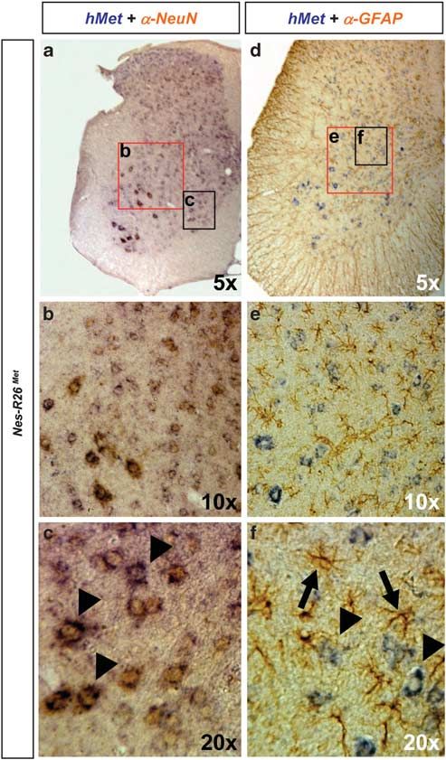
Genetically enhancing Met levels in ALS neurons
                                                                   M Genestine et al
2

          Consistently, transgenic mice expressing the mutant forms                    Results
          of human SOD1 recapitulate a number of ALS symptoms
          and have been instrumental in evaluating the molecular                       Generation        of     conditional      Rosa26LacZstopMet
          and cellular events underlying ALS pathology.7 Moreover,                     transgenic mice. To enhance Met signalling levels in a
          genetic and cell biological studies based on the differential                temporally and spatially regulated manner, we generated
          expression of mutant SOD1 in distinct cell types have                        mice carrying a conditional mouse–human chimeric met
          demonstrated that although death of MNs causes ALS                           transgene (met tg). In particular, we engineered a mouse
          symptoms, the disease also renders other cells, such as                      strain in which a cytomegalovirus (CMV)-enhancer/b-actin-
          astrocytes and microglia, dysfunctional. Thus, MN loss, in                   promoter controls the expression of a floxed-b-geo-reporter
          addition to a cell-autonomous origin, is also triggered by non-              gene followed by a met tg. Such a strategy was chosen to
          cell-autonomous defects involving toxicity of other unhealthy                keep the met tg silent, unless the stop cassette is excised by
          cells.8,9 Notably, these different cell types play distinct roles in         cre-mediated recombination. To avoid integration site effects
          ALS pathogenesis. Damage within MNs is primarily asso-                       and favour loxP-site accessibility, the construct was inserted
          ciated with disease onset and its early progression phase,                   into the Rosa26 locus (Rosa26LacZstopMet, referred to
          whereas damage within microglia and astrocytes accelerates                   R26LacZstopMet; Figures 1a–c).
          MN degeneration and ALS progression.8–12 The recognition                        To specifically gain expression in neural cells, we crossed
          that multiple cell types determine ALS evolution has boosted                 the R26LacZstopMet mice with the nestin-cre transgenics25
          the need to understand the relative contribution of providing                (nestin-cre;Rosa26LacZstopMet, referred to as Nes-R26Met),
          beneficial signals to different cell types involved in the                   leading to the excision of the lacZ-floxed cassette and
          disease.                                                                     consequently expression of the Met chimeric protein (Mettg)
             Among several strategies to alleviate ALS symptoms, a                     in neural tissues (Figures 1c and d). Western blot analysis of
          major hope has been placed on the ability of trophic factors,                E15.5 embryos and P7 mice revealed the presence of Mettg in
          acting on MN in culture or during development,4,7 to activate                dissected brains and spinal cords only after recombination
          endogenous RTKs. Initial results were disappointing as                       (Figure 1d). To estimate Mettg levels versus endogenous Met,
          infusion of specific trophic factors had little or no beneficial             we performed western blot analysis of brain and spinal cord
          effects. The weakness of these approaches appeared to be                     protein extracts at different developmental stages using
          inadequate delivery of these factors to the right cells. This                antibodies recognizing the kinase domain of both endogenous
          possibility was further supported by genetic studies assessing               and Mettg. Quantification analyses indicated that Mettg levels
          growth factor efficacy depending on the delivery site.13,14                  were at least 5/7-fold increased in brains and spinal cords of
          Therefore, understanding the relative contribution of enhan-                 heterozygous Nes-R26Met, when compared with endogenous
          cing RTK signalling in distinct cell types is needed to further              mouse Met (Figures 1e and f). Consistently, Mettg levels were
          clarify the ALS biology and to evaluate how beneficial signals               twofold higher in homozygous mice compared to hetero-
          should be delivered for therapy.                                             zygous littermates (Figures 1d–f).
             In this study, we assessed the biological and functional                     We next characterized the R26LacZstopMet mice before
          significance of enhanced signalling, above endogenous                        and after recombination by following the expression of the
          levels, downstream of the Met RTK specifically in neurons                    lacZ-stop cassette and met tg transcript in adult brains and
          during neuro-degenerative diseases, such as ALS. A number                    spinal cords. We found a decrease in b-galactosidase activity
          of features makes the Met receptor of special interest to                    and lacZ transcripts in Nes-R26 Met compared with R26 LacZ-
                                                                                       stopMet
          address RTK signalling levels during neuro-degenera-                                  transgenics, indicating that recombination in several
          tion.15,16 During development, activation of the Met receptor                brain regions occurred as expected (Figures 2a–d, Supple-
          by its ligand HGF regulates MN fate at multiple levels,                      mentary Figures 1 and 2). Conversely, mettg was expressed
          including identity acquisition, axonal growth, and survival.2,17–20          in brains only after recombination in a pattern complementary
          During injury, HGF is able to enhance regeneration of the                    to lacZ distribution (Figures 2e and f). High levels were found
          lesioned spinal cord and of crushed peripheral nerves.21,22                  in the hippocampus, cerebellum, cerebral cortex, and cervical
          Intracerebral delivery or transgenic-mediated neuronal                       spinal cord (Figures 2g–i). Expression studies performed
          expression of HGF in mutant SOD1 animal models acts                          on adult lumbar spinal cord sections revealed also a
          simultaneously on different dysfunctional cell types.23,24                   complementary distribution pattern. In particular, lacZ or
          However, the contribution of enhancing Met signalling                        mettg were found in cells with large nuclei resembling MNs
          uniquely in neurons and its relative impact on ALS                           of R26LacZstopMet and Nes-R26Met, respectively (Figure 3).
          onset and progression remains to be established. To enhance                  Quantification analysis revealed that cre-mediated excision
          Met signalling above a threshold level in a temporal                         occurred in approximately 56% of these cells (56.3±1.1;
          and tissue-specific manner, we generated conditional met                     Po0.0001; Figure 3g).
          transgenic mice using the cre-loxP system. Here, we show
          that transgene-mediated neuronal expression of the Met RTK                   Molecular and cellular characterization of Nes-R26Met
          in SOD1G93A mice selectively delays disease onset,                           mice. Although the Rosa26 locus drives gene expression
          without slowing down its progression. Our findings show that                 ubiquitously,26 we observed that the lacZ distribution in
          boosting RTK signalling in a cell-type-restricted manner can                 brains and spinal cords of adult R26LacZstopMet mice
          have a distinct beneficial impact in counteracting the                       appeared restricted to distinct cell types (Figures 2a–d
          processes underlying the evolution of neurodegenerative                      and 3a–f, Supplementary Figures 1 and 2). Colocalization
          diseases.                                                                    studies revealed b-galactosidase activity predominantly in

Cell Death and Disease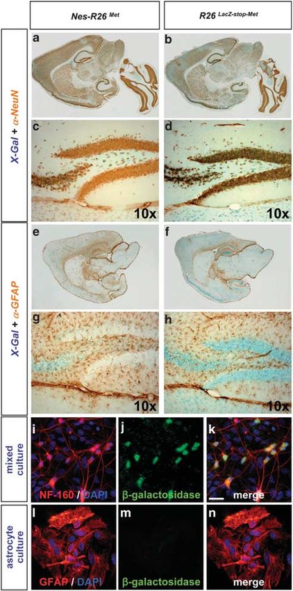
Genetically enhancing Met levels in ALS neurons
                                                                                           M Genestine et al
                                                                                                                                                                                       3

Figure 1 Temporal and tissue-specific Met transgene activation using Cre-mediated recombination. (a) The R26LacZstopMet construct targeted in the Rosa26 locus is
shown. The targeting construct consists of the CMV-enhancer and the chicken b-actin promoter (CMV/b-actin) driving the b-geo reporter and a Met chimeric cDNA. The
reporter gene is followed by three copies of the SV40 polyadenylation signal (3xpA) and flanked by loxP sites. The Met chimeric cDNA consists of a 50 portion encoding the
mouse extracellular domain fused to a 30 region coding for the intracellular human portion. Positions of the probes used for Southern analysis (a and b) as well as primers used
for PCR (1, 2, 3) are indicated. (b) Southern blots of neo-resistant ES clones analyzed with probe a (EcoRI digestion; top) and probe b (EcoRV digestion; bottom). The 15.5 Kb
band corresponds to the wild-type allele whereas the 5.5 Kb band depicts the recombinant allele in clones 1 and 2. (c) PCR of mouse-tail genomic DNAs showing the mutant
allele before and after Cre recombination (top). The presence of an amplicon in the Nes-R26Met mice with the 1 þ 2 primers is detected because genotypes are performed
using DNA extracted from tails, which include tissues with and without recombination. PCR showing the genotypes of heterozygous and homozygous Nes-R26Met mice
(bottom). (d) Western blots showing the expression of the Met chimeric protein (using anti-human Met antibodies) in brain and spinal cord extracts from embryos (E15.5) and
newborn mice (P7) with the indicated genotypes. (e) Quantification analysis of Met chimeric protein levels versus endogenous Met. Western blot analysis of protein extracts
from dissected brains and spinal cords at different developmental stages using antibodies recognizing the Met kinase domain (MetKD). (f) Quantification analyses revealed
that levels of Met chimeric proteins were at least five- and sevenfolds increased in brains and spinal cords of heterozygous Nes-R26Met, respectively, when compared with
the endogenous mouse Met. Met chimeric levels were double in homozygous mice. Numbers on columns indicate fold of increase in Met protein levels over the endogenous
Met. In the graph, Nes-R26Met and Nes-R26Met/Met genotypes are indicated as M/ þ and M/M, respectively. Values are expressed as means±S.E.M. Tub, tubulin; hMet,
human-Met

NeuN-positive neurons, but not in glial fibrillary acidic protein                          indicating that mettg should be predominantly confined
(GFAP)-positive astrocytes (Figures 4a–h). The restricted                                  to neurons after nestin-cre-mediated recombination. Consis-
neuronal lacZ expression was also observed in cultured cells                               tently, mettg transcripts colocalized with Smi32-positive
(Figures 4i–n). Thus, the genetic setting we adopted (CMV-                                 neurons, but not with GFAP-positive astrocytes (Supple-
enhancer/b-actin-promoter in Rosa26) results in an animal                                  mentary Figure 3). The restricted expression of mettg in
model with a restricted expression of the transgene,                                       neurons was also observed in Nes-R26Met adult spinal cords,

                                                                                                                                                                   Cell Death and Disease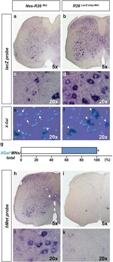
Genetically enhancing Met levels in ALS neurons
M Genestine et al
4
Figure 2 Molecular characterization of Nes-R26Met and R26LacZstopMet mice in adult brains. (a–d) Expression of the lacZ reporter gene was followed by measuring
b-galactosidase (X-Gal) activity (a and b) or its transcript levels (c and d) on sagittal sections. Decrease in X-Gal and lacZ transcripts were found after nestin-cre-mediated
recombination. (e and f) Expression of the met chimeric transgene in sagittal section of adult Nes-R26Met and R26LacZstopMet brains. (g–i) High levels of transgenic met
transcripts were detected in the hippocampus (g), cerebellum (h), cervical spinal cord (i). Panels (g–i) show enlarged views of brain areas delineated by the rectangles in (e).
cb, cerebellum; cc, cerebral cortex; h, hippocampus; sc, spinal cord
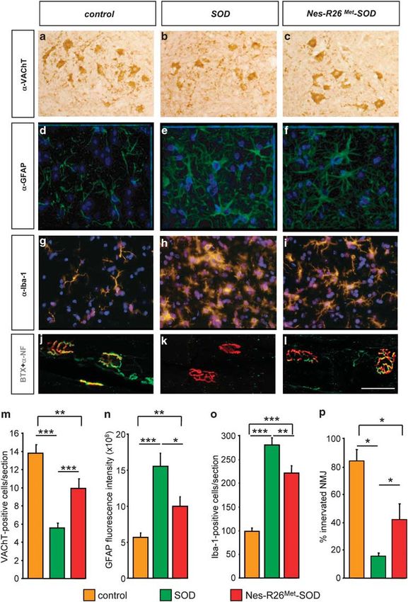
where it was found in dorsal horn neurons, intermediate modulators). Altogether these data show that Nes-R26Met
lateral neurons, and MNs, but not in GFAP-positive astro- mice can be a useful animal model to investigate the
cytes (Figure 5). Thus, mettg is predominantly restricted to functional consequences of enhancing Met levels in distinct
neurons in Nes-R26Met mice. cell types, using available tissue-specific cre-lines.
We next examined whether Mettg protein was active by
following its phosphorylation state using anti-phospho-Met Phenotypical characterization of Nes-R26Met mice. As
antibodies. High levels of phosphorylated Mettg were found in previously discussed, Met regulates specification, axonal
the pons, medulla, lateral ventricles, rostral-migratory stream, growth, and survival of MN subtypes during development.17–20
olfactory bulbs, cerebral and lumbar spinal cords (Figure 6 We therefore evaluated whether enhanced Met levels
and data not shown). These results show that Mettg is influence MN numbers in Nes-R26Met mice. By staining
predominantly functional in spatially restricted domains, which spinal cord sections either with cresyl violet or with vesicular-
possibly correlate to a map of cellular competence for Mettg acetylcholie-transporter (VAChT) antibodies, similar MN
activation influenced by a combination of parameters, such numbers were found at thoracic (data not shown) or lumbar
as environmental contexts (e.g., endogenous HGF levels) spinal cord levels in Nes-R26Met and control animals
or permissive intracellular mechanisms (e.g., signalling (P40.05; Figures 7a–d and g). As expected by the
Cell Death and Disease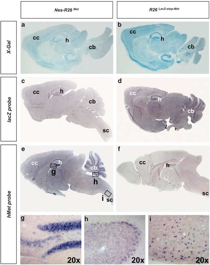
Genetically enhancing Met levels in ALS neurons
                                                                M Genestine et al
                                                                                                                                                                 5

colocalization studies, no differences in GFAP-fluorescence     evaluate the overall motor function and no significant
intensity were observed (P40.05; Figures 7e, f and h).          differences were found in Nes-R26Met versus controls
   As body weight is a generic indicator of animal physiology   (P40.05; Figure 7i). Altogether, these studies show that
influenced by body metabolism, activity, and feeding beha-      enhancing Met levels in neurons does not cause gross
viour, the weight of Nes-R26Met mice was followed over-time     physiological abnormalities.
and no significant differences were found versus controls
(P40.05; Figure 7j). We next performed the rotarod test to      Neuronal-enhanced Met levels counteract ALS
                                                                symptoms in SOD transgenic mice. We next
                                                                investigated the functional relevance of genetically
                                                                enhancing Met signalling levels in the context of a
                                                                neurodegenerative disorder, such as the ALS. The
                                                                Nes-R26Met mice were therefore crossed with a strain
                                                                carrying high copy numbers of the SOD1G93A transgene to
                                                                generate an ALS animal model with increased Met levels in
                                                                neurons. Five groups of mice were generated: (a) wild-type;
                                                                (b) Rosa26 LacZstopMet, (c) Nes-R26Met, (d) SOD1G93A
                                                                (referred to as SOD), and (e) Nes-R26 Met;SOD1G93A
                                                                (referred to as Nes-R26Met-SOD). As no significant
                                                                changes        were      observed     between     wild-type,
                                                                Rosa26 LacZstopMet, and Nes-R26Met for all parameters
                                                                examined, results of control animals included these three
                                                                groups.
                                                                   By evaluating the life span of SOD and Nes-R26Met-SOD
                                                                mice, we found that neuronal-enhanced Met levels prolonged
                                                                the survival by 13 days (mean age, SOD: 136±2 days;
                                                                Nes-R26Met-SOD: 149±3 days; P ¼ 0.0021; Figure 8a). As
                                                                reduction of body weight is an objective measure of ALS
                                                                disease, we examined its evolution in SOD and Nes-R26Met-
                                                                SOD mice, and found that neuronal-enhanced Met levels
                                                                delayed body weight loss. In particular, loss of 10% body
                                                                weight was retarded for 13 days (SOD: 123±3 days; Nes-
                                                                R26Met-SOD: 136±5 days; P ¼ 0.0094; Figure 8b), whereas
                                                                no significant differences were observed during disease
                                                                progression (SOD: 12.9±2.5 days; Nes-R26Met-SOD:
                                                                13±2.4 days; P40.05; Figure 8c). Together, these results
                                                                indicate that enhanced Met levels in neurons counteract ALS
                                                                symptoms in SOD transgenic mice.

                                                                Improved motor performance and delayed onset of
                                                                paralysis in SOD mice with neuronal-enhanced Met
                                                                levels. The neuro-degeneration defects causing ALS
                                                                disease lead to progressive muscle weakness, atrophy,
                                                                and paralysis. Screwed hindlimbs and locomotor defects are
                                                                among the first symptoms affecting transgenic ALS mice. We

                                                                Figure 3 Molecular analysis of Nes-R26Met and R26LacZstopMet mice in adult
                                                                lumbar spinal cords on transversal sections. (a and b) In situ hybridization using lacZ
                                                                riboprobes. (c and d) Enlarged view of the ventral regions, showing lacZ expression in
                                                                distinct cell types characterized by large cell nuclei resembling MNs in R26LacZstopMet
                                                                and a decrease in its levels after nestin-cre-mediated recombination in Nes-R26Met.
                                                                (e and f) Section showing the overlap of X-Gal activity and DAPI staining in lumbar
                                                                spinal cords. X-Gal-negative cells with large nuclei were predominantly found in
                                                                Nes-R26Met, but not in R26LacZstopMet transgenics. Arrows and arrowheads points to
                                                                X-Gal-negative and X-Gal-positive cells with large nuclei, respectively. (g) Quantification
                                                                analysis of X-Gal-positive (blue) and X-Gal-negative cells with large nuclei (resembling
                                                                MNs; white) in Nes-R26Met mice, showing that nestin-cre-mediated lacZ excision
                                                                occurred in approximately 56% of these cells. Values are expressed as means±S.E.M.
                                                                (h–k) Expression of the met chimeric transgene in lumbar spinal cords. Panels (j and k)
                                                                show an enlarged view of ventral spinal cords and met transgenic expression in cells
                                                                resembling MNs

                                                                                                                                             Cell Death and Disease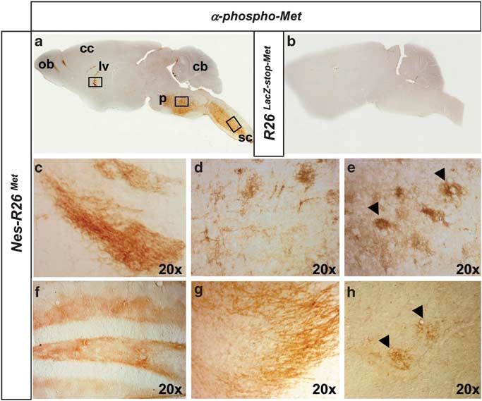
Genetically enhancing Met levels in ALS neurons
                                                                         M Genestine et al
6

                                                                                                  Figure 5 Subcellular localization of chimeric met transcripts in lumbar spinal
                                                                                                  cords of adult Nes-R26Met mice. (a–c) Colocalization studies of exogenous met
                                                                                                  transcript with NeuN protein showing met expression in neuronal cell types.
                                                                                                  (d–f) Colocalization studies of exogenous met transcript with GFAP protein showing
                                                                                                  that transgenic met is not predominantly expressed in astrocytes. Panels (b and e),
                                                                                                  panels (c and f) correspond to an enlarged view of spinal cord areas indicated by
                                                                                                  red and black rectangles in (a and b), respectively. Arrowheads in (c) point to MNs
                                                                                                  co-expressing chimeric met transcript and NeuN protein. Arrows and arrowheads in
          Figure 4 Subcellular localization of the lacZ reporter gene in Nes-R26Met and           (f) indicate GFAP-positive astrocytes and MN expressing chimeric met transcripts,
          R26LacZstopMet mice. (a and b) Colocalization studies of X-Gal activity with NeuN     respectively
          protein showing the reporter activity in neuronal cell types. (c and d) Enlarged view
          of the hippocampus area. (e–h) Colocalization studies of X-Gal activity with GFAP
          protein showing that the lacZ reporter gene is not predominantly expressed in
          astrocytes. Panels (g and h) correspond to an enlarged view of the hippocampus.         the increased time that mice needed to execute this motor
          (i–k) Mixed cell cultures derived from E12.5 R26LacZstopMet spinal cords were         task (SOD: 113±3 days; Nes-R26Met-SOD: 127±3 days;
          immunostained for neurofilament-160 (red) and b-galactosidase (green) proteins.         P ¼ 0.0015; Figure 8d), whereas disease progression was
          (i–n) Astrocyte cultures from P2 R26LacZstopMet spinal cords immunostained for        unchanged (SOD: 30±3.9 days; Nes-R26Met-SOD: 22±2.7
          GFAP (red) and b-galactosidase (green). DAPI was used to counterstain nuclei            days; P40.05; Figure 8e). Therefore, neuronal-enhanced
          (blue), scale bar: 20 mm
                                                                                                  Met improved motor strength and swimming performance of
                                                                                                  SOD mice by acting specifically on disease onset.
                                                                                                    The motor capability of Nes-R26Met-SOD versus SOD mice
          monitored the appearance and progression of motor defects                               was further evaluated by performing footprint studies (Sup-
          in SOD versus Nes-R26Met-SOD compared with controls by                                  plementary Figure 4). The forepaw/hindpaw overlap analysis
          employing swimming tank and footprint assays.27 Onset of                                revealed a delay of 19 days in the locomotor gait dysfunction
          swimming defects was delayed by 14 days, as estimated by                                (SOD: 108±3 days; Nes-R26Met-SOD: 127±3 days;

Cell Death and Disease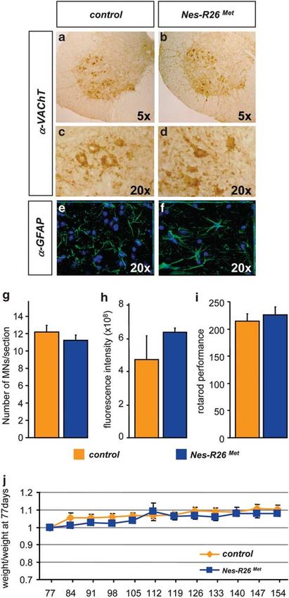
Genetically enhancing Met levels in ALS neurons
                                                                                           M Genestine et al
                                                                                                                                                                                       7

Figure 6 Tissue-distribution of activated chimeric Met in adult brains and spinal cords. (a and b) Sagittal sections of adult brains showing phospho-Met staining in
Nes-R26Met, but not in R26LacZstopMet mice. (c–h) Enlarged view of phospho-Met distribution in the lateral ventricle (c), pons (d), cervical spinal cord (e), corresponding to
regions indicated by squares in panel (a). Distribution of phospho-Met in the hippocampus (f), olfactory bulb (g), and lumbar spinal cord (h). Arrowheads in (e and h) point to
phospho-Met-positive cells with large nuclei resembling MNs. cb, cerebellum; cc, cerebral cortex; lv, lateral ventricle; ob, olfactory bulb; p, pons; sc, spinal cord

P ¼ 0.0009; Figure 8f), accompanied by a delay of 9 days in                                which appear at disease onset and become more prominent
the appearance of step-length defects (SOD: 106±2 days;                                    during progression.9 In contrast to controls, activated
Nes-R26Met-SOD: 115±2 days; P ¼ 0.0136; Figure 8h). The                                    GFAP-positive astrocytes (changes in fluorescence intensity)
motor performance decline rate was not appreciably different                               were detected in lumbar spinal cords, although reduced
between SOD and Nes-R26Met-SOD mice, showing that when                                     in Nes-R26Met-SOD compared with SOD (SOD: 15.5±
the disease has started, it progresses at the same rate                                    1.9  108; Nes-R26Met-SOD: 9.9±1.4  108; P ¼ 0.0164;
(overlap, SOD: 30±3.9 days; Nes-R26Met-SOD: 22±22.7                                        Figures 9d–f and n). Similarly, the number of microglial cells
days; P40.05; step-length, SOD: 32±3.1 days; Nes-R26Met-                                   was reduced in Nes-R26Met-SOD mice compared with SOD
SOD: 34±1.7 days; P40.05; Figures 8g and i).                                               (SOD:     281.7±14.6; Nes-R26Met-SOD:                222.1±15.5;
                                                                                           P ¼ 0.0016; Figures 9g–i and o). Analysis of muscle innerva-
Neuronal-enhanced Met levels attenuate MN loss in                                          tion revealed that the increased MN numbers in Nes-R26Met-
spinal cords of SOD mice. As motor performances were                                       SOD was accompanied by an enhanced integrity of neuro-
transiently improved in Nes-R26Met-SOD mice by delaying                                    muscular junctions (NMJs; SOD: 16±1.8%; Nes-R26Met-
disease onset, we evaluated the neuroprotective effects of                                 SOD: 41.7±11.4%; P ¼ 0.041; controls: 84.4±7.9%;
increased Met levels by quantifying lumbar spinal cord MNs.                                Figure 9p). Thus, increased neuronal Met levels elicit a
For these studies, we selected three animals among the                                     combination of protective effects in different cell types: (1) cell-
Nes-R26Met-SOD, SOD, and controls at the symptomatic                                       autonomous protective effects on spinal cord MNs and for
disease phase (120 days). This stage was chosen because                                    NMJ maintenance; (2) non-cell-autonomous delay of astro-
all behavioural studies showed significant differences                                     cyte activation and increased microglia cell numbers.
between groups. Lumbar spinal cord sections were stained
with VAChT antibodies and MN numbers were determined
                                                                                           Discussion
(Figures 9a–c and m). As expected, we observed a
significant 60% MN loss in SOD mice compared with                                          Most of neurodegenerative diseases result from a combina-
controls (P ¼ 0.0065). By contrast, neuronal-enhanced Met                                  torial action of pathological signals produced by neurons
in Nes-R26Met-SOD mice led to an improvement of MN                                         themselves and by neighbouring cells acting in a non-cell-
maintenance as the surviving MN numbers increased by                                       autonomous manner.7 A number of molecules including
32% compared with SOD (P ¼ 0.0002; Figure 9m).                                             trophic factors and their receptors can elicit beneficial effects
   We next assessed to what extent enhancing Met function in                               on disease-related cells when applied in vitro and/or when
neurons influenced astrogliosis and microglia activation,                                  delivered in disease animal models. Understanding how these

                                                                                                                                                                   Cell Death and Disease
Genetically enhancing Met levels in ALS neurons
                                                                        M Genestine et al
8

                                                                                                  Figure 7 Molecular and functional characterization of Nes-R26Met mice versus
                                                                                                  controls. (a–f) Transversal sections through lumbar spinal cords showing similar
                                                                                                  numbers of VAChT-positive (a–d) and GFAP-positive (e and f) cells. (g and h)
                                                                                                  Quantification studies showed no significant difference in numbers of VAChT-
                                                                                                  immunopositive cells (g; P40.05) and GFAP-fluorescence intensity (h; P40.05)
                                                                                                  among the two groups of mice (n ¼ 3). (i) Rotarod behavioural analysis in
                                                                                                  Nes-R26Met and control littermate mice showing that enhanced neuronal Met levels
                                                                                                  did not significantly influence locomotor performance (P40.05; n ¼ 10). (j) Body
                                                                                                  weight analysis in Nes-Mettg and control mice showed no significant differences
                                                                                                  overtime. Body weight is expressed as weight evolution compared with that of mice
                                                                                                  at age 77 days. Values are expressed as means±S.E.M.

                                                                                                  molecules act on dysfunctional cells remains a key topic to
                                                                                                  clarify disease mechanisms and to evaluate their use for
                                                                                                  therapies. We show here that enhanced signalling by Met has
                                                                                                  an impact on a specific stage of ALS pathology when it is
                                                                                                  selectively upregulated in neurons. Indeed, transgene-
                                                                                                  mediated neuronal expression of Met elicits a beneficial
                                                                                                  effect in SOD1G93A by delaying disease onset, but not
                                                                                                  progression.
                                                                                                     Our results are based on a genetic approach involving the
                                                                                                  generation of conditional mettg mice, in which Met signalling
                                                                                                  levels is modulated in a temporally and spatially regulated
                                                                                                  manner. Such an approach offers the possibility of exploring
                                                                                                  how enhanced Met signalling above endogenous levels
                                                                                                  influences cell fate in developmental events, in adult
                                                                                                  physiology, and in pathological conditions. Loss of Met
                                                                                                  function during development interferes with identity acquisi-
                                                                                                  tion, axonal growth, and survival of MN subsets.2,17–20 We
                                                                                                  show here that neuronal-enhanced Met signalling levels do
                                                                                                  not affect either MN development or function in adulthood,
                                                                                                  whereas it influences MN maintenance in ALS pathological
                                                                                                  conditions. Thus, it is likely that excessive Met functions are
                                                                                                  restrained by mechanisms such as tissue homeostasis or by
                                                                                                  limiting amounts of ligand. Importantly, the dispensable
                                                                                                  function of Met in several adult tissues is in contrast to its
                                                                                                  requirement in counteracting degenerative processes follow-
                                                                                                  ing injuries, such as axotomy,21,22 hepatectomy,28 and skin-
                                                                                                  wound.29 These regenerative studies together with our
                                                                                                  findings indicate that in a pathological context, cell types like
                                                                                                  neurons become sensitive to the beneficial effects provided
                                                                                                  by Met. The generation of compound transgenics by crossing
                                                                                                  the R26LacZstopMet mice with available cre-lines will offer a
                                                                                                  unique genetic setting for determining tissue-specific sensi-
                                                                                                  tiveness to enhanced Met signalling either during develop-
                                                                                                  ment, in adulthood, or in pathologies.
                                                                                                     The pleiotropic functions elicited by the HGF/Met system in
                                                                                                  neurons have boosted the interest in exploring its potential for

          Figure 8 Neuronal-enhanced Met signalling levels in ALS mice improves survival and locomotor performance by delaying disease onset. (a) Survival analysis of
          Nes-R26Met-SOD (red) and SOD (green) mice (n ¼ 40) showing a delay of 13 days of average life span. (b) The loss of 10% body weight was 13 day delayed in Nes-R26Met-
          SOD (n ¼ 11) compared with SOD (n ¼ 17) mice. (c) Body weight loss was not significantly different in Nes-R26Met-SOD compared with SOD mice once the disease started
          (P40.05). (d and e) Motor performance analysis using a 1 meter swimming tank device. Panel (d) shows a delay of 14 days in motor defect onset of Nes-R26Met-SOD mice
          (n ¼ 11) compared with SOD transgenics (n ¼ 17). Panel (e) shows the analysis of the duration of locomotor defects with not significant differences between groups
          (P40.05). (f–i) Comparison of motor performance using the footprint test. Analysis of the forepaw/hindpaw overlap revealed a 19 day improvement in Nes-R26Met-SOD mice
          (n ¼ 11) compared with SOD transgenics (n ¼ 17; f). No significant differences were found during disease progression (g; P40.05). Step length defects also appeared with a
          delay of 9 days in Nes-R26Met-SOD compared with SOD mice (h). Again, no changes were found during disease progression (i; P40.05). a, b, d, f and h are Kaplan–Meier
          curves. Values are expressed as means±S.E.M.

Cell Death and Disease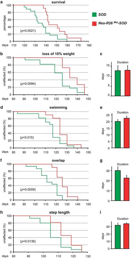
Genetically enhancing Met levels in ALS neurons
M Genestine et al
                                                                      9

                                                  Cell Death and Disease
Genetically enhancing Met levels in ALS neurons
M Genestine et al
10
Figure 9 Effects of enhanced Met signalling in MN maintenance and astrogliosis at ALS symptomatic phase (120 days). (a–c) VAChT-stained sections through the ventral horn of
the lumbar spinal cord showed an increase in MN maintenance in Nes-R26Met-SOD compared with SOD transgenics. (d–f) GFAP-stained sections through the ventral horn of the
lumbar spinal cords showing astrogliosis in Nes-R26Met-SOD and SOD mice. (g–i) Iba-1-stained sections through the ventral horn of the lumbar spinal cords showing an increase in the
number of microglial cells in Nes-R26Met-SOD and SOD mice. (j–l) Immunofluorescence staining of gastrocnemius muscle with a-bungarotoxin-tetramethylrhodamine (red), anti-
neurofilament (green). Scale bar 50 mm. (m) Quantification of MN numbers (VAChT-positive) among the three groups of mice, showing a 32% increase in Nes-R26Met-SOD compared
with SOD mice (Nes-R26Met-SOD versus SOD : P ¼ 0.0002; SOD versus control: Po0.0001; Nes-R26Met-SOD versus control: P ¼ 0.006; n ¼ 3). (n) Quantification of fluorescence
intensity of GFAP-immunopositive cells. Significant differences were observed between the three groups (Nes-R26Met-SOD versus SOD : P ¼ 0.0164; SOD versus control:
P ¼ 0.0002; Nes-R26Met-SOD versus control: P ¼ 0.0084; n ¼ 3). (o) Quantification of Iba-1-positive cells. Significant differences in microglia numbers were observed between the
three groups (Nes-R26Met-SOD versus SOD : P ¼ 0.0016; SOD versus control and Nes-R26Met-SOD versus control: Po0.0001; n ¼ 3). (p) The percentage of a-Bungarotoxin-
stained end-plates showing complete or partial colocalization with neurofilament staining was evaluated in the three groups (Nes-R26Met-SOD versus SOD: P ¼ 0.0416; controls
versus SOD : P ¼ 0.0143; controls versus Nes-R26Met-SOD : P ¼ 0.0256). Values are expressed as means±S.E.M. ***Po0.001, **Po0.01, *Po0.05
Cell Death and DiseaseGenetically enhancing Met levels in ALS neurons
M Genestine et al
11
ALS therapy through several strategies. HGF intrathecal presynaptic terminals in ALS MNs, either by providing RTK
administration at disease onset provided evidence that, when support (our studies), or by removing signals such as BAX
present at high doses and accessible to different disease influencing NMJ integrity,12 or by depleting mutant SOD,8,9
cells, HGF/Met attenuates MN degeneration and retards ameliorates ALS by selectively delaying disease onset. It is
disease progression by 11 days.30 However, these studies did known that the initial MN damage is followed by a progressive
not clarify whether, and to what extent, the HGF/Met system increase in cytotoxic and inflammatory mediator levels, which
exerts support on MNs in a cell-autonomous manner. Insights further affect MN themselves and neighbouring cells. Astro-
to this issue come from studies based on genetic neuronal cytes and microglia are the predominant cell types driving
delivery of HGF in the low copy number SOD1G93A mice. In disease progression towards death. Although disease pro-
particular, hgf expression driven by the neuron-specific gression is unchanged in Nes-R26Met mice, our genetic
enolase-promoter delayed disease onset by approximately analysis show that enhanced-neuronal Met in SOD mice
28 days, rather than influencing its progression.23 However, impacts on distinct cell types involved in the ALS disease:
as exogenous HGF is expressed and secreted by neurons, MNs, in a cell-autonomous manner, possibly by providing
these hgf transgenic mice did not allow discriminating trophic support and/or maintenance of NMJ integrity and
between the HGF effects on MNs versus those elicited on astrocytes and microglia, in a non-cell-autonomous manner,
astrocytes, which in turn influence MNs and microglia by delaying their activation. Future studies will establish the
function. Consistently, a decrease in the number of microglia, relative contribution of each individual event in delaying the
reactive astrocytes, and MN loss was observed in hgf onset of the disease by neuronal Met signalling and uncover
transgenics. Our mouse model allowed discrimination the underlying mechanisms.
between the cell-autonomous effects elicited by enhanced Understanding how therapeutic reagents act at cellular
Met signalling in MNs and those influenced by HGF on other levels is needed to accelerate the progress of promising
dysfunctional cells. Moreover, the Nes-R26Met mice estab- treatments towards the clinic. Our findings emphasize the
lished that Met signalling in MNs selectively counteracts ALS relevance of genetically assessing the effects of agents on
disease onset. It remains to be investigated what contribution distinct cell types implicated in a disease and during its
enhanced Met signalling, above endogenous levels in evolution. Although successful therapies for ALS will possibly
astrocytes and microglia, could have with respect to disease require concomitant actions on distinct dysfunctional cells, our
evolution. results highlight the considerable therapeutic potential of
The severity of ALS and the lack of effective therapeutic modulating RTK signalling in MNs to combat degenerative
strategies are driving efforts to explore agents, applied either signals.
separately or in combination, to counteract disease symp-
toms. Concerning trophic factors, HGF could offer therapeutic Materials and Methods
advantages at multiple levels. It is noteworthy that endogen- Generation of R26LacZstopMet mice. The R26LacZstopMet mice were
ous Met is upregulated starting from ALS onset in ventral generated by taking advantage of the loxP-flanked (‘floxed’)-stop cassette system.
spinal cords of SOD1G93A mice (data not shown and see Sun To generate the chimeric mouse–human gene, the human Met cDNA encoding the
et al. 23). These observations indicate that endogenous Met is transmembrane and cytoplasmic portion was fused in-frame with the mouse
turned on to counteract ALS symptoms, but endogenous extracellular cDNA sequence using the PvuII site present in both sequences. We
chose such a strategy to ensure that the Met chimeric protein interacts efficiently
HGF levels may not be sufficient to reverse the damage, as
with the endogenous mouse HGF, but can still be identified from the endogenous
was found studying regeneration after optic-nerve axotomy mouse Met protein. The fusion product was subsequently subcloned into the
(unpublished results). Moreover, as HGF/Met elicits functions pCALL2 vector downstream of the insert containing the CMV enhancer-chicken
in muscle cells,31,32 it is possible that ectopic HGF in muscles b-actin promoter followed by the loxP-flanked b-geo/3xpA cassette. This vector is
would also counteract neuro-muscular-junction denervation referred to as pCALL-Met. To generate the Rosa26 targeting construct, the insert
and muscle atrophy. Indeed, HGF favours the formation of containing the CMV-enhancer/chicken-b-actin-promoter-loxP-flanked b-geo/3xpA-
Met was subcloned into the XhoI site of pRosa-1 vector previously modified using a
NMJs during development.33 Although enhanced Met activa-
poly-linker. The targeting vector was electroporated into R1 ES cell lines. Cell
tion is often associated with tumour formation and metastasis, culture, electroporation, selection, and Southern blot analyses were performed as
we did not observe side effects such as neoplasia in previously described.32 To identify recombined clones, genomic DNA was digested
Nes-R26Met mice despite Mettg expression levels in regions with EcoRI or EcoRV and probed with an external or internal probe, respectively.
of the nervous system. Thus, the beneficial effects of HGF/ Two selected ES cell lines carrying the homologous recombination were used to
Met on ALS degenerative processes appear to be well generate the R26LacZstopMet mice through blastocyst injections.
tolerated in healthy nervous system tissues.
The use of the conditional ALS mouse model, in which the Transgenic mice. The mouse line expressing cre recombinase under the
nestin promoter was previously described.25 The B6SJL-Tg(SOD1*G93A)
mutant SOD1 gene can be deleted according to the 1Gur(SOD1G93A (SOD1G93A) mouse line was used as ALS disease model.34
cre-transgenic line used, has been instrumental in clarifying Both R26LacZstopMet and nestin-cre mice were backcrossed into the B6SJL
the influence of distinct cell types during ALS evolution. In genetic background before breeding with the SOD1G93A transgenics. The number of
particular, selective reduction of mutant SOD1 in MNs mice used for behavioural and immuno-histochemical studies are indicated in figure
predominantly impacts on disease onset and its early phase. legends. Each genetic group consisted of a mixed population of equal numbers of
In contrast, reduced mutant SOD1 either in astrocytes or in males and females. The presence of a vaginal plug in the morning was considered
as 0.5 embryonic day (E0.5). All procedures involving the use of animals were
microglia mainly influences the late stage of disease progres-
performed in accordance with the European Community Council Directive of 24
sion.8,9 Thus, although recombination in Nes-R26Met mice did November 1986 on the protection of animals used for experimental purposes
not occur in 44% of lumbar spinal cord MNs, rebalancing the (86/609/EEC). The experimental protocols were carried out in compliance with
levels of stress and survival signals and/or protecting institutional ethical committee guidelines for animal research. All efforts were made
Cell Death and DiseaseGenetically enhancing Met levels in ALS neurons
M Genestine et al
12
to minimize the number of animals used and their suffering. When paralysis started, performed four trials at each time point for each animal and recorded the three best
food and water were placed directly into the cage. To reduce animal pain, mice were performances for statistical analysis. Briefly, the rotarod test was performed by
killed when they were unable to right themselves within 30 s when placed on their placing mice on an accelerating rod (3 cm diameter) and by recording the time each
back. animal took to fall from the rod. The speed of the rotarod accelerated from 4 to
40 r.p.m. over a 5-min period. The swimming tank device allows evaluation of the
Antibodies. Antibodies used were anti-tubulin and anti-GFAP and anti-VAChT hindlimb strength and performance; it is suitable for assessing onset and
(1 : 1000; Sigma-Aldrich, St. Louis, MO, USA), anti-MetKD (1 : 1000) and anti- progression of ALS symptoms. For the swimming tank, each mouse performed four
phospho Y1234-1235-Met (1 : 50; Cell Signaling, Danvers, MA, USA), anti-human trials and the time needed to reach the platform was recorded. A mouse was
Met (1 : 500; Santa-Cruz Biotechnology Inc., Santa Cruz, CA, USA), anti-Smi32 considered at the onset of motor defect when it needed 6.8 s to perform the
(1 : 500; Sternberger monoclonals, Covance, Dallas, TX, USA), anti-NeuN (1 : 200; swimming tank test. The 6.8 s were chosen as reference as this value corresponds
Chemicon, Millipore, Billerica, MA, USA), and anti-neurofilament-145 (1 : 1000; to the first significant difference versus control mice. For the footprint test, the fore-
AB1987; Millipore), anti-mouse or rabbit fluorescent-coupled secondary antibodies toes and the hind-toes were labelled in blue and red, respectively. Weekly
(1 : 400; Jackson, West Grove, PA, USA), anti-mouse or rabbit biotin-coupled monitoring of walking patterns allows assessment of motor coordination and
secondary antibodies (1 : 500; Jackson). For western blot analyses, the following synchrony through the evaluation of several parameters, including toe spread,
secondary antibodies were used: anti-rabbit IgG-peroxidase or anti-mouse IgG- forepaw/hindpaw overlap, and step-length. Two parameters were measured: (1) the
peroxidase (1 : 4000, Jackson). overlap distance between forepaw and hindpaw on the same side; (2) the stride
length as the distance of the hindpaw on the same side between each step. For the
Histological analysis. Anesthetized mice were intra-cardiacally perfused first overlap, the Kaplan–Meier curve indicates the mice when the distance between
with PBS then with 4% para-formaldehyde (PFA, Sigma) in PBS. Brains, spinal forepaw and hindpaw increased by 45%. For the step length, the Kaplan–Meier
cords, and muscles were dissected, postfixed in 4% PFA and embedded. For brain curve indicates when the distance of the hindpaw for each step decreased by 40%.
and spinal cords, 16 or 30-mm thick cryo-sections were performed (Leica, Wetzlar, To reduce mouse stress and fatigue, the swimming and the footprint behavioural
Germany). In situ hybridization, immuno-histochemistry, and X-Gal staining were tests were carried out on different days.
performed as previously described.27,35,36 MN numbers were determined on 16-mm
thick lumbar spinal cord sections stained with VAChT antibodies or cresyl violet. Statistical analysis. Results were expressed as the mean±S.E.M.
A total of 10 sections per mouse were analyzed. Astrogliosis was monitored by Statistically significant differences on cell counts were assessed by the Student’s
measuring fluorescence-levels of sections stained with anti-GFAP antibodies t-test. Statistically significant differences among the groups of mice were assessed
(Image J software, ImageJ 1.41, NIH, Bethesda, MD, USA). For NMJ staining, by two-way ANOVA. Post hoc Bonferroni’s correction was also used to test all pair-
35-mm thick longitudinal sections were collected on Superfrost Plus Slides wise comparisons between groups and time points per group. A log-rank test was
(CML, Thermo Scientific, Braunscheweig, Germany). Tissue sections were used to calculate the statistical differences in the onset and survival of the different
incubated in blocking solution (0.5% Triton X-100, 5% BSA in PBS) at 37 1C for mouse cohorts. Statistical significance was defined as ns: P40.05; *Po0.05;
2 h. Rabbit polyclonal anti neurofilament-145 antibodies were diluted in the same **Po0.01; ***Po0.001.
blocking solution and incubated overnight at 4 1C. Anti-rabbit-alexa 488-conjugated
secondary antibody (Invitrogen, Life Technologies, Carlsbad, CA, USA) and
a-bungarotoxin-tetramethylrhodamine-conjugate (1 : 1000; Invitrogen) were Conflict of interest
incubated for 2 h at room temperature (1% BSA in PBS), before washing and The authors declare no conflict of interest.
mounting. Stained end-plates on sections were examined under an Axio
microscope (Zeiss, Oberkochen, Germany). Innervated (yellow) or denervated
(red) end-plates were counted on apoptome (Zeiss) 35-mm Z-stacks.
Acknowledgements. We are particularly grateful to C Henderson, A Moqrich,
K Dudley, and all lab members for discussions and comments. We thank: V Girod-
Biochemical studies. Protein extracts were prepared from freshly dissected David, L Jullien, staff members at IBDML and CIML animal house and transgenic
brains and spinal cords at the appropriate stages and western blot analyses were facilities for help with mouse husbandry; IBDML imaging platform; CG Lobe for the
performed as previously described.37–39 Quantifications were done by measuring pCALL2 vector. This work was supported by funds from INCa, ARC, FRM, AFM,
band intensities with the Image J software. FdF, Fondation Bettencourt-Schueller, Marie Curie Host Fellowship for the Transfer-
of-Knowledge to FM and RD. MG was supported by University-Franco-Italy
Cultures. Cell culture procedures were previously described.40 Briefly, E12.5 fellowship, EC by AFM, AF by FRM, HH by FdF, ARC.
spinal cords from R26LacZstopMet transgenic embryos were dissected in Hank’s
Balanced Salt Solution containing 7 mM HEPES pH7.4 and 4.5 g/l glucose. Cells
were dissociated in Ham-F10 medium (Invitrogen) with 0.025% Trypsin (Sigma) and
1. Lemmon MA, Schlessinger J. Cell signaling by receptor tyrosine kinases. Cell 2010; 141:
centrifuged over a 4% (w/v) BSA cushion at 800 g for 5 min. Cells were plated on 1117–1134.
poly-ornithine/laminin-treated coverslips in supplemented Neurobasal medium 2. Maina F, Pante G, Helmbacher F, Andres R, Porthin A, Davies AM et al. Coupling
(Invitrogen) containing neurotrophic factors (0.1 ng/ml GDNF, 1 ng/ml BDNF, and Met to specific pathways results in distinct developmental outcomes. Mol Cell 2001; 7:
10 ng/ml CNTF) and maintained at 37 1C in 7.5% CO2 atmosphere for 3 days before 1293–1306.
being processed for immunochemistry. Astrocytes were prepared from P2 3. Furlan A, Stagni V, Hussain A, Richelme S, Conti F, Prodosmo A et al. Abl interconnects
R26LacZstopMet spinal cords. When confluent, cells were trypsinized and plated oncogenic Met and p53 core pathways in cancer cells. Cell Death Differ 2011 in press.
onto coverslips in Dulbecco’s modified Eagle’s (DMEM, Invitrogen) medium 4. Connor B, Dragunow M. The role of neuronal growth factors in neurodegenerative
supplemented with 10% fetal bovine serum and penicillin–streptomycin. Cells were disorders of the human brain. Brain Res Brain Res Rev 1998; 27: 1–39.
5. Snider WD, Zhou FQ, Zhong J, Markus A. Signaling the pathway to regeneration. Neuron
then cultured at 37 1C in 5% CO2 atmosphere for 3 days before immunostaining.
2002; 35: 13–16.
Cultured cells were processed for immunocytochemistry as above described. We 6. Siegel GJ, Chauhan NB. Neurotrophic factors in Alzheimer’s and Parkinson’s disease
used the following primary antibodies diluted in PBS containing 4% BSA, 2% brain. Brain Res Brain Res Rev 2000; 33: 199–227.
donkey serum: anti-b-Gal (1 : 4000; Cappel, MP Biomedicals, Ullkirch, France), anti- 7. Kanning KC, Kaplan A, Henderson CE. Motor neuron diversity in development and
neurofilament 160 (1 : 600; NN18, Sigma), anti-GFAP (1 : 500; MAB360, Millipore). disease. Annu Rev Neurosci 2010; 33: 409–440.
Alexa Fluor 488 (1 : 500; A21206; Invitrogen) and 555 (1 : 1000; A31570; 8. Boillee S, Yamanaka K, Lobsiger CS, Copeland NG, Jenkins NA, Kassiotis G et al. Onset
Invitrogen)-conjugated donkey anti-rabbit and anti-mouse were used as and progression in inherited ALS determined by motor neurons and microglia. Science
secondary antibodies (Invitrogen). 2006; 312: 1389–1392.
9. Yamanaka K, Chun SJ, Boillee S, Fujimori-Tonou N, Yamashita H, Gutmann DH et al.
Astrocytes as determinants of disease progression in inherited amyotrophic lateral
Behavioural tests. Body weight measurements and all behavioural tests sclerosis. Nat Neurosci 2008; 11: 251–253.
began when mice reached the age of 70 days and recording was performed weekly. 10. Miller TM, Kaspar BK, Kops GJ, Yamanaka K, Christian LJ, Gage FH et al. Virus-delivered
To analyze motor functions, locomotor tests included the rotarod, the 1 m swimming small RNA silencing sustains strength in amyotrophic lateral sclerosis. Ann Neurol 2005;
tank and the footprint, which have been done as previously described.27 We 57: 773–776.
Cell Death and DiseaseGenetically enhancing Met levels in ALS neurons
M Genestine et al
13
11. Ralph GS, Radcliffe PA, Day DM, Carthy JM, Leroux MA, Lee DC et al. Silencing mutant 27. Raoul C, Abbas-Terki T, Bensadoun JC, Guillot S, Haase G, Szulc J et al. Lentiviral-
SOD1 using RNAi protects against neurodegeneration and extends survival in an ALS mediated silencing of SOD1 through RNA interference retards disease onset and
model. Nat Med 2005; 11: 429–433. progression in a mouse model of ALS. Nat Med 2005; 11: 423–428.
12. Gould TW, Buss RR, Vinsant S, Prevette D, Sun W, Knudson CM et al. Complete 28. Huh CG, Factor VM, Sanchez A, Uchida K, Conner EA, Thorgeirsson SS. Hepatocyte
dissociation of motor neuron death from motor dysfunction by Bax deletion in a mouse growth factor/c-met signaling pathway is required for efficient liver regeneration and repair.
model of ALS. J Neurosci 2006; 26: 8774–8786. Proc Natl Acad Sci USA 2004; 101: 4477–4482.
13. Guillot S, Azzouz M, Deglon N, Zurn A, Aebischer P. Local GDNF expression mediated by 29. Chmielowiec J, Borowiak M, Morkel M, Stradal T, Munz B, Werner S et al. c-Met is
lentiviral vector protects facial nerve motoneurons but not spinal motoneurons in essential for wound healing in the skin. J Cell Biol 2007; 177: 151–162.
SOD1(G93A) transgenic mice. Neurobiol Dis 2004; 16: 139–149. 30. Ishigaki A, Aoki M, Nagai M, Warita H, Kato S, Kato M et al. Intrathecal delivery of
14. Li W, Brakefield D, Pan Y, Hunter D, Myckatyn TM, Parsadanian A. Muscle-derived but not hepatocyte growth factor from amyotrophic lateral sclerosis onset suppresses disease
centrally derived transgene GDNF is neuroprotective in G93A-SOD1 mouse model of ALS. progression in rat amyotrophic lateral sclerosis model. J Neuropathol Exp Neurol 2007; 66:
Exp Neurol 2007; 203: 457–471. 1037–1044.
15. Maina F, Hilton MC, Andres R, Wyatt S, Klein R, Davies AM. Multiple roles for hepatocyte 31. Bladt F, Riethmacher D, Isenmann S, Aguzzi A, Birchmeier C. Essential role for the c-met
growth factor in sympathetic neuron development. Neuron 1998; 20: 835–846. receptor in the migration of myogenic precursor cells into the limb bud. Nature 1995; 376:
16. Maina F, Klein R. Hepatocyte growth factor—a versatile signal for developing neurons. 768–771.
Nat Neurosci 1999; 2: 213–217. 32. Maina F, Casagranda F, Audero E, Simeone A, Comoglio P, Klein R et al. Uncoupling
17. Ebens A, Brose K, Leonardo ED, Hanson MG, Bladt F, Birchmeier C et al. Hepatocyte of Grb2 from the Met receptor in vivo reveals complex roles in muscle development.
growth factor/Scatter factor is an axonal chemoattractant and a neurotrophic factor for Cell 1996; 87: 531–542.
spinal motor neurons. Neuron 1996; 17: 1157–1172. 33. Madhavan R, Peng HB. HGF induction of postsynaptic specializations at the
18. Maina F, Hilton MC, Ponzetto C, Davies AM, Klein R. Met receptor signaling is required for neuromuscular junction. J Neurobiol 2006; 66: 134–147.
sensory nerve development and HGF promotes axonal growth and survival of sensory 34. Chiu AY, Zhai P, Dal Canto MC, Peters TM, Kwon YW, Prattis SM et al. Age-dependent
neurons. Genes Dev 1997; 11: 3341–3350. penetrance of disease in a transgenic mouse model of familial amyotrophic lateral
19. Yamamoto Y, Livet J, Pollock RA, Garces A, Arce V, deLapeyriere O et al. Hepatocyte sclerosis. Mol Cell Neurosci 1995; 6: 349–362.
growth factor (HGF/SF) is a muscle-derived survival factor for a subpopulation of 35. Dono R. Fibroblast growth factors as regulators of central nervous system development
embryonic motoneurons. Development 1997; 124: 2903–2913. and function. Am J Physiol Regul Integr Comp Physiol 2003; 284: R867–R881.
20. Helmbacher F, Dessaud E, Arber S, deLapeyriere O, Henderson CE, Klein R et al. 36. Zuniga A, Michos O, Spitz F, Haramis AP, Panman L, Galli A et al. Mouse limb deformity
Met signaling is required for recruitment of motor neurons to PEA3-positive motor pools. mutations disrupt a global control region within the large regulatory landscape required for
Neuron 2003; 39: 767–777. Gremlin expression. Genes Dev 2004; 18: 1553–1564.
21. Kato N, Nemoto K, Nakanishi K, Morishita R, Kaneda Y, Uenoyama M et al. 37. Segarra J, Balenci L, Drenth T, Maina F, Lamballe F. Combined Signaling through ERK,
Nonviral HVJ (hemagglutinating virus of Japan) liposome-mediated retrograde PI3K/AKT, and RAC1/p38 Is Required for Met-triggered Cortical Neuron Migration. J Biol
gene transfer of human hepatocyte growth factor into rat nervous system Chem 2006; 281: 4771–4778.
promotes functional and histological recovery of the crushed nerve. Neurosci Res 2005; 38. Moumen A, Ieraci A, Patane S, Sole C, Comella JX, Dono R et al. Met signals hepatocyte
52: 299–310. survival by preventing Fas-triggered FLIP degradation in a PI3k-Akt-dependent manner.
22. Kitamura K, Iwanami A, Nakamura M, Yamane J, Watanabe K, Suzuki Y et al. Hepatocyte Hepatology 2007; 45: 1210–1217.
growth factor promotes endogenous repair and functional recovery after spinal cord injury. 39. Moumen A, Patane S, Porras A, Dono R, Maina F. Met acts on Mdm2 via mTOR to signal
J Neurosci Res 2007; 85: 2332–2342. cell survival during development. Development 2007; 134: 1443–1451.
23. Sun W, Funakoshi H, Nakamura T. Overexpression of HGF retards disease progression 40. Aebischer J, Cassina P, Otsmane B, Moumen A, Seilhean D, Meininger V et al. IFNgamma
and prolongs life span in a transgenic mouse model of ALS. J Neurosci 2002; 22: triggers a LIGHT-dependent selective death of motoneurons contributing to the non-cell-
6537–6548. autonomous effects of mutant SOD1. Cell Death Differ, e-pub ahead of print 12 November 2010.
24. Kadoyama K, Funakoshi H, Ohya W, Nakamura T. Hepatocyte growth factor (HGF)
attenuates gliosis and motoneuronal degeneration in the brainstem motor nuclei of a
transgenic mouse model of ALS. Neurosci Res 2007; 59: 446–456. Cell Death and Disease is an open-access journal
25. Tronche F, Kellendonk C, Kretz O, Gass P, Anlag K, Orban PC et al. Disruption of the published by Nature Publishing Group. This work is
glucocorticoid receptor gene in the nervous system results in reduced anxiety. Nat Genet licensed under the Creative Commons Attribution-Noncommercial-No
1999; 23: 99–103.
26. Soriano P. Generalized lacZ expression with the ROSA26 Cre reporter strain. Nat Genet Derivative Works 3.0 Unported License. To view a copy of this license,
1999; 21: 70–71. visit http://creativecommons.org/licenses/by-nc-nd/3.0/
Supplementary Information accompanies the paper on Cell Death and Disease website (http://www.nature.com/cddis)
Cell Death and DiseaseYou can also read