IRE1α regulates skeletal muscle regeneration through myostatin mRNA decay
←
→
Page content transcription
If your browser does not render page correctly, please read the page content below
IRE1α regulates skeletal muscle regeneration through myostatin
mRNA decay
Shengqi He, … , Zhenji Gan, Yong Liu
J Clin Invest. 2021;131(17):e143737. https://doi.org/10.1172/JCI143737.

 Research Article   Muscle biology

Skeletal muscle can undergo a regenerative process in response to injury or disease to preserve muscle mass and
function, which are critically influenced by cellular stress responses. Inositol-requiring enzyme 1 (IRE1) is an ancient
endoplasmic reticulum stress sensor and mediates a key branch of the unfolded protein response. In mammals, IRE1α is
implicated in the homeostatic control of stress responses during tissue injury and regeneration. Here, we show that IRE1α
serves as a myogenic regulator in skeletal muscle regeneration in response to injury and muscular dystrophy. We found
in mice that IRE1α was activated during injury-induced muscle regeneration, and muscle-specific IRE1α ablation resulted
in impaired regeneration upon cardiotoxin-induced injury. Gain- and loss-of-function studies in myocytes demonstrated
that IRE1α acts to sustain both differentiation in myoblasts and hypertrophy in myotubes through regulated IRE1-
dependent decay (RIDD) of mRNA encoding myostatin, a key negative regulator of muscle repair and growth.
Furthermore, in the mouse model of Duchenne muscular dystrophy, loss of muscle IRE1α resulted in augmented
myostatin signaling and exacerbated the dystrophic phenotypes. These results reveal a pivotal role for the RIDD output of
IRE1α in muscle regeneration, offering insight into potential therapeutic strategies for muscle loss diseases.

Find the latest version:
https://jci.me/143737/pdf
The Journal of Clinical Investigation                                                                                                                             RESEARCH ARTICLE

IRE1α regulates skeletal muscle regeneration through
myostatin mRNA decay
Shengqi He,1 Tingting Fu,2 Yue Yu,3 Qinhao Liang,1 Luyao Li,1 Jing Liu,2 Xuan Zhang,1 Qian Zhou,2 Qiqi Guo,2 Dengqiu Xu,2
Yong Chen,1 Xiaolong Wang,4 Yulin Chen,4 Jianmiao Liu,5 Zhenji Gan,2 and Yong Liu1
Hubei Key Laboratory of Cell Homeostasis, College of Life Sciences, The Institute for Advanced Studies, Frontier Science Center for Immunology and Metabolism, Wuhan University, Wuhan, China. 2State
1

Key Laboratory of Pharmaceutical Biotechnology and MOE Key Laboratory of Model Animals for Disease Study, Department of Spine Surgery, Nanjing Drum Tower Hospital, The Affiliated Hospital of
Nanjing University Medical School, Jiangsu Key Laboratory of Molecular Medicine, Chemistry and Biomedicine Innovation Center (ChemBIC), Model Animal Research Center, Nanjing University Medical
School, Nanjing University, Nanjing, China. 3Division of Ophthalmology, Beth Israel Deaconess Medical Center, Boston, Massachusetts, USA. 4Key Laboratory of Animal Genetics, Breeding and Reproduction
of Shaanxi Province, College of Animal Science and Technology, Northwest A&F University, Yangling, China. 5Cellular Signaling Laboratory, Key Laboratory of Molecular Biophysics of Ministry of Education,
Huazhong University of Science and Technology, Wuhan, China.

             Skeletal muscle can undergo a regenerative process in response to injury or disease to preserve muscle mass and function,
             which are critically influenced by cellular stress responses. Inositol-requiring enzyme 1 (IRE1) is an ancient endoplasmic
             reticulum stress sensor and mediates a key branch of the unfolded protein response. In mammals, IRE1α is implicated in the
             homeostatic control of stress responses during tissue injury and regeneration. Here, we show that IRE1α serves as a myogenic
             regulator in skeletal muscle regeneration in response to injury and muscular dystrophy. We found in mice that IRE1α was
             activated during injury-induced muscle regeneration, and muscle-specific IRE1α ablation resulted in impaired regeneration
             upon cardiotoxin-induced injury. Gain- and loss-of-function studies in myocytes demonstrated that IRE1α acts to sustain
             both differentiation in myoblasts and hypertrophy in myotubes through regulated IRE1-dependent decay (RIDD) of mRNA
             encoding myostatin, a key negative regulator of muscle repair and growth. Furthermore, in the mouse model of Duchenne
             muscular dystrophy, loss of muscle IRE1α resulted in augmented myostatin signaling and exacerbated the dystrophic
             phenotypes. These results reveal a pivotal role for the RIDD output of IRE1α in muscle regeneration, offering insight into
             potential therapeutic strategies for muscle loss diseases.

Introduction                                                                                             degrade select mRNA species in a process referred to as “regulated
The endoplasmic reticulum (ER) is an essential membrane net-                                             IRE1-dependent decay” (RIDD) (7–9). In mammals, IRE1α signal-
work that governs the folding and secretion of nearly one-third                                          ing has been implicated in regulating a wide range of biological
of the cellular proteome in eukaryotes. Increased protein fold-                                          processes, including determination of cell fate, metabolic homeo-
ing demand or accumulation of unfolded/misfolded proteins                                                stasis, and immune responses (10–14). Whereas XBP1s serves as a
within the ER lumen causes ER stress, activating the adaptive                                            crucial effector of IRE1α’s RNase activation, recent studies have
cell response termed the unfolded protein response (UPR) (1–3).                                          also revealed implications of its RIDD activity in the control of
Among the 3 ER-resident transmembrane UPR sensors (3), inositol-                                         secretory cargo proteins (8, 15), insulin production in pancreatic β
requiring enzyme 1 (IRE1) is the most conserved signal transducer,                                       cells (16), hepatic lipid metabolism (17), and dendritic cell homeo-
with dual enzyme activities, i.e., Ser/Thr protein kinase and                                            stasis in immunity (18). Moreover, a growing body of evidence
endoribonuclease (RNase), within its cytoplasmic portion (1, 2, 4).                                      suggests that dysregulated activation of IRE1α contributes to the
Upon ER stress, IRE1 is activated through dimerization/oligomer-                                         pathogenesis of many chronic diseases, including metabolic disor-
ization and autophosphorylation, mediating a key branch of the                                           ders, cancer, and neurodegeneration (1, 11, 19–22).
UPR signaling through its RNase activity (1–3, 5, 6). Activated IRE1                                          Skeletal muscles have the remarkable capacity to undergo
catalyzes the unconventional splicing of the mRNA encoding the                                           regenerative growth in response to injury or other external stim-
transcription factor X-box binding protein 1 (XBP1) to generate a                                        uli. Muscle repair is a highly coordinated process involving mus-
spliced active form of XBP1 (XBP1s), thereby driving a major UPR                                         cle damage, regeneration, and myofiber remodeling (23–25).
program involved in modulating protein folding, secretion, and                                           Muscle regeneration relies on activation and expansion of myo-
ER-associated degradation (5, 6). In addition, IRE1 can also directly                                    genic stem cells, known as satellite cells, residing beneath the
                                                                                                         basal lamina of myofibers, followed by myoblast differentiation
                                                                                                         and ultimate fusion into multinucleated myotubes (26–28). At
Conflict of interest: The authors have declared that no conflict of interest exists.
                                                                                                         the final stage of muscle repair, myotubes undergo hypertrophy
Copyright: © 2021, American Society for Clinical Investigation.
Submitted: August 31, 2020; Accepted: July 14, 2021; Published: September 1, 2021.
                                                                                                         remodeling to generate mature muscle fibers and restore their
Reference information: J Clin Invest. 2021;131(17):e143737.                                              contractile capacity. Multiple myogenic signaling pathways
https://doi.org/10.1172/JCI143737.                                                                       serve to control muscle regeneration and hypertrophy (27–29),

                                                                                                                                                                                                      1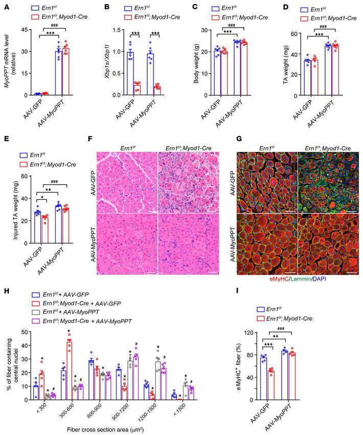
RESEARCH ARTICLE The Journal of Clinical Investigation
mainly mediated by orchestrated activation of the transcript suggest that IRE1α may act as a component in the control mecha-
ional regulatory factors MYF5, MyoD, and myogenin, along with nism during muscle repair and regenerative growth response.
regulated expression of the myosin heavy chain (MyHC) genes To determine the importance of IRE1α in skeletal mus-
(27, 28). In addition, a number of cytokines and growth factors cle regeneration, we used Ern1fl/fl mice with 2 loxP sites flanking
are known to be critically involved in controlling muscle repair exon 2 of the Ern1 gene (38) and Myod1-Cre mice to generate
and growth, as exemplified by IL-6, myostatin/TGF-β, and insu- skeletal muscle–specific IRE1α-knockout (referred to as Ern1fl/fl
lin-like growth factor-1 (IGF-1) (30, 31). Impairment of muscle Myod1-Cre) mice, which were born at normal Mendelian ratios and
regeneration and growth leads to muscle atrophy or dystrophy, appeared to be grossly normal. Immunoblot analysis showed sub-
which represents a key pathological feature of many human dis- stantial reductions in IRE1α protein levels from multiple muscle
eases with muscle loss (32). For instance, Duchenne muscular types in Ern1fl/fl Myod1-Cre mice relative to their Ern1fl/fl littermates
dystrophy (DMD), caused by mutations in the X-linked gene or Myod1-Cre controls, whereas no changes were detected in their
dystrophin, is the most common and severe form of human mus- hearts (Supplemental Figure 1A; supplemental material available
cular dystrophy and remains incurable to date (33, 34). A better online with this article; https://doi.org/10.1172/JCI143737DS1).
understanding of the complex interplays between the intrinsic As expected, markedly reduced Xbp1 mRNA splicing was detected
cell signaling network and microenvironmental cues will uncover in the Ern1fl/fl Myod1-Cre muscles relative to their control groups
the molecular regulatory mechanisms in the maintenance of (Supplemental Figure 1B). Ern1fl/fl Myod1-Cre mice exhibited no
muscle mass, thus facilitating the development of regenerative overt differences in their body weight, muscle weight, or myofi-
therapeutics for muscle degeneration disorders. ber morphology (Supplemental Figure 2, A–D), and showed no
As a critical regulator of ER stress response in restoration of significant alterations in the abundance of Atf4 or Chop mRNA
proteostatic homeostasis, IRE1α has been documented as promot- (Supplemental Figure 2E) or of Myh1, Myh2, or Myh4 mRNA
ing cell proliferation during the reparative regeneration of the liver (Supplemental Figure 2F) in their TA muscles. This suggests that
(35) and the compensatory growth of pancreatic islets under meta- ablation of IRE1α neither provokes ER stress, nor affects muscle
bolic stress (36). However, it has yet to be explored whether IRE1α development. When subjected to CTX-induced injury in their TA
plays a role in sensing skeletal muscle stress and regulating its muscles, animals of all 3 genotypes showed markedly reduced TA
regeneration process. In this study, we used a cardiotoxin-induced muscle weight, presumably due to extensive myofiber degenera-
(CTX-induced) acute muscle injury model as well as the genetic tion, at 4 days after CTX injection (Figure 1D). Whereas gradual
mdx model of DMD in mice, and found that IRE1α acts to sustain restoration of TA muscle mass was seen in all 3 genotypes at 8 and
muscle repair and growth, largely through its RIDD activity to 12 days after CTX injection (Figure 1D), a significant reduction
downregulate the expression of the mRNA encoding myostatin, in the weight of regenerating TA muscle was observed in Ern1fl/fl
a critical negative regulator of muscle regeneration and growth. Myod1-Cre mice relative to their Ern1fl/fl or Myod1-Cre counterparts
Our findings demonstrate the physiological importance of IRE1α’s at 12 days after CTX-induced injury (Figure 1D). This indicates
RIDD activity in the control of muscle regeneration, and its dys- that loss of IRE1α in muscle cells resulted in impaired regenera-
regulation may represent an unrecognized mechanism linking ER tive response to acute muscle injury. Indeed, histological analysis
stress to muscle degenerative diseases. revealed that at 8 days after CTX injection, while TA muscles from
all 3 genotypes showed newly formed myofibers containing cen-
Results tralized nuclei that replaced the damaged myofibers (Figure 1E), a
Activation of the IRE1α signaling branch is implicated in skeletal significantly lower percentage of large regenerated myofibers was
muscle regeneration. To investigate whether IRE1α is implicated in found in Ern1fl/fl Myod-Cre TA muscles than in those of Ern1fl/fl or
regulating muscle regeneration, we first examined its activation Myod1-Cre control mice at 8 or 12 days after CTX-induced injury
states during the regenerative response to acute skeletal muscle (Figure 1, E and F, and Supplemental Figure 3A). This defect of
injury by injecting CTX into the tibialis anterior (TA) muscles of reparative regeneration in Ern1fl/fl Myod1-Cre TA muscles was fur-
adult mice. In line with previously reported findings (37), CTX- ther reflected by analysis of muscle sections stained for eMyHC
induced myofiber degeneration triggered robust muscle regener- (Figure 1, G and H, and Supplemental Figure 3B). At 8 days after
ation responses, as indicated by prominently elevated expression CTX injection, whereas Ern1fl/fl or Myod1-Cre TA muscles exhibited
of embryonic MyHC (eMyHC) at 2 days after CTX treatment (Fig- strongly expressed eMyHC in most myofibers, Ern1fl/fl Myod1-Cre
ure 1A). Starting at 2 days after CTX-elicited injury, we observed TA muscles showed marked reductions in eMyHC expression in
significant increases in the phosphorylation at Ser724 within the many myofibers, along with eMyHC-positive fibers displaying
kinase domain of IRE1α that peaked at 4 and 8 days (Figure 1A). apparent heterogeneity in size (Figure 1, G and H). At 12 days after
Consistent with activated IRE1α’s RNase activity, significantly CTX injection, however, Ern1fl/fl or Myod1-Cre TA muscles had
increased Xbp1 mRNA splicing and decreased abundance of Blos1 weaker eMyHC signals and larger myofibers, but Ern1fl/fl Myod1-
mRNA, a typical RIDD substrate of IRE1α, were detected during Cre TA muscles displayed accumulation of eMyHC in smaller
the regenerative response to CTX-induced injury (Figure 1B), in myofibers, likely reflecting an earlier stage of delayed regenera-
parallel with marked activation of the Myog and Myh8 gene expres- tion (Supplemental Figure 3B). Thus, loss of muscle IRE1α leads to
sion program (Figure 1C). Notably, increased eIF2α phosphoryla- impaired skeletal muscle regeneration after acute injury.
tion and BiP protein expression levels were also seen following Skeletal muscle regeneration in response to injury involves
CTX treatment (Figure 1A). These results indicate that the IRE1α highly orchestrated myogenic transcriptional network and cyto-
branch of the UPR is activated in response to muscle damage, and kine signaling. Interestingly, we detected markedly decreased
2 J Clin Invest. 2021;131(17):e143737 https://doi.org/10.1172/JCI143737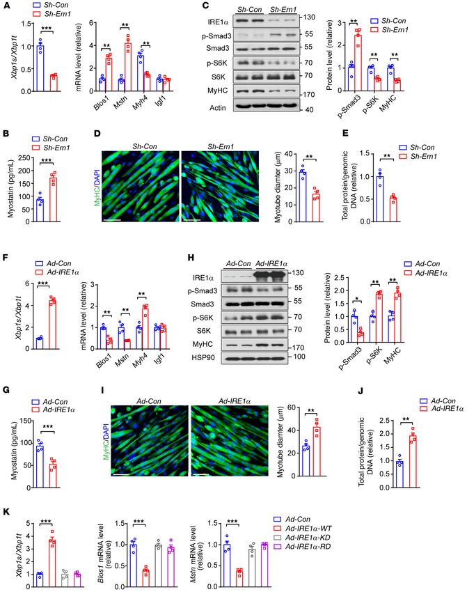
The Journal of Clinical Investigation                                                                    RESEARCH ARTICLE

                                 J Clin Invest. 2021;131(17):e143737 https://doi.org/10.1172/JCI143737                3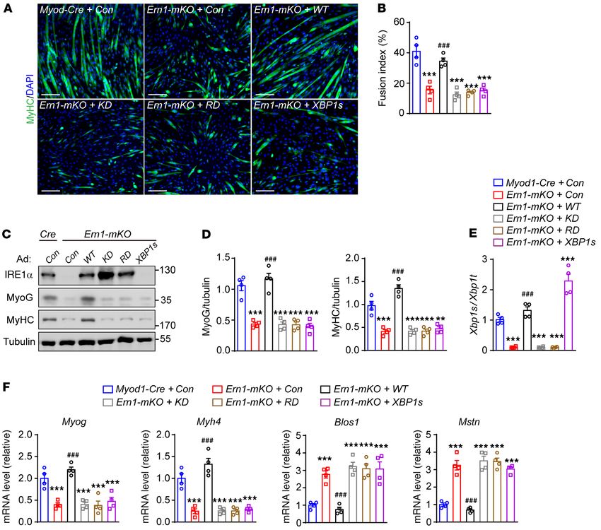
RESEARCH ARTICLE The Journal of Clinical Investigation
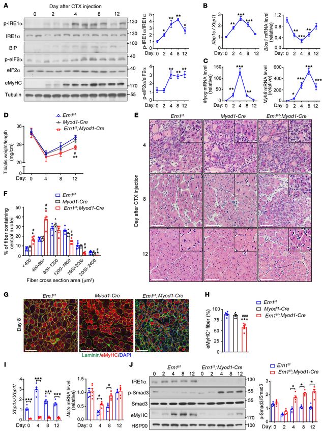
Figure 1. Injury-induced IRE1α activation affects skeletal muscle regener- We then investigated the requirement for IRE1α in myogenesis
ation and myostatin signaling. (A–C) Muscle injury induces IRE1α activa- using primary myoblasts isolated from Ern1fl/fl Myod1-Cre and
tion. TA muscle of adult male mice was injected with CTX to induce acute
Myod1-Cre mice. During the early period of myoblast differenti-
muscle injury (n = 5 mice at each time point). (A) Immunoblot analysis of
the phosphorylation of IRE1α and eIF2α, and protein expression of BiP and ation into myotubes, we observed significantly induced IRE1α
eMyHC in muscle extracts. Each lane represents 1 individual mouse. Aver- phosphorylation (Figure 2, A and B), in parallel with increased
aged p-IRE1α/IRE1α and p-eIF2α/eIF2α ratios are shown from densitomet- Xbp1 mRNA splicing and markedly induced Myh4 expression
ric quantification. (B and C) Quantitative RT-PCR analysis of Xbp1 mRNA (Supplemental Figure 5A). Consistently, loss of IRE1α in prima-
splicing and the abundance of mRNAs encoding the indicated genes. (D–J)
ry myoblasts resulted in significantly upregulated expression of
IRE1α abrogation impairs muscle regeneration and enhances myostatin
signaling. TA muscles of male Ern1fl/fl and Myod1-Cre control and Ern1fl/fl Mstn mRNA (Figure 2C), along with elevated Smad3 phosphory-
Myod1-Cre mice were subjected to CTX-induced injuries (n = 5 mice at each lation and reduced MyHC protein level (Figure 2, D and E). At 3
time point). (D) TA muscle weight relative to tibia length. (E) Represen- days after differentiation, increased myostatin protein was detect-
tative H&E staining of TA muscles from mice of the indicated genotypes. ed in the culture medium of Ern1-mKO (i.e. Ern1fl/fl Myod1-Cre)
(F) Percentage of regenerated myofibers in the indicated cross-sectional
myotubes (Figure 2F); and most Myod1-Cre myoblasts became
areas of TA muscles at 12 days after CTX injection. Myofibers containing
centralized nuclei were quantified by ImageJ from 500 myofibers of the TA elongated and fused with expression of the differentiation marker
muscle in each mouse. (G) Representative laminin (green) and eMyHC (red) MyHC (Figure 2, G–I). By contrast, Ern1-mKO myoblasts showed
immunostaining of TA muscles at 8 days after CTX injection. (H) Quanti- significant decreases in cell fusion and multinuclear myotube for-
fication of the percentage of eMyHC+ myofibers within laminin staining. mation (Figure 2, G–I), with fewer and shorter myotubes observed
(I) Quantitative RT-PCR analysis of Xbp1 mRNA splicing and Mstn mRNA
after differentiation for 5 days (Supplemental Figure 5B). More-
abundance in TA muscles. Data are shown as relative to the value for
Ern1fl/fl TA muscle at day 0 after normalization to Gapdh as the internal over, Ern1-mKO myocytes had significant reductions in the mRNA
control. (J) Immunoblot analysis of TA muscle lysates. Averaged p-Smad/ and protein expression levels of the differentiation markers
Smad ratios are shown after normalization to the value for Ern1fl/fl TA myogenin and MyHC when compared with their Myod1-Cre con-
muscle at day 0. All data are presented as mean ± SEM. Significance was trols (Figure 2, J–L). These results suggest that IRE1α ablation
calculated by 1-way (A–C and H) or 2-way (D, F, I, and J) ANOVA with Bon-
leads to upregulation of myostatin expression and impairment of
ferroni’s multiple-comparison test. *P < 0.05, **P < 0.01, ***P < 0.001 vs.
day 0 or Ern1fl/fl. #P < 0.05, ###P < 0.001 vs. Myod1-Cre. Scale bars: 100 μm. myoblast differentiation in a cell-autonomous manner.
To further test whether IRE1α’s RNase activity is required for
myoblast differentiation, we examined the effects of restored expres-
sion of WT or mutant IRE1α using adenoviruses on the defective
Xbp1 mRNA splicing but significantly increased abundance of differentiation of Ern1-mKO myoblasts. Immunostaining of MyHC
Mstn mRNA, encoding myostatin (Figure 1I), along with a con- showed that overexpression of IRE1α-WT effectively restored the
siderable reduction in Igf1 mRNA expression (Supplemental Fig- differentiation capacity (Figure 3, A and B) as well as the protein and
ure 3C), in TA muscles of Ern1fl/fl Myod1-Cre mice relative to their mRNA expression levels of Myog and Myh4 in Ern1-mKO myoblasts
Ern1fl/fl counterparts during the regenerative response at 4 and 8 (Figure 3, C–F) to an extent similar to that in Myod1-Cre controls,
days after CTX injection. Given that myostatin is a secreted TGF-β whereas overexpression of the IRE1α kinase-dead (KD, K599A) or
family member protein that functions as a potent negative regula- RNase-dead (RD, K907A) mutant versions (41) lost these effects
tor of myogenic differentiation and muscle growth (30), this sug- (Figure 3, A–F). Supporting the role of IRE1α in suppression of myo-
gests that IRE1α ablation affected the signaling pathways in the statin, overexpression of IRE1α-WT, but not its kinase- or RNase-
control of muscle regeneration. Indeed, we observed prominently deficient mutants, substantially blunted the upregulated expres-
elevated phosphorylation of Smad3, the downstream transcription sion of Mstn in Ern1-mKO myocytes, which resembled the changes
factor of the myostatin signaling cascade (39), in Ern1fl/fl Myod1- of the typical RIDD substrate, Blos1 (Figure 3F). Notably, adenoviral
Cre TA muscles (Figure 1J). In accordance with poorer muscle overexpression of XBP1s did not significantly affect the differentia-
regeneration, this enhanced myostatin signaling was associated tion capacity or Mstn expression in Ern1-mKO myocytes (Figure 3,
with marked reductions in eMyHC protein levels (Figure 1J). A–F). These data demonstrate an IRE1α RNase-dependent, XBP1-
Together, these results demonstrate that IRE1α abrogation leads independent regulation of myoblast differentiation, suggesting that
to impaired muscle regeneration and enhanced myostatin signal- IRE1α acts as a negative regulator of myostatin to control myoblast
ing in response to acute muscle injury, indicating that IRE1α may differentiation during muscle regeneration.
function as a myogenic regulator of muscle repair and growth. IRE1α downregulates myostatin expression and promotes myotube
IRE1α RNase regulates myostatin expression and myoblast differ- hypertrophy. Because myostatin is also known as an autocrine fac-
entiation. Because skeletal muscle regeneration involves the acti- tor to suppress hypertrophy in myotubes (30, 42, 43), we next tested
vation, proliferation, and differentiation of muscle satellite cells whether IRE1α could impact myotube hypertrophy remodeling
(MuSCs) (28), we first examined whether IRE1α ablation affected by downregulating myostatin expression. In differentiated C2C12
MuSCs in Ern1fl/fl Myod1-Cre mice. Using a FACS-based method myotubes, adenoviral expression of a short hairpin RNA (shRNA)
(40) for quantitative analysis of either quiescent or activated directed against IRE1α (Sh-Ern1), in comparison with the scramble
MuSCs (VCAM1+CD45 –CD31– Sca1– cells), we observed no signif- shRNA control (Sh-Con), resulted in substantially decreased Xbp1
icant differences in the number of MuSCs from CTX-injured TA mRNA splicing, but significantly increased mRNA abundance of
muscles of Ern1fl/fl, Myod1-Cre, and Ern1fl/fl Myod1-Cre mice (Sup- Blos1 and Mstn (Figure 4A), accompanied by a significant reduction
plemental Figure 4, A and B). This suggests that the satellite cell in the mRNA level of Myh4 but not Igf1 (Figure 4A). In line with
niche was presumably not affected by muscle IRE1α deficiency. enhanced myostatin signaling and impaired myotube growth,
4 J Clin Invest. 2021;131(17):e143737 https://doi.org/10.1172/JCI143737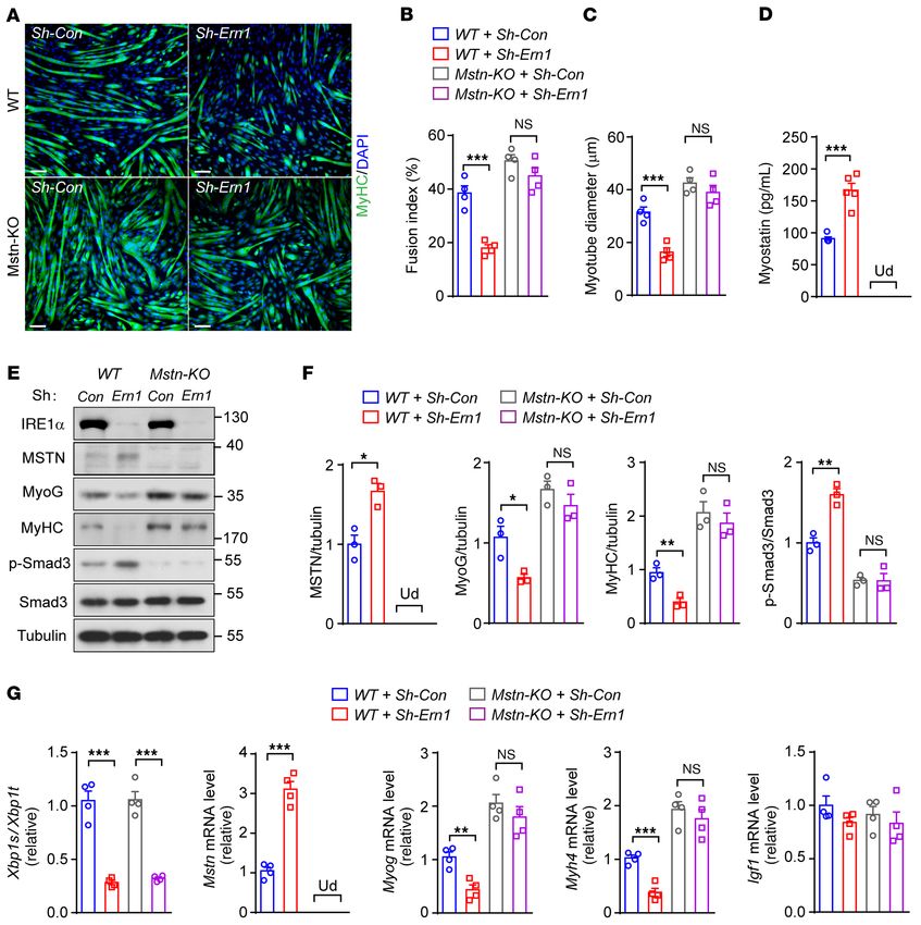
The Journal of Clinical Investigation RESEARCH ARTICLE
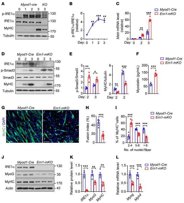
Figure 2. IRE1α ablation increases myostatin expression and impairs myoblast differentiation. (A–E) Primary myoblasts isolated from TA muscle
of male Myod1-Cre and Ern1fl/fl Myod1-Cre (KO or Ern1-mKO) mice were differentiated for up to 3 days as indicated (n = 3–5 independent experiments).
(A and B) Immunoblot analysis of IRE1α phosphorylation and MyHC expression (A) at the indicated time of differentiation, and quantification of
averaged p-IRE1α/IRE1α ratios (B). (C) Quantitative RT-PCR analysis of the mRNA abundance of Mstn. (D and E) Immunoblot analysis of cell lysates
(D) using the indicated antibodies, and averaged p-Smad3/Smad3 and MyHC/tubulin levels (E). (F–L) Primary TA myoblasts were differentiated into
myotubes for 3 days (n = 4–6 independent experiments). (F) ELISA analysis of myostatin protein in culture medium. (G–I) Representative images
(G) of immunostaining of MyHC (green) and DAPI staining of nuclei (blue), with quantification of the fusion index (H) and the number of nuclei per
MyHC+ fiber (I). (J–L) Immunoblot analysis (J) and quantification (K) of MyoG and MyHC in differentiated myocytes, and quantitative RT-PCR analysis
(L) of their mRNA expression levels. All data are presented as mean ± SEM. Significance was calculated by unpaired 2-tailed Student’s t test (F, H, I,
K, and L), 1-way ANOVA (B), or 2-way ANOVA (C and E) with Bonferroni’s multiple-comparison test. *P < 0.05, **P < 0.01, ***P < 0.001 vs. Myod1-Cre
group, or vs. day 0 for B. Scale bars: 100 μm.
knockdown of IRE1α expression led to significantly higher produc- sistent with the phenotypes of smaller myofibers in mice with mus-
tion of myostatin protein in the medium (Figure 4B) and increased cle IRE1α abrogation following CTX-induced injury. Conversely,
phosphorylation of Smad3 (Figure 4C), along with decreases in when compared with the control, adenoviral IRE1α overexpression
S6K phosphorylation and MyHC protein levels. Moreover, this was in differentiated C2C12 myotubes resulted in significantly elevated
in parallel with significantly reduced diameters of myotubes (Fig- Xbp1 mRNA splicing (Figure 4F), reduced mRNA abundance of
ure 4D) and total cellular protein contents (Figure 4E). This is con- Blos1 and Mstn (Figure 4F), and lower myostatin protein produc-
J Clin Invest. 2021;131(17):e143737 https://doi.org/10.1172/JCI143737 5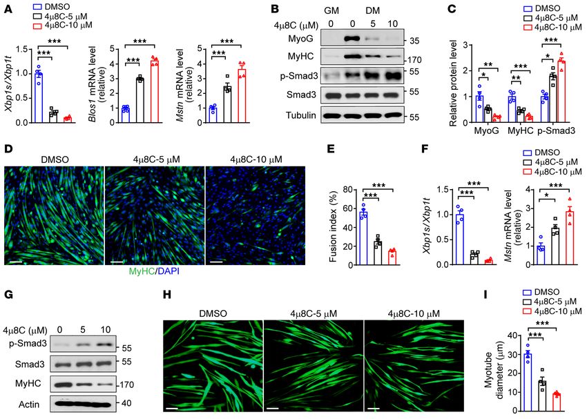
RESEARCH ARTICLE                                                                                           The Journal of Clinical Investigation

Figure 3. IRE1α RNase blunts myostatin signaling and promotes myoblast differentiation. Primary Myod1-Cre and Ern1-mKO myoblasts were infected
with control recombinant adenovirus or adenoviruses expressing human WT or mutant (KD, kinase dead) IRE1α protein or XBP1s and then differentiated
for 3 days (n = 4 independent experiments). (A) Representative images of anti-MyHC and DAPI staining. (B) Quantification of the fusion index. (C)
Immunoblot analysis of the indicated proteins. (D) Quantification of MyoG/tubulin and MyHC/tubulin after normalization to the value of control. (E and
F) Quantitative RT-PCR analysis of Xbp1 mRNA splicing (E) and the mRNA abundance of the indicated genes (F). All data are presented as mean ± SEM.
Significance was calculated by 1-way ANOVA with Bonferroni’s multiple-comparison test. **P < 0.01, ***P < 0.001 vs. Myod1-Cre group. ###P < 0.001 vs.
Ern1-mKO + control adenovirus. Scale bars: 100 μm.

tion (Figure 4G) and signaling (Figure 4H), along with enlarged               liferation (44), we also evaluated whether IRE1α exerts a regulatory
myotubes (Figure 4I) and higher cellular protein contents (Figure             action in this regard. MTT analyses showed that neither deficiency
4J). Notably, adenoviral shRNA knockdown of XBP1 expression                   nor overexpression of IRE1α had significant effects on the prolifer-
caused a slight reduction in Mstn mRNA level (Supplemental Fig-               ation rate of primary or C2C12 myoblasts (Supplemental Figure 7,
ure 6A), while showing no significant effect on myotube growth                A–C). Moreover, FACS analyses showed that knockdown of IRE1α
(Supplemental Figure 6B). In addition, adenoviral overexpression              expression in C2C12 myoblasts did not affect cell death, which was
of IRE1α-WT, but not its KD or RD mutant versions, significantly              increased upon differentiation stimuli (Supplemental Figure 7D).
increased Xbp1 mRNA splicing and decreased the mRNA abun-                     Thus, IRE1α-directed regulation of myostatin is implicated in both
dance of Mstn and Blos1 in C2C12 myotubes (Figure 4K). These                  myocyte differentiation and hypertrophy growth, consistent with
in vitro analyses via loss- and gain-of-function strategies in myo-           our observation in vivo that loss of IRE1α led to impairment in mus-
tubes strongly suggest that IRE1α downregulation of myostatin                 cle regeneration upon injury.
expression also contributes to the control of myotube hypertrophy                  Myostatin mediates IRE1α’s regulation of muscle cell differentia-
remodeling. Since myostatin was reported to impact myoblast pro-              tion and growth. We then determined whether myostatin is required

   6                                     J Clin Invest. 2021;131(17):e143737 https://doi.org/10.1172/JCI143737
The Journal of Clinical Investigation                                                                    RESEARCH ARTICLE

                                 J Clin Invest. 2021;131(17):e143737 https://doi.org/10.1172/JCI143737                7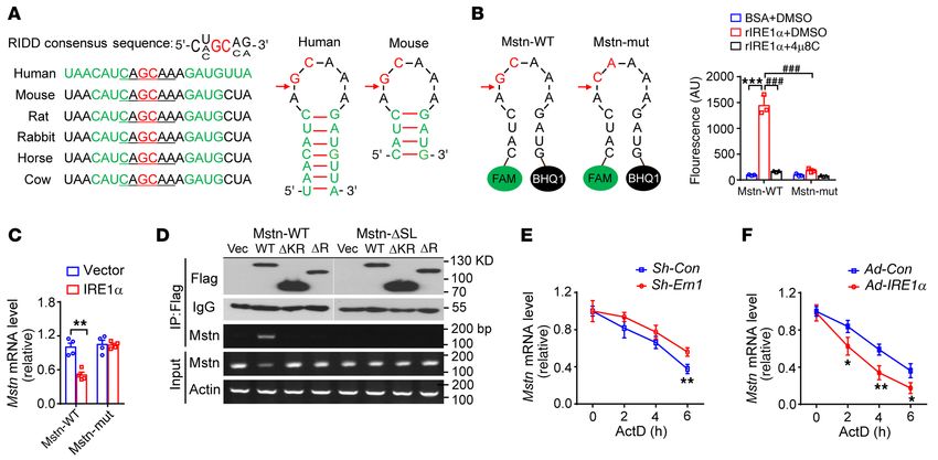
RESEARCH ARTICLE                                                                                          The Journal of Clinical Investigation

Figure 4. IRE1α downregulates myostatin expression and promotes                 muscle weight in both groups (Figure 6, C and D). Importantly, at
hypertrophy of differentiated myotubes. (A–J) C2C12 myoblast cells              8 days after CTX-induced injury, MyoPPT not only promoted the
were differentiated for 4 days into myotubes and then infected for 48           reparative regeneration of TA muscles of the Ern1fl/fl control group,
hours with adenoviruses expressing a scramble control shRNA (Sh-Con)
or shRNA directed against IRE1α (Sh-Ern1) (A–E), or with empty control
                                                                                but also prominently rescued the regenerative defect of Ern1fl/fl
adenovirus (Ad-Con) or that expressing human IRE1α (Ad-IRE1α) (F–J) (n          Myod1-Cre TA muscles, as evidenced by significantly increased
= 4 independent experiments). (A and F) Quantitative RT-PCR analysis of         TA muscle weight (Figure 6E), more newly formed myofibers
Xbp1 mRNA splicing and the mRNA abundance of the indicated genes. (B            containing centralized nuclei (Figure 6F), increased eMyHC
and G) ELISA analysis of myostatin protein in culture medium. (C and H)         expression (Figure 6G), and higher percentages of large regener-
Immunoblot analysis of the indicated proteins from myotube extracts.
Averaged MyHC/actin, p-S6K/S6K, and p-Smad3/Smad3 ratios were
                                                                                ated myofibers (Figure 6H) and eMyHC-positive fibers (Figure 6I)
normalized to the value of Sh-Con or Ad-Con myotubes. (D and I) MyHC            when compared with the GFP control group (Figure 6, E–I). These
immunostaining of myotubes. Myotube diameters were quantified using             results further demonstrate in vivo that myostatin is a crucial
ImageJ software. (E and J) Total cellular protein content relative to genomic   player in mediating IRE1α ablation–associated impairment of
DNA was measured in myotubes. (K) C2C12 myotubes were infected with             skeletal muscle regeneration upon acute injury.
empty control or adenoviruses expressing the WT or indicated mutant
IRE1α protein. Quantitative RT-PCR analysis of Xbp1 mRNA splicing and
                                                                                     IRE1α suppresses myostatin expression through its RIDD activity.
the mRNA abundance of Blos1 and Mstn (n = 4 independent experiments).           We next asked whether the mRNA expression of Mstn is subjected
All data represent mean ± SEM. Significance was calculated by unpaired          to the control of IRE1α’s RIDD activity. To determine whether
2-tailed Student’s t test (A–J) or 1-way ANOVA (K) with Bonferroni’s            Mstn mRNA represents a valid RIDD substrate, we conducted bio-
multiple-comparison test. *P < 0.05, **P < 0.01, ***P < 0.001 vs.               informatics analysis combined with prediction of mRNA second-
Sh-Con or Ad-Con. Scale bars: 100 μm.
                                                                                ary structures. Indeed, we identified a highly conserved region in
                                                                                mammalian mRNAs encoding myostatin (spanning nucleotides
                                                                                335–349 downstream of the putative transcription start site in
for IRE1α-mediated effect on myoblast differentiation using                     mouse Mstn mRNA) that possesses the postulated RIDD consen-
Mstn-KO C2C12 cells in which Mstn was abrogated by a CRISPR/                    sus sequence (CNGCNN) (9) within a predicted hairpin struc-
Cas9–based strategy (45). Immunostaining of MyHC in differen-                   ture containing a potential IRE1α cleavage site as found in Xbp1
tiated myotubes showed slightly increased fusion index and myo-                 mRNA (Figure 7A). Then, we tested whether this region of Mstn
tube diameter in Mstn-KO cells relative to the WT control cells                 mRNA could be directly cleaved by purified recombinant IRE1α
(Figure 5, A–C). Whereas knockdown of IRE1α expression resulted                 protein, using a synthetic 14-nucleotide Mstn mRNA molecule
in lower cell fusion and smaller myotube formation in parallel with             labeled with 5′-FAM fluorophore and 3′-black hole quencher
higher myostatin production (Figure 5D) in WT myoblasts, loss of                (BHQ) as the substrate in an in vitro biochemical assay (Figure
myostatin diminished these effects of IRE1α deficiency in Mstn-                 7B). Incubation with IRE1α of this Mstn mRNA substrate abolished
KO cells (Figure 5, A–D). Consistently, immunoblotting and quan-                the quenching effect of BHQ, resulting in a dramatic increase in
titative reverse transcription PCR (qRT-PCR) analyses showed                    its fluorescent output signal (Figure 7B). By contrast, addition
that ablation of myostatin restored the protein and mRNA expres-                of 4μ8C, a pharmacological inhibitor of IRE1α RNase activity
sion of the myogenic markers myogenin and MyHC in the face of                   (51, 52), markedly reduced this cleavage-based fluorescent sig-
IRE1α knockdown (Figure 5, E–G), with no significant alterations                nal (Figure 7B). To affirm IRE1α’s action at the putative cleavage
observed in Igf1 mRNA level (Figure 5G). Moreover, the promot-                  site, we generated a synthetic mutant Mstn mRNA substrate by
ing effects of IRE1α overexpression on muscle cell differentiation              replacing the conserved GC with CA, which completely abolished
and growth were also diminished in Mstn-KO cells (Supplemental                  the cleavage-based fluorescent signal in comparison with its WT
Figure 8, A–E). These data revealed the importance of myostatin                 version (Figure 7B). Furthermore, quantitative RT-PCR analysis
suppression, at least in large part, in mediating IRE1α regulation              in HEK293T cells showed that cotransfected expression of IRE1α
of myocyte differentiation and growth. Together, our results sup-               resulted in significantly decreased abundance of the WT but not
port a model in which IRE1α suppresses myostatin expression,                    the GC-to-CA mutant Mstn mRNA spanning the entire coding
thereby promoting myoblast differentiation and myotube hyper-                   region of myostatin (Figure 7C). Interestingly, RT-PCR analyses
trophy growth during skeletal muscle regeneration.                              showed that this Mstn mRNA could be immunoprecipitated along
     Myostatin mediates IRE1α’s regulatory action during skeletal               with the WT but not the truncation mutant IRE1α protein lacking
muscle regeneration. To determine the importance of myostatin                   its RNase domain from cotransfected HEK293T cells (Figure 7D);
in mediating IRE1α’s regulatory effect in vivo on muscle regen-                 by contrast, deletion of the stem-loop structure of the Mstn mRNA
eration, we generated the recombinant adeno-associated virus                    abolished its ability to associate with IRE1α protein (Figure 7D).
2/9 (AAV2/9) expressing the N-terminal propeptide (MyoPPT)                      This indicates the structural involvement of IRE1α RNase domain
of myostatin, which is capable of inhibiting the biological activ-              and the stem-loop of the Mstn mRNA substrate in this particular
ity and function of mature myostatin (46–48). Systemic delivery                 RIDD machinery. We further determined whether IRE1α could
of MyoPPT (49, 50) to neonatal Ern1fl/fl and Ern1fl/fl Myod1-Cre                affect the stability of Mstn mRNA using C2C12 myotubes follow-
mice, relative to the GFP control, resulted in efficient MyoPPT                 ing treatment with actinomycin D. Knockdown of IRE1α resulted
expression in their TA muscles and did not influence IRE1α’s Xbp1               in a decrease in the degradation rate of Mstn mRNA (Figure 7E),
mRNA splicing activity (Figure 6, A and B); and in accordance                   whereas IRE1α overexpression significantly accelerated it (Fig-
with reported findings (49, 50), blocking myostatin signaling by                ure 7F). Collectively, these results demonstrate that Mstn mRNA
MyoPPT caused significant increases in their body weight and TA                 could indeed serve as a bona fide RIDD substrate of IRE1α.

   8                                      J Clin Invest. 2021;131(17):e143737 https://doi.org/10.1172/JCI143737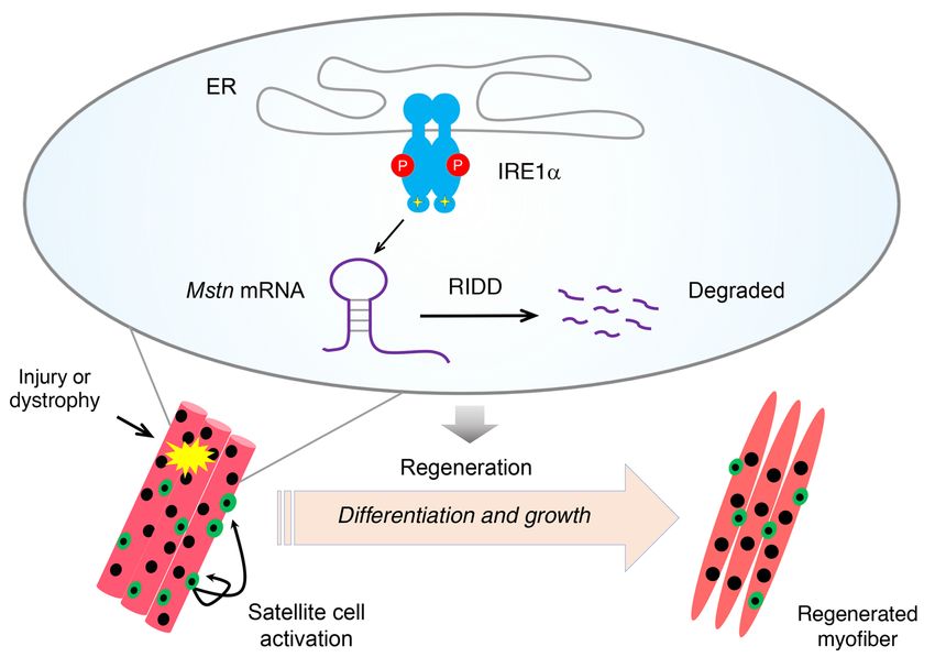
The Journal of Clinical Investigation                                                                                   RESEARCH ARTICLE

Figure 5. Myostatin ablation reverses the effect of IRE1α deficiency on muscle cell differentiation and growth. WT and Mstn-KO C2C12 myoblast cells
were infected for 24 hours with Sh-Con or Sh-Ern1 adenoviruses and then differentiated for 3–4 days in myogenic medium. (A–C) Representative images of
MyHC immunostaining (A), and quantification of the fusion index (B) and myotube diameters (C) (n = 4 independent experiments). (D) ELISA analysis of
myostatin protein in culture medium (n = 3 independent experiments). (E and F) Immunoblot analysis of the indicated proteins (E), and quantification of
averaged MSTN/tubulin, MyoG/tubulin, MyHC/tubulin, and p-Smad3/Smad3 levels (F) (n = 3 independent experiments). (G) Quantitative RT-PCR analysis
of Xbp1 mRNA splicing and the mRNA abundance of the indicated genes (n = 4 independent experiments). All results represent mean ± SEM. *P < 0.05,
**P < 0.01, ***P < 0.001 by 2-way ANOVA with Bonferroni’s multiple-comparison test. Ud, undetectable. Scale bars: 100 μm.

    We further validated that pharmacological inhibition by                  resulted in decreased Xbp1 mRNA splicing and elevated mRNA
4μ8C of IRE1α RNase activity in primary myoblasts was able to,               abundance of Mstn (Figure 8F), enhanced myostatin signaling
in a dose-dependent fashion, suppress Xbp1 mRNA splicing and                 and lower levels of myogenic markers (Figure 8G), and decreased
elevate the mRNA abundance of Blos1 and Mstn (Figure 8A),                    diameters of myotubes (Figure 8, H and I). Together, these data
leading to increased activation of myostatin signaling, reduced              clearly demonstrate that IRE1α could employ its RNase-depen-
protein expression of myogenic markers, and prominently lower                dent RIDD activity to blunt the mRNA expression of myostatin
myotube formation (Figure 8, B–E). Consistently, 4μ8C inhibition             and exert its myogenic effect during the differentiation of myo-
of IRE1α RNase activity in differentiated C2C12 myotubes also                blasts and hypertrophic growth of myotubes.

                                        J Clin Invest. 2021;131(17):e143737 https://doi.org/10.1172/JCI143737                                     9
RESEARCH ARTICLE The Journal of Clinical Investigation 10 J Clin Invest. 2021;131(17):e143737 https://doi.org/10.1172/JCI143737
The Journal of Clinical Investigation RESEARCH ARTICLE
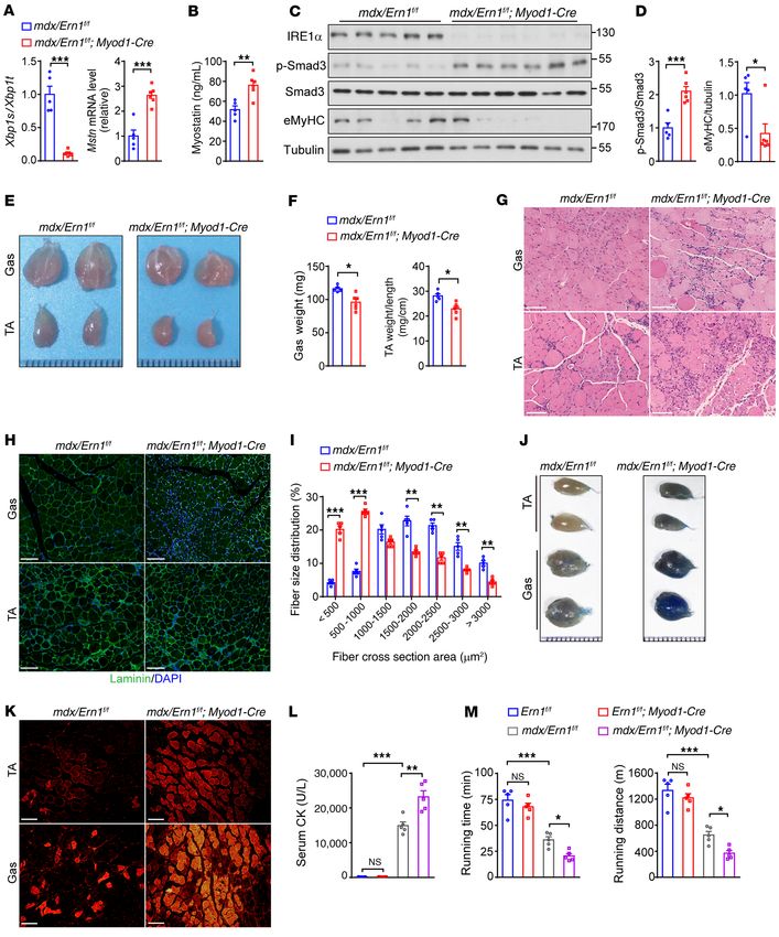
Figure 6. Inhibition in vivo of myostatin signaling rescues the impairment to assess muscle fiber integrity, we further detected more pro-
of muscle regeneration resulting from IRE1α deficiency. Male Ern1fl/fl or nounced damage with higher dye uptake in mdx/Ern1fl/fl Myod1-
Ern1fl/fl Myod1-Cre mice were injected intraperitoneally on postnatal day 3
Cre muscles (Figure 9, J and K). In addition, we observed no overt
(P3) and P6 with adeno-associated virus 2/9 (AAV2/9) expressing GFP
(AAV-GFP) or MyoPPT (AAV-MyoPPT). TA muscles of indicated mice were changes in the serum levels of creatine kinase (CK), a hallmark
injected with PBS or CTX at 7 weeks of age and analyzed at 8 days after indicator of damaged muscles, in Ern1fl/fl Myod1-Cre mice, but
injection. (A and B) Quantitative RT-PCR analysis of the mRNA abundance of found marked elevations in serum CK levels in mdx/Ern1fl/fl mice
MyoPPT (A) and Xbp1 mRNA splicing (B) in PBS-treated uninjured TA mus- relative to their Ern1fl/fl controls (Figure 9L). Moreover, mdx/Ern1fl/fl
cles (n = 6 mice per group). (C) Body weight of mice of the indicated groups.
Myod1-Cre mice displayed more pronounced increases in their
(D) Weight of PBS-treated uninjured TA muscles. (E) Weight of CTX-injured
TA muscles from mice of the indicated groups (n = 5–7 mice per group). (F serum CK levels relative to mdx/Ern1fl/fl mice (24,109 ± 3797 U/L
and G) Representative H&E staining (F) and laminin (green) and eMyHC vs. 14,437 ± 2719 U/L, n = 5 mice per genotype, P = 0.0013). To
(red) immunostaining (G) of injured TA muscles (n = 5 mice per group). (H) further evaluate the impact of IRE1α deficiency on muscle func-
Percentage of regenerated myofibers in the indicated cross-sectional areas tionality and performance, we challenged the animals with tread-
of TA muscles. Myofibers containing centralized nuclei were quantified by
mill exercise and measured the running time and distance before
ImageJ from 500 myofibers in each mouse (n = 5 mice per group). (I) Quanti-
fication of the percentage of eMyHC+ myofibers within laminin staining exhaustion. Whereas Ern1fl/fl Myod1-Cre and Ern1fl/fl mice displayed
(n = 5 mice per group). All data are presented as mean ± SEM. Significance no significant difference in their exercise performance, mdx/
was calculated by 2-way ANOVA with Bonferroni’s multiple-comparison test. Ern1fl/fl animals showed a significantly shorter running time and
*P < 0.05, **P < 0.01, ***P < 0.001 vs. Ern1fl/fl + AAV-GFP. #P < 0.05, ###P < distance (Figure 9M), and mdx/Ern1fl/fl Myod1-Cre mice exhibit-
0.001 vs. Ern1fl/fl Myod1-Cre + AAV-GFP. Scale bars: 100 μm.
ed a more pronounced impairment in their running ability (mdx/
Ern1fl/fl, 651 ± 88 m, vs. mdx/Ern1fl/fl Myod1-Cre, 364 ± 90 m; n =
5 mice per genotype, P = 0.004). These results demonstrate that
Loss of IRE1α exacerbates the dystrophic phenotypes in mdx mice. ablation of IRE1α aggravates the severity of muscle dystrophy
DMD is the most common and severe form of muscular dystrophy, stemming from the lack of dystrophin in mdx mice, further estab-
and it remains incurable to date (33, 34). The mdx mouse model lishing the IRE1α-myostatin axis as an important regulatory loop
of DMD exhibits extensive muscle degeneration and regeneration during regeneration in the setting of muscle dystrophy.
due to a null mutation in the Dmd gene, coding for the dystrophin
protein, that causes higher sensitivity of myofibers to contractile Discussion
stress (53). We wondered whether the IRE1α-myostatin regulatory Skeletal muscle relies critically on its self-regeneration for pre-
circuit also plays a protective role in the setting of muscular dystro- serving muscle mass and ensuring muscle function in response to
phy. To this end, we generated mdx/Ern1fl/fl Myod1-Cre mice with injury and disease. Muscle regenerative capacity declines with age
muscle-specific IRE1α deletion in the mdx background through and is severely compromised in many muscular dystrophies (39).
genetic intercrossing. As expected, we detected significantly Delineation of the molecular mechanisms involved in regulating
increased abundance of TA muscle Mstn mRNA as well as higher skeletal muscle regeneration and repair is of great importance to
circulating myostatin protein in mdx/Ern1fl/fl Myod1-Cre mice rela- development of therapeutic approaches for muscle degenerative
tive to their mdx/Ern1fl/fl littermates (Figure 9, A and B). This was diseases. Herein, using 2 mouse models, we have uncovered a
associated with elevated Smad3 phosphorylation and reduced critical role for the UPR sensor IRE1α in promoting skeletal mus-
eMyHC protein level (Figure 9, C and D). Supporting a defective cle regeneration in response to CTX-induced acute muscle injury
IRE1α-mediated suppression of myostatin and weakened muscle or genetic muscle dystrophy. Ablation of IRE1α specifically in
regeneration, mdx/Ern1fl/fl Myod1-Cre mice at 6 weeks, as well as at skeletal muscles results in impaired muscle regenerative repair
21 weeks, of age had grossly smaller gastrocnemius (Gas) and TA or worsened progression of muscle dystrophy. Importantly, our
muscles, and the weight of these muscles was significantly lower data demonstrate that IRE1α employs its RNase-dependent RIDD
than in their mdx/Ern1fl/fl counterparts (Figure 9, E and F, and action to downregulate the level of mRNA encoding myostatin,
Supplemental Figure 9A). Histological analyses revealed more a key inhibitory regulator of muscle growth and repair. Our find-
severe muscle dystrophic characteristics in mdx/Ern1fl/fl Myod1- ings reveal a physiological function of the RIDD output of IRE1α
Cre animals, as indicated by higher variations in their fiber size, enzyme in the control of muscle regeneration and growth (Figure
apparently elevated number of small fibers, and more infiltration 10), suggesting that IRE1α may serve as an important modifier in
of inflammatory immune cells as a result of degenerated myofi- the disease progression of muscular dystrophy or muscle loss.
bers (Figure 9G and Supplemental Figure 9B). Immunostaining IRE1α is the most ancient ER stress sensor, conveying a crit-
of laminin in muscle sections showed many smaller regenerat- ical signaling response through its RNase activity to generate
ing myofibers in Gas and TA muscles of mdx/Ern1fl/fl Myod1-Cre an active transcription factor, XBP1s (3, 5). While IRE1α is best
mice at 6 or 21 weeks of age compared with their mdx/Ern1fl/fl known to exert its prosurvival actions through XBP1s when cop-
counterparts (Figure 9H and Supplemental Figure 9C), with a ing with ER stress, emerging lines of evidence also indicate that
significantly lower percentage of larger regenerating myofibers activated IRE1α RNase activity may have a broad range of func-
observed in TA muscles (Figure 9I and Supplemental Figure 9D). tions, as reflected by an increasing number of identified putative
This indicates that loss of IRE1α results in impaired maturation of RIDD substrates (8, 15). However, the physiologically relevant
myofibers during the regenerative growth of mdx/Ern1fl/fl Myod1- RIDD actions of IRE1α have yet to be established in vivo. In this
Cre muscles, similar to its effect on reparative muscle growth fol- study, we provide physiological evidence for a RIDD-mediated
lowing CTX-induced acute injury. Using Evans blue dye staining mechanism by which IRE1α regulates myostatin production and
J Clin Invest. 2021;131(17):e143737 https://doi.org/10.1172/JCI143737 11RESEARCH ARTICLE The Journal of Clinical Investigation
Figure 7. Myostatin mRNA is a RIDD target of IRE1α. (A) Sequence alignment of Mstn mRNAs from the indicated species with the putative conserved
RIDD region, with the predicted stem-loop secondary structure shown for human and mouse Mstn mRNA. The potential IRE1α cleavage site is indicated by
an arrow. (B) Fluorescence-based analysis of IRE1α cleavage of the synthetic WT or mutant (mut, GC to CA) Mstn mRNA (n = 3 independent experiments).
(C) Quantitative RT-PCR analysis of the abundance of Mstn mRNA in HEK293T cells cotransfected with plasmids expressing the entire coding region of WT
or mutant myostatin (GC to CA) together with empty vector or human IRE1α expression plasmid (n = 4 independent experiments). (D) HEK293T cells were
cotransfected with plasmids expressing the WT or stem-loop deletion (ΔSL) mutant myostatin together with vector (Vec) control or plasmids expressing
FLAG-tagged human WT or deletion mutant (KR, kinase and RNase domain; R, RNase domain) IRE1α proteins. Representative RT-PCR analysis of Mstn
mRNA from immunoprecipitates (IP) using anti-FLAG antibody or from total cellular RNA (Input). Shown also are representative immunoblots of immu-
noprecipitated IRE1α proteins and IgG control (n = 3 independent experiments). (E and F) The stability of the Mstn mRNA was determined by quantitative
RT-PCR in C2C12 myotubes infected with Sh-Con or Sh-Ern1 adenoviruses (E), or Ad-Con or Ad-IRE1α adenoviruses (F), after treatment with actinomycin D
(ActD) for the indicated time intervals. Data are shown relative to the value at time 0 of ActD treatment (set as 1) after normalization to the GAPDH mRNA
level as internal control (n = 3 independent experiments). All data are shown as mean ± SEM. Significance was calculated by 2-way ANOVA with Bonfer-
roni’s multiple-comparison test. *P < 0.05, **P < 0.01, ***P < 0.001. ###P < 0.001. Scale bars: 100 μm.
regenerative maturation of myofibers in response to muscle injury accordance with the fact that myostatin can regulate both differ-
or degenerative dystrophy. It is notable that ablation of IRE1α entiation in myoblasts and hypertrophy in myotubes (55). More-
did not cause changes in muscle and myofiber sizes or myosta- over, inhibition of myostatin has been shown to improve the
tin expression levels in Ern1fl/fl Myod1-Cre mice in the absence of hallmarks of the muscular dystrophy in mdx mice (56, 57). This
injury or dystrophy. Moreover, IRE1α phosphorylation is relatively also supports our model that the lack of IRE1α-myostatin regula-
low in muscle cells under basal conditions, but is highly induced tory axis contributes to the more severe dystrophic phenotypes in
during myoblast differentiation or upon muscle injury. This sug- mdx/Ern1fl/fl Myod1-Cre mice.
gests that activation of the IRE1α-myostatin regulatory circuit is XBP1 has been recently reported to be dispensable for sat-
coupled to the stress condition induced by muscle injury or dam- ellite cell–mediated muscle regeneration, whereas the PERK/
age, conceivably reflecting the multifunctional property of IRE1α eIF2α arm of the UPR may play an important role in this process
in utilizing its RIDD activity to handle various types of stress con- (58). Documented studies have also suggested that myostatin
ditions in a tissue- or cell type–specific fashion. can suppress satellite cell activation and self-renewal (43, 59,
It is worth noting that we used Myod1-Cre mice to create mice 60). While our results indicate that IRE1α deficiency caused no
with IRE1α deletion specifically in skeletal muscles. Because of significant changes in the number of TA muscle satellite cells
the activated expression pattern of Myod1 during myogenesis upon acute injury, it has yet to be more thoroughly interrogated
(54), IRE1α abrogation most likely occurred in muscle stem cells whether IRE1α can regulate satellite cells through a myosta-
(i.e., satellite cells) at particular differentiation stages as well as tin-mediated mechanism or cell-intrinsically in an XBP1-
in mature muscle fibers. Our results from analyses of muscle cell independent manner, e.g., by affecting the STAT3 pathway (35,
differentiation and growth in vitro suggest that IRE1α can promote 61), which was reported to critically regulate the proliferation
both myoblast differentiation and myotube hypertrophy growth. and self-renewal of muscle stem cells (62).
Thus, the impairment of injury-induced regeneration in Ern1fl/fl Myostatin, GDF11, and activin A are the 3 circulating factors
Myod1-Cre mice can be largely attributed to the cell-autonomous that belong to the TGF-β family in the negative control of skeletal
action of IRE1α in myocyte differentiation and growth. This is in muscle mass (30). It remains to be dissected whether IRE1α also
12 J Clin Invest. 2021;131(17):e143737 https://doi.org/10.1172/JCI143737The Journal of Clinical Investigation RESEARCH ARTICLE
Figure 8. Pharmacological inhibition of IRE1α RNase activity impairs muscle cell differentiation and growth. (A–E) Primary myoblasts isolated from WT
mice were cultured in growth medium (GM), and subsequently incubated in differentiation medium (DM) with or without 4μ8C for 3 days (n = 4 indepen-
dent experiments). (A) Quantitative RT-PCR analysis of Xbp1 mRNA splicing and the mRNA abundance of Blos1 and Mstn. (B) Immunoblot analysis of
the expression of the indicated proteins. (C) Quantification of MyoG/tubulin, MyHC/tubulin, and p-Smad3/Smad3 levels. (D) MyHC immunostaining. (E)
Quantification of the fusion index. (F and G) Differentiated C2C12 myotubes were treated for 24 hours with DMSO versus 5 μM or 10 μM 4μ8C. (F) Quanti-
tative RT-PCR analysis of Xbp1 mRNA splicing and the Mstn mRNA abundance (n = 4 independent experiments). (G) Immunoblot analysis of the indicated
proteins (n = 3 independent experiments). (H and I) C2C12 myoblasts infected by EGFP-expressing adenovirus were differentiated for 4 days and likewise
treated with 4μ8C (n = 4 independent experiments). (H) Representative images of myotubes by fluorescence microscopy. (I) Quantification of myotube
diameters using ImageJ software. All data are shown as mean ± SEM. Significance was calculated by 1-way ANOVA with Bonferroni’s multiple-comparison
test. *P < 0.05, **P < 0.01, ***P < 0.001. Scale bars: 100 μm.
contributes in the regulation of GDF11 or activin A production. In Methods
addition, even though IRE1α did not directly, or through myostatin Detailed methods can be found in Supplemental Methods.
signaling, affect the expression of IGF1, a potent inducer of muscle Animal studies. C57BL/6J mice were obtained from Shanghai
hypertrophy (30), in differentiated myotubes, and XBP1s showed a Laboratory Animal Co. Ltd. Generation of Ern1fl/fl mice has been
merely marginal effect on myotube growth in vitro, it warrants fur- described previously in detail (38). Ern1fl/fl mice were intercrossed
ther investigation whether IRE1α can simultaneously act to positive- with mice expressing Cre recombinase under the control of a Myod1
ly regulate Igf1 expression through the XBP1s pathway in vivo. promoter (The Jackson Laboratory, stock 014140) to create mice
In summary, our results have unmasked a crucial role of IRE1α with muscle-specific deletion of IRE1α. The mdx mice were pur-
in regulating skeletal muscle regeneration through RIDD control chased from The Jackson Laboratory (stock 000476). Male Ern1fl/fl
of myostatin expression. Given its well-established central role in mice were first bred with homozygote female Dmdmdx/mdx mice to
suppressing muscle regeneration and growth (30, 42, 43, 59, 60, generate Ern1fl/+ Dmdmdx/Y males and Ern1fl/+ Dmdmdx/+ females. Male
63–65), myostatin has been shown to represent a valuable target Ern1fl/+ Dmdmdx/Y mice were then bred to female Dmdmdx/mdx mice to
for improving muscular dystrophy in mouse models. In this sce- generate Ern1fl/+ Dmdmdx/mdx females and Ern1fl/+ Dmdmdx/Y males,
nario, targeted modulation of the RIDD output of IRE1α may rep- which were subsequently intercrossed to obtain Ern1fl/fl Dmdmdx/mdx
resent a potential avenue for therapeutic intervention in DMD or females. Female Ern1fl/fl Dmdmdx/mdx mice were finally bred with
other skeletal muscle degenerative diseases. male Ern1fl/fl Myod1-Cre mice to produce Dmdmdx/Y/Ern1fl/fl Myod1-
J Clin Invest. 2021;131(17):e143737 https://doi.org/10.1172/JCI143737 13RESEARCH ARTICLE The Journal of Clinical Investigation 14 J Clin Invest. 2021;131(17):e143737 https://doi.org/10.1172/JCI143737
The Journal of Clinical Investigation                                                                                  RESEARCH ARTICLE

Figure 9. Loss of IRE1α exacerbates the dystrophic phenotypes in mdx              Study approval. All animal studies were carried out in strict accor-
mice. (A) Quantitative RT-PCR analysis of Xbp1 mRNA splicing and the         dance with the institutional guidelines for the humane treatment of
mRNA abundance of Mstn in TA muscles from male mdx/Ern1fl/fl
                                                                             animals, and all experimental protocols were approved by the IACUCs
and mdx/Ern1fl/fl Myod1-Cre mice at 6 weeks of age (n = 5–6 mice per
genotype). (B) ELISA analysis of serum myostatin levels (n = 5–6 mice        at the College of Life Sciences, Wuhan University, and the Model Ani-
per genotype). (C) Immunoblot analysis of TA muscle lysates using the        mal Research Center of Nanjing University.
indicated antibodies. (D) Averaged p-Smad3/Smad3 and eMyHC/tubu-
lin ratios after normalization to the value in mdx muscle (n = 5–6 mice      Author contributions
per genotype). (E and F) Representative pictures (E) and weight (F) of
                                                                             SH, ZG, and YL conceived and designed the studies. SH performed
gastrocnemius (Gas) and TA muscles from mdx/Ern1fl/fl and mdx/Ern1fl/fl
Myod1-Cre mice at 6 weeks of age (n = 5–6 mice per genotype). (G and H)      most of the experiments and analyzed the data. TF, YY, QL, LL,
Representative images of H&E staining of Gas and TA muscles (G) and          Jing Liu, XZ, QZ, QG, DX, and Yong Chen conducted some of the
laminin (green) immunostaining of Gas and TA muscles (H) (n = 5 mice         animal studies and cellular experiments. SH, ZG, and YL analyzed
per genotype). (I) Percentage of myofibers in the indicated cross-section-   data. XW, Yulin Chen, and Jianmiao Liu provided assistance with
al areas of TA muscle. Quantification was conducted by ImageJ from 500
                                                                             the experimental design, reagents and data analysis. SH, ZG, and
myofibers of the TA muscle from each mouse (n = 5 mice per genotype).
(J and K) Analysis of Evans blue dye uptake of Gas and TA muscles (n =       YL wrote the manuscript.
5 mice per genotype). (J) Representative images of Gas and TA muscles.
(K) Representative fluorescent micrographs of muscle sections. (L and        Acknowledgments
M) Serum creatine kinase (CK) activity (L) and mean running time and         We thank Dahai Zhu, Chinese Academy of Medical Sciences,
distance on a motorized treadmill (M) for mice of the indicated geno-
                                                                             and Zhen-Guo Wu, Hong Kong University of Science and Tech-
types at 6 weeks of age (n = 5 mice per genotype). All data are shown as
mean ± SEM. Significance was calculated by unpaired 2-tailed Student’s       nology, for insightful discussions. This work was supported by
t test (A, B, D, and F), 2-way ANOVA (I), or 1-way ANOVA (L and M) with      grants from the Ministry of Science and Technology of China
Bonferroni’s multiple-comparison test. *P < 0.05, **P < 0.01, ***P <         (National Key R&D Program of China 2018YFA0800700 and
0.001. Scale bars: 100 μm.                                                   2016YFA0500100) to YL and ZG, and from the National Natural
                                                                             Science Foundation of China (31690102, 91857204, 32021003,
                                                                             31922033, 91857105, 31771291, 91739303, and 32071136) to YL,
Cre or Dmdmdx/Y/Ern1fl/fl males. Male offspring were genotyped, ran-         ZG, Jianmiao Liu, and TF; and by the Natural Science Founda-
domly assigned, and used at 5–21 weeks of age. Littermate controls           tion of Jiangsu Province (BK20170014, to ZG) and Fundamental
were used in all cases, and histological analysis was performed by           Research Funds for the Central Universities (021414380511 and
investigators who were blinded to the experimental groups.                   2042020kf1056, to ZG and YL).
     Statistics. All data represent the mean SEM, with P less than
0.05 considered as a statistically significant difference. Differences       Address correspondence to: Yong Liu, College of Life Sciences,
between groups were tested using the unpaired 2-tailed Student’s t           Wuhan University, 299 Bayi Road, Wuhan 430072, China. Phone:
test, or by ANOVA with Bonferroni’s post hoc test when more than             86.27.68753463; Email: liuyong31279@whu.edu.cn. Or to: Zhenji
2 groups were compared, as indicated in the figure legends. All data         Gan, Model Animal Research Center of Nanjing University, 12 Xue-
points were used in statistical analyses. Statistical analyses were          fu Road, Pukou, Nanjing 210061, China. Phone: 86.25.58641546;
carried out using Prism 8 software (GraphPad Software).                      Email: ganzj@nju.edu.cn.

                                                                                                    Figure 10. IRE1α regulates skeletal muscle regen-
                                                                                                    eration through RIDD suppression of myostatin.
                                                                                                    Upon injury-induced or dystrophy-associated
                                                                                                    stress, IRE1α RNase is activated in muscle cells
                                                                                                    to decay the mRNA encoding myostatin, thereby
                                                                                                    promoting myoblast differentiation and myotube
                                                                                                    growth during the skeletal muscle regeneration
                                                                                                    response.

                                        J Clin Invest. 2021;131(17):e143737 https://doi.org/10.1172/JCI143737                                  15
You can also read