U-Pb Zircon Dating of Migmatitic Paragneisses and Garnet Amphibolite from the High Pressure Seve Nappe Complex in Kittelfjäll, Swedish Caledonides ...
←
→
Page content transcription
If your browser does not render page correctly, please read the page content below
minerals
Article
U-Pb Zircon Dating of Migmatitic Paragneisses and
Garnet Amphibolite from the High Pressure Seve
Nappe Complex in Kittelfjäll, Swedish Caledonides
Michał Bukała 1,2, * , Jarosław Majka 1,3 , Katarzyna Walczak 1 , Adam Włodek 1 ,
Melanie Schmitt 4 and Anna Zagórska 5
 1   Faculty of Geology, Geophysics and Environmental Protection, AGH-University of Science and Technology,
     30-059 Kraków, Poland; jmajka@agh.edu.pl (J.M.); kwalczak@agh.edu.pl (K.W.); wlodek@agh.edu.pl (A.W.)
 2   Instituto Andaluz de Ciencias de la Tierra, CSIC & Universidad de Granada, 18100 Armilla, Granada, Spain
 3   Department of Earth Sciences, Uppsala University, 752 36 Uppsala, Sweden
 4   Department of Geosciences, Swedish Museum of Natural History, 104 05 Stockholm, Sweden;
     Melanie.Schmitt@nrm.se
 5   Institute of Geological Sciences, Polish Academy of Sciences, Kraków Research Centre, 31-002 Kraków,
     Poland; a.zagorska@ingpan.krakow.pl
 *   Correspondence: bukala@agh.edu.pl

 Received: 17 February 2020; Accepted: 23 March 2020; Published: 25 March 2020

 Abstract: The Seve Nappe Complex exposed in the Kittelfjäll area of the northern Scandinavian
 Caledonides comprises a volcano-sedimentary succession representing the Baltica passive margin,
 which was metamorphosed during the Iapetus Ocean closure. Garnet amphibolites, together with
 their host migmatitic paragneisses, record a potential (U)HP event followed by decompression-driven
 migmatization. The garnet amphibolites were originally thought to represent retrogressively altered
 granulites. The petrological and geochemical features of a studied garnet amphibolite allow for
 speculation about a peridotitic origin. Zirconium (Zr) content in rutile inclusions hosted in garnet in
 paragneisses points to near-peak temperatures between 738 ◦ C and 780 ◦ C, which is in agreement
 with the c. 774 ◦ C obtained from the matrix rutile in the garnet amphibolite. The matrix rutile in
 multiple paragneiss samples records temperatures below 655 ◦ C and 726 ◦ C. Whereas the LA-ICP-MS
 U-Pb dating of zircon cores revealed the age spectrum from Paleoproterozoic to early Paleozoic,
 suggesting a detrital origin of zircon cores in paragneisses, the metamorphic zircon rims show
 an Early Ordovician cluster c. 475–469 Ma. Additionally, zircon cores and rims from the garnet
 amphibolite yielded an age of c. 473 Ma. The REE patterns of the Caledonian zircon rims from the
 paragneisses show overall low LREE concentrations, different from declining to rising trends in HREE
 (LuN /GdN = 0.49–38.76). Despite the textural differences, the cores and rims in zircon from the garnet
 amphibolite show similar REE patterns of low LREE and flat to rising HREE (LuN /GdN = 3.96–65.13).
 All zircon rims in both lithologies display a negative Eu anomaly. Hence, we interpret the reported
 ages as the growth of metamorphic zircon during migmatization, under granulite facies conditions
 related to exhumation from (U)HP conditions.

 Keywords: geochronology; subduction; metamorphism; migmatite; decompression

1. Introduction
     The Scandinavian Caledonides represent a Paleozoic collisional orogen formed due to closure of
the Iapetus ocean and subsequent collision between Laurentia and Baltica [1,2]. Prior to the collision,
the Baltica passive margin (profusely intruded by dolerite dykes [3,4]), as well as the Iapteus plate,
were subducted and subsequently exhumed and thrust onto the Baltic Shield (Gee et al., [5] and references

Minerals 2020, 10, 295; doi:10.3390/min10040295                                www.mdpi.com/journal/minerals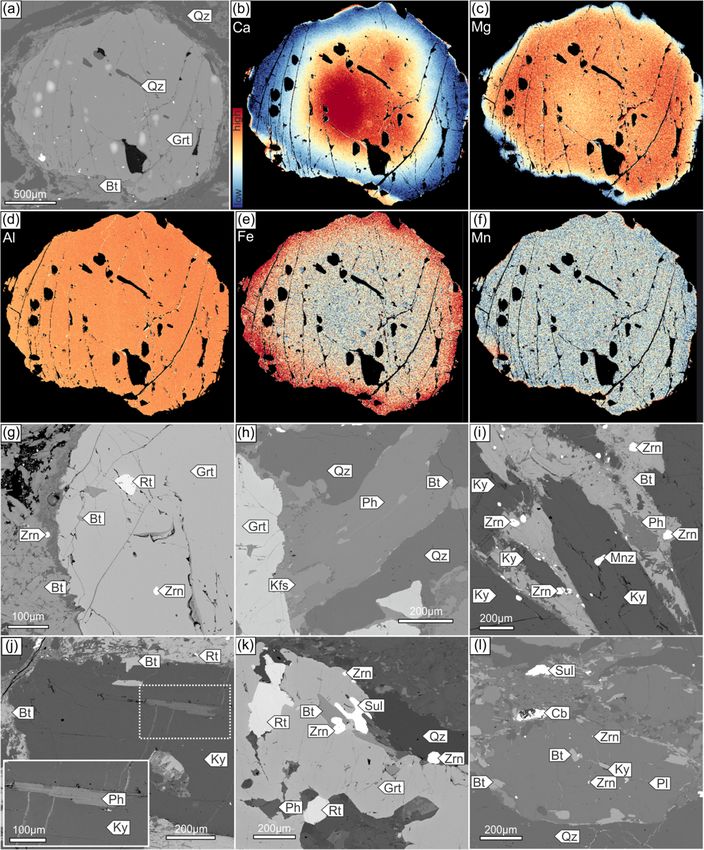
Minerals 2020, 10, 295                                                                             2 of 24

therein). Structurally, the Scandinavian Caledonides comprise a series of allochthonous thrust sheets
emplaced in an eastward direction, in contrast to the Greenland Caledonides, which record the opposite,
westward direction of thrusting onto Laurentia [6]. The tectonostratigraphy of the Scandinavian
Caledonides is divided into the Autochthon and the Lower, Middle, Upper and Uppermost allochthons.
The Autochthon represents the Baltic Shield, while the Lower and Middle allochthons constitute the
inner and outer Baltica passive margin, respectively. The Upper Allochthon comprises rocks with
Iapetus affinity, while the Uppermost Allochthon is composed of Laurentia-derived terranes (Figure 1a).
     (Ultra) high-pressure ((U)HP) rocks, which are important tracers of subduction processes, have been
reported in the Scandinavian Caledonides from different tectonostratigraphic levels including the Middle
and Uppermost allochthons, as well as the Parautochthon (i.e., the Autochthon buried and reworked
during the latest stages of the Caledonian orogeny) [5]. In the Parautochthon, (U)HP rocks have been
recognized in the Western Gneiss Region and Lofoten [7,8]. On the other hand, the (U)HP Tromsø Nappe
of the Uppermost Allochthon is thought to represent either an exotic terrane of near-Laurentia origin [9]
or an out-of-sequence thrust with its counterparts in the Middle Allochthon [10,11]. The unit with the
most frequent occurrences of (U)HP rock is the upper part of the Middle Allochthon, particularly the
Seve Nappe Complex (SNC) in Sweden, and its Norwegian equivalent, the Blåhø Nappe. The Middle
Allochthon records an increasing metamorphic grade (from greenschist to amphibolite/granulite facies)
towards the top of this sequence, but the highest grade metamorphic conditions (reaching up to
eclogite facies) have been identified only within the SNC [5]. The SNC can be traced at a distance of
approximately 800 km along the strike of the orogen [12,13]; thus, it provides a unique insight into a
record of subduction processes operating along the entire orogen.
     While the SNC lithologies exposed in Jämtland have an established history of (U)HP metamorphism
of Late Ordovician/ early Silurian age, e.g., [14–18], the (U)HP rocks known from Norrbotten [19,20]
recording late Cambrian/Early Ordovician metamorphism are less extensively dated [21–23]. Even less
well constrained are the timing and conditions of metamorphism within the SNC in Västerbotten
(Figure 1a). The U-Pb dating of zircon from Gardiken, c. 25 km north of the study area, suggested a Late
Ordovician age of granulite facies metamorphism [24]. Recently, Petrík et al. [25] provided evidence
for Early Ordovician (U)HP metamorphism in Saxnäs, located c. 30 km south of the target area,
while Grimmer et al. [26] estimated that exhumation to mid-crustal levels took place in the Llandovery
and Wenlock. Thus, the main aim of this study is to fill this age gap of (U)HP metamorphism in
the Middle Seve Nappe of Västerbotten, by coupling U-Pb zircon dating with rare earth elements
(REE) analyses and trace elements (Zr-in-rutile) thermometry. We also provide insight into zircon
behaviour during the migmatization of SNC paragneisses and garnet amphibolite. In a broader
perspective, these age estimates allow for the revision of already existing models for subduction along
the Baltoscandian margin and, more precisely, for defining the spatial and temporal boundary between
the northern and central parts of the SNC in the Scandinavian Caledonides.

2. Geological Setting
      The (U)HP metamorphic conditions recorded by SNC cropping out in west–central Jämtland
have been established by the discovery of metamorphic microdiamonds in paragneisses at
Tväråklumparna [27] (Figure 1a), and subsequently in a similar migmatitic paragneisses at Åreskutan.
The latter experienced (U)HP metamorphism at 4.1–4.2 GPa and 830–840 ◦ C, followed by a granulite
facies overprint at 1.0–1.1 GPa and 850–860 ◦ C [14,28]. The age of the prograde to near-peak
metamorphism has been estimated to c. 455 Ma, based on in-situ monazite dating [29]. Several zircon
U-Pb studies have shown that decompression-driven migmatization took place around 442–440 Ma in
migmatites and leucogranites and around 436 Ma in partially melted amphibolites [18,24,30], which is
in line with titanite U-Pb ages [31]. Recently, Klonowska et al. [14] reported c. 445 Ma and c. 441 Ma
Th-U-Pb monazite ages from both, Tväråklumparna and Åreskutan, reflecting post-(U)HP exhumation
of paragneisses. These ages corroborate c. 439–438 Ma monazite ages interpreted as the timing of
late melt crystallization [29]. The youngest group of zircon U-Pb ages (c. 430–428 Ma) was obtained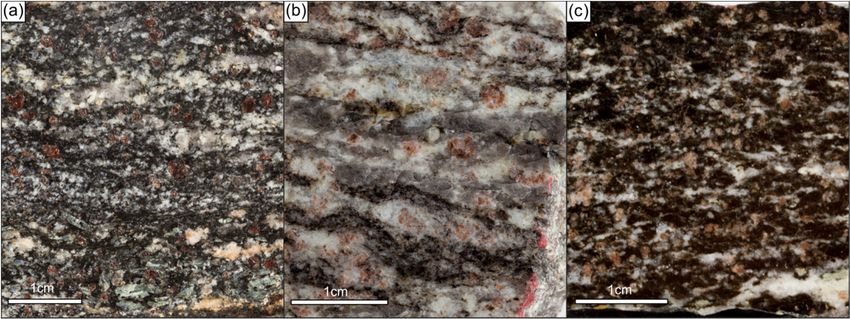
Minerals 2020, 10, 295                                                                            3 of 24

from pegmatites, discordant to the migmatitic foliation that are deformed by later thrusting [18].
This thrusting event was dated to c. 424–423 Ma using Th-U-Pb monazite geochronology and reflects
the timing of tectonic stacking of the Seve nappes [29].
      Recently, (U)HP metamorphism has been confirmed also farther north in Norrbotten, on eclogites
and metasedimentary rocks from the so-called Vaimok lens with peak pressure conditions at 2.8–3.1 GPa
and 660–780 ◦ C [20] (Figure 1a). This confirmed previous speculations about the possible continuation
of the SNC (U)HP lithologies north of the Arctic circle [19]. While the overlying Sarek lens does not
show obvious signs of metamorphism exceeding amphibolite facies, the Tsäkkok lens records blueschist
to eclogite facies metamorphism with a minimum pressure of 1.2–1.4 GPa and 500–630 ◦ C [32–34],
more recently updated to 2.2 GPa and 590 ± 60 ◦ C [35]. The previous studies established the age
of prograde metamorphism of the Vaimok lens at 500–475 Ma [36], based on titanite U-Pb dating
in calc-silicate rocks. This age was recently confirmed by Barnes et al. [23] who reported c. 498 Ma
prograde monazite growth in similar metasediments. Mørk et al. [21] estimated the timing of
eclogite facies metamorphism at c. 505 Ma using the garnet–omphacite–whole rock Sm-Nd isochrone.
However, zircon U-Pb dating of eclogites yielded a younger age of c. 482 Ma from both the Vaimok
and Tsäkkok lenses [22]. Such age, in turn, is in line with those derived for the Vaimok metasediments
showing a formation of HP metamorphic zircon rims at c. 480–475 Ma [23]. The timing of subsequent
exhumation and related cooling was estimated with 40 Ar/39 Ar geochronology at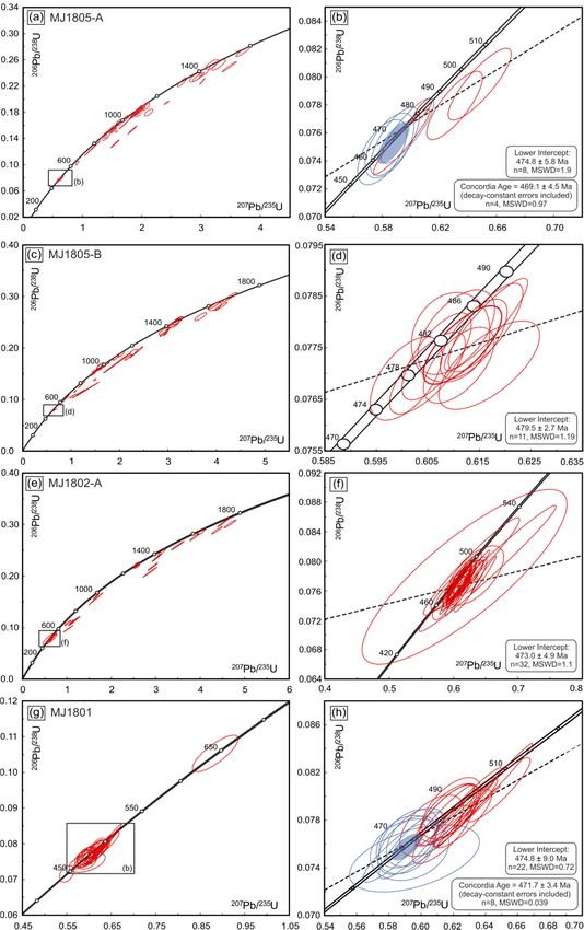
Minerals 2020, 10, 295 4 of 24
Minerals 2020, 10, 295 3 of 24
Figure 1.
1. Geological
Geological map
mapof
ofthe
thestudy
studyarea:
area:(a)
(a)Simplified
Simplifiedtectonostatigraphic
tectonostatigraphic map map of
of the
the Scandinavian
Scandinavian
Caledonides (after Gee et al., [6]). The
The diamond
diamond symbols
symbols indicate
indicate localities
localities within
within the Seve Nappe
Complex with
with confirmed
confirmedmetamorphic
metamorphicmicrodiamonds,
microdiamonds, i.e.,i.e.,
fromfrom south
south to north:
to north: Tväråklumparna
Tväråklumparna [27],
[14], Åreskutan
Åreskutan [15]
[14] and and [14,25].
Saxnäs Saxnäs The
[15,16]. The shows
rectangle rectangle shows
the area the area
presented onpresented on map (b).
map (b). Abbreviations:
Abbreviations: Western
Western Gneiss Region Gneiss
(WGR). Region
(b) A (WGR).
simplified (b) A simplified
tectonostratigraphic maptectonostratigraphic map of the
of the northern Jämtland/southern
northern Jämtland/southern
Västebotten Västebotten
area. The yellow star marks thearea. The yellow
sampling star marks
area within the sampling
the high-grade area unit.
Marsfjället within the
high-grade Marsfjället unit.
The Marsfjället unit (Figure 1b) consists of migmatitic paragneisses and garnet amphibolites that
haveWhile
undergone
the amphibolite to granulite
SNC lithologies exposed facies
in metamorphism
Jämtland have[46,47]. However, Grimmer
an established history ofet al. [26]
(U)HP
showed evidence
metamorphism of eclogite
of Late facies
Ordovician/ metamorphism
early Silurian age, e.g., of [15,17–20],
garnet–kyanite mica rocks
the (U)HP schists fromfrom
known the
Middle Seve Nappe, recording a decompression from HP conditions
Norrbotten [21,22] recording late Cambrian/Early Ordovician metamorphism are less extensively of at least 1.7 GPa at
~670 ◦ C. Subsequently, a microdiamond
dated [23–25]. Even less well constrained areof themetamorphic origin has
timing and conditions been reported within
of metamorphism from the
kyanite–garnet-bearing
SNC Marsfjället
in Västerbotten (Figure 1a). gneiss
The U-Pbcropping
dating outofnear Saxnäs
zircon from [25], showingc.that
Gardiken, 25 metasedimentary
km north of the
rocks area,
study of Middle Seve ainLate
suggested Västerbotten
Ordovicianunderwent (U)HPfacies
age of granulite metamorphism.
metamorphism The[26].
diamond hasPetrík
Recently, been
identified
et within inclusions
al. [16] provided evidenceinfor
garnet,
Earlykyanite and zircon,
Ordovician (U)HP whereas in the west-central
metamorphism in Saxnäs,Jämtland
located localities
c. 30 km
diamonds
south have
of the been
target found
area, onlyGrimmer
while in garnetet[14,27].
al. [27]Despite
estimated the lithological
that exhumation similarities, monazitelevels
to mid-crustal from
took place in the Llandovery and Wenlock. Thus, the main aim of this study is to fill this age gapthe
Saxnäs yields a significantly older Th-U-Pb age of c. 472 Ma, which is interpreted as reflecting of
timing of
(U)HP (U)HP metamorphism
metamorphism [25]. In
in the Middle the Nappe
Seve Kittelfjäll
of region, the Marsfjället
Västerbotten, by couplingunit forms
U-Pb approximately
zircon dating
a 4 km
with wide
rare N–Selements
earth trending(REE)
belt ofanalyses
extensivelyandmigmatized
trace elements paragneisses,
(Zr-in-rutile)hosting abundant lenses
thermometry. We alsoof
garnet amphibolites
provide insight into and ultramafic
zircon behaviour rocks (Figure
during the 1b). In the west,ofitSNC
migmatization is overlain by a thin
paragneisses andsliver of
garnet
amphibolite. In a broader perspective, these age estimates allow for the revision of already existing
models for subduction along the Baltoscandian margin and, more precisely, for defining the spatial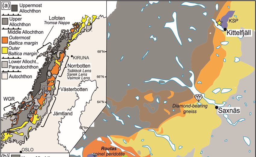
Minerals 2020, 10, 295                                                                                             5 of 24

mica schists intercalated with amphibolites. In the east, the Marsfjället unit occurs together with
garnet–clinopyroxene–plagioclase-bearing Kittelfjäll amphibolites and a huge body of the Kittelfjäll
spinel peridotite (KSP—Figure 1b; e.g., Clos et al. [15] and references therein). Despite the lack of
reported eclogites in the studied area and precise P-T estimates of the peak metamorphic conditions,
HP metamorphic conditions of Kittelfjäll paragneisses have been speculated as they share a common
exhumation history with the KSP from P-T conditions of c. 1.0–2.0 GPa and 650–830 ◦ C [15].

3. Materials and Methods
     For this study, four rock samples were collected from the high-grade Marsfjället gneiss unit exposed
to the north of Borkasjön lake, west of the Kittelfjäll village (Figure 1b). Three samples, namely MJ18-02A,
MJ18-05A and MJ18-05B, represent macroscopically different subtypes of migmatitic paragneisses,
whereas the sample MJ18-01 represents a garnet-bearing amphibolite hosted by migmatitic paragneiss
(Figure  2).2020,
   Minerals   GPS 10, coordinates
                      295         of all outcrops are provided in Table S1.                         6 of 24

        Figure
      Figure     2. Photographs
              2. Photographs       of the
                               of the     studied
                                      studied     samples
                                              samples       collected
                                                       collected fromfrom   the Marsfjället
                                                                      the Marsfjället       high-grade
                                                                                      high-grade         unit.
                                                                                                  unit. (a)    (a)
                                                                                                            Kyanite-
        Kyanite- biotite-rich
      biotite-rich migmatiticmigmatitic
                                paragneissparagneiss (sample MJ1805-A).
                                            (sample MJ18-05A).            (b) Quartz-rich
                                                                  (b) Quartz-rich         migmatitic
                                                                                  migmatitic         paragneiss
                                                                                               paragneiss   (sample
        (sample MJ1802-A).
      MJ18-02A).    (c) Garnet(c) Garnet amphibolite
                               amphibolite            (sample MJ1801).
                                             (sample MJ18-01).

         The
       The    chemicalcomposition
           chemical       compositionof  ofthe
                                            the minerals
                                                minerals was
                                                          was obtained
                                                                obtainedby bywavelength
                                                                              wavelengthdispersive
                                                                                              dispersivespectroscopy
                                                                                                           spectroscopy
   (WDS)
(WDS)       using
         using       a Jeol
                 a Jeol      Superprobe
                        Superprobe     82308230  electron
                                             electron      microprobe
                                                       microprobe        (JEOL,
                                                                    (JEOL,  Tokyo,Tokyo,
                                                                                    Japan) Japan)
                                                                                             at theat the Faculty
                                                                                                    Faculty          of
                                                                                                             of Geology,
   Geology,    Geophysics      and  Environment     Protection,   AGH—University       of
Geophysics and Environment Protection, AGH—University of Science and Technology (AGH-UST)  Science  and   Technology
in (AGH-UST)
   Kraków, Poland. in Kraków,    Poland. The
                           The operating       operatingwere
                                             conditions   conditions   were as
                                                                as follows:  anfollows:   an accelerating
                                                                                 accelerating    voltage of voltage
                                                                                                              15 kVof and
   15  kV  and    beam     current  of  15–20  nA   for aluminosilicates.   For  the  analyses   of  Zr
beam current of 15–20 nA for aluminosilicates. For the analyses of Zr in rutile, an accelerating voltage in rutile, an
of accelerating
   15 kV and avoltage        of 15 kVof
                    beam current       and a beam
                                         120  nA werecurrent of 120
                                                         used.  ThenA    were used.
                                                                     counting   timesThe   counting
                                                                                        were         times
                                                                                               20 s on  peakswere
                                                                                                                and20 10
                                                                                                                      s s
   on peaks and 10 s on background positions for aluminosilicates. For Zr in rutile, counting times were
on background positions for aluminosilicates. For Zr in rutile, counting times were 150–300 s on
   150–300 s on peaks and 75–150 s on background positions. The following natural minerals and
peaks and 75–150 s on background positions. The following natural minerals and synthetic standards
   synthetic standards were used for calibration: albite (Si, Al, Na), diopside (Ca, Mg), sanidine (K),
were used for calibration: albite (Si, Al, Na), diopside (Ca, Mg), sanidine (K), rutile (Ti), fayalite (Fe),
   rutile (Ti), fayalite (Fe), rhodonite (Mn), vanadinite (V), Cr2O3 (Cr), tugtupite (Cl), fluorite (F), YPO4
rhodonite (Mn), vanadinite (V), Cr2 O3 (Cr), tugtupite (Cl), fluorite (F), YPO4 (P), barite (Ba), celestine
   (P), barite (Ba), celestine (Sr). Additional standard materials used for the analysis of Zr in rutile were:
(Sr).  Additional standard materials used for the analysis of Zr in rutile were: zircon (Zr), LiNbO3 (Nb),
   zircon (Zr), LiNbO3 (Nb), and tantalite-(Mn) (Ta). The WDS/energy dispersive spectroscopy (EDS)
and   tantalite-(Mn)
   chemical mapping      (Ta).  The WDS/energy
                            of garnet was performed dispersive
                                                        under thespectroscopy   (EDS) chemical
                                                                    following conditions:           mapping
                                                                                             accelerating      of garnet
                                                                                                            voltage  of
was   performed      under   the following    conditions:
   15kV, beam current of 100 nA and dwell time of 100 ms.  accelerating   voltage  of 15  kV, beam   current   of 100 nA
and dwellU–Pbtime    of 100
                dating    wasms.
                               performed on carefully separated zircon grains that were mounted in an epoxy
   mount and then polished to expose the internal parts of the crystals. The cathodoluminescence (CL)
   images of all mounted grains were obtained using a Hitachi SU3500 Scanning Electron Microscope
   at the Polish Geological Institute—National Research Institute, Warsaw Poland, to reveal the internal
   structure of zircon. U-Pb dating and rare earth elements (REE) analyses were completed using a Nu
   Plasma II multi-collector inductively coupled plasma mass spectrometer (MC-ICPMS) coupled to an
   ESI NWR193UC (Elemental Scientific Lasers, Bozeman, MT, USA) excimer based laser ablation
   system at the Vegacenter at the Swedish Museum of Natural History, Stockholm, Sweden. For U-Pb
   analyses, the m/z (mass-to-charge ratios) corresponding to masses 202, 204, 206, 207 and 208 were
   measured on ion counters and those corresponding to 232, 235 and 238 were measured on Faraday
   collectors. Spots of 20 or 25 µm in diameter were ablated at a frequency of 7 Hz and laser fluence of
   1.7 J/cm2. Helium was used as a sample carrier gas with a flow rate of 0.3 L/min, which was then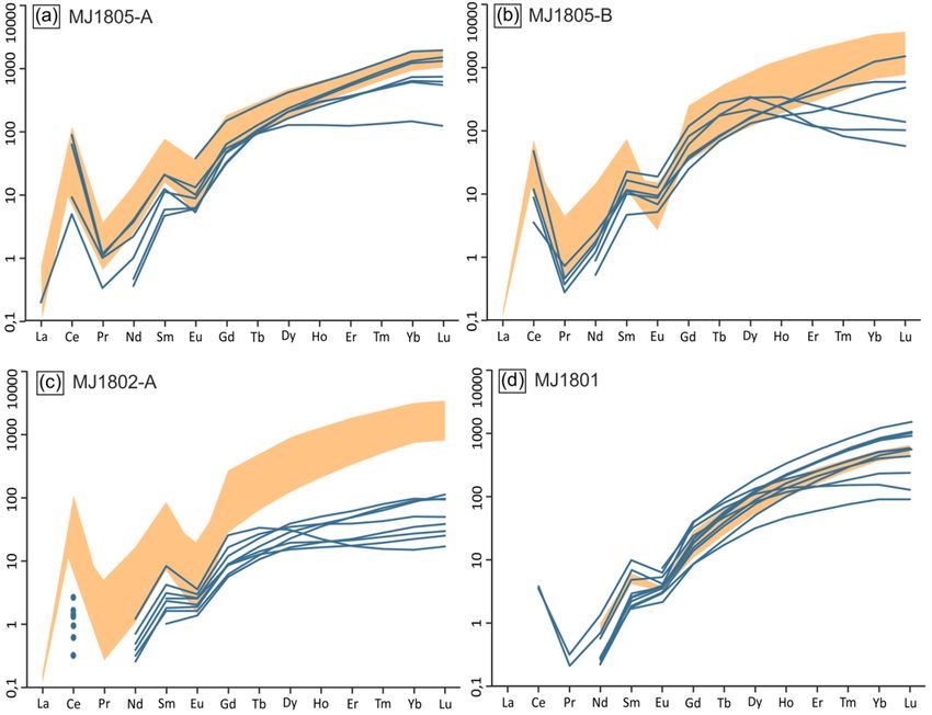
Minerals 2020, 10, 295                                                                              6 of 24

     U–Pb dating was performed on carefully separated zircon grains that were mounted in an epoxy
mount and then polished to expose the internal parts of the crystals. The cathodoluminescence (CL)
images of all mounted grains were obtained using a Hitachi SU3500 Scanning Electron Microscope at
the Polish Geological Institute—National Research Institute, Warsaw Poland, to reveal the internal
structure of zircon. U-Pb dating and rare earth elements (REE) analyses were completed using a Nu
Plasma II multi-collector inductively coupled plasma mass spectrometer (MC-ICPMS) coupled to an
ESI NWR193UC (Elemental Scientific Lasers, Bozeman, MT, USA) excimer based laser ablation system
at the Vegacenter at the Swedish Museum of Natural History, Stockholm, Sweden. For U-Pb analyses,
the m/z (mass-to-charge ratios) corresponding to masses 202, 204, 206, 207 and 208 were measured
on ion counters and those corresponding to 232, 235 and 238 were measured on Faraday collectors.
Spots of 20 or 25 µm in diameter were ablated at a frequency of 7 Hz and laser fluence of 1.7 J/cm2 .
Helium was used as a sample carrier gas with a flow rate of 0.3 L/min, which was then mixed with Ar
gas at a flow rate (mix gas) of 0.9 L/min. The data collection procedure included a 20 s ablation followed
by 20 s washout time. The data was processed using the Iolite add-on “VizualAge” [48]. All isotope
ratios were normalized to the zircon 91500 reference material with an age of 1065 Ma [49]. The GJ-1
zircon (609 Ma; [50]; obtained age of 604 Ma, 2SD = 12), Plešovice zircon (337 Ma; [51]; obtained age of
337 Ma, 2SD = 8) and Temora 2 zircon (417 Ma: [52]; obtained age of 420 Ma, 2SD = 8) were utilized as
secondary standards. Age calculations and construction of concordia diagrams were prepared using
the Excel extension Isoplot 4.15 [53]. All uncertainties are reported at the 2σ level.
     Rare earth elements concentrations in zircon were obtained by ablating spots of 20 µm in diameter
within both, zircon cores and rims of known U-Pb age. This approach allowed us to distinguish
between REE patterns displayed by Caledonian and older zircon rims, most frequently of Proterozoic
age. The spots were ablated at a frequency of 8 Hz and fluence of 3 J/cm2 . The 25 s ablation time
with 40 ms dwell time/isotope was followed by 20 s washout time. The results were quantified using
the reference zircon 91500 (concentration values from Wiedenbeck et al. [54]). Plešovice [51] and GJ-I
zircons [50] were used as validation material. A  p precision of better than 10% RSE was achieved for
most REE. Reported values of [Eu/Eu* = EuN / (SmN × GdN )] and LuN /GdN have been calculated
based on the REE concentrations normalized to Cl Chondrite [55].

4. Results

4.1. Petrography and Mineral Chemistry

4.1.1. Migmatitic Paragneisses
     All three samples of the studied migmatitic paragneisses exhibit a foliation marked by the
alignment of leucocratic layers and melanocratic bands. Even though paragneisses seem to show
macroscopically significant differences, the mineral composition and mode of mineral occurrence are
similar in all samples. The leucocratic layers are dominated by quartz, K-feldspar, plagioclase and
scarce white mica, whereas the melanocratic bands are dominated by biotite and kyanite. Minor phases
such as rutile, monazite, zircon, Fe-sulfides, carbonates, and ilmenite are randomly distributed within
the samples.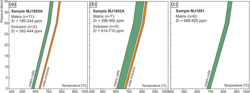
Minerals 2020, 10, 295                                                                                   7 of 24

      In the samples MJ18-05A and MJ18-05B garnet forms subhedral porphyroblasts that show
almandine-rich composition varying from Alm60 Prp25 Grs12 Sps3 (i.e., Almandine–Pyrope–Grossular–
Spessartine) in the core to Alm68 Prp23 Grs6 Sps3 in the rim (Figure 3a–f; Table S2a). The garnet displays
zoning marked mostly by Ca, which decreases rimwards, whereas the opposite trend is well-marked by
Mg and Fe (Figure 3b,c,e). Some grains preserve a lobate outermost rim, which is significantly enriched
in Ca and simultaneously depleted in Mg (Figure 3b,c) and has a composition of Alm70 Prp17 Grs10 Sps3 .
Notably, the outermost rim is preserved only along the garnet crystal faces parallel to the foliation
(Figure 3a,b). The manganese content is constant throughout the entire grain (Figure 3f), except for
the discontinuous outermost rim, which is slightly enriched in spessartine compared to the garnet
core (Table S2a). Garnet contains scarce inclusions of quartz, rutile, zircon, and biotite (Figure 3a,g).
Garnet porphyroblasts are usually enveloped by biotite and K-feldspar, which also forms grains in
the garnet pressure shadows (Figure 3h) as well as large, elongated grains with a lobate boundary,
most frequently associated with quartz within leucocratic bands. K-feldspar shows a minor compositional
change from the core (Or90.0 Ab9.8 An0.2 ) towards the rim (Or90.7 Ab9.0 An0.3 ). Plagioclase forms large,
rounded, and lobate grains in leucocratic bands and also shows minor compositional zoning from
Ab71.3 An25.9 Or2.7 in the core to Ab74.2 An24.7 Or1.0 in the rim (Table S2b). Kyanite forms prismatic grains
concentrated within dark, melanocratic bands, and shows only minor or no alteration to sillimanite at
the grain boundaries. Kyanite hosts numerous inclusions of rutile, monazite, white mica, and zircon
(Figure 3i,j). White mica occurs in two textural positions, either as single, partially decomposed grains
enveloped by later biotite (Figure 3i) or as inclusions in kyanite. White mica grains located in the
rock matrix contain 3.15–3.18 Si a.p.f.u. (atom per formula unit) compared to 3.07–3.11 Si a.p.f.u. in
the inclusions in kyanite. Biotite shows a constant composition with XMg(Mg/Mg + Fe) = 0.53−0.58
(Table S2c). Rutile forms abundant single grains that are randomly distributed within the matrix or
aggregates concordant to the foliation. It shows no signs of reaction to titanite, but it is commonly
associated with ilmenite. Rutile has also been identified in inclusions in garnet (Figure 3g). Zircon forms
rare inclusions in garnet (Figure 3g), but most frequently, it is associated with biotite, kyanite, and monazite
within the melanocratic bands in the matrix (Figure 3i).
      In the sample MJ18-02A garnet forms anhedral to subhedral grains containing numerous inclusions
composed predominantly of quartz ± plagioclase ± biotite and minor zircon, rutile, sulfides, and white
mica (Figure 3k). Large kyanite and plagioclase porphyroblasts are elongated concordant to the
foliation defined by the alignment of K-feldspar, biotite, and white mica. Sporadic, single grains of
REE-bearing carbonates can be found within the matrix (Figure 3l). Plagioclase shows compositional
zoning marked by increasing Na content towards the rim from Ab53.4 An45.3 Or1.2 to Ab70.9 An27.8 Or1.2 .
White mica forms large flakes with Si content varying from 2.99 to 3.12 Si a.p.f.u and is partially
decomposed to K-feldspar ± biotite ± quartz (Figure 3h). Biotite forms only small grains in the
matrix with a constant composition of XMg = 0.60–0.64. Rutile forms inclusions in garnet (Figure 3k)
and kyanite as well as grains disseminated in the matrix. Rutile contains thin lamellae of ilmenite,
but·shows no signs of replacement by titanite, despite the fact that idiomorphic titanite grains have
also been found in the matrix. Monazite forms anhedral grains (up to 200 µm in diameter) widespread
in the matrix. Zircon is widespread within the whole sample. It has been found as inclusions in garnet
and plagioclase (Figure 3l) and as single grains randomly distributed in the matrix. Locally, a series of
zircon grains have been identified within the outermost rim of the garnet as well as in the close vicinity
of the garnet grain boundaries, preserving features of partial melting (Figure 3k).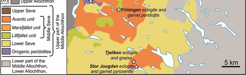
Minerals 2020, 10, 295                                                                                                                  8 of 24

      Minerals 2020, 10, 295                                                                                                  9 of 24

            Figure 3. Back-scattered electron (BSE) and wavelength-dispersive X-ray spectrometry (WDS) maps
      Figure 3. Back-scattered electron (BSE) and wavelength-dispersive X-ray spectrometry (WDS) maps
            of the rock forming minerals in migmatitic paragneisses. (a) BSE image of the garnet (MJ1805-A)
      of the rock forming minerals in migmatitic paragneisses. (a) BSE image of the garnet (MJ18-05A)
            presented in the WDS maps in (b–f). The bright spots on the BSE image mark the area of removed
      presented in the WDS maps in (b–f). The bright spots on the BSE image mark the area of removed
            graphite coating. These areas were removed from the X-ray maps during data treatment in
      graphite    coating.v.2.3.1
            XMapTools       These[56].
                                     areas
                                         WDSwere
                                               mapsremoved
                                                        show thefrom
                                                                   peakthecounts
                                                                            X-raymeasured
                                                                                   maps during     data
                                                                                              only in thetreatment    in XMapTools
                                                                                                           garnet grain. (b) X-
      v.2.3.1
            ray[56].
                 mapWDS
                      of Ca,maps     show
                              (c) X-ray     theofpeak
                                          map     Mg. counts
                                                         (d) X-raymeasured
                                                                    map of Al. only   in themap
                                                                                 (e) X-ray    garnet  grain.
                                                                                                  of Fe.      (b) X-ray
                                                                                                         (f) X-ray  map ofmap
                                                                                                                           Mn.of Ca,
      (c) X-ray   map
            (g) BSE     of Mg.
                     image        (d) X-ray
                             of garnet        mapinclusions
                                          hosting    of Al. (e) ofX-ray   map
                                                                     zircon  andof rutile
                                                                                    Fe. (f)(MJ1805-A).
                                                                                            X-ray map(h)  of A
                                                                                                             Mn.   (g) BSEgrain
                                                                                                                phengite    image of
      garnet   hostingdown
            breaking    inclusions     of zircon
                               to biotite         and rutile in
                                            and K-feldspar      (MJ18-05A).
                                                                   the garnet (h)   A phengite
                                                                                pressure    shadowgrain   breaking(i)
                                                                                                     (MJ1802-A).     down   to biotite
                                                                                                                        Kyanite
            porphyroblasts
      and K-feldspar          enveloped
                          in the   garnet by   biotite and
                                            pressure          partially
                                                         shadow          decomposed
                                                                    (MJ18-02A).          phengite.porphyroblasts
                                                                                    (i) Kyanite    Note the abundant     zircon by
                                                                                                                      enveloped
            and  monazite   grains   (MJ1805-A).    (j)  A phengite   grains hosted    in kyanite porphyroblast
      biotite and partially decomposed phengite. Note the abundant zircon and monazite grains (MJ18-05A).          (MJ1805-A).
      (j) A(k)  Partiallygrains
             phengite     meltedhosted
                                    garnet with   abundant
                                            in kyanite         zircon grains located
                                                           porphyroblast                at the garnet
                                                                              (MJ18-05A).             grain edge
                                                                                               (k) Partially       (MJ1802-A).
                                                                                                               melted   garnet with
            (l) Plagioclase grain in the neosome. Note the unusually bright a REE-bearing carbonate (MJ1802-A).
      abundant zircon grains located at the garnet grain edge (MJ18-02A). (l) Plagioclase grain in the neosome.
            Mineral abbreviations: garnet (Grt), rutile (Rt), biotite (Bt), zircon (Zr), quartz (Qz), K-feldspar (Kfs),
      Note the unusually bright a REE-bearing carbonate (MJ18-02A). Mineral abbreviations: garnet (Grt),
            phengite (Ph), kyanite (Ky), monazite (Mnz), sulfide (Sul), carbonate (Cb), plagioclase (Pl).
      rutile (Rt), biotite (Bt), zircon (Zr), quartz (Qz), K-feldspar (Kfs), phengite (Ph), kyanite (Ky), monazite
      (Mnz), sulfide (Sul), carbonate (Cb), plagioclase (Pl).
Minerals 2020, 10, 295 9 of 24
4.1.2. Garnet Amphibolite
The sample MJ18-01 shows a foliation defined by parallel alignment of quartz and plagioclase
aggregates and a concordant elongation of amphibole grains. Amphibole is the most abundant
phase, forming large, subhedral grains. Amphibole has been identified in rare inclusions in garnet.
Despite its abundance, amphibole shows a fairly uniform chemical composition encompassing pargasite
and magnesio-hornblende species (Table S3a). Garnet is randomly distributed within the matrix
and shows a variety of crystal shapes. It forms either subhedral grains with preserved crystal
faces or anhedral grains with a highly irregular shape resembling a lobate texture (Figure 4a,g).
Frequently, garnet is surrounded by irregularly shaped domains that are composed of quartz
and plagioclase; additionally, in some places, garnet is replaced by biotite at the grain boundary
(Figure 4h). Despite these various shapes, the vast majority of garnet grains show similar compositional
differences between the core and rim. Chemical zoning within the core is well-marked by Ca
and Mg distribution and changes rimward from Alm50 Grs28 Prp17 Sps5 to Alm52 Grs31 Prp14 Sps3
(Table S3b). The garnet rim displays the high-Ca composition of Alm45 Grs41 Prp11 Sps3 . Garnet seldom
hosts inclusions of quartz, plagioclase, apatite, and amphibole. However, some garnet grains
have an additional domain within the core that has numerous chromite inclusions (Figure 4a–f).
This domain is enriched in Cr and Ca compared to the typical core domain and shows the composition
of Alm48–45 Grs27–22 Prp16–14 Uvr14–9 Sps~2 AdrMinerals 2020, 10, 295 10 of 24
Minerals 2020, 10, 295 10 of 24
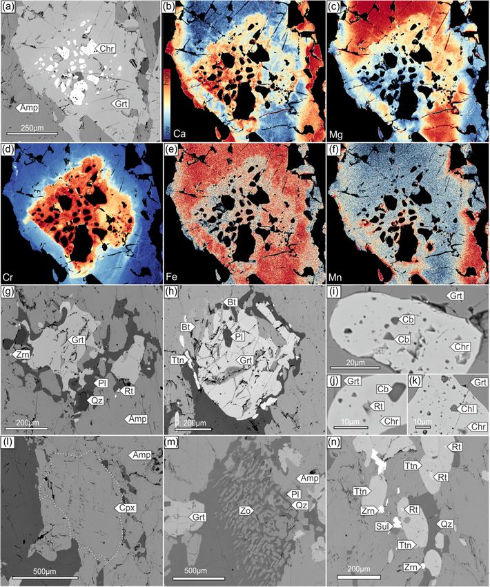
Figure
Figure Back-scattered
4. 4. Back-scattered electron
electron(BSE)
(BSE)andand
wavelength-dispersive
wavelength-dispersive X-ray spectrometry
X-ray spectrometry(WDS) mapsmaps
(WDS) of
theofrock forming
the rock minerals
forming in garnet
minerals amphibolite
in garnet (MJ18-01).
amphibolite (a) BSE
(MJ1801). image
(a) BSE of the
image ofgarnet presented
the garnet in
presented
theinWDS mapsmaps
the WDS in (b–f). WDSWDS
in (b–f). mapsmapsshow show
the peak
the counts measured
peak counts only inonly
measured the garnet. (b) X-ray
in the garnet. (b)map
X-ray
ofmap
Ca, (c)
of X-ray
Ca, (c)map X-ray ofmap
Mg. of (d)Mg.
X-ray(d)map
X-rayofmap
Cr. (e)
of X-ray
Cr. (e)map
X-rayofmap
Fe. (f)
of X-ray
Fe. (f)map
X-rayofmap
Mn. of(g)Mn.
BSE(g)
image of partially
BSE image melted melted
of partially garnet with
garnet a lobate
with agrain
lobateedge. (h)edge.
grain Garnet(h)grain
Garnetpartially replaced replaced
grain partially by biotite.by
(i)biotite.
Chromite inclusion in
(i) Chromite garnet hosting
inclusion in garnet numerous
hosting inclusions
numerous of carbonates.
inclusions (j) Chromite(j)inclusion
of carbonates. Chromite
with rutile inclusion. (k) Chromite inclusion hosting a chlorite inclusion. (l) Clinopyroxene
inclusion with rutile inclusion. (k) Chromite inclusion hosting a chlorite inclusion. (l) Clinopyroxene of diopside
composition
of diopsidepreserved
composition in the matrix.in
preserved Thethewhite
matrix.dotted line outlines
The white dotted the
lineboundary
outlines the of boundary
the Cpx grain.
of the
(m) Poikiloblastic
Cpx intergrowth of
grain. (m) Poikiloblastic zoisite andofplagioclase.
intergrowth zoisite and(n) Scarce rutile
plagioclase. (n) grains
Scarce replaced by titanite.
rutile grains replaced
Note the abundant
by titanite. Note the zircon in the vicinity
abundant zircon in ofthe
titanite. Mineral
vicinity abbreviations:
of titanite. garnet (Grt), zircon
Mineral abbreviations: garnet(Zr),
(Grt),
plagioclase (Pl), quartz (Qz), amphibole (Amp), biotite (Bt), titanite (Ttn), carbonate
zircon (Zr), plagioclase (Pl), quartz (Qz), amphibole (Amp), biotite (Bt), titanite (Ttn), carbonate (Cb), chromite
(Cb),
(Chr), chlorite
chromite (Chl),
(Chr), clinopyroxene
chlorite (Cpx), zoisite
(Chl), xlinopyroxene (Zo), zoisite
(Cpx), sulfide(Zo),
(Sul).sulfhide (Sul).(i) in inclusions in garnet, (ii) within poikiloblastic domains with quartz and zoisite, and (iii) within
abundant plagioclase and quartz domains within the matrix. In all positions, plagioclase has a similar
composition of Ab70–75An24–30Or0.5–1.3. Rutile forms rare, small (yielded
rutile inclusions 738 ◦ C and > 780
T > 738 °C◦ C
and
(at >a fixed
780 °C (at a fixed
pressure of
25pressure
kbar) inof 25samples
the kbar) in MJ18-05A
the samples andMJ1805-A
MJ18-02A,andrespectively.
MJ1802-A, respectively. The matrix
The matrix rutile rutile
displays < 655
a Tdisplays
◦CaTand < 726
< 655 °C ◦and
C in< the
726samples
°C in theMJ18-05A
samples MJ1805-A and MJ1802-A,
and MJ18-02A, respectively respectively
(at a fixed(at a fixedof
pressure pressure
15 kbar;of
15 kbar;
Figure Figure
5; Table 5; Table S4).
S4).
Figure5.5.P-T
Figure P-Tdiagrams
diagrams showing
showing thethe results
results of Zr-in-rutile
of Zr-in-rutile geothemometry
geothemometry for afor a given
given textural
textural types
types of
of rutile.
rutile. GreenGreen fields the
fields show show thefor
results results
matrixfor matrix
rutile, rutile,
whereas thewhereas the orange
orange fields fields
represent represent
temperature
temperature
range calculatedrange
for calculated for rutile(a)
rutile inclusions. inclusions. (a) Sample migmatitic
Sample MJ18-05A: MJ1805-A: migmatitic
paragneiss.paragneiss.
(b) Sample (b)
Sample MJ1802-A: migmatitic paragneiss. (c) Sample MJ1801:
MJ18-02A: migmatitic paragneiss. (c) Sample MJ18-01: garnet amphibolite. garnet amphibolite.
4.3.
4.3.Zircon
ZirconU-Pb
U-PbDating
Dating
Zircon
Zirconseparated
separatedfrom
frommigmatitic
migmatiticparagneisses
paragneissesoccurs
occursasassub-idomorphic,
sub-idomorphic,nearly
nearlyisometric
isometricoror
elongated
elongated grains, most frequently not exceeding 200 µm in length. The CL imaging revealedthe
grains, most frequently not exceeding 200 µm in length. The CL imaging revealed the
complex
complex internal structure
internal of of
structure thethe
studied grains,
studied with
grains, cores
with enveloped
cores envelopedby zircon overgrowths
by zircon (Figure
overgrowths 6).
(Figure
The zircon cores most frequently preserve oscillatory and sector zoning, and likely represent former
detrital grains of the protolith. The overgrowths are significantly darker in CL compared to the cores.
The majority of the overgrowths are homogenous with no visible internal structure and only some
show subtle concentric and cloudy zoning (e.g., the first grain in section MJ18-02A in Figure 6).
U-Pb isotope analyses of zircon cores and non-Caledonian zircon rims from the migmatitic
paragneisses (MJ18-05A, n = 37; MJ18-05B, n = 36; MJ18-02A, n = 21) reveal a wide age spectrum
from Paleo- and Mesoproterozoic to early Paleozoic, reflecting the heterogeneous provenance of the
sediment protolith (Figure 7a,c,e). The vast majority of the zircon core and non-Caledonian zircon rim
analyses are moderately discordant (Table S5); however, in all samples, concordant analyses (less than
10% discordance) of Meso-to-Neoproterozoic age yield clusters between c. 1100–900 Ma, and between
c. 1450–1350 Ma in samples MJ18-05A and MJ18-05B (Figure 7). The obtained results do not differ
from previously published provenance studies of other Seve Nappe Complex rocks [58,59].Minerals 2020, 10, 295 12 of 24
6). The zircon cores most frequently preserve oscillatory and sector zoning, and likely represent
former detrital grains of the protolith. The overgrowths are significantly darker in CL compared to
the cores. The majority of the overgrowths are homogenous with no visible internal structure and
Minerals
only2020, 10, 295
some show subtle concentric and cloudy zoning (e.g., the first grain in section MJ1802-A in12 of 24
Figure 6).
Figure
Figure 6. Cathodoluminescence(CL)
6. Cathodoluminescence (CL)images
images of
of representative
representativezircon grains
zircon displaying
grains a variety
displaying of
a variety of
microtextures. The white circles mark the analytical spots with obtained
206 206Pb/238U ages (for
238
microtextures. The white circles mark the analytical spots with obtained Pb/ U ages (for Caledonian
Caledonian ages) and 207Pb/206Pb for ages >900 Ma.
ages) and 207 Pb/206 Pb for ages >900 Ma.
U-Pb isotope analyses of zircon cores and non-Caledonian zircon rims from the migmatitic
In the sample MJ18-05A, 25 zircon rims have been analysed. Only nine of the analysed rims
paragneisses (MJ1805-A, n = 37; MJ1805-B, n = 36; MJ1802-A, n = 21) reveal a wide age spectrum from
are Caledonian. One discordant analysis was excluded from age calculation (Table S5a). Eight spot
Paleo- and Mesoproterozoic to early Paleozoic, reflecting the heterogeneous provenance of the
analyses representing
sediment Caledonian
protolith (Figure 7a,c,e).age
Thecluster yield aoflower
vast majority intercept
the zircon agenon-Caledonian
core and Ma (n = 8,
of 474.8 ± 5.8 zircon
MSWD = 1.9; Figure 7b; Table S5a) with the 206 Pb/238 U ages ranging from 493.1 ± 13 to 464.0 ± 11 Ma.
rim analyses are moderately discordant (Table S5); however, in all samples, concordant analyses (less
Within the10%
than cluster, the subgroup
discordance) of four concordant dates
of Meso-to-Neoproterozoic givesclusters
age yield a concordia 469.0 ± 4.5
agec.of1100–900
between Ma,Maand(n = 4,
MSWD = 0.97;
between c. 1450–1350
Figure 7b).Ma in samples MJ1805-A and -B (Figure 7). The obtained results do not differ
from previously
Twenty-six published
zircon rims haveprovenance studies in
been analysed of other Seve Nappe
the sample Complex
MJ18-05B. rocks [58,59].
Thirteen of the analysed rims
In the sample MJ1805-A, 25 zircon rims have been analysed.
are Caledonian. Two discordant analyses (above 3% discordance) were excluded from age Only nine of the analysed rims are
calculation
Caledonian. One discordant analysis was excluded from age calculation (Table S5a). Eight spot
(Table S5b). Eleven spot analyses form a close group yielding a lower intercept at 479.5 ± 2.7 Ma (n =
analyses representing Caledonian age cluster yield a lower intercept age of 474.8 ± 5.8 Ma (n = 8,
11, MSWD = 1.19; Figure 7d), with the 206 Pb/238 U ages varying over a range of 483.9 ± 4–477.6 ± 4 Ma.
MSWD = 1.9; Figure 7b; Table S5a) with the 206Pb/238U ages ranging from 493.1 ± 13 to 464.0 ± 11 Ma.
However, it was impossible to calculate concordia age for this group.
Within the cluster, the subgroup of four concordant dates gives a concordia age of 469.0 ± 4.5 Ma (n
In the sample
= 4, MSWD MJ18-02A,
= 0.97; Figure 7b).50 zircon rims have been analysed. Out of these rims, 37 are Caledonian,
while fiveTwenty-six
discordant analyses
zircon rims havewerebeen
excluded
analysed from agesample
in the calculation (Table
MJ1805-B. S5c).ofThe
Thirteen the remaining
analysed 32
Caledonian
rims areanalyses
Caledonian.yieldTwo
a lower intercept
discordant age of(above
analyses 473.0 3%± 4.9 Ma (MSWD
discordance) = 1.1;
were Figurefrom
excluded 7f; Table
age S5c)
the 206 Pb/238
with calculation (Table S5b).ranging
U ages Eleven spot
from analyses
492.0 form
± 32 atoclose group
462.4 ± 11yielding
Ma. a lower intercept at 479.5 ± 2.7
Ma
The(nmajority
= 11, MSWD of =the
1.19; Figuregrains
zircon 7d), with the 206the
from Pb/238garnet
U ages varying over a range
amphibolite MJ18-01of 483.9
show ± 4–477.6 ±4
well-defined
Ma. However, it was impossible to calculate concordia age for this group.
concentric zoning within the cores that are overgrown by CL-bright thin rims. Nevertheless, some of
In the sample MJ1802-A, 50 zircon rims have been analysed. Out of these rims, 37 are
the analysed grains are much darker in CL and show no internal zoning (Figure 6). Twelve zircon cores,
Caledonian, while five discordant analyses were excluded from age calculation (Table S5c). The
10 zircon rims and five homogenous grains have been analysed. Only three discordant (above 3%
discordance) analyses were excluded from age calculation (Table S5d). Zircon cores and rims, as well
as the homogenous grains, yielded a wide group of Caledonian dates with a total span of >40 Ma
(Figure 7g). The whole Caledonian cluster of dates yields a lower intercept age of 474.8 ± 9.0 Ma
(n = 22, MSWD = 0.72; Figure 7h), but the zircon rims show a span of 206 Pb/238 U dates from 501.0 ± 17
to 464.8 ± 14 Ma (Figure 7h; Table S5d). The subset of concordant analyses plotted a concordia age of
471.7 ± 3.4 Ma (n = 8, MSWD = 0.039; Figure 7h). The two single concordant dates stand out from theMinerals 2020, 10, 295 13 of 24
Caledonian U-Pb dataset, i.e., the 646 ± 25 Ma and 445 ± 11 Ma dates (Figure 7g). These outliers were
not included in the age calculations.
Minerals 2020, 10, 295 14 of 24
Figure 7. Concordia diagrams of zircon U-Pb analyses. (a) All analyses from sample MJ1805-A. (b)
Figure 7. Concordia diagrams of zircon U-Pb analyses. (a) All analyses from sample MJ18-05A.
Caledonian age cluster from sample MJ1805-A. The lower intercept calculated for the whole cluster,
(b) Caledonian age cluster from sample MJ18-05A. The lower intercept calculated for the whole
concordia age calculated for the analyses marked by blue circle. (c) All analyses from sample MJ1805-
cluster, concordia age calculated
B. (d) Caledonian age cluster for
fromthe analyses
sample marked
MJ1805-B. (e)by
Allblue circle.from
analyses (c) All analyses
sample from sample
MJ1802-A. (f)
MJ18-05B. (d) Caledonian age cluster from sample MJ18-05B. (e) All analyses from sample
Caledonian age cluster from sample MJ1802-A. (g) All analyses from sample MJ1801. (h) Caledonian MJ18-02A.
(f) Caledonian
age clusterage cluster
from fromMJ1801.
sample sampleThe MJ18-02A. (g) All analyses
lower intercept from sample
was calculated MJ18-01.
for the (h) Caledonian
whole cluster, the
age cluster fromage
concordia sample MJ18-01.for
was calculated The
thelower intercept
analyses markedwas
by calculated
blue circle. for the whole cluster, the concordia
age was calculated for the analyses marked by blue circle.Minerals 2020, 10, 295 14 of 24
4.4. Trace Element Chemistry of Zircon
The zircon from migmatitic paragneisses preserves distinctive, apparently detrital, cores which
display broadly similar REE patterns (Figure 8a–c). The studied cores show significant enrichment
in heavy rare earth elements (HREE, average LuN /GdN = 21.37). Light rare earth elements (LREE)
concentrations are commonly below the detection limit (Table S6). In some of the studied grains,
even though measured LREE values were above the detection limit, the analyses with errors exceeding
50% were not plotted and excluded
p from further discussion and interpretation. A pronounced negative
Eu anomaly [Eu/Eu* = EuN / (SmN × GdN )] is well marked in all cores, with average values of Eu/Eu*
Minerals 2020, 10, 295 15 of 24
= 0.29 (n = 10; MJ18-05A), 0.28 (n = 7; MJ18-05B) and 0.14 (n = 9; MJ18-02A).
Figure8.8. Chondrite-normalized
Figure Chondrite-normalizedREEREEpatterns
patternsin
inzircon.
zircon. Orange
Orange and
and blue
blue lines
lines represent
representzircon
zirconcores
cores
and
andrims,
rims,respectively.
respectively.(a)
(a)Sample
SampleMJ18-05A:
MJ1805-A:migmatitic
migmatiticparagneiss.
paragneiss.(b)
(b)Sample
SampleMJ18-05B:
MJ1805-B:migmatitic
migmatitic
paragneiss.
paragneiss.(c)
(c)Sample
SampleMJ18-02A:
MJ1802-A:migmatitic
migmatiticparagneiss.
paragneiss. (d)
(d) Sample
Sample MJ18-01: garnet amphibolite.
MJ1801: garnet amphibolite.
Caledonian zircon rims from the samples MJ18-05A and MJ18-05B display two different REE
5. Discussion
patterns (Figure 8a,b); some of the studied rims mimic the REE pattern of the cores, while others
differ in HREE content
5.1. Metamorphic and HREE to MREE ratios. In the sample MJ18-05A, the rims exhibit a rising
Evolution
trend in HREE with LuN /GdN ratios range from 13.6 to 28.1, except one analysis showing a flat HREE
Notwithstanding
pattern (LuN /GdN = 2.6;the pervasive
Figure 8a). Inmigmatization of the studied
the sample MJ18-05B, rocks,
part of the remnants
analysis showsofa constant
the peak
pressure mineral assemblage have been preserved in both the paragneisses and the
increase in HREE with LuN /GdN ratio range from 16.4 to 38.7, whereas the other part shows a relative amphibolite. In
the paragneisses, garnet,
(Gd, kyanite, quartz, phengite, rutile and zircon represent the Lu
assemblage stable
enrichment in MREE Tb and Dy) followed by negative slope towards Lu, with N /GdN ranging
at HP conditions. The presence of the latter two as inclusions within the outer part
from 0.5 to 2.3, but Lu/DyMinerals 2020, 10, 295 15 of 24
(n = 8; Table S6c; Figure 8c). The HREE/MREE ratios are characterized by a nearly flat pattern with
LuN /GdN = 2.90–15.61 (Figure 8c). Only one analysed rim shows a different trend marked by a high
content of Gd and a subsequent decrease towards Lu (LuN /GdN = 0.65). Additionally, the same analysis
shows the strongest Eu anomaly of Eu/Eu* = 0.24.
Despite the CL-visible textural boundary between the cores and rims of zircons from the garnet
amphibolite (MJ18-01), there are no differences between their REE patterns (Figure 8d). The zircon
rims, represented by three analyses, show a negative Eu anomaly of Eu/Eu* = 0.28–0.34, indistinctive
to the zircon cores with values varying over a broad range of Eu/Eu* = 0.25–0.76 (Table S6d). The vast
majority of analyses show the same trend marked by increasing HREE content; however, the LuN /GdN
ratio varies over a range of 10.04–65.13. Noteworthy, one rim analysis shows the flat HREE pattern
(LuN /GdN = 3.96).
5. Discussion
5.1. Metamorphic Evolution
Notwithstanding the pervasive migmatization of the studied rocks, remnants of the peak pressure
mineral assemblage have been preserved in both the paragneisses and the amphibolite. In the
paragneisses, garnet, kyanite, quartz, phengite, rutile and zircon represent the assemblage stable at HP
conditions. The presence of the latter two as inclusions within the outer part of the garnet core implies
that they were stable during the prograde and near-peak part of the P-T path. Even though the white
mica has been identified in inclusions either in the kyanite (Figure 3j; sample MJ18-05A and -B) or in the
garnet (Figure 3k; sample MJ18-02A), only some of the mica grains preserve phengitic composition (>3.1
Si a.p.f.u.). Most of the partially decomposed grains, especially those that break down to K-feldspar,
have a muscovite composition. The garnet preserves a prograde chemical zoning marked by decreasing
grossular towards the rim. However, the flat spessartine profile throughout the garnet grain suggests
homogenization of the primary garnet composition in granulite facies. The outermost lobate rim
of garnet grains displaying a sharp increase in Ca and Fe content (Figure 3b,e) was interpreted as a
product of melt crystallization during the partial melting of metapelites [60]. The interaction with
melt prompted a back-diffusion of Mn, leading to enrichment in spessartine content at the grain
boundary (Figure 3f). The outermost rim is exceptionally well marked at the planes parallel to the
foliation, where garnet is in contact with a neosome (Figure 3a). This observation additionally attests
to the modification of garnet edge during melting. As discussed above, partial melting and significant
modification of the primary composition of minerals from the peak pressure assemblage calls into
question applicability of conventional geothermobarometry methods to estimate the P-T conditions of
the peak pressure stage. However, rutile inclusions in garnet, once entrapped, are believed to retain the
recorded temperature expressed by Zr content [57]. Since the rutile inclusions are not recrystallized,
we do interpret the obtained temperatures as indicative of the near-peak temperatures as >738 ◦ C and
>780 ◦ C (at the pressure of 2.5 GPa) in the samples MJ18-05A and MJ18-02A, respectively (Figure 5a,b).
The peak pressure stage, which presumably took place at (U)HP conditions based on the estimations
provided by Petrík et al., [25] from the nearby Saxnäs locality, was followed by an extensive granulite
facies overprint and related migmatization. The latter caused formation of the garnet (outermost
rim)–K-feldspar–plagioclase–quartz–biotite–ilmenite–titanite–zircon assemblage. Based on the textural
observations, it is inferred that partial melting was driven by coupled processes of decompression and
dehydration. The dehydration melting has been deduced from the white mica breakdown commonly
observed in garnet pressure shadows, where mica is replaced by K-feldspar accompanied by biotite and
quartz (Figure 3h). Plagioclase is a common phase involved in a melt-generating reaction during white
mica breakdown in metapelites at conditions within the kyanite stability field [61–63]. Plagioclase
is unstable at (U)HP conditions, hence the occurrence of its large porphyroblasts, which suggests
formation at the earliest stages of decompression melting. This interpretation is favoured by the
observation of increased content of Na at the plagioclase grain boundary in contact with a meltedMinerals 2020, 10, 295 16 of 24 neosome (Figure 3l), implying that the composition of the plagioclase core was not in equilibrium with the partial melt. The direct evidence for decompression at relatively high-temperature conditions is provided by the exsolution of ilmenite from the matrix rutile, which designates a decompression to pressures below c. 1.4 GPa (Meinhold [64] and references therein). Further recrystallization to titanite implies a continuous decrease in temperature and represents the late stage of exhumation and cooling down to amphibolite facies. Additionally, the matrix rutile records temperatures of
Minerals 2020, 10, 295                                                                               17 of 24

opening of the Iapetus ocean [3,4]. However, the close association of eclogites, garnet–pyroxenites and
peridotites hosted in the Seve metasediments is well-documented [42,43], and observations from the
Kittelfjäll amphibolite call into careful reconsideration of the source of at least some garnet amphibolites
throughout the SNC. Nevertheless, unravelling such complex origin of the garnet amphibolite protolith
is beyond the scope of this paper, yet outlines an attractive future research target.

5.2. U-Pb Ages and Conditions of Zircon Formation
     Zircon is known to crystallize in a broad P-T space encompassing low grade [71] to (U)HP and
UHT conditions [72,73], but its affiliation to a given mineral assemblage based solely on textural
observations can be a challenging task. However, coupling zircon U-Pb isotopic analyses with
detailed textural observations and REE signatures can help to decipher zircon growth history during a
metamorphic cycle.
     The zircon cores from paragneisses display a multiplicity of internal textures (Figure 6). The textural
heterogeneity, variable Eu anomalies and broad U-Pb age spectra reflect a heterogeneous source of
zircon in the protolith. The zircon rims define the idio- and subidiomorphic shape of grains. They also
show significantly lower luminescence than the cores, which is typical for metamorphic zircon [74].
Faint concentric and cloudy zoning, observed in some of the studied rims, suggests the presence of
melt during zircon growth or its precipitation from a fluid [75,76]. Additionally, some of the zircon
rims form lobate-shaped intergrowths with cores, which may advocate for the replacement of cores
by the dissolution–reprecipitation process (Figure 6; [77,78]). Thus, the partial corrosion of the older
cores possibly provided an excess in Zr for new zircon growth [79]. The flat and even negative
slope of the M-HREE pattern observed in several zircon rims (Figure 8) likely reflects the growth of
zircon within the stability field of garnet, which is responsible for HREE fractionation (e.g., Kohn [80]
and references therein). The other zircon rims display a positive slope of HREE, mimicking zircon
cores’ REE patterns, which is especially well-pronounced in the samples MJ18-05A and MJ18-05B
(Figure 8a,b). Such patterns may be interpreted as an effect of the zircon growth during the partial
dissolution of the garnet, resulting in HREE release to the system or the growth of new zirconat the
expense of zircon cores, thus retaining the zircon core REE pattern. Such a broad variation of HREE
content observed within the zircon rims in one sample could be associated with small scale variations
in REE availability in the system. This interpretation is favoured by the abundance of zircon identified
at the garnet grain boundary in contact with partial melt (Figure 3k). Thus, we conclude that the
formation of the zircon rims in the studied paragneisses took place under granulite facies conditions in
the presence of partial melt.
     Pronounced negative Eu anomalies observed in all zircon rims imply their formation in the presence
of plagioclase [75] (Figure 8). However, the weaker Eu anomalies observed in some rims in samples
MJ18-05A and MJ18-02A may indicate that plagioclase was a minor constituent of the assemblage.
The depletion in LREE, commonly with concentrations below the detection limit, is observed in all
samples. Abnormally low LREE concentration was noticed only in the sample MJ18-02A (Figure 8c,
Table S6). Such a pattern is typically developed during zircon growth in an assemblage rich in
LREE-bearing minerals like monazite, allanite and/or titanite [81]. Since the monazite is present in all
three samples of migmatitic paragneisses (Figure 3i), it can control the LREE budget to some extent.
Moreover, to a lesser extent, titanite controls LREE uptake in metamorphic rocks [81], which may be
a key factor at the late stage of exhumation, since it widely replaces rutile in the vicinity of zircon
in all the studied samples. Additionally, in the sample MJ18-02A, LREE-bearing carbonates have
been found within a neosome (Figure 3l). Interestingly, zircon rims in this sample display the lowest
concentrations of REE (Figure 8c), suggesting that the presence of the LREE-bearing carbonates could
have also affected the overall REE distribution.
     Although zircon from the amphibolite (MJ18-01) is commonly overgrown by a CL-bright rim,
the textural resemblance with zircon from paragneisses (Figure 6) suggests a similar process leading
to the formation of these rims in the presence of melt/fluid [82]. Interestingly, in this lithology,
You can also read