Mouse Model of Alagille Syndrome and Mechanisms of Jagged1 Missense Mutations
←
→
Page content transcription
If your browser does not render page correctly, please read the page content below
Europe PMC Funders Group
                                        Author Manuscript
                                        Gastroenterology. Author manuscript; available in PMC 2020 February 07.
                                              Published in final edited form as:
                                               Gastroenterology. 2018 March 01; 154(4): 1080–1095. doi:10.1053/j.gastro.2017.11.002.
Europe PMC Funders Author Manuscripts

                                              Mouse Model of Alagille Syndrome and Mechanisms of Jagged1
                                              Missense Mutations
                                              Emma R. Andersson1,2,*, Indira V. Chivukula1,11,*, Simona Hankeova1,2,3,*, Marika Sjöqvist2,
                                              Yat Long Tsoi1, Daniel Ramsköld4, Jan Masek2, Aiman Elmansuri2, Anita Hoogendoorn2,
                                              Elenae Vazquez5, Helena Storvall9, Julie Netušilová3, Meritxell Huch6,7, Björn Fischler8,
                                              Ewa Ellis9, Adriana Contreras5, Antal Nemeth8, Kenneth C. Chien1, Hans Clevers6, Rickard
                                              Sandberg10, Vitezslav Bryja3, Urban Lendahl1

                                              1Department   of Cell and Molecular Biology, Karolinska Institutet, Stockholm, Sweden
                                              2Department   of Biosciences and Nutrition, Karolinska Institutet, Stockholm, Sweden 3Institute of
                                              Experimental Biology, Faculty of Science, Masaryk University, Brno, Czech Republic
                                              4Rheumatology Unit, Department of Medicine Solna, Karolinska Institutet, Karolinska University

                                              Hospital, Stockholm, Sweden 5Unidad de Investigación Biomédica en Cáncer, Instituto Nacional
                                              de Cancerología-Instituto de Investigaciones Biomédicas, Universidad Nacional Autónoma de
                                              México, México City, México 6Hubrecht Institute for Developmental Biology and Stem Cell
                                              Research, University Medical Centre Utrecht, Netherlands 8Karolinska University Hospital,
                                              Department of Pediatrics, CLINTEC, Karolinska Institutet, Stockholm, Sweden 9Karolinska
                                              University Hospital, CLINTEC, Karolinska Institutet, Stockholm, Sweden 10Ludwig Institute for
                                              Cancer Research, Karolinska Institutet, Stockholm, Sweden
Europe PMC Funders Author Manuscripts

                                              Abstract

                                              This is an open access article under the CC BY-NC-ND license (http://creativecommons.org/licenses/by-nc-nd/4.0/).
                                              Address requests for reprints to: Urban Lendahl, PhD, Karolinska Institutet, CMB, von Eulers väg 3, 17177 Stockholm, Sweden.
                                              urban.lendahl@ki.se. or, Emma R. Andersson, PhD, Karolinska Institutet, BioNut, Novum, 14183 Huddinge, Sweden.
                                              emma.andersson@ki.se.
                                              7Present address: The Wellcome Trust/CRUK Gurdon Institute, Tennis Court Road, CB2, 1QN, Cambridge, UK
                                              11Current affiliation: Integrated Cardio Metabolic Centre (ICMC), Karolinska Institutet, Huddinge, Sweden
                                              *Authors share co-first authorship.
                                              Author contributions: E.R.A., U.L.: Author study concept and design; acquisition of data; analysis and interpretation of data; drafting
                                              of the manuscript; critical revision of the manuscript for important intellectual content; statistical analysis; obtained funding;
                                              administrative, technical, or material support; study supervision. V.B.: Critical revision of the manuscript for important intellectual
                                              content; obtained funding; technical or material support; study supervision. I.V.C., S.H.: Acquisition of data; analysis and
                                              interpretation of data; drafting of the manuscript; critical revision of the manuscript for important intellectual content; statistical
                                              analysis. D.R., Y.L.T., R.S., E.V., H.S.: Analysis and interpretation of data; statistical analysis. M.S.: Acquisition of data; analysis and
                                              interpretation of data; statistical analysis; study supervision. J.N., A.E.: Acquisition of data; analysis and interpretation of data. A.H.,
                                              J.M.: Acquisition of data; analysis and interpretation of data; statistical analysis. M.H.: Technical and material support; critical
                                              revision of the manuscript for important intellectual content. B.F., E.E., A.N., K.C., H.C., A.P.: Critical revision of the manuscript for
                                              important intellectual content; intellectual development of project, and material support (samples and/or methods).
                                              Deposited data: GSE104876 is the reference series for this manuscript (https://www.ncbi.nlm.nih.gov/geo/query/acc.cgi?
                                              acc=GSE104876). GSE104873: RNA seq of livers from patients with Alagille syndrome (Figures 4 and 5) (https://
                                              www.ncbi.nlm.nih.gov/geo/query/acc.cgi?acc=GSE104873). GSE104874: Species specific transcriptomic data (mouse) of mouse
                                              C2C12 cells cocultured with human JAG1- or JAG1Ndr-expressing cells (Figure 5) (https://www.ncbi.nlm.nih.gov/geo/query/acc.cgi?
                                              acc=GSE104874). GSE104875: RNA Seq of Jag1Ndr/Ndr and Jag1+/+ liver (Figure 6) (https://www.ncbi.nlm.nih.gov/geo/query/
                                              acc.cgi?acc=GSE104875).
                                              Conflicts of interest
                                              The authors disclose no conflicts. A separate project in ERA lab is funded by ModeRNA.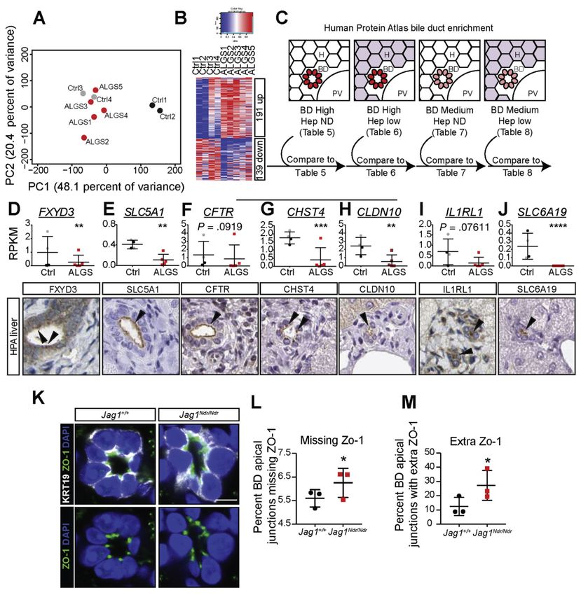
Andersson et al.                                                                                          Page 2

                                                 Background & Aims—Alagille syndrome is a genetic disorder characterized by cholestasis,
                                                 ocular abnormalities, characteristic facial features, heart defects, and vertebral malformations.
                                                 Most cases are associated with mutations in JAGGED1 (JAG1), which encodes a Notch ligand,
                                                 although it is not clear how these contribute to disease development. We aimed to develop a mouse
                                                 model of Alagille syndrome to elucidate these mechanisms.
Europe PMC Funders Author Manuscripts

                                                 Methods—Mice with a missense mutation (H268Q) in Jag1 (Jag1+/Ndr mice) were outbred to a
                                                 C3H/C57bl6 background to generate a mouse model for Alagille syndrome (Jag1Ndr/Ndr mice).
                                                 Liver tissues were collected at different timepoints during development, analyzed by histology,
                                                 and liver organoids were cultured and analyzed. We performed transcriptome analysis of
                                                 Jag1Ndr/Ndr livers and livers from patients with Alagille syndrome, cross-referenced to the Human
                                                 Protein Atlas, to identify commonly dysregulated pathways and biliary markers. We used species-
                                                 specific transcriptome separation and ligand-receptor interaction assays to measure Notch
                                                 signaling and the ability of JAG1Ndr to bind or activate Notch receptors. We studied signaling of
                                                 JAG1 and JAG1Ndr via NOTCH 1, NOTCH2, and NOTCH3 and resulting gene expression patterns
                                                 in parental and NOTCH1-expressing C2C12 cell lines.

                                                 Results—Jag1Ndr/Ndr mice had many features of Alagille syndrome, including eye, heart, and
                                                 liver defects. Bile duct differentiation, morphogenesis, and function were dysregulated in newborn
                                                 Jag1Ndr/Ndr mice, with aberrations in cholangiocyte polarity, but these defects improved in adult
                                                 mice. Jag1Ndr/Ndr liver organoids collapsed in culture, indicating structural instability. Whole-
                                                 transcriptome sequence analyses of liver tissues from mice and patients with Alagille syndrome
                                                 identified dysregulated genes encoding proteins enriched at the apical side of cholangiocytes,
                                                 including CFTR and SLC5A1, as well as reduced expression of IGF1. Exposure of Notch-
                                                 expressing cells to JAG1Ndr, compared with JAG1, led to hypomorphic Notch signaling, based on
                                                 transcriptome analysis. JAG1-expressing cells, but not JAG1Ndr-expressing cells, bound soluble
                                                 Notch1 extracellular domain, quantified by flow cytometry. However, JAG1 and JAG1Ndr cells
                                                 each bound NOTCH2, and signaling from NOTCH2 signaling was reduced but not completely
Europe PMC Funders Author Manuscripts

                                                 inhibited, in response to JAG1Ndr compared with JAG1.

                                                 Conclusions—In mice, expression of a missense mutant of Jag1 (Jag1Ndr) disrupts bile duct
                                                 development and recapitulates Alagille syndrome phenotypes in heart, eye, and craniofacial
                                                 dysmorphology. JAG1Ndr does not bind NOTCH1, but binds NOTCH2, and elicits hypomorphic
                                                 signaling. This mouse model can be used to study other features of Alagille syndrome and organ
                                                 development.

                                           Keywords
                                                 Notch; Jagged1; Alagille; Heart; Liver; Kidney; Vertebrae; Development

                                                           Notch signaling is a highly conserved cell-contact–dependent signaling pathway used
                                                           reiteratively in many developmental processes. Mutations in the Notch pathway lead to
                                                           numerous diseases,1 including Alagille syndrome (ALGS1; Online Mendelian Inheritance in
                                                           Man/OMIM no. 118450, and ALGS2, OMIM no. 610205).2 ALGS is an autosomal
                                                           dominant genetic disorder that in more than 90% of patients is caused by mutations in
                                                           JAGGED1 (JAG1),3,4 while about 1% harbor NOTCH2 mutations.5 Alagille syndrome often

                                                             Gastroenterology. Author manuscript; available in PMC 2020 February 07.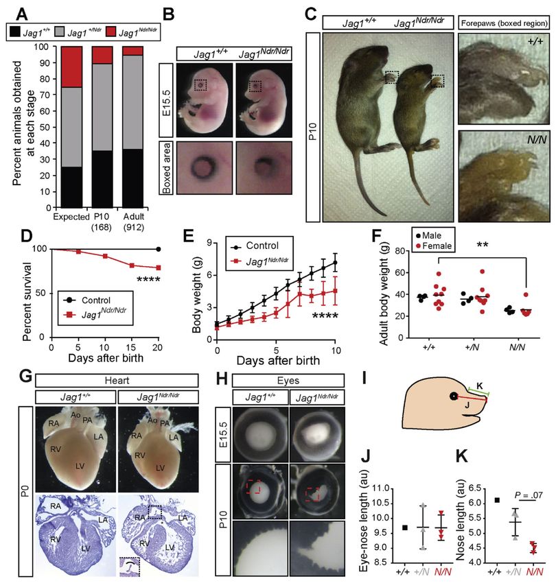
Andersson et al.                                                                                               Page 3

                                                           presents early in life with severe liver and heart defects,6 but also affects vertebrae, eyes, and
                                                           craniofacial morphology.

                                                           How Alagille JAG1 mutations affect signaling through different Notch receptors is poorly
                                                           understood. The JAG1 ligand is expressed on a signal-sending cell that activates signaling
                                                           upon contact with a Notch receptor on a juxta-posed signal-receiving cell. Missense
Europe PMC Funders Author Manuscripts

                                                           mutations in ALGS are enriched in the receptor-binding DSL and DOS domains1 of JAG1,
                                                           and analysis of the crystallized JAG1 receptor-binding domain and an extracellular portion
                                                           of NOTCH17 shows that the previously described Jag1Ndr mutation8 maps to this interaction
                                                           domain, but how the JAG1Ndr mutation mechanistically affects Notch signaling through
                                                           receptors other than NOTCH1 remains to be established.

                                                           Deletions in a single JAG1 allele are sufficient to cause ALGS, suggesting
                                                           haploinsufficiency is the disease-causing mechanism. This is also supported by various
                                                           mouse models, based on targeting Jag1 and/or Notch2 (for review, see 1). Mouse models for
                                                           ALGS liver disease include conditional Jag1 ablation in portal vein mesenchyme,9 and Jag1/
                                                           Notch2 compound heterozygous mice.10,11 However, the first model does not mimic the full
                                                           syndrome, and the Jag1/Notch2 model biases our understanding of ALGS towards Jag1/
                                                           Notch2-regulated conditions, though NOTCH2 mutations are observed in only a fraction of
                                                           ALGS cases.12 Also, NOTCH2-related ALGS2 presents differently from JAG1-related
                                                           ALGS1; for example, patients with NOTCH2 mutations less frequently display heart
                                                           defects.13 Jag1+/dDSL mice on a C57bl6 background display bile duct paucity but are not
                                                           jaundiced,14 and it is unknown whether these mice recapitulate other major features of
                                                           ALGS. Thus, the link between missense Jag1 mutations and ALGS has not yet been possible
                                                           to address in vivo.

                                                           In this report, we show that a missense mutation in Jag1 (H268Q; Nodder, Jag1Ndr 8)
Europe PMC Funders Author Manuscripts

                                                           generates a mouse model for ALGS, mimicking disease pathology in eye, craniofacial
                                                           morphology, heart, and liver. By investigating liver development at different stages, and
                                                           using liver organoids from Jag1Ndr/Ndr mice, we show that while biliary differentiation is
                                                           delayed, biliary morphogenesis and maintenance are disrupted. In line with dysregulated
                                                           morphogenesis, whole transcriptome analysis of ALGS liver biopsies and Jag1Ndr/Ndr livers
                                                           confirms dysregulated expression of cell polarity genes, but not of key regulators of bile duct
                                                           differentiation at postnatal or adult stages. At the molecular level, the JAG1Ndr mutation
                                                           generates a hypomorphic ligand that is unable to bind to specific Notch receptors: JAG1Ndr
                                                           binds NOTCH2, but not NOTCH1, and to a lesser degree NOTCH3. Collectively, we show
                                                           that a missense mutation in Jag1 is sufficient to invoke an ALGS phenotype in mice, and
                                                           provide the first evidence that a Jag1 missense mutation can impact differentially on
                                                           different Notch receptor interactions.

                                           Methods
                                           Mouse Maintenance, Breeding, and Genetics
                                                           Jag1+/Ndr mice have been described previously,8 and for the present study were maintained
                                                           in a mixed C3H/C57bl6 genetic background. For details, see Supplementary Materials.

                                                             Gastroenterology. Author manuscript; available in PMC 2020 February 07.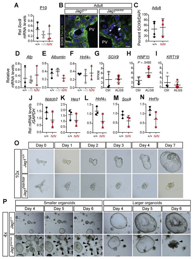
Andersson et al.                                                                                           Page 4

                                           Measurement of Craniofacial Proportions
                                                           The distance from the eye to the snout tip and from the snout/forehead bridge to the snout tip
                                                           were measured using ImageJ in images of E15.5 embryos taken from the animal’s right side.
                                                           All measurements were performed by experimenters blinded to the genotype.

                                           Antibodies, Immunohistochemistry, and Staining
Europe PMC Funders Author Manuscripts

                                                           Fourteen-μm cryosections of liver were stained using routine staining protocols. For
                                                           antibodies and staining details, see Supplementary Materials.

                                           Bile Duct Quantification
                                                           Bile ducts in 10–100 portal triads were quantified per stage in Jag1+/+ and Jag1Ndr/Ndr mice.
                                                           For details, see Supplementary Materials.

                                           Blood Chemistry Analysis
                                                           Plasma and serum were sent to the Swedish University of Agricultural Sciences for analysis
                                                           of blood chemistry. For details, see Supplementary Materials.

                                           Quantitative real-time polymerase chain reaction (qPCR)
                                                           qPCR was performed, as described.15 For primers see Supplementary Materials.

                                           Liver Organoid Cell Culture
                                                           Liver organoids were isolated and cultured, as described,16 in the presence of R-spondin.

                                           Collection of Human Samples for RNA Sequencing
                                                           Human liver needle biopsies were collected for clinical purposes, and a small part (3–5 mm
Europe PMC Funders Author Manuscripts

                                                           x 1 mm) was snap-frozen and stored at -80°C. Diagnosis details are in Supplementary
                                                           Materials.

                                           Tissue Dissection, Homogenization, RNA Extraction, and cDNA Library Preparation
                                                           Liver was homogenized and RNA from liver or cells was extracted using Direct-zol RNA
                                                           MiniPrep (cat. no. R2050; Zymo Research, Irvine, CA) or the RNeasy Mini Kit (cat. no.
                                                           74104; Qiagen, Hilden, Germany).

                                                           cDNA libraries for all samples were created using the TruSeq RNA Sample Prep Kit v2–48,
                                                           Set A (cat. no. RS-122-2001; Illumina, San Diego, CA) and Set B (cat. no. RS-122-2002;
                                                           Illumina). For specifics, see Supplementary Materials.

                                           Alignment, Analysis of Technical Performance, and Bioinformatics
                                                           The cDNA libraries were sequenced on a HiSeq 2000 with a 50–52 read length, single-end,
                                                           for different samples.17 Bioinformatics and sequencing details are provided in
                                                           Supplementary Materials.

                                                             Gastroenterology. Author manuscript; available in PMC 2020 February 07.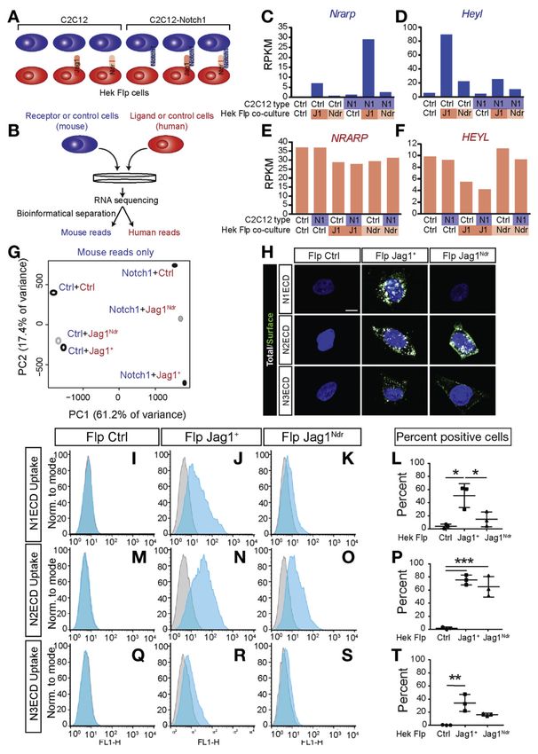
Andersson et al.                                                                                             Page 5

                                           Human Protein Atlas Cross-referencing Enrichment of Bile Duct Genes
                                                           Proteins expressed in bile ducts were identified using the Human Protein Atlas (HPA, http://
                                                           www.proteinatlas.org/),18 using the following search string: Field: Tissue expression (IHC),
                                                           Tissue: Liver, Cell Type: Bile duct cells, Expression: High or Medium AND Field: Tissue
                                                           expression (IHC), Tissue: Liver, Cell Type: Bile duct cells, Expression: Not detected or low.
                                                           Supplementary Tables 5–8. For details, see Supplementary Materials.
Europe PMC Funders Author Manuscripts

                                           Bile Duct Orientation by ZO-1 Staining in Adult Mice
                                                           ZO-1 orientation analysis was carried out in 6–12 well-formed/functional bile ducts per
                                                           animal (n=3).

                                           IGF1 ELISA
                                                           IGF1 in serum was detected using ELISA according to manufacturer’s instructions (cat. no.
                                                           EMIGF1; Thermo Fisher Scientific, Waltham, MA).

                                           Cell Lines and Cell Culture
                                                           Mouse C2C12 control and C2C12-FLNotch1 and human HEK-293-Flp-In cells8: HEK293-
                                                           Flp control (Flp Ctrl), HEK293-Flp-Jag1WT (Flp JAG1+), HEK293-Flp-Jag1Ndr (Flp
                                                           JAG1Ndr) were used. For culture conditions and luciferase experiments, see Supplementary
                                                           Materials.

                                           Notch ECD Uptake Experiments
                                                           NOTCH1-Fc, NOTCH2-Fc, and NOTCH3-Fc (R&D Systems, Minneapolis, MN) was
                                                           coupled to Alexa 488 anti-Fc (Invitrogen, Carlsbad, CA). Flp Ctrl, Flp JAG1+, and Flp
                                                           JAG1Ndr cells were treated with the tagged proteins for 1 hour at 37°C. Cells were stained
                                                           for confocal imaging or trypsinized for fluorescence-activated cell sorter fluorescence-
Europe PMC Funders Author Manuscripts

                                                           activated cell sorting (FACS) analysis, as described in Supplementary Materials.

                                           Statistical Analysis
                                                           Differences between control and experimental conditions were tested using t test, 1-way
                                                           ANOVA, or 2-way ANOVA. For specifics, see Supplementary Materials.

                                           Results
                                           Jag1Ndr/Ndr Mice Recapitulate Alagille Syndrome
                                                           We previously described a mouse Jag1 mutation (H268Q) in the second epidermal growth
                                                           factor (EGF)-like repeat of JAG1,8 a region enriched for missense mutations in ALGS.1 This
                                                           allele is nicknamed Nodder (Jag1Ndr) because of a head-nodding phenotype in heterozygous
                                                           C3H mice. Jag1Ndr/Ndr mice are embryonic lethal on this genetic background8 and, because
                                                           the phenotype of other Jag1 heterozygous mice depends on genetic background,14,19 we
                                                           asked whether mixed Jag1Ndr/Ndr mice bypass C3H lethality and recapitulate ALGS.

                                                           On a mixed C3H/C57bl6 background, viability was considerably improved: Jag1Ndr/Ndr
                                                           embryos were recovered at a rate of 20% at embryonic day (E) 15.5, 10% from postnatal day

                                                             Gastroenterology. Author manuscript; available in PMC 2020 February 07.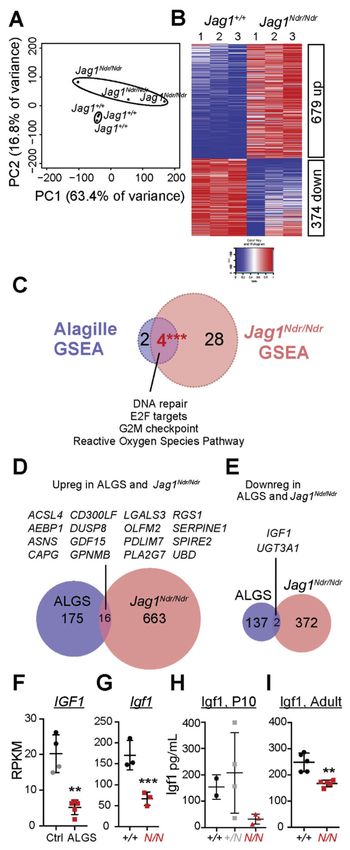
Andersson et al.                                                                                             Page 6

                                                           (P) 0, and 5% in adults (Figure 1A, Supplementary Table 1, and data not shown). At E15.5,
                                                           Jag1Ndr/Ndr embryos appeared grossly normal, and only exhibited a mild iris dysmorphology
                                                           (Figure 1B). In contrast, postnatal Jag1Ndr/Ndr pups were jaundiced (Figure 1C), excreted
                                                           yellow stools (data not shown), exhibited partial post-natal mortality (Figure 1D), and failed
                                                           to thrive (Figure 1E). Adult Jag1Ndr/Ndr mice were 30% smaller than Jag1+/+ and Jag1+/Ndr
                                                           mice (Figure 1F).
Europe PMC Funders Author Manuscripts

                                                           ALGS is diagnosed based on the presence of cholestasis, ocular abnormalities, characteristic
                                                           facial features, heart defects, and vertebral malformations.6 The heart defects range from
                                                           pulmonary artery stenosis to tetralogy of Fallot, a severe defect encompassing pulmonary
                                                           stenosis, overriding aorta, ventricular septal defect, and right ventricular hypertrophy.20 Both
                                                           atrial and ventricular septation defects were present in E15.5 and P0 Jag1Ndr/Ndr mice
                                                           (Figure 1G and Supplementary Figure 1A).

                                                           Patients with ALGS display posterior embryotoxon,12 a malformation attributed to neural
                                                           crest defects,21 and a smaller cornea.22 The first obvious phenotype in Jag1Ndr/Ndr mice was
                                                           bilateral iris deformation with dorsal constriction at E13.5, which progressed to severe
                                                           deformities and occasionally micropthalmia by P10 (Figure 1B,H, and Supplementary
                                                           Figure 1B). Jag1Ndr/Ndr lenses were similar to wild types in size at E15.5 and were only
                                                           slightly smaller at P10 (Supplementary Figure 1C,D), while 30% of adult Jag1Ndr/Ndr mice
                                                           exhibited micropthalmia (data not shown).

                                                           Craniofacial alterations, including a broad prominent forehead, deep-set eyes, and a pointy
                                                           chin, are seen in 77%–96% of patients.12 Jag1Ndr/Ndr mice similarly displayed a tendency
                                                           toward altered craniofacial proportions with a wild type eye-nose length (Figure 1I,J), but a
                                                           reduced snout length (bridge to tip, Figure 1I,K), supporting a role for Jag1 in craniofacial
                                                           development, in line with previous reports.23 Alcian blue/Alizarin red staining of cartilage
Europe PMC Funders Author Manuscripts

                                                           and bone at P0 and P10 did not reveal obvious vertebral malformations (data not shown),
                                                           indicating that butterfly vertebrae12 are probably not present in Jag1Ndr/Ndr mice.

                                                           In conclusion, Jag1Ndr/Ndr mice recapitulate cardinal features of ALGS, including ocular,
                                                           craniofacial, and cardiac defects. Jaundice indicates liver dysfunction, and we therefore next
                                                           asked whether Jag1Ndr/Ndr mice display ductopenia.

                                           Jag1Ndr/Ndr Mice Exhibit Early Life Biliary Dysmorphogenesis and Dysfunction With Later
                                           Rescue
                                                           A crippling ALGS symptom is cholestatic liver disease, with conjugated hyperbilirubinemia
                                                           and decreased liver function, which histologically is associated with paucity of intrahepatic
                                                           bile ducts. Thus, liver transplantation is frequently required. The pathomechanisms for
                                                           ductopenia are poorly understood and it is unclear why, in some patients, cholestasis
                                                           diminishes with time.24,25 Because some Jag1Ndr/Ndr mice survive to adulthood, the model
                                                           provides an opportunity to elucidate disease development across different stages.

                                                           Jag1Ndr/Ndr mice displayed strong jaundice at neonatal stages (Figure 1C), whereas
                                                           surviving adult Jag1Ndr/Ndr mice did not display jaundice, nor excrete yellow feces (data not
                                                           shown). To determine whether Jag1Ndr/Ndr mice manifest a transient biliary phenotype, we

                                                             Gastroenterology. Author manuscript; available in PMC 2020 February 07.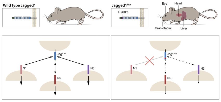
Andersson et al.                                                                                              Page 7

                                                           analyzed biliary histology and marker expression in portal regions during development. Both
                                                           Sox9 (a Notch target gene that regulates bile duct development26) and Hnf1β were present in
                                                           Jag1+/+ periportal areas, but absent in Jag1Ndr/Ndr mice at E18.5. At p0, faintly positive cells
                                                           were detected around portal tracts in Jag1Ndr/Ndr mice, at levels far weaker than the clusters
                                                           of Sox9/Hnf1β-positive cells undergoing lumen formation in Jag1+/+ livers. At this stage,
                                                           Jag1Ndr/Ndr hilar portal regions had no morphologically discernible mature bile ducts, while
Europe PMC Funders Author Manuscripts

                                                           3.3% of Jag1+/+ hilar portal veins had adjoining mature bile ducts (Figure 2C,D). The
                                                           majority of P0 portal veins in Jag1Ndr/Ndr livers contained either no KRT19+ cells or
                                                           disorganized clusters of KRT19+ cells (Figure 2B–D, Supplementary Figure 2A). At P10,
                                                           bile ducts were rarely found in Jag1Ndr/Ndr livers, while portal veins in Jag1+/+ livers
                                                           manifested 1 or 2 adjacent mature bile ducts (Figure 2E,F; Supplementary Figure 2B,C).
                                                           Hepatoblast and hepatocyte marker expression levels were unaltered (data not shown), but
                                                           serum biochemistry at P10 confirmed that Jag1Ndr/Ndr liver function was severely
                                                           compromised (Figure 2G–J, Supplementary Figure 2D–F).

                                                           In contrast, lumenized bile ducts could be found in both Jag1+/+ and Jag1Ndr/Ndr adult mice,
                                                           though with disrupted morphology in Jag1Ndr/Ndr livers (Figure 2K–M, Supplementary
                                                           Figure 2G,H). We classified and quantified pan-cytokeratin+ and KRT19+ bile ducts as
                                                           “well-formed” (1 layer of biliary cells, a round lumen), “functional” (1 or more layers of
                                                           biliary cells, a discernible lumen) or “clusters” (clusters of biliary cells, no discernible
                                                           lumen) (Figure 2L). Adult Jag1Ndr/Ndr mice harbor fewer “well-formed” bile ducts and
                                                           instead contain “clusters” of biliary cells (Figure 2M, Supplementary Figure 2G). However,
                                                           there was no significant difference between Jag1+/+ and Jag1Ndr/Ndr mice when grouping
                                                           well-formed and functional bile ducts (Supplementary Figure 2H). Serum analysis
                                                           confirmed a full functional recovery in adult Jag1Ndr/Ndr mice (Figure 2N, Supplementary
                                                           Figure 2I,J), with only a small difference in aspartate aminotransferase levels still detectable.
Europe PMC Funders Author Manuscripts

                                                           In contrast to the transient biliary phenotype, there was a persistent absence of hepatic
                                                           arteries in Jag1Ndr/Ndr mice (Figure 2K, Supplementary Figure 2K,L). In conclusion, the
                                                           Jag1Ndr/Ndr mice display a biliary phenotype that is severe at early postnatal stages but that
                                                           improves during adulthood.

                                           Disrupted Bile Duct Morphogenesis and Delayed Differentiation
                                                           It is unclear whether the ALGS biliary defects are because of disrupted morphogenesis,11
                                                           differentiation defects,27 or both.28 To address this question, we analyzed the expression of
                                                           key genes regulating differentiation. Expression of Sox9, and Hnf4α, a transcription factor
                                                           required for hepatocyte differentiation,29 as well as alfa-fetoprotein (a marker of
                                                           hepatoblasts) and albumin (a marker for hepatocyte function), was unaffected in P10 and
                                                           adult Jag1Ndr/Ndr mice (Figure 3A–F, data not shown). Similarly, SOX9 mRNA levels were
                                                           not affected in ALGS liver biopsies (Figure 3G), nor were the well-characterized biliary
                                                           markers HNF1β or KRT19 (Figure 3H,I). This is in contrast to the early absence of SOX9-
                                                           and HNF1β-positive cells at E18.5 and P0 (Figure 2A,B).

                                                           To assess adult bile duct development and morphology, we used a recently developed model
                                                           for long-term in vitro expansion of bile duct-derived progenitor cells.16,30 Bile duct
                                                           fragments were hand-picked and cultured in vitro as liver organoids, forming readily from

                                                             Gastroenterology. Author manuscript; available in PMC 2020 February 07.
Andersson et al. Page 8
both adult control and Jag1Ndr/Ndr mice. Notch2, Hes1, Hnf4α, Sox9, and Hnf1β mRNA
expression were not altered (Figure 3J–N), further supporting that differentiation was
delayed, but not completely inhibited (see Figure 2). However, liver organoids from
Jag1Ndr/Ndr mice grew less well than Jag1+/+ organoids (Figure 3O). Importantly, a number
of Jag1Ndr/Ndr organoids collapsed in culture after 5–6 days (Figure 3P), demonstrating
structural instability. Jag1Ndr/Ndr biliary cells from adult mice are therefore similar to Jag1+/+
Europe PMC Funders Author Manuscripts
biliary cells in terms of cell identity, but exhibit differences in structural stability. In
conclusion, the data argue for morphologic as well as differentiation defects.
Novel Biomarkers for Alagille Syndrome Reveal Dysregulation of Apical Proteins
To further assess the molecular basis for ALGS, we performed genome-wide transcriptome
studies of liver biopsies from 5 patients with ALGS. Control samples from pediatric patients
with liver disease and/or cholangiopathies allow us to detect genes specifically dysregulated
in ALGS, rather than cholestasis pathways or general liver disease mechanisms. Principal
component analysis (PCA) showed that the 5 ALGS liver transcriptomes clustered with the 2
cholangiopathy samples: autoimmune hepatitis with primary sclerosing cholangitis, and
progressive familial intrahepatic cholestasis type 2. In contrast, the transcriptomes from 2
patients with autoimmune hepatitis segregated more distinctly (Figure 4A, Supplementary
Table 2). There were 191 up-regulated and 139 down-regulated genes (adjusted P value 1.5, Figure 4B, Supplementary Tables 3 and 4, Supplementary Figure 3).
The transcriptome data are derived from bulk liver. We therefore devised a strategy to cross-
reference the transcriptome data with protein expression patterns from the HPA (http://
www.proteinatlas.org), a map of the human proteome,18 allowing us to identify genes
encoding proteins expressed in bile ducts (Figure 4C, Supplementary Tables 5–8). This
strategy identified the well-established biliary markers HNF1β, KRT19, and SOX9,
confirming strategy validity and specificity, and HPA data showed the expected biliary
Europe PMC Funders Author Manuscripts
expression (Supplementary Figure 4A).
Comparison of HPA bile duct-enriched proteins to ALGS transcriptomes revealed 5 up-
regulated and 7 down-regulated novel bile duct markers in ALGS (Figure 4D,E,
Supplementary Figure 4B–L). Of these, FXYD domain containing ion transport regulator 3
(FXYD3, Figure 4D) and Solute carrier family 5 (sodium/glucose cotransporter) member 1
(SLC5A1, Figure 4E) showed the highest significance, and encode proteins enriched at the
apical surface of bile ducts. Manual comparison of the protein localization of the top 30
down-regulated genes to the HPA revealed 5 additional bile duct-specific genes with apical
cholangiocyte staining including Cystic Fibrosis Transmembrane Conductance Regulator
(CFTR, Figure 4F), Carbohydrate Sulfotransferase 4 (CHST4, Figure 4G), Claudin 10
(CLDN10, Figure 4H), Interleukin 1 receptor-like 1 (IL1RL1, Figure 4I) and Solute Carrier
Family 6 (Neutral Amino Acid Transporter) member 19 (SLC6A19, Figure 4J). Given this
link to aberrant cell polarity, we assessed the distribution of Zona occludens 1 (ZO-1, a.k.a.
TJP-1), a marker of apical junctions in cholangiocytes that is not down-regulated in ALGS
or in Jag1Ndr/Ndr mice (data not shown). Even in the best-formed bile ducts in Jag1Ndr/Ndr
mice, ZO-1 was mis-localized, confirming polarity defects (Figure 4K–M). In conclusion,
Gastroenterology. Author manuscript; available in PMC 2020 February 07.Andersson et al. Page 9
the most highly down-regulated biliary genes encode proteins enriched at the apical surface
of bile ducts, corroborating morphogenesis disruption in ALGS.
Igf1 is Down-regulated in Patients With ALGS and Jag1Ndr/Ndr Mice
We next compared the transcriptomic changes in Jag1Ndr/Ndr livers to ALGS livers. RNA
sequencing of Jag1Ndr/Ndr and Jag1+/+ livers yielded distinct transcriptional profiles (Figure
Europe PMC Funders Author Manuscripts
5A, Supplementary Table 9), with 679 up-regulated and 374 down-regulated genes (Figure
5B, Supplementary Tables 10 and 11, and Supplementary Figure 5).
We assessed changes in signaling pathways and major cellular programs using gene set
enrichment analyses (GSEA), which identified 35 sets significantly enriched in Jag1Ndr/Ndr
livers at False Discovery RateAndersson et al. Page 10
9), which separates more than 99% of a mixed transcriptome to the correct species.17 Notch
target gene responses were specifically detectable in the Notch receptor-expressing (mouse)
cell reads in conditions with Notch activation (Figure 6C–F, Supplementary Figure 10A–D).
We co-cultured ligand-expressing HEK293 Flp cells with control C2C12 cells (which
express Notch2 and Notch3 at twice the levels of Notch1 and with almost undetectable
Europe PMC Funders Author Manuscripts
Notch4 [Supplementary Figure 10E]), or with NOTCH1-overexpressing C2C12 cells, to
examine whether NOTCH1 modified the transcriptomic response induced by ligand-
expressing cells. PCA of the mouse transcriptomes showed that the JAG1Ndr-induced
transcriptome is intermediate between response to control cells, and response to JAG1+-
expressing cells (Figure 6G, Supplementary Table 24). However, in C2C12 cells, which
predominantly express Notch2 and Notch3, the JAG1Ndr-induced transcriptome clustered
with the JAG1+-induced transcriptome, whereas for NOTCH1-overexpressing C2C12 cells,
the JAG1Ndr transcriptome lay closer to the Ctrl-induced transcriptome. This suggests that
JAG1Ndr signals weakly or not at all through NOTCH1, in line with our previous report.8 In
addition, Notch target genes showed weak (10%–80%) up-regulation by the JAG1Ndr ligand
(Supplementary Figure 10F). In sum, the JAG1Ndr allele is hypomorphic at the global
transcriptome level with regard to its ability to elicit Notch signaling.
JAG1Ndr Induces Receptor-selective Binding
The differentially hypomorphic signaling elicited by JAG1Ndr suggested that JAG1Ndr-
mediated signaling through Notch receptor paralogs may be altered. Because the H268Q
mutation resides in the Notch receptor-interacting domain, we tested whether JAG1Ndr
exhibited receptor paralog-specific binding. Flp JAG1+ or Flp JAG1Ndr ligand-expressing
cells were treated with fluorescently tagged soluble NOTCH1-3 receptor extracellular
domain peptides (N1-3ECD, Figure 6H). Immunocytochemistry for the NECD-Fc was
performed, without permeabilization, to detect extracellular ECDs (Figure 6H). N2ECD and
Europe PMC Funders Author Manuscripts
N3ECD were bound by Flp JAG1+ and Flp JAG1Ndr cells, whereas N1ECD only interacted
with Flp Jag1+ (Figure 6H); the latter in keeping with our previous report.8 FACS analysis
showed that N1ECD was internalized by 50% of Flp JAG1+ cells, while Flp JAG1Ndr cells
did not significantly internalize N1ECD (Figure 6I–L, Supplementary Figure 10G). In
contrast, N2ECD was internalized by 80% of Flp JAG1+ cells, and by 70% of Flp JAG1Ndr
cells (Figure 6M–P). However, the amount of N2ECD internalized by Flp JAG1Ndr cells was
lower than by Flp JAG1+ cells (Supplementary Figure 10H). N3ECD was internalized by
35% of Flp Jag1+ cells, and by 20% of Flp Jag1Ndr cells (Figure 6Q–T, Supplementary
Figure 10I). Because N2ECD internalization was reduced, we next tested the extent of
activation of cells expressing Notch2 receptors, in response to co-culture with cells
expressing Flp JAG1+ or Flp JAG1Ndr. Co-culture with Flp JAG1Ndr cells resulted in
reduced Notch activation (as defined by 12XCSL-luciferase activation), as compared with
co-culture with Flp JAG1+ cells (Supplementary Figure 10J). In conclusion, Flp JAG1Ndr
exhibits a selective loss of interaction with NOTCH1, but the interaction with NOTCH2 and
NOTCH3 is partially retained, although NOTCH2-mediated signaling elicited by Flp
JAG1Ndr is reduced.
Gastroenterology. Author manuscript; available in PMC 2020 February 07.Andersson et al. Page 11
Discussion
ALGS is usually caused by mutations in the JAG1 gene, but how dysregulated Notch
signaling links to phenotypic consequences has been enigmatic. In this report, we provide
evidence that a Jag1 missense mutation (Jag1H268Q) recapitulates Alagille symptoms in a
number of organs. Jag1H268Q elicits a reduced Notch transcriptomic response and Notch
Europe PMC Funders Author Manuscripts
receptor-selective binding (schematically depicted in Figure 7).
Disturbed Morphogenesis in Jag1Ndr Bile Ducts
The Jag1Ndr/Ndr mouse demonstrates that a Jag1 missense mutation can recapitulate ALGS.
Other models based on loss-of-function Jag1 and/or Notch2 alleles do not display the entire
spectrum of disease phenotypes. The Jag1Ndr/Ndr mouse displays Alagille-like phenotypes in
several organs, including heart, lens, and craniofacial structures, as well as liver, and thus
represents a clinically relevant mouse model for ALGS. An important distinction must be
made, however, with regards to genetics: ALGS in humans is generally caused by
heterozygosity for JAG1, while in mice the phenotype arises in the homozygous Jag1Ndr/Ndr
state.
The fact that Jag1Ndr/Ndr mice survive to adulthood provides an opportunity to explore liver
pathology over a lifetime. Interestingly, despite neonatal ductopenia, the number of bile
ducts increases in the adult – although with aberrant morphology. In keeping with this,
cholestasis was pronounced in pups, while adults display a full recovery regarding
cholestasis, suggesting compensatory mechanisms rescue ductopenia in Jag1Ndr/Ndr mice. To
what extent recuperation of the liver occurs also in ALGS is contested because some patients
recover from cholestasis with time32 while in others biliary breakdown continues.24,25 Some
patients display regenerating liver nodules with normal bile duct numbers.24,33,34 The RNA
sequencing of ALGS liver samples (Figure 4) showed some heterogeneity, and in light of the
Europe PMC Funders Author Manuscripts
variable liver disease severity and progression or reversal, additional analyses on bigger
cohorts of patients will be necessary to determine whether there is a unifying molecular
mechanism leading to bile duct abnormalities and whether diseases progression can be
predicted based on transcriptomic data. The notion of a transient liver phenotype is also of
interest for therapy; if cholestasis could be temporarily treated, then harnessing endogenous
repair mechanisms could reduce the need for liver transplantation. In this context, the
Jag1Ndr/Ndr mouse may serve as an important tool to explore novel therapeutic strategies, to
test if transplanted cells can give rise to bile ducts, or which treatments induce repair
mechanisms.
The Jag1Ndr/Ndr mice also shed light on the nature of the ALGS biliary pathology, attributed
to disrupted morphogenesis of the bile ducts,11 defective bile duct maintenance, or
differentiation defects.27,28 Our data indicate that morphogenesis and maintenance of bile
ducts are affected, in addition to differentiation. This notion is supported by profound
changes in gene expression in ALGS affecting cell polarity, at postnatal and adult stages.
Further support for disturbed morphogenesis comes from the liver organoid data, in which
Jag1Ndr/Ndr organoids initiated growth but collapsed a few days later, supporting structural
rather than developmental defects. A previous report showed that liver organoids from
Gastroenterology. Author manuscript; available in PMC 2020 February 07.Andersson et al. Page 12
human Alagille livers showed no phenotype in the undifferentiated state, but underwent
collapse and apoptosis upon R-spondin withdrawal.30
Dysregulated cell polarity by disrupted Notch signaling has been shown in other polarized
structures. Removal of CSL, the canonical Notch transcription factor, in embryonic stem
cells disrupts neural rosettes,35 a lumenized and polarized colony of neural cells modeling
Europe PMC Funders Author Manuscripts
the neural tube in vitro. Similarly, in the zebrafish lateral line, Notch is required for apical
constriction of proneuromast rosettes,36 and regulates apical junction-associated genes,
which together with our results, indicates a more general interaction between Notch and the
cell polarity machinery.
The Jag1Ndr Mutation Causes Hypomorphic Signaling and Receptor Selectivity
Our data lend support to the hypomorphic view of ALGS mutations. The transcriptome data
from co-cultured ligand- and receptor-expressing cells indicate that Jag1Ndr induces a
hypomorphic Notch signaling response.
The finding that the H268Q mutation yields a JAG1 ligand that selectively loses its ability to
interact with and activate NOTCH1, while maintaining interaction with NOTCH2 and 3,
provides a novel facet of Notch signaling. Modifications of NOTCH receptors by Fringe
fine-tunes their interaction with JAG or DLL ligands, but that a ligand mutation is sufficient
to select for NOTCH2 and 3, but not NOTCH1, interaction is unprecedented. The H268Q
missense mutation falls in the second EGF repeat, a region involved in Notch receptor
activation,37 close to a number of patient-specific JAG1 mutations,1 and close to 3
interacting amino acids in NOTCH1, in EGF9, and EGF10,7 2 of which are conserved in
NOTCH2 (L368 and P391); whereas the Valine 392 in NOTCH1 is instead a Leucine in
NOTCH2. Whether this difference will explain the differential effect of the JAGGED1
H268Q mutation on NOTCH1 and 2 activation, however, remains to be tested. This
Europe PMC Funders Author Manuscripts
difference cannot explain differences in binding to NOTCH3 because all 3 amino acids are
conserved in NOTCH3 (Supplementary Materials).
The Recovery of Jag1Ndr/Ndr Mice Suggests Endogenous Mechanisms can Rescue Alagille
Syndrome
Why certain patients with ALGS recover liver biliary function while others progress to liver
transplantation is currently unknown. Jag1Ndr/Ndr pups display a severe biliary phenotype
that is functionally rescued in adults. This is in contrast to Jag1+/dDSL mice, which display
ALGS-like liver phenotypes in a C57bl6 background, but are not reported to improve with
age.14
Sox9 is expressed not only in cholangiocytes, but also in stem-like hepatocytes upon insult,
9,38,39 which can trans-differentiate into cholangiocytes. A similar rescue in adult albumin-
Cre Hnf6flox/flox Rbpjflox/flox mice40 suggests Notch-independent mechanisms induce
ductular reaction and hepatocyte trans-differentiation. The recovery in Jag1Ndr/Ndr mice may
also be because of hepatocyte trans-differentiation, which can be induced with Notch
activation.28 Thus, although Notch is not required for rescue, it may be sufficient. Transient
activation of Notch may therefore – in principle – be feasible as therapy, though the role of
Notch signaling in cancer suggests this could be associated with significant risks.41,42 The
Gastroenterology. Author manuscript; available in PMC 2020 February 07.Andersson et al. Page 13
loss of Igf1 also presents an interesting therapeutic target because Igf1 stimulates
cholangiocyte proliferation.43
While Jag1Ndr/Ndr mice recovered biliary function they did not recover hepatic artery
numbers, suggesting that the absence of proper portal triad vascular architecture does not
preclude recovery of a functional biliary tree. Thus, cholangiocytes are not completely
Europe PMC Funders Author Manuscripts
dependent on the presence of a hepatic artery, which would otherwise be suggested by the
biliary breakdown induced by hepatic artery ligation, and which would preclude cell
replacement therapy. Instead, our results suggest cell replacement therapies to replace absent
cholangiocytes may be feasible, even in the absence of normal hepatic vasculature.
Our data, showing a selective loss of primarily Notch1-mediated signaling, may be at
apparent odds with the fact that ALGS can be caused by NOTCH2 mutations,12 and that
compound heterozygous Jag1/Notch2 mice are also growth delayed, display jaundice, and
recapitulate hepatic, cardiac, renal, and ocular defects.10 Moreover, Notch2 is required for
bile duct morphogenesis and differentiation in vivo, 44–46 while Notch1 is dispensable.44
However, the mildly reduced Jag1Ndr-Notch2 signaling described here may be sufficient to
cause a pathologic outcome in keeping with the dose-sensitive nature of the Notch signaling
pathway.
In sum, the Nodder mouse provides a clinically relevant model for ALGS and allows for the
first time a Jag1 missense mutation to be linked both to phenotypic traits typical for the
disease and to dysregulated Notch signaling, manifested by hypomorphic signaling and
receptor-selectivity.
Supplementary Material
Refer to Web version on PubMed Central for supplementary material.
Europe PMC Funders Author Manuscripts
Acknowledgments
Funding
U.L. acknowledges support from the Swedish Research Council (project grant and the Linnaeus Center DBRM),
European Research Council Marie Curie ITN-FP7 NotchIT (IVC), the Swedish Cancer Society, Hjärnfonden, Knut
och Alice Wallenbergs Stiftelse, and ICMC (the Integrated Cardio Metabolic Center). E.R.A. and members of
Andersson lab were supported by a Center of Innovative Medicine (CIMED) Grant, the Daniel Alagille Award, KI
Funding, and the Alex and Eva Wallström Foundation. S.H. was supported by Wera Ekströms Stiftelse and a grant
for KI-MU exchange (see below), and grants in ERA lab. M.S. was supported by an OSK Huttunen post doc
fellowship. J.M. was supported by a WennerGren Fellowship and grants in ERA lab. V.B. and S.H. were supported
by “KI-MU” program (CZ.1.07/2.3.00/20.0180) co-financed from European Social Fund and the state budget of the
Czech Republic.
The funders had no role in study design, analysis, or interpretation of data.
Abbreviations used in this paper
ALGS Alagille syndrome
ECD extracellular domain
EGF epidermal growth factor
Gastroenterology. Author manuscript; available in PMC 2020 February 07.Andersson et al. Page 14
FACS fluorescence-activated cell sorting
GSEA gene set enrichment analyses
HPA Human Protein Atlas
PCA principal component analysis
Europe PMC Funders Author Manuscripts
qPCR quantitative real-time polymerase chain reaction
References
1. Mašek J, Andersson ER. The developmental biology of genetic Notch disorders. Development.
2017; 144:1743–1763. [PubMed: 28512196]
2. Grochowski CM, Loomes KM, Spinner NB. Jagged1 (JAG1): structure, expression, and disease
associations. Gene. 2016; 576:381–384. [PubMed: 26548814]
3. Li L, Krantz ID, Deng Y, et al. Alagille syndrome is caused by mutations in human Jagged1, which
encodes a ligand for Notch1. Nat Genet. 1997; 16:243–251. [PubMed: 9207788]
4. Oda T, Elkahloun AG, Pike BL, et al. Mutations in the human Jagged1 gene are responsible for
Alagille syndrome. Nat Genet. 1997; 16:235–242. [PubMed: 9207787]
5. McDaniell R, Warthen DM, Sanchez-Lara PA, et al. NOTCH2 mutations cause Alagille syndrome, a
heterogeneous disorder of the notch signaling pathway. Am J Hum Genet. 2006; 79:169–173.
[PubMed: 16773578]
6. Alagille D, Odièvre M, Gautier M, et al. Hepatic ductular hypoplasia associated with characteristic
facies, vertebral malformations, retarded physical, mental, and sexual development, and cardiac
murmur. J Pediatr. 1975; 86:63–71. [PubMed: 803282]
7. Luca VC, Kim BC, Ge C, et al. Notch-Jagged complex structure implicates a catch bond in tuning
ligand sensitivity. Science. 2017; 1:1–24.
8. Hansson EM, Lanner F, Das D, et al. Control of Notch-ligand endocytosis by ligand-receptor
interaction. J Cell Sci. 2010; 123:2931–2942. [PubMed: 20720151]
9. Hofmann JJ, Zovein AC, Koh H, et al. Jagged1 in the portal vein mesenchyme regulates intrahepatic
bile duct development: insights into Alagille syndrome. Development. 2010; 137:4061–4072.
Europe PMC Funders Author Manuscripts
[PubMed: 21062863]
10. McCright B, Lozier J, Gridley T. A mouse model of Alagille syndrome: Notch2 as a genetic
modifier of Jag1 haploinsufficiency. Development. 2002; 129:1075–1082. [PubMed: 11861489]
11. Lozier J, McCright B, Gridley T. Notch signaling regulates bile duct morphogenesis in mice. PLoS
One. 2008; 3:e1851. [PubMed: 18365007]
12. Spinner, NB, Leonard, LD, Krantz, ID. Alagille Syndrome. University of Washington; Seattle:
2013.
13. Kamath BM, Bauer RC, Loomes KM, et al. NOTCH2 mutations in Alagille syndrome. J Med
Genet. 2012; 49:138–144. [PubMed: 22209762]
14. Thakurdas SM, Lopez MF, Kakuda S, et al. Jagged1 heterozygosity in mice results in a congenital
cholangiopathy which is reversed by concomitant deletion of one copy of Poglut1 (Rumi).
Hepatology. 2016; 63:550–565. [PubMed: 26235536]
15. Jin S, Mutvei AP, Chivukula IV, et al. Non-canonical Notch signaling activates IL-6/JAK/STAT
signaling in breast tumor cells and is controlled by p53 and IKKα/IKKβ. Oncogene. 2013;
32:4892–4902. [PubMed: 23178494]
16. Huch M, Dorrell C, Boj SF, et al. In vitro expansion of single Lgr5+ liver stem cells induced by
Wnt-driven regeneration. Nature. 2013; 494:247–250. [PubMed: 23354049]
17. Chivukula IV, Ramsköld D, Storvall H, et al. Decoding breast cancer tissue-stroma interactions
using species-specific sequencing. Breast Cancer Res. 2015; 17:109. [PubMed: 26265142]
18. Uhlen M, Oksvold P, Fagerberg L, et al. Towards a knowledge-based Human Protein Atlas. Nat
Biotechnol. 2010; 28:1248–1250. [PubMed: 21139605]
Gastroenterology. Author manuscript; available in PMC 2020 February 07.Andersson et al. Page 15
19. Kiernan AE, Li R, Hawes NL, et al. Genetic background modifies inner ear and eye phenotypes of
Jag1 heterozygous mice. Genetics. 2007; 177:307–311. [PubMed: 17890364]
20. McElhinney DB, Krantz ID, Bason L, et al. Analysis of cardiovascular phenotype and genotype-
phenotype correlation in individuals with a JAG1 mutation and/or Alagille syndrome. Circulation.
2002; 106:2567–2574. [PubMed: 12427653]
21. Williams AL, Bohnsack BL. Neural crest derivatives in ocular development: discerning the eye of
the storm. Birth Defects Res C Embryo Today. 2015; 105:87–95. [PubMed: 26043871]
Europe PMC Funders Author Manuscripts
22. Kim BJ, Fulton AB. The genetics and ocular findings of Alagille syndrome. Semin Ophthalmol.
2007; 22:205–210. [PubMed: 18097983]
23. Humphreys R, Zheng W, Prince LS, et al. Cranial neural crest ablation of Jagged1 recapitulates the
craniofacial phenotype of Alagille syndrome patients. Hum Mol Genet. 2012; 21:1374–1383.
[PubMed: 22156581]
24. Jinguji M, Tsuchimochi S, Nakajo M, et al. Scintigraphic progress of the liver in a patient with
Alagille syndrome (arteriohepatic dysplasia). Ann Nucl Med. 2003; 17:693–697. [PubMed:
14971613]
25. Sparks EE, Perrien DS, Huppert KA, et al. Defects in hepatic Notch signaling result in disruption
of the communicating intrahepatic bile duct network in mice. Dis Model Mech. 2011; 4:359–367.
[PubMed: 21282722]
26. Antoniou A, Raynaud P, Cordi S, et al. Intrahepatic bile ducts develop according to a new mode of
tubulogenesis regulated by the transcription factor SOX9. Gastroenterology. 2009; 136:2325–
2333. [PubMed: 19403103]
27. Tanimizu N, Miyajima A. Notch signaling controls hepatoblast differentiation by altering the
expression of liver-enriched transcription factors. J Cell Sci. 2004; 117:3165–3174. [PubMed:
15226394]
28. Zong Y, Panikkar A, Xu J, et al. Notch signaling controls liver development by regulating biliary
differentiation. Development. 2009; 136:1727–1739. [PubMed: 19369401]
29. Li J, Ning G, Duncan SA. Mammalian hepatocyte differentiation requires the transcription factor
HNF-4alpha. Genes Dev. 2000; 14:464–474. [PubMed: 10691738]
30. Huch M, Gehart H, Van Boxtel R, et al. Long-term culture of genome-stable bipotent stem cells
from adult human liver. Cell. 2015; 160:299–312. [PubMed: 25533785]
31. Bucuvalas JC, Horn JA, Carlsson L, et al. Growth hormone insensitivity associated with elevated
circulating growth hormone-binding protein in children with Alagille syndrome and short stature. J
Europe PMC Funders Author Manuscripts
Clin Endocrinol Metab. 1993; 76:1477–1482. [PubMed: 8501153]
32. Riely CA, Cotlier E, Jensen PS, et al. Arteriohepatic dysplasia: A benign syndrome of intrahepatic
cholestasis with multiple organ involvement. Ann Intern Med. 1979; 91:520–527. [PubMed:
484950]
33. Torizuka T, Tamaki N, Fujita T, et al. Focal liver hyperplasia in Alagille syndrome: assessment
with hepatoreceptor and hepatobiliary imaging. J Nucl Med. 1996; 37:1365–1367. [PubMed:
8708775]
34. Tuset E, Ribera JM, Doménech E, et al. Pseudotumorous hyperplasia of the caudate lobe of the
liver in a patient with Alagille syndrome. Med Clin (Barc). 1995; 104:420–422. [PubMed:
7715262]
35. Main H, Radenkovic J, Jin SB, et al. Notch signaling maintains neural rosette polarity. PLoS One.
2013; 8:e62959. [PubMed: 23675446]
36. Kozlovskaja-Gumbrienė A, Yi R, Alexander R, et al. Proliferation-independent regulation of organ
size by Fgf/Notch signaling. Elife. 2017; 6:e21049. [PubMed: 28085667]
37. Kovall RA, Blacklow SC. Mechanistic insights into notch receptor signaling from structural and
biochemical studies. Curr Top Dev Biol. 2010; 92:31–71. [PubMed: 20816392]
38. Fan B, Malato Y, Calvisi DF, et al. Cholangiocarcinomas can originate from hepatocytes in mice. J
Clin Invest. 2012; 122:2911–2915. [PubMed: 22797301]
39. Tanimizu N, Nishikawa Y, Ichinohe N, et al. Sry HMG box protein 9-positive (Sox9+) epithelial
cell adhesion molecule-negative (EpCAM-) biphenotypic cells derived from hepatocytes are
involved in mouse liver regeneration. J Biol Chem. 2014; 289:7589–7598. [PubMed: 24482234]
Gastroenterology. Author manuscript; available in PMC 2020 February 07.Andersson et al. Page 16
40. Walter TJ, Vanderpool C, Cast AE, et al. Intrahepatic bile duct regeneration in mice does not
require Hnf6 or notch signaling through Rbpj. Am J Pathol. 2014; 184:1479–1488. [PubMed:
24631193]
41. Nowell CS, Radtke F. Notch as a tumour suppressor. Nat Rev Cancer. 2017; 17:145–159.
[PubMed: 28154375]
42. Andersson ER, Lendahl U. Therapeutic modulation of Notch signalling–are we there yet? Nat Rev
Drug Discov. 2014; 13:357–378. [PubMed: 24781550]
Europe PMC Funders Author Manuscripts
43. Alvaro D, Metalli VD, Alpini G, et al. The intrahepatic biliary epithelium is a target of the growth
hormone/insulin-like growth factor 1 axis. J Hepatol. 2005; 43:875–883. [PubMed: 16083987]
44. Geisler F, Nagl F, Mazur PK, et al. Liver-specific inactivation of Notch2, but not Notch1,
compromises intrahepatic bile duct development in mice. Hepatology. 2008; 48:607–616.
[PubMed: 18666240]
45. Jeliazkova P, Jörs S, Lee M, et al. Canonical Notch2 signaling determines biliary cell fates of
embryonic hepatoblasts and adult hepatocytes independent of Hes1. Hepatology. 2013; 57:2469–
2479. [PubMed: 23315998]
46. Falix FA, Weeda VB, Labruyere WT, et al. Hepatic Notch2 deficiency leads to bile duct agenesis
perinatally and secondary bile duct formation after weaning. Dev Biol. 2014; 396:201–213.
[PubMed: 25446530]
Europe PMC Funders Author Manuscripts
Gastroenterology. Author manuscript; available in PMC 2020 February 07.Andersson et al. Page 17
Editor’s Notes
Background and Context
Alagille syndrome is caused by mutations in JAGGED1, but it is unclear how specific
mutations impact on Notch signaling and affect development, and how faithfully Jag1
Europe PMC Funders Author Manuscripts
mutant models mimic the human condition.
New Findings
A receptor-selective missense mutation in mouse Jag1 (H268Q) recapitulates Alagille
syndrome phenotypes in mice. RNA seq of mouse liver samples and samples from
patients, showed that apical polarity of bile ducts is severely disrupted.
Limitations
The mouse model is based on homozygous mutation of Jag1, while human patients are
heterozygous for JAG1 mutations.
Impact
This new mouse model will allow development and testing of new therapies for Alagille
syndrome, as well as a new model in which to study how Notch signaling controls
development of organs affected by Alagille syndrome.
Europe PMC Funders Author Manuscripts
Gastroenterology. Author manuscript; available in PMC 2020 February 07.Andersson et al. Page 18
Europe PMC Funders Author Manuscripts
Europe PMC Funders Author Manuscripts
Figure 1.
Jag1Ndr/Ndr C3H/C57bl6 mice survive to adulthood with Alagille-like phenotypes. (A)
Jag1+/Ndr mice were mated to generate Jag1+/+, Jag1+/Ndr and Jag1Ndr/Ndr offspring. At P10
and adult stages, fewer than the expected 25% of Jag1Ndr/Ndr mice were observed. (B, C) At
E15.5 Jag1Ndr/Ndr mice appear grossly normal, with a mild eye defect (B), and by P10 are
smaller and jaundiced (C). (D) After birth, 20% of Jag1Ndr/Ndr mice die within the first 20
days. (E) At birth, Jag1Ndr/Ndr mice are of normal size, but fail to gain weight as rapidly, a
difference that is significant from P2, and (F) persistently weigh less than wild types. (G)
Gastroenterology. Author manuscript; available in PMC 2020 February 07.Andersson et al. Page 19
Jag1Ndr/Ndr hearts are somewhat smaller than wild type hearts, likely corresponding to the
smaller size of Jag1Ndr/Ndr mice. Hematoxylin staining of cryosections reveals ventricular
(asterisk) and atrial (boxed) septation defects. (H) Iris dysmorphologies are manifested in
Jag1Ndr/Ndr mice as early as E15.5. (I) Craniofacial proportions were measured in photos of
E15.5 embryos, measuring the distance from (J) the eye to the tip of the snout and (K) the
snout bridge to the tip of the snout, revealing a tendency towards altered proportions. For J
Europe PMC Funders Author Manuscripts
and K, 3 animals were measured for Jag1+/Ndr and Jag1Ndr/Ndr, but only 1 Jag1+/+. Error
bars indicate s.d.; **PAndersson et al. Page 20
Europe PMC Funders Author Manuscripts
Europe PMC Funders Author Manuscripts
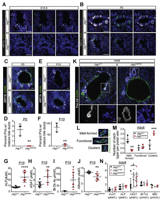
Figure 2.
Postnatal Jag1Ndr/Ndr mice display ductopenia, which is rescued in adults. (A, B) HNF1β,
SOX9, and KRT19 staining show a marked absence of biliary cells at E18.5 (A) and weak
staining at P0 (B) near the hilum in Jag1Ndr/Ndr liver. (C, D) KRT19+ cell clusters appear
around ASMA+ periportal regions near the hilum of wild type Jag1+/+ mice at P0, but are
absent in Jag1Ndr/Ndr mice. (E, F) By P10, clusters of biliary cells have lumenized to form
ducts in Jag1+/+ mice, but not in Jag1Ndr/Ndr mice. Jag1Ndr/Ndr mice display increased (G)
alkaline phosphatase (ALP), (H) aspartate aminotransferase (ASAT), (I) direct bilirubin (Bil
Gastroenterology. Author manuscript; available in PMC 2020 February 07.Andersson et al. Page 21
Dir), and (J) decreased albumin. (K) At adult stages, lumenized bile ducts are present in both
Jag1+/+ and Jag1Ndr/Ndr mice, though classification (L) of structures shows (M) significantly
more clusters in Jag1Ndr/Ndr mice and fewer well-formed bile ducts. (N) Nevertheless,
markers of liver function demonstrate a rescue of bile duct function in adult Jag1Ndr/Ndr
mice in most serum chemistry markers. A small difference in aspartate aminotransferase
levels persists. Error bars indicate s.d.; *PAndersson et al. Page 22
Europe PMC Funders Author Manuscripts
Europe PMC Funders Author Manuscripts
Figure 3.
Jag1Ndr/Ndr biliary cells express the expected markers but display structural instability. Sox9
levels are unchanged at P10 at the mRNA level (A), and at adult stages at protein levels (B,
C). qPCR for (D) alpha-fetoprotein, (E) albumin, and (F) Hnf4α show no significant
differences in Jag1Ndr/Ndr mice at P10. Similarly, RNA sequencing of ALGS livers shows no
difference in (G) SOX9, (H) HNF1β, or (I) KRT19 levels. Organoids derived from adult
Jag1Ndr/Ndr livers expressed normal levels of (J) Notch2, (K) Hes1, (L) Hnf4α, (M) Sox9,
and (N) Hnf1β as assessed by qPCR, but (O) grew slowly and (P) sometimes spontaneously
Gastroenterology. Author manuscript; available in PMC 2020 February 07.Andersson et al. Page 23
collapsed. Collapse was not related to organoid size because both smaller and larger
organoids collapsed. No differences were significant. Scale bar: (B) 10 μm.
Europe PMC Funders Author Manuscripts
Europe PMC Funders Author Manuscripts
Gastroenterology. Author manuscript; available in PMC 2020 February 07.Andersson et al. Page 24
Europe PMC Funders Author Manuscripts
Europe PMC Funders Author Manuscripts
Figure 4.
RNA sequencing of ALGS liver reveals a specific decrease in apical markers of biliary cells.
(A) Principle component analysis (PCA) of RNA sequencing of liver biopsies from patients
with ALGS or control patients. A comparison with non-cholestatic control samples (Ctrl 1
and 2) and with cholestatic control samples (Ctrl 3 and 4) shows that ALGS samples cluster
with cholestatic liver samples. (B) Heatmap shows 191 significantly up-regulated and 139
down-regulated genes in ALGS samples. (C) Dysregulated genes were compared with
protein lists generated using the HPA (www.proteinatlas.org)18 for genes with high/medium
Gastroenterology. Author manuscript; available in PMC 2020 February 07.You can also read