Retinal degeneration in mice and humans with neuronal ceroid lipofuscinosis type 8
←
→
Page content transcription
If your browser does not render page correctly, please read the page content below
Original Article
                                                                                                                                        Page 1 of 15

Retinal degeneration in mice and humans with neuronal ceroid
lipofuscinosis type 8
Elyse M. Salpeter1, Brian C. Leonard1, Antonio J. Lopez2, Christopher J. Murphy1,2, Sara Thomasy1,2,
Denise M. Imai3, Kristin Grimsrud4,5, K. C. Kent Lloyd4,6, Jiong Yan7, Rossana Sanchez Russo8,
Suma P. Shankar9, Ala Moshiri2^
1
William R. Pritchard Veterinary Medical Teaching Hospital, School of Veterinary Medicine, University of California, Davis, Davis, California, USA;
2
Department of Ophthalmology & Vision Science, School of Medicine, University of California, Davis, Sacramento, California, USA; 3Comparative
Pathology Laboratory, School of Veterinary Medicine, UC Davis, Davis, California, USA; 4Mouse Biology Program, University of California, Davis,
Davis, California, USA; 5Department of Pathology and Laboratory Medicine, School of Medicine, University of California, Davis, Sacramento,
California, USA; 6Department of Surgery, School of Medicine, University of California, Davis, Sacramento, California, USA; 7Department
of Ophthalmology, Emory University, Atlanta, Georgia, USA; 8Department of Human Genetics, Emory University, Atlanta, Georgia, USA;
9
    Department of Pediatrics & Department of Ophthalmology & Vision Science, School of Medicine, University of California, Davis, Sacramento,
California, USA
Contributions: (I) Conception and design: EM Salpeter, A Moshiri, SP Shankar; (II) Administrative support: BC Leonard, CJ Murphy, S Thomasy,
DM Imai; (III) Provision of study materials or patients: K Grimsrud, KCK Lloyd, J Yan, R Sanchez Russo, SP Shankar; (IV) Collection and assembly
of data: EM Salpeter, AJ Lopez; (V) Data analysis and interpretation: EM Salpeter, A Moshiri; (VI) Manuscript writing: All authors/Both authors; (VII)
Final approval of manuscript: All authors/Both authors.
Correspondence to: Ala Moshiri, MD, PhD. Department of Ophthalmology and Vision Science, School of Medicine, University of California, Davis
Eye Center, 4860 Y. Street, Suite 2400, Sacramento, CA 95817, USA. Email: amoshiri@ucdavis.edu.

                  Background: Ceroid lipofuscinosis type 8 belongs to a heterogenous group of vision and life-threatening
                  neurodegenerative diseases, neuronal ceroid lipofuscinosis (NCL). Effective therapy is limited to a single
                  drug for treatment of ceroid lipofuscinosis type 2, necessitating animal disease models to facilitate further
                  therapeutic development. Murine models are advantageous for therapeutic development due to easy
                  genetic manipulation and rapid breeding, however appropriate genetic models need to be identified and
                  characterized before being used for therapy testing. To date, murine models of ocular disease associated with
                  ceroid lipofuscinosis type 8 have only been characterized in motor neuron degeneration mice.
                  Methods: Cln8−/− mice were produced by CRISPR/Cas9 genome editing through the International Mouse
                  Phenotyping Consortium. Ophthalmic examination, optical coherence tomography, electroretinography, and
                  ocular histology was performed on Cln8−/− mice and controls at 16 weeks of age. Quantification of all retinal
                  layers, retinal pigmented epithelium, and the choriocapillaris was performed using images acquired with
                  ocular coherence tomography and planimetry of histologic sections. Necropsy was performed to investigate
                  concurrent systemic abnormalities. Clinical correlation with human patients with CLN8-associated
                  retinopathy is provided.
                  Results: Retinal degeneration characterized by retinal pigment epithelium mottling, scattered drusen, and
                  retinal vascular attenuation was noted in all Cln8−/− mice. Loss of inner and outer photoreceptor segment
                  demarcation was noted on optical coherence tomography, with significant thinning of the whole retina
                  (P=1e-9), outer nuclear layer (P=1e-9), and combined photoreceptor segments (P=1e-9). A global reduction
                  in scotopic and photopic electroretinographic waveforms was noted in all Cln8−/− mice. Slight thickening of
                  the inner plexiform layer (P=0.02) and inner nuclear layer (P=0.004), with significant thinning of the whole
                  retina (P=0.03), outer nuclear layer (P=0.01), and outer photoreceptor segments (P=0.001) was appreciated
                  on histologic sections. Scattered lipid vacuoles were noted in splenic red pulp of all Cln8−/− mice, though no

^ ORCID: 0000-0002-4277-8094.

© Annals of Translational Medicine. All rights reserved.                             Ann Transl Med 2021 | http://dx.doi.org/10.21037/atm-20-4739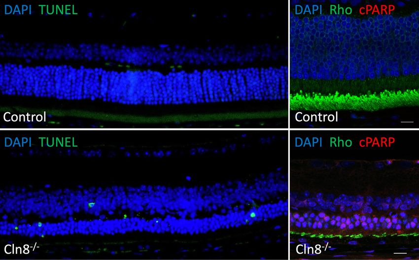
Page 2 of 15                                                                     Salpeter et al. Retinal degeneration in Cln8−/− mouse model

                gross systemic abnormalities were detected on necropsy. Retinal findings are consistent with those seen in
                patients with ceroid lipofuscinosis type 8.
                Conclusions: This study provides detailed clinical characterization of retinopathy in adult Cln8−/− mice.
                Findings suggest that Cln8−/− mice may provide a useful murine model for development of novel therapeutics
                needed for treating ocular disease in patients with ceroid lipofuscinosis type 8.

                Keywords: Neuronal ceroid lipofuscinosis; retinal degeneration; murine model

                Submitted Jun 16, 2020. Accepted for publication Mar 19, 2021.
                doi: 10.21037/atm-20-4739
                View this article at: http://dx.doi.org/10.21037/atm-20-4739

Introduction                                                               subtypes, with effective pharmacologic therapy limited
                                                                           to a single clinically approved drug, Cerliponase alfa
The International Mouse Phenotyping Consortium (IMPC)
                                                                           (Brineura™, BioMarin Pharmaceutical Inc., Novato, CA,
is a cooperative association that employs high throughput
                                                                           USA), for treatment of CLN2 (12,13).
mouse mutagenesis and comprehensive phenotyping with
                                                                              Murine models of human disease are an important
the goal of creating the first functional encyclopedia of the
                                                                           component in therapeutic development due to the relative
mammalian genome (1,2). Additionally, CRISPR/Cas9
                                                                           ease of genetic manipulation and short generational interval.
genome editing through the IMPC facilitates production
                                                                           These models contribute to therapeutic development
and identification of murine models for human disease,
                                                                           through facilitating investigation of disease pathophysiology,
such as the recent creation of a bio-engineered neuronal
ceroid lipofuscinosis type 8 (CLN8) murine model with                      aiding in identification of therapeutic targets, and facilitating
ocular manifestation. Neuronal ceroid lipofuscinosis (NCL)                 initial assessment of efficacy of therapeutic approaches. To
is a heterogenous group of progressive, life-threatening,                  date, murine models have been identified for CLN1 (14-17),
neurodegenerative disorders characterized by lysosomal                     CLN2 (18), CLN3 (17,19-28), CLN5 (29), CLN6 (30),
accumulation in multiple tissues of the central nervous                    CLN8 (31-35), and CLN10 (36,37). Reports of retinopathy
system (3). Progressive epilepsy with mental retardation                   characterization in murine models of NCL predominantly
was recognized as a NCL subtype (CLN8) (4,5). CLN8                         describe findings in CLN3 (19,28), CNL5 (38), CNL6 (39),
is an endoplasmic reticulum (ER) associated membrane                       and CLN8 (32-35), though the available descriptions of ocular
protein required for transfer of lysosomal enzymes from the                disease associated with CLN8 are limited to homozygous
ER to the Golgi complex; deficiency leads to depletion of                  motor neuron degeneration (mnd) murine models. Neural
soluble enzymes in the lysosome, thus impairing lysosome                   degeneration in the mnd model results from spontaneous
biogenesis and lysosomal enzyme transport (6). Metabolic                   defect in the murine orthologue of CLN8 and exhibits
radiolabeling has highlighted delayed lysosomal enzyme                     pathology similar to human NCLs (32, 40).
maturation with CLN8 deficiency, which was rescued by
CLN8 re-expression in CLN8-deficient cells obtained by                     Spontaneous mutations in CLN8 have also been
CRISPR/Cas9 genome editing (6).                                            identified in canines (41-43)
    Ocular symptoms such as retinal degeneration can be the
first sign of NCL disease and facilitate an early diagnosis (7).           To the authors’ knowledge, phenotypic characterization of
Ocular manifestations of NCL are most commonly noted                       retinopathy in CLN8 murine models is limited to a report
with CLN3 (8,9), though retinopathy and visual deficits                    of indirect ophthalmoscopic and electroretinographic
associated with CLN8 have also been reported (10).                         findings, and limited reports of histopathological changes in
    All NCL subtypes are unique inherited metabolic                        mnd mice (33-35,40). The aim of this study was therefore to
disorders, limiting specific pharmacologic treatments to                   define the clinical, electrophysiologic, and anatomic retinal
single genetic forms, or groups of disorders that share an                 changes in a novel mouse model with targeted deletion
underlying metabolic pathway defect (11). Development of                   of Cln8. Complete characterization will better inform
improved therapeutics is needed for the majority of NCL                    the use of Cln8−/− mice as animal models for development

© Annals of Translational Medicine. All rights reserved.                          Ann Transl Med 2021 | http://dx.doi.org/10.21037/atm-20-4739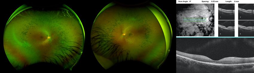
Annals of Translational Medicine, 2021                                                                            Page 3 of 15

of improved therapeutics and management of CLN8               causes a frameshifted transcript of all known isoforms,
retinopathy in humans.                                        effectively resulting in a null allele by nonsense protein/
   We present the following article in accordance with the    early termination and potential for nonsense mediated
ARRIVE reporting checklist (available at http://dx.doi.       decay (NMD) of the transcript. Embryos were surgically
org/10.21037/atm-20-4739).                                    transferred into the oviducts of pseudopregnant CD1
                                                              female mice. After gestation and littering, pre-weaned
                                                              mouse pups were sampled and genetically tested by end-
Methods
                                                              point PCR and sequencing to definitively identify founder
Study design                                                  mice harboring the desired internal frameshift deletion.
                                                              Subsequent germline transmission mice (N1) were further
The study included one experimental group with Cln8−/−
                                                              sequenced to confirm the desired deletion which consists
mice, and one control group with C57BL/6N wildtype.
                                                              of a 53 bp deletion (GGTCTTCTGGAACCTGGCGG
All mice underwent an ophthalmic examination, ocular
                                                              CGACGCGTGCTGTCTTCGGCGTCCAGAGCAC
imaging, electroretinography, and necropsy at 16 weeks of     AA) of the 183rd to 235th coding nucleotides which creates
age. Investigators were blinded to genetic profiles when      an early stop signal within exon 1, preceded by 28 amino
performing ophthalmic examinations, analysis of ocular        acid nonsensical sequence. After weaning, sexually mature
images, analysis of electroretinograms, and necropsies.       mice with confirmed germline transmission of the desired
This report also reviews two cases of CLN8 in humans, to      deletion allele were backcrossed two generations before
highlight similarities with the murine model in this study.   intercrossing heterozygous mice to generate homozygous
                                                              knockouts. Sex (male and female) and Mendelian (zygosity)
Animals                                                       ratios were normal as expected. In total, five heterozygous
                                                              breeding trios produced a total of 133 pups with normal
Experiments were performed under a project license (NO.       male and female distribution. The zygosity distribution was
IACUC # 21824) granted by the Institutional Animal Care       reported to be 45.9% heterozygous, 29.3% homozygous
and Use Committee at UC Davis, in compliance with the         and 24.8% wildtype. Overall, breeding and animal health
National Institutes of Health (NIH) Guide for the Care        were within normal expectations for this mouse line and no
and Use of Laboratory Animals and the guidelines of the       observable abnormalities were observed during production
Association for Research in Vision and Ophthalmology          or while maintaining these mice. For this study, 3 Cln8−/−
Statement for the Use of Animals in Ophthalmic and Vision     mice were available for analysis, and 2 wildtype, and 1
Research.                                                     Cln8+/− mouse were chosen as age-matched controls.
   All mice were housed and cared for in accordance with
the recommendations provided in the Guide for the Care
and Use of Laboratory Animals Eighth Edition. Mice were       Ophthalmic examination
cohoused by gender groups of 4–5 animals per cage in          Complete ophthalmic examinations were performed on
individually ventilated cages (optimice IVC, Animal Care      both eyes of 3 Cln8−/−, 1 Cln8+/−, and C57BL/6N wildtype
Systems, Centenniel, CO) on a 12:12 hour (06:00–18:00)        mice at 16 weeks of age. A standardized operating protocol
light cycle. Temperature ranges in the room are maintained    for evaluation of ocular and adnexal structures was followed
between 68–79 ℉ and mice are maintained on standard           at the UCDavis study site (https://www.mousephenotype.
laboratory rodent chow (Rodent chow, Envigo 2918).            org/impress/protocol/267/7) and were carried out by
   To study the in vivo function of Cln8, gene-specific       experienced technical support staff trained to identify and
knockout mice (Cln8−/−) were generated using CRISPR/          differentiate background lesions common in the C57BL/6N
Cas9 genome editing. Briefly, one-cell state C57BL/6N         strain (44). Examiners were overseen by lead site scientists
zygotes were harvested from super ovulated female             who subsequently reviewed all phenotypes. Pupillary
mice and electroporated using standard protocols with a       light reflexes were evaluated, the eyelids, third eyelid,
ribonucleoprotein (RNP) consisting of Cas9 protein and        conjunctiva, sclera, cornea, iris, and anterior chamber were
two synthetic guide RNAs (gRNA) internal to the coding        examined using broad beam illumination at the highest
region of exon 1 (CGGCCAAAGAGAAGGTCTTC                        intensity setting (Kowa SL-15, Kowa, Tokyo, Japan, or
& GGCGTCCAGAGCACAACTGC). The deletion                         equivalent) with magnification set at 16X. The irides of all

© Annals of Translational Medicine. All rights reserved.           Ann Transl Med 2021 | http://dx.doi.org/10.21037/atm-20-4739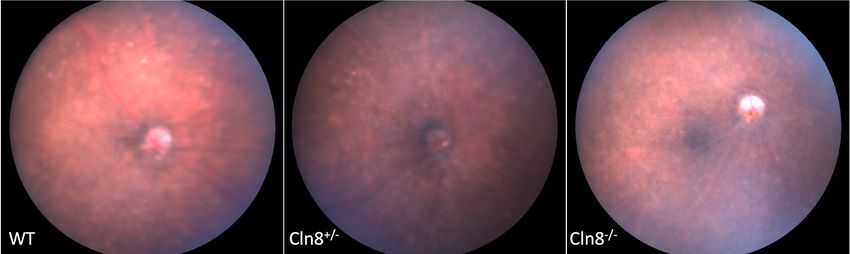
Page 4 of 15                                                           Salpeter et al. Retinal degeneration in Cln8−/− mouse model

mice were then pharmacologically dilated with a solution of        between the two eyes of each animal.
1:7 10% phenylephrine HCl (Akorn Inc., Lake Forest, IL,
USA, or equivalent): 1% tropicamide (Bausch & Lomb Inc.,
                                                                   Histology
Tampa, FL, USA, or equivalent). A 0.1 mm slit beam at the
highest intensity setting was used to evaluate the anterior        Eyes were removed and immediately fixed in 2.5%
segment (cornea, anterior chamber, and lens), followed by          glutaraldehyde and 2% paraformaldehyde in 0.1 M
evaluation of the retrolental region of the vitreous.              sodium phosphate buffer. Parasagittal sections of eyes
   Fundus examinations were performed via indirect                 were processed routinely for histopathology, embedded in
ophthalmoscopy using a 60 Diopter double aspheric                  paraffin, sectioned at 5 µm, and stained with hematoxylin-
handheld lens (Volk Optical Inc, Mentor, OH, USA or                eosin (H&E). Retinal layer measurements were taken of
equivalent) and a portable indirect headset (Keeler AllPupil       total retina, nerve fiber layer, retinal ganglion cell layer,
II LED Vantage Plus Wireless Headset, Keeler Instruments           inner plexiform layer, inner nuclear layer, outer plexiform
Inc., Broomall, PA, USA).                                          layer, outer nuclear layer, inner photoreceptor segment,
                                                                   outer photoreceptor segment, retinal pigmented epithelium,
                                                                   and choriocapillaris on imaged H&E sections as described
Ocular imaging
                                                                   above for OCT images.
A cocktail of ketamine/medetomidine (100/0.3 mg/kg)
was administered intraperitoneally to induce anesthesia in
                                                                   Immunohistochemistry
all mice undergoing ocular imaging. Fundus photographs
were acquired with the Micron III (Phoenix Research                Sections were deparaffinized using three changes of xylenes
Laboratories, Pleasanton, CA, USA).                                for 5 minutes each. The tissue sections were then stepwise
   OCT imaging was performed with an Envisu R2200 SD-              hydrated by submersion in 100% ethanol (twice), 95%
OCT (spectral-domain OCT; Bioptigen-Leica, Wetzlar,                ethanol, 75% ethanol, and distilled water—5 minutes per
Germany), after dilatation with both tropicamide 1% and            step. Heat-induced epitope retrieval was then performed
phenylephrine 2.5%. Thickness of the total retina, nerve           with 1mM EDTA, pH 8.0. The tissue sections were then
fiber layer, retinal ganglion cell layer, inner plexiform layer,   blocked for 30min in blocking solution (4% BSA and 0.5%
inner nuclear layer, outer plexiform layer, outer nuclear          Triton X-100 in PBS) at ambient temperature. Primary
layer, combined inner and outer photoreceptor segments,            antibodies (rabbit anti-cleaved PARP-94885S Cell Signaling
retinal pigmented epithelium, and choriocapillaris were            @ 1:100, mouse anti-rhodopsin-MABN15 Sigma-Aldrich
manually measured at a distance of ~0.2 mm from each               @ 1:1,000) were then incubated overnight at 4C. The slides
side of the optic nerve using calipers in ImageJ software          were washed three times in with PBS for five minutes each.
according to established guidelines (45). Measurements             Alexa Fluor conjugated secondary antibodies (A32723 and
from both sides of the optic nerve head and between left           A32740- ThermoFisher Scientific) were incubated for an
and right eyes were averaged for each animal.                      hour followed by a 5-minute incubation in DAPI (1 μg/mL).
                                                                   The tissue sections were washed with PBS three times for
                                                                   five minutes each. Finally, the slides were cover slipped with
Electroretinography
                                                                   FluorSave Reagent (Millipore, 345789).
Following a 12-hour dark adaptation period, the mice received
a cocktail of ketamine/medetomidine (100/0.3 mg/kg)
                                                                   TUNEL
intraperitoneally to induce anesthesia. A single drop of
both tropicamide 1% and phenylephrine 2.5% was used                Detection of apoptotic cells was conducted using ApopTag®
for dilation, and the ocular surface was lubricated with           Fluorescein In Situ Apoptosis Detection Kit (S7110, Sigma-
methylcellulose-containing artificial tears. Standard full         Aldrich). Sections were deparaffinized and hydrated like
field scotopic and photopic electroretinography (ERG) was          the immunohistochemistry method and then treated with
performed on both eyes with LKC Big Shot with a Ganzfeld           proteinase K (20 μg/mL) at ambient temperature. The tissue
dome (LKC Technologies Inc., Gaithersburg, MD, USA).               sections were then washed twice with PBS for 2 minutes
For quantitative comparisons, measurements were averaged           each. The tissue sections were equilibrated and the TdT

© Annals of Translational Medicine. All rights reserved.                 Ann Transl Med 2021 | http://dx.doi.org/10.21037/atm-20-4739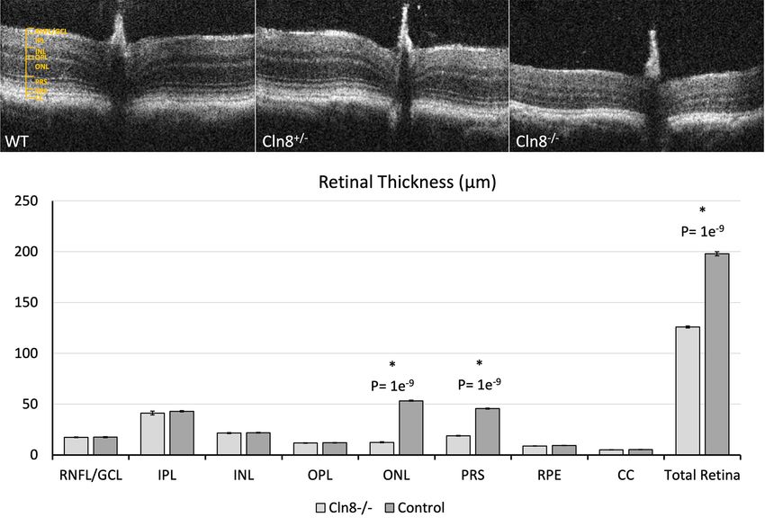
Annals of Translational Medicine, 2021                                                                                         Page 5 of 15

        A                                            B                                      C

Figure 1 Fundus images of wildtype, Cln8+/−, and Cln8−/− mice. (A) Fundus of wildtype C57BL/6N rd8 mouse and (B) Cln8+/− mouse with
mild retinal dysplasia and normal retinal vasculature. (C) Fundus of Cln8−/− mouse with optic nerve pallor, retinal vascular attenuation, and
diffuse retinal pigment epithelium mottling.

enzyme was applied for an hour at 37 ℃. The reaction was                 instrument, Germany).
then stopped with the stop buffer and the sections were
washed three times in PBS for 1 minute each. The anti-
                                                                         Statistical analysis
digoxigenin conjugate was then applied for 30 minutes at
ambient temperature followed by a 5-minute incubation                    For comparisons of retinal layer thicknesses between Cln8−/−
with DAPI (1 μg/mL). The sections were washed 4 time in                  and control (Cln8+/− and/or wildtype) mice on OCT images
PBS for 2 minutes each. Finally, the slides were cover slipped           and H&E sections, a 2-tailed Student’s t-test was performed.
with FluorSave Reagent (Millipore, 345789). TdT enzyme,                  A P value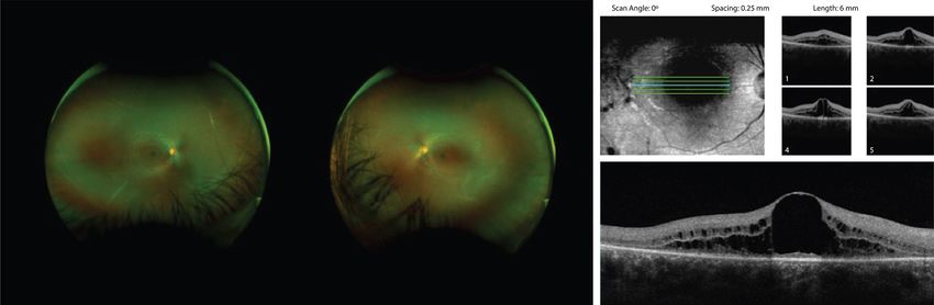
Page 6 of 15                                                                      Salpeter et al. Retinal degeneration in Cln8−/− mouse model

 Table 1 Using optical coherence tomography, thickness measurements of the entire retina, individual retinal layers and the choriocapillaris were
 acquired from Cln8−/− and control mice
 Measurement                                                       Group                  Mean (µm)             P value         Standard error
                                                                            −/−                                      -9
 Whole retina                                                 Control, Cln8                198, 126              1.0e *              10.4
                                                                            −/−
 Retinal nerve fiber layer/ganglion cell layer                Control, Cln8                 18, 17               0.31                 0.9
                                                                            −/−
 Inner plexiform layer                                        Control, Cln8                 43, 41               0.21                 4.5

 Inner nuclear layer                                          Control, Cln8−/−              22, 22               0.23                 1.1
                                                                            −/−
 Outer plexiform layer                                        Control, Cln8                 12, 12               0.17                 0.6
                                                                            −/−                                      -9
 Outer nuclear layer                                          Control, Cln8                 53, 12               1.0e *               1.2
                                                                            −/−                                      -9
 Combined inner and outer photoreceptor segments              Control, Cln8                 46, 19               1.0e *               0.8
                                                                            −/−
 Retinal pigmented epithelium                                 Control, Cln8                  9, 9                0.06                 0.3
                                                                            −/−
 Choriocapillaris                                             Control, Cln8                  5, 5                0.23                 0.2
 Measurements were taken at approximately 0.2 mm on either side of the optic nerve and averaged for each eye. N=3 for each group. *,
 significant differences are demarcated.

collected from Cln8 −/− and control mice using OCT                          in Figure 3D,E.
are represented in Table 1. Distinct inner and outer
photoreceptor segments could not be distinguished in
                                                                            Histopathology
images from any eye of Cln8−/− mice, whereas a clear IS/
OS junction (aka ellipsoid zone) distinction was notable                    Retinal measurements and their associated P values
between segments in images from control eyes. Whole                         collected from histologic sections of Cln8−/− and control
retinal thickness in Cln8−/− was significantly thinner than                 mice are presented in Table 2. Whole retinal thickness in
controls (P=1e-9, SE =10.4), with significant differences                   Cln8 −/− was significantly thinner than controls (P=0.03,
noted in outer nuclear layer (P=1e -9 , SE =0.1.2), and                     SE =14.0), with statistically significant thinning of the
combined photoreceptor inner and outer segments (P=1e-                      outer nuclear layer (P=0.01, SE =2.3), and photoreceptor
9
  , SE =0.8). There was no significant difference noted in                  outer segments (P=0.001, SE =2.8) in Cln8−/− mice. Slight
retinal nerve fiber layer (P=0.3, SE =0.9), inner plexiform                 increases in thickness were noted in the inner plexiform
layer (P=0.2, SE =4.5), inner nuclear layer (P=0.2, SE                      layer (P=0.02, SE =3.0) and inner nuclear layer (P=0.004,
=1.1), outer plexiform layer (P=0.2, SE =0.6), or retinal                   SE =1.5) of Cln8−/− mice. There was no significant difference
pigmented epithelium (P=0.06, SE =0.3) thickness between                    noted in retinal nerve fiber layer (P=0.5 SE =2.6), outer
groups. Comparative OCT b-scans from Cln8 −/−, Cln8+/−,                     plexiform layer (P=0.1 SE =0.8), photoreceptor inner
and wildtype mice with a bar graph illustrating mean                        segments (P=0.5, SE =1.4), or retinal pigmented epithelium
retinal layer measurements are represented in Figure 2.                     (P=0.17, SE =0.3) thickness measurements between groups.
                                                                            Comparative values obtained from H&E sections from
                                                                            Cln8−/− and control mice are represented in Figure 4.
Electroretinography
                                                                               To determine the retinal neurons most susceptible to cell
A globally reduced ERG waveform, with severely reduced                      death in the absence of Cln8, TUNEL labeling and cPARP
a- and b-waves, was noted in all Cln8−/− mice compared to                   immunohistochemistry were used to identify apoptotic cells.
those of wildtype and Cln8 +/− mice. Representative ERG                     TUNEL labeling was found in the outer nuclear layers in
waveforms from Cln8 −/−, Cln8 +/−, and wildtype mice are                    Cln8−/− mice but not in controls. Many rhodopsin positive
shown in Figure 3A,B,C.                                                     rod photoreceptors were positive for cPARP in Cln8 −/−
    Quantitative comparisons of the stimulus-response curve                 mice, but not in controls (Figure 5). These findings suggest
illustrating mean a- and b-wave amplitudes are represented                  rod photoreceptors are the most vulnerable in the absence

© Annals of Translational Medicine. All rights reserved.                           Ann Transl Med 2021 | http://dx.doi.org/10.21037/atm-20-4739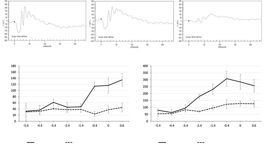
Annals of Translational Medicine, 2021                                                                                          Page 7 of 15

            A                                         B                                    C

            D

Figure 2 Comparison of OCT between wildtype, Cln8+/−, and Cln8−/− mice. (A) Retinal architecture in a wildtype C57BL/6N rd8 mouse, and
(B) in a Cln8+/− mouse. (C) OCT of a Cln8−/− mouse retina demonstrating marked outer nuclear layer thinning, a loss of definition in the inner
and outer segments of the photoreceptors, and diffuse retinal thinning. (D) Bar graph showing mean retinal layer thickness measurements
in Cln8−/− and control mice on OCT images. *, statistical significance, PT
scattered lipid vacuoles were noted in splenic red pulp of all            (p.A67V) variant of unknown significance on this gene
knockout mice. Observations in control mice was limited to                confirmed in trans, and electron microscopy of lymphocytes
identifying an enlarged heart in 1/3 control mice.                        showing intracytoplasmic inclusions. She was noted to have
                                                                          cystoid macular edema (CME) bilaterally, with visual acuity
                                                                          of 20/40 OU at time of initial report and more recently OD
Clinical correlation
                                                                          20/80 +2 and OS 20/100. On follow up ophthalmologic
The abnormalities seen on retinal imaging of Cln8−/− mice                 evaluation she had attenuated vasculature with cystoid
were compared with humans with confirmed CLN8-                            macular edema and diffuse retinal pigment epithelium

© Annals of Translational Medicine. All rights reserved.                        Ann Transl Med 2021 | http://dx.doi.org/10.21037/atm-20-4739
Page 8 of 15 Salpeter et al. Retinal degeneration in Cln8−/− mouse model
A B C
WT Cln8+/− Cln8+/−
D E
ERG a-wave ERG b-wave
Ampltiude (μM)
Ampltiude (μV)
Stimulus Intensity (cd·s/m2) Stimulus Intensity (cd·s/m2)
WT a-wave Cln8+/− a-wave WT b-wave Cln8+/− b-wave
Figure 3 Comparison of electroretinography tracings in wildtype, Cln8+/−, and Cln8−/−mice. With scotopic bright flash (0 dB), normal
tracings are present in a (A) wildtype C57BL/6N rd8 mouse and (B) Cln8+/− mouse, with reduced a- and b-wave amplitudes in a Cln8−/− mouse
(C). Line graphs denote mean a-wave (D), and b-wave (E) amplitudes at varying stimulus intensities in Cln8−/− (n=3) and wildtype (n=4) mice;
note the severely reduced a- and b- wave amplitudes in Cln8−/− (dashed lines) compared to wildtype (solid lines) mice. Error bars represent
SEM.
Table 2 Histology measurements collected from Cln8−/− (n=3) and control (n=3) mice
Measurement Group Mean (µm) P value Standard error
Whole retina Control, Cln8−/− 156, 124 0.03* 14.0
−/−
Retinal nerve fiber layer/ganglion cell layer Control, Cln8 12, 11 0.53 2.6
−/−
Inner plexiform layer Control, Cln8 22, 36 0.02* 3.0
−/−
Inner nuclear layer Control, Cln8 27, 32 0.004* 1.5
−/−
Outer plexiform layer Control, Cln8 10, 8 0.14 0.8
−/−
Outer nuclear layer Control, Cln8 46, 23 0.01* 2.3
−/−
Inner photoreceptor segment Control, Cln8 14, 12 0.46 1.4
Outer photoreceptor segment Control, Cln8−/− 15, 4 0.001* 2.8
Retinal pigmented epithelium Control, Cln8−/− 6, 6 0.17 0.3
−/−
Choriocapillaris Control, Cln8 3, 5 0.3 0.6
Histologic sections were chosen at the level of the optic nerve and measurements were taken approximately adjacent to the optic nerve
head in the posterior retina. *, statistical significance, PAnnals of Translational Medicine, 2021 Page 9 of 15
A B
Control Cln8+/−
C Retinal thickness (μm)
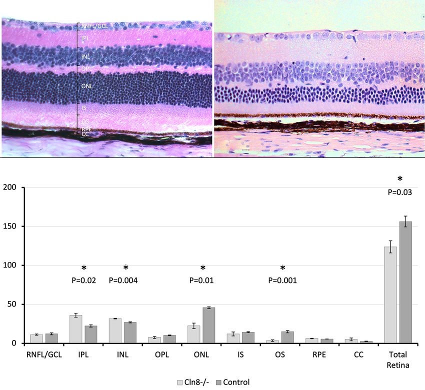
Figure 4 Comparison of histologic retinal sections between Cln8−/− and control mice. (A) Retinal architecture in control C57BL/6N rd8
mouse. (B) H&E of a Cln8−/− mouse retina demonstrating mild thickening of the inner plexiform and inner nuclear layer, with marked
outer nuclear layer thinning and whole retinal thinning. (C) Bar graph showing mean retinal layer thickness measurements in histologic
sections of Cln8−/− (n=3) and control (n=4) mice. *, statistical significance, PPage 10 of 15 Salpeter et al. Retinal degeneration in Cln8−/− mouse model
Figure 5 Photoreceptors undergo apoptosis in the absence of Cln8. Control retinal sections (top row) show no TUNEL-positive nuclei and
no cPARP-positive cells in the rhodopsin labeled outer nuclear layer. Cln8−/− mice (bottom row) have numerous TUNEL-positive cells in
the outer nuclear layer. Many rhodopsin-positive rod photoreceptors are also cPARP-positive in Cln8 knockouts. Furthermore, rhodopsin
positive outer segments are much shorter in Cln8−/− mice than controls. Scale bar in right column represents 10 microns.
Scan Angle: 0° Spacing: 0.25 mm Length: 6 mm
Figure 6 Bilateral fundus images showing attenuated vasculature and diffuse retinal pigment epithelium mottling. OCT of the right eye
showing cystoid macular edema.
vasculature, peripheral bone spicules and diffuse retinal he has had one witnessed generalized tonic clonic seizure
pigment epithelial mottling. On recent follow up, fundus lasting less than one minute in the setting of weaning
examination and ultrawide field color fundus imaging of his anti-epileptic medication which has since been
showed mid-peripheral bone spicules and mild attenuation adjusted.
of vasculature (Figure 7). OCT showed no CME and He has not had any recent brain imaging and has not had
fundus autofluorescence showed scattered macular areas of a formal psychoeducational evaluation, but he has a master’s
decreased and increased intensity. Since the initial report degree in engineering and continues to work full-time job
© Annals of Translational Medicine. All rights reserved. Ann Transl Med 2021 | http://dx.doi.org/10.21037/atm-20-4739Annals of Translational Medicine, 2021 Page 11 of 15
Figure 7 Fundus images showing pallor of optic nerves, attenuated vasculature, mid-peripheral bone spicules and diffuse retinal pigment
epithelial mottling. The nasal and temporal sides of the macular OCT show outer retinal thinning and loss of the IS/OS junction similar to
that seen in mice
living independently. electroretinography abnormalities in mnd mice closely
resemble those found in this study, with barely detectable
waveforms only with the brightest stimulus by 5 months of
Discussion
age, and undetectable waveforms with single-flash methods
This study details the phenotypic attributes of retinal at 6 months of age (33).
degeneration in Cln8−/− mice using several testing modalities Previous histologic evaluation of retinal changes in
at 16 weeks postnatal age, providing a promising murine mnd mice noted reduced numbers of photoreceptors and
model for CLN8 ophthalmic disease in humans. significant reduction in outer nuclear layer thickness, with
The single previous report with indirect ophthalmoscopic one study noting the onset of these changes at 5 weeks of
findings in mnd mice reports similar retinal pathology to age (33), while another study documented photoreceptor
that noted in this study: severe retinal vessel attenuation, layer atrophy in mice older than 3 months of age (40).
focal and diffuse loss of pigment epithelium, and patches of These changes are consistent with the reduced outer
pigment deposits (33). These lesions were not appreciable nuclear layer and photoreceptor segment thinning
until approximately 5 months of age in the aforementioned appreciated on OCT and histology in this study. Another
study, while similar abnormalities were detected in this study found that a prominent feature of photoreceptor
study by 4 months of age. This variation in timeline may degeneration in mnd mice was the persistent and progressive
be due to differences between the spontaneous mutation in shortening of photoreceptor outer segments, while
mnd mice and the targeted Cln8 deletion in this study, or shortening of inner segments was less pronounced and
due to strain-specific background differences. Chang et al. occurred at a much later stage of degeneration (35). The
used backcrossing of mice with a spontaneously occurring histologic findings in the current study support this finding,
Cln8 mutation (33), while the current study employed in which significant thinning was noted in outer but not
engineered Cln8 gene knockout. Moreover, genetic inner photoreceptor segments of Cln8−/− mice.
background has been shown to affect the age of onset, While this study and previous reports (33,40) noted
speed of progression, precise neurological symptoms and significant thinning of the outer retina in CLN8 murine
type of cells affected in mnd mice (46). The aforementioned models, Groh et al. (17) described predominant thinning
study used outcrossed C57BL/6J × AKR/J mice. A slightly of the inner retina in CLN1 and CLN3 murine models.
more rapid retinal degeneration may be secondary to Significant thinning of the nerve fiber layer, ganglion cell
the rd8 mutation present in the C57BL/6N mice used layer, inner plexiform layer, and inner nuclear layer was
in the current study, though it is difficult to definitively appreciated in 7-month-old CLN1 mice and 16 month old
determine the secondary effect of the background strain, CLN3 mice (17). Although this variation in retinal layer
as the C57BL/6J strain wildtype mice also possess the rd8 predilection may reflect differences in pathophysiology
mutation without displaying the phenotype (44). Published amongst CNL diseases, it is interesting to note that PAS
© Annals of Translational Medicine. All rights reserved. Ann Transl Med 2021 | http://dx.doi.org/10.21037/atm-20-4739Page 12 of 15 Salpeter et al. Retinal degeneration in Cln8−/− mouse model
staining inclusions have been noted in retinal ganglion these modalities is small and could be related to tissue
cells of mnd mice (33,34). Ultrastructural examination fixation and processing. Significant thinning of the outer
of the retina was not available in the current study and nuclear layer, outer photoreceptor segments, and whole
was thus unable to investigate intracellular changes such retina in mice with Cln8−/− were detected with confidence.
as inclusions. Further studies are warranted to evaluate Additionally, a clear clinical and electrophysiologic
intracellular changes in CLN8 retinal layers. phenotype could be described with the small number of
Variation in phenotypic presentation of retinopathy has study animals.
been noted in humans with mutations in CLN8, with main Despite minor limitations of this study, the overall
ocular lesions ranging from cystoid macular edema, diffuse similarities of ocular pathology in Cln8−/− mice with the
macular schisis, optic disk atrophy, peripheral bone spicules, human disease suggests strong potential as a murine
and retinal atrophy (10,47,48). Significant similarities are model for testing therapies in preclinical models. Future
noted between the limited descriptive reports available of longitudinal studies with a larger study population are
retinopathy in humans with CLN8 and Cln8−/− mice of this warranted to determine developmental onset and possible
study, including attenuated retinal vasculature and diffuse progression of retinal degeneration. Such studies would
retinal pigment epithelial mottling (10). inform ideal ages for assessment of therapeutic strategies
Additionally, attenuated rod and cone photoreceptor aimed at preventing or mitigating the visual consequences
function was noted in numerous reports of CLN8 in humans, in this murine model that would be most translatable to the
similar to that noted in the current study (10,47-49). treatment of the human disease.
No gross abnormalities were noted in the brain or other
extraocular organs in Cln8−/− mice on necropsy in this study,
Conclusions
though scattered lipid vacuoles were noted in splenic red
pulp of all Cln8−/− mice. This contrasts with reports of non- In conclusion, Cln8−/− mice provide a potential alternative
ophthalmic cellular pathology in mnd mice, encompassing murine model for retinopathy in patients with CLN8 due to
cytoplasmic inclusion material in liver, heart, smooth and similar anatomic and electrophysiologic retinal pathology.
skeletal muscle, lamina propria of the intestine, pancreatic This animal model may serve as a tool for therapeutic
ducts, uterine glands, endothelial cells in the skin, and renal development and disease management.
tubular epithelial cells (40). This difference in affected
organs may be due to different levels of tissue evaluation,
Acknowledgments
as ultrastructural analysis in the current study may have
identified inclusions in organs lacking gross histologic The authors wish to acknowledge and thank Monica Motta
abnormalities. In addition, specialized systems within each and Brad Shibata for outstanding technical support and
organ were not individually analyzed, such as the retinotectal the NEI Microscopy Core (P30EY12576) led by Dr. Paul
system, which may have improved our ability to detect FitzGerald. The authors also thank Brandon Willis for
subtle lesions. None of the Cln8−/− mice displayed obvious his contributions in the genetic construct design and all
CNS (Central Nervous System) abnormalities however, the MBP staff for their assistance. The authors thank the
such as premature death or seizures, which may support International Mouse Phenotyping Consortium.
the absence of lesions noted in CNS organs on necropsy Funding: The study was supported by NIH funding
and histopathology in the current study. Non-ophthalmic source of the KOMP (UM1OD023321). SPS receives
abnormalities well documented in humans include cerebral grant support from Children’s Miracle Endowed Chair in
and cerebellar atrophy, as well as white matter changes Pediatric Genetics.
(47-50). Further studies are warranted to investigate gross
CNS pathology in murine models of CLN8.
Footnote
Limitations of the current study include a small study
population and the cross-sectional nature of data collection Provenance and Peer Review: This article was commissioned
at a single time point. Although a small study population by the Guest Editor (Susanna S Park) for the series “Novel
may have resulted in discordant inner nuclear and inner Tools and Therapies for Ocular Regeneration” published
plexiform layer measurements between groups in OCT in Annals of Translational Medicine. The article has
and H&E images, the difference in layer thickness between undergone external peer review.
© Annals of Translational Medicine. All rights reserved. Ann Transl Med 2021 | http://dx.doi.org/10.21037/atm-20-4739Annals of Translational Medicine, 2021 Page 13 of 15
Reporting Checklist: The authors have completed the Phenotyping Consortium. Dis Model Mech 2012;5:289-92.
ARRIVE reporting checklist. Available at http://dx.doi. 2. Brown SD, Moore MW. The International Mouse
org/10.21037/atm-20-4739 Phenotyping Consortium: past and future perspectives on
mouse phenotyping. Mamm Genome 2012;23:632-40.
Data Sharing Statement: Available at http://dx.doi. 3. Mole SE, Williams RE, Goebel HH. The Neuronal
org/10.21037/atm-20-4739 Ceroid Lipofuscinoses (Batten Disease). 2ed. Oxford:
UKL Oxford University Press, 2011.
Conflicts of Interest: All authors have completed the ICMJE 4. Herva R, Tynela J, Hirvasniemi A, et al. Northern
uniform disclosure form (available at http://dx.doi. epilepsy: a novel form of neuronal ceroid lipofuscinosis.
org/10.21037/atm-20-4739). The series “Novel Tools and Brain Pathol 2000;10:215-22.
Therapies for Ocular Regeneration” was commissioned 5. Ranta S, Lehesjoki AE. Northern epilepsy, a new member
by the editorial office without any funding or sponsorship. of the NCL family. Neurol Sci 2000;21:S43-7.
Dr. CJM reports grants from NIH, during the conduct of 6. di Ronza A, Bajaj L, Sharma J, et al. CLN8 is an ER cargo
the study. Dr. AM reports grants from NIH National Eye receptor that regulates lysosome biogenesis. Nat Cell Biol
Institute (K08EY027463), during the conduct of the study. 2018;20:1370-7.
The authors have no other conflicts of interest to declare. 7. Quagliato EMAB, Rocha DM, Sacai PY, et al. Retinal
function in patients with the neuronal ceroid lipofuscinosis
Ethical Statement: The authors are accountable for all phenotype. Arq Bras Oftalmol 2017;80:215-9.
aspects of the work in ensuring that questions related 8. Pagon RA, Adam MP, Ardinger HH, et al. editor.
to the accuracy or integrity of any part of the work are GeneReviews. Seattle (WA): University of Washington,
appropriately investigated and resolved. The study was Seattle, 2001.
conducted in accordance with the Declaration of Helsinki 9. Williams RE, Aberg L, Autti T, et al. Diagnosis of the
(as revised in 2013). The study was approved by the neuronal ceroid lipofuscinoses: an update. Biochim
Institutional Review Board of Emory University (NO. Biophys Acta 2006;1762:865-72.
MODCR001-IRB00024817) and informed consent was 10. Sanchez RL, Yan J, Sarah Richards, et al. Atypical
taken from both patients. Experiments were performed presentation of neuronal ceroid lipofuscinosis type 8 in a
under a project license (NO. IACUC # 21824) granted by sibling pair and review of the eye findings and neurological
the Institutional Animal Care and Use Committee at UC features. Am J Ophthalmol Case Rep 2016;4:50-3.
Davis, in compliance with the National Institutes of Health 11. Kohlschütter A, Schulz A, Bartsch U, et al. Current
(NIH) Guide for the Care and Use of Laboratory Animals and emerging treatment strategies for Neuronal Ceroid
and the guidelines of the Association for Research in Vision Lipofuscinoses. CNS Drugs 2019;33:315-25.
and Ophthalmology Statement for the Use of Animals in 12. Markham A. Cerliponase alfa: first global approval. Drugs
Ophthalmic and Vision Research. 2017;77:1247-9.
13. Schulz A, Ajayi T, Specchio N, et al. Study of
Open Access Statement: This is an Open Access article intraventricular cerliponase alfa for CLN2 disease. N Engl
distributed in accordance with the Creative Commons J Med 2018;378:1898-907.
Attribution-NonCommercial-NoDerivs 4.0 International 14. Ahtiainen L, Luiro K, Kauppi M, et al. Palmitoyl protein
License (CC BY-NC-ND 4.0), which permits the non- thioesterase 1 (PPT1) deficiency causes endocytic defects
commercial replication and distribution of the article with connected to abnormal saposin processing. Exp Cell Res
the strict proviso that no changes or edits are made and the 2006;312:1540-53.
original work is properly cited (including links to both the 15. Gupta P, Soyombo AA, Atashband KE, et al.
formal publication through the relevant DOI and the license). Disruption of PPT1 or PPT2 causes neuronal ceroid
See: https://creativecommons.org/licenses/by-nc-nd/4.0/. lipofuscinosis in knockout mice. Proc Natl Acad Sci
USA 2001;98:13566-71.
16. Jalanko A, Vesa J, Manninen T, et al. Mice with
References
Ppt1Deltaex4 mutation replicate the INCL phenotype
1. Brown SD, Moore MW. Towards an encyclopaedia of and show an inflammation-associated loss of interneurons.
mammalian gene function: the International Mouse Neurobiol Dis 2005;18:226-41.
© Annals of Translational Medicine. All rights reserved. Ann Transl Med 2021 | http://dx.doi.org/10.21037/atm-20-4739Page 14 of 15 Salpeter et al. Retinal degeneration in Cln8−/− mouse model
17. Groh J, Stadler D, Buttman M, et al. Non-invasive Hum Mol Genet 2004;13:2893-906.
assessment of retinal alterations in mouse models of 30. Gao H, Boustany RM, Espinola JA, et al. Mutations in
infantile and juvenile neuronal ceroid lipofuscinosis by a novel CLN6-encoded transmembrane protein cause
spectral domain optical coherence tomography. Acta variant neuronal ceroid lipofuscinosis in man and mouse.
Neuropathol Commun 2014;2:54. Am J Hum Genet 2002;70:324-35.
18. Sleat DE, El-Banna M, Sohar I, et al. Residual levels of 31. Faust JR, Rodman JS, Daniel PF, et al. Two related
tripeptidyl-peptidase I activity dramatically ameliorate proteolipids and dolichol-linked oligosaccharides
disease in late-infantile neuronal ceroid lipofuscinosis. Mol accumulate in motor neuron degeneration mice (mnd/
Genet Metab 2008;94:222-33. mnd), a model for neuronal ceroid lipofuscinosis. J Biol
19. Cotman SL, Vrbanac V, Lebel LA, et al. Cln3(Deltaex7/8) Chem 1994;269:10150-5.
knock-in mice with the common JNCL mutation exhibit 32. Ranta S, Zhang Y, Ross B, et al. The neuronal ceroid
progressive neurologic disease that begins before birth. lipofuscinoses in human EPMR and mnd mutant mice
Hum Mol Genet 2002;11:2709-21. are associated with mutations in CLN8. Nat Genet
20. Ding SL, Tecedor L, Stein CS, et al. A knock-in reporter 1999;23:233-6.
mouse model for Batten disease reveals predominant 33. Chang B, Bronson RT, Hawes NL, et al. Retinal
expression of Cln3 in visual, limbic and subcortical motor degeneration in motor neuron degeneration: a mouse
structures. Neurobiol Dis 2011;41:237-48. model of ceroid lipofuscinosis. Invest Ophthalmol Vis Sci
21. Eliason SL, Stein CS, Mao Q, et al. A knock-in reporter 1994;35:1071-6.
model of Batten disease. J Neurosci 2007;27:9826-34. 34. Seigel GM, Wagner J, Wronska A, et al. Progression of
22. Finn R, Kovacs AD, Pearce DA. Altered sensitivity early postnatal retinal pathology in a mouse model of
of cerebellar granule cells to glutamate receptor neuronal ceroid lipofuscinosis. Eye 2005;19:1306-12.
overactivation in the Cln3(Deltaex7/8)-knockin mouse 35. Messer A, Plummer J, Wong V, et al. Retinal degeneration
model of juvenile neuronal ceroid lipofuscinosis. in motor neuron degeneration (mnd) mouse. Exp Eye Res
Neurochem Int 2011;58:648-55. 1993;57:637-41.
23. Greene ND, Bernard DL, Taschner PE, et al. A murine 36. Koike M, Shibata M, Waguri S, et al. Participation of
model for juvenile NCL: gene targeting of mouse Cln3. autophagy in storage of lysosomes in neurons from mouse
Mol Genet Metab 1999;66:309-13. models of neuronal ceroid-lipofuscinoses (Batten disease).
24. Katz ML, Johnson GS, Tullis GE, et al. Phenotypic Am J Pathol 2005;167:1713-28.
characterization of a mouse model of juvenile neuronal 37. Koike M, Nakanishi H, Saftig P, et al. Cathepsin, D
ceroid lipofuscinosis. Neurobiol Dis 2008;29:242-53. deficiency induces lysosomal storage with ceroid lipofuscin
25. Katz ML, Shibuya H, Liu PC, et al. A mouse gene in mouse CNS neurons. J Neurosci 2000;20:6898-906.
knockout model for juvenile ceroid-lipofuscinosis (Batten 38. Leinonen H, Keksa-Goldstein V, Ragauskas S, et al.
disease). J Neurosci Res 1999;57:551-6. Retinal degeneration in a mouse model of CLN5 Disease
26. Mitchison HM, Bernard DJ, Greene ND, et al. Targeted is associated with compromised autophagy. Sci Rep
disruption of the Cln3 gene provides a mouse model for 2017;7:1597.
Batten disease. The Batten Mouse Model Consortium. 39. Bronson RT, Donahue LR, Johnson KR, et al. Neuronal
Neurobiol Dis 1999;6:321-34. ceroid lipofuscinosis (nclf), a new disorder of the mouse
27. Osório NS, Sampaio-Marques B, Chan CH, et al. linked to chromosome 9. Am J Med Genet 1998;77:289-97.
Neurodevelopmental delay in the Cln3Deltaex7/8 40. Bronson RT, Lake BD, FRC Path, et al. Motor
mouse model for Batten disease. Genes Brain Behav neuron degeneration of mice is a model of neuronal
2009;8:337-45. ceroid lipofuscinosis (Batten’s Disease). Ann Neurol
28. Seigel GM, Lotery A, Kummer A, et al. Retinal pathology 1993;33:381-5.
and function in a cln3- knockout mouse model of juvenile 41. Guo J, Johnson GS, Brown HA, et al. A CLN8 nonsense
ceroid lipofuscinosis (Batten Disease). Mol Cell Neurosci mutation in the whole genome sequence of a mixed breed
2002;19:515-27. dog with neuronal ceroid lipofuscinosis and Australian
29. Kopra O, Vesa J, von Schantz C, et al. A mouse model for Shepherd ancestry. Mol Genet Metab 2014;112:302-9.
Finnish variant late infantile neuronal ceroid lipofuscinosis 42. Lingaas F, Guttersrud O-A, Arnet E, et al. Neuronal
CLN5, reveals neuropathology associated with early aging. ceroid lipofuscinosis in Salukis is caused by a single base
© Annals of Translational Medicine. All rights reserved. Ann Transl Med 2021 | http://dx.doi.org/10.21037/atm-20-4739Annals of Translational Medicine, 2021 Page 15 of 15
pair insertion in CLN8. Anim Genet 2018;49:52-8. 47. Mahajnah M, Zelnik N. Phenotypic heterogeneity in
43. Guo J, Johnson GS, Cook J, et al. Neuronal ceroid consanguineous patients with a common CLN8 mutation.
lipofuscinosis in a German Shorthaired Pointer associated Pediatr Neurol 2012;47:303-5.
with a previously reported CLN8 nonsense variant. Mol 48. Allen NM, O'hlci B, Anderson G, et al. Variant late
Genet Metab Rep 2019;21:100521. infantile neuronal ceroid lipofuscinosis due to a novel
44. Moore BA, Roux MJ, Sebbag L, et al. A population study heterozygous CLN8 mutation and de novo 8p23.3
of common ocular abnormalities in C57BL/6N rd8 mice. deletion. Clin Genet 2012;81:602-4.
Invest Ophthalmol Vis Sci 2018;59(6):2252-61. 49. Striano P, Specchio N, Biancheri R, et al. Clinical and
45. Abramoff MD, Magalhaes PJ, Ram SJ. Image Processing electrophysiological features of epilepsy in Italian patients
with ImageJ. Biophotonics Int 2004;11:36-42. with CLN8 mutations. Epilepsy Behav 2007;10:187-91.
46. Messer A, Plummer J, Macmillen MC, et al. Genetics of 50. Jadav RH, Sinha S, Yasha TC, et al. Magnetic resonance
primary and timing effects in the mnd mouse. Am J Med imaging in neuronal ceroid lipofuscinosis and its subtypes.
Genet 1995;57:361-4. Neuroradiol J 2012;25:755-61.
Cite this article as: Salpeter EM, Leonard BC, Lopez AJ,
Murphy CJ, Thomasy S, Imai DM, Grimsrud K, Lloyd KCK,
Yan J, Sanchez Russo R, Shankar SP, Moshiri A. Retinal
degeneration in mice and humans with neuronal ceroid
lipofuscinosis type 8. Ann Transl Med 2021. doi: 10.21037/atm-
20-4739
© Annals of Translational Medicine. All rights reserved. Ann Transl Med 2021 | http://dx.doi.org/10.21037/atm-20-4739You can also read