CROSS-SPECIES HIGHER SENSITIVITIES OF FCΓ RIIIA/FCΓ RIV TO AFUCOSYLATED IGG FOR ENHANCED ADCC - OXFORD ACADEMIC JOURNALS
←
→
Page content transcription
If your browser does not render page correctly, please read the page content below
Antibody Therapeutics, 2021, Vol. 4, No. 3 159–170
                                                                                                         https://doi.org/10.1093/abt/tbab016
                                                                                              Advance Access Publication on 19 August 2021
Original Research Article

Cross-species higher sensitivities of Fcγ RIIIA/Fcγ RIV
to afucosylated IgG for enhanced ADCC
Changchuin Mao1 , Richard Near2 , Xuemei Zhong2 and Wenda Gao1 , *
1 AntagenPharmaceuticals, Inc., Canton, MA 02021, USA, and 2 Department of Medicine, Boston University Medical
Center, Boston, MA 02118, USA

Received: June 30, 2021; Revised: July 26, 2021; Accepted: August 13, 2021

                                                                                                                                                                 Downloaded from https://academic.oup.com/abt/article/4/3/159/6354750 by guest on 21 November 2021
Abstract
Background: Expressing afucosylated human IgG1 antibodies with Chinese hamster ovary (CHO) cells
deficient of α-(1,6)-fucosyltransferase (FUT8) is being more and more accepted as a routine method to
enhance antibody-dependent cellular cytotoxicity (ADCC) of therapeutic antibodies, especially for anti-cancer
regimens. However, in pre-clinical studies relying on disease models other than mice and primates, e.g., those
underrepresented species for infectious diseases, it is less clear whether such afucosylated antibodies can
demonstrate enhanced therapeutic index. This is because the orthologues of human Fcγ RIIIA or mouse
Fcγ RIV from those species have not been well characterized.
Methods: We set up a luciferase-based ADCC assay with Jurkat reporter cells expressing Fcγ RIIIA/Fcγ RIV
from human, mouse, rat, hamster, guinea pig, ferret, rabbit, cat, dog, pig and monkey, and also produced
human, mouse, hamster, rabbit and pig IgG from wild type and Fut8−/− CHO cells or hybridomas.
Results: We confirmed that enhanced stimulation through Fcγ RIIIA/Fcγ RIV by afucosylated IgG, as compared
with wild type IgG, is a cross-species phenomenon.
Conclusions: Thus, efficacy and toxicology studies of the next generation afucosylated therapeutic IgG and
Fc fusion proteins in these underrepresented animal models should be expected to generate translatable
data for treating human diseases, leading to the expanded applications of this new class of glycoengineered
biologics.

   Statement of Significance: Orthologues of hFcγ RIIIA from mouse, rat, hamster, guinea pig, ferret, rabbit,
   cat, dog, pig and monkey exhibit higher sensitivities towards afucosylated IgG for enhanced ADCC,
   facilitating efficacy, toxicity and MOA studies of the next generation afucosylated therapeutic IgG and
   Fc fusion proteins directly in underrepresented animal models of human diseases.

KEYWORDS: IgG; afucosylation; Fcγ R; ADCC; cross-species

INTRODUCTION                                                                      e.g., are via ADCC and ADCP. To enhance ADCC/ADCP,
Antibodies through their fragment crystallizable (Fc)                             one approach is to introduce mutations in IgG Fc region
region engagement of Fc receptors can exert potent                                to increase its binding towards human Fcγ RIIIA [1–
antibody-dependent cellular cytotoxicity (ADCC) and                               3]. However, this may also create antigenic epitopes and
phagocytosis (ADCP) activities towards cancerous and/or                           trigger anti-drug antibodies. Another approach is through
infected cells. The major mechanisms of action (MOA)                              glycoengineering to generate afucosylated IgG antibodies
for most therapeutic IgG antibodies in cancer treatment,                          [4, 5]. The Asn297-linked N-glycans in human IgG1 Fc are

∗ To whom correspondence should be addressed. Wenda Gao, Antagen Pharmaceuticals, Inc., 780 Dedham St., STE 800, Canton, MA 02021, USA. Tel:
(617) 347-3705; Email: wgao@antagenpharm.com
© The Author(s) 2021. Published by Oxford University Press on behalf of Antibody Therapeutics. All rights reserved. For Permissions, please email: jour-
nals.permissions@oup.com
This is an Open Access article distributed under the terms of the Creative Commons Attribution Non-Commercial License (https://creativecommons.org/license
s/by-nc/4.0/), which permits non-commercial re-use, distribution, and reproduction in any medium, provided the original work is properly cited. For commercial
re-use, please contact journals.permissions@oup.com
160 Antibody Therapeutics, 2021

bi-antennary complex-type composed of a tri-mannosyl             have similar enhancement in ADCC/ADCP observed in
core structure with or without core fucose residues.             the human system.
Afucosylated forms of human IgG1 are observed as natural            When examining the homologues of human Fcγ RIIIA
variants (∼10%) in normal human serum IgGs, hence                in various animal species, we noticed that there is ample
are not immunogenic. Afucosylated human IgG1 exhibit             sequence information derived from automatic bioinfor-
dramatically increased ADCC due to the enhancement of            matics annotation, but scarce functional data are available
Fcγ RIIIA binding capacity without any detectable change         on this low affinity immunoglobulin gamma Fc receptor
in complement-dependent cytotoxicity or antigen binding          from underrepresented animal species. Some homologues
capability [6–9]. Recent findings indicate that the lack of      are mistakenly annotated as different family members,
core fucose also promotes ADCP mediated by Fcγ RIIIA-            whereas others are not even characterized and curated.
positive monocytes and macrophages, especially under             This status quo severely hampered the application of
conditions that more closely resemble the physiologic            afucosylated IgGs as the next generation therapeutics. In
settings when the competing endogenous serum IgG is              2016, we identified a previously uncharacterized protein

                                                                                                                                 Downloaded from https://academic.oup.com/abt/article/4/3/159/6354750 by guest on 21 November 2021
present [10, 11].                                                (H0VDZ8 from UNIPROT database) as the guinea pig
   Although it has been strongly proposed that the next          (Cavia porcellus) Fcγ RIV that exhibits enhanced binding
generation anti-cancer IgGs should all be afucosylated           to afucosylated human and mouse IgG [29]. This crucial
if their MOAs involve ADCC/ADCP [12], there are                  information directly supported the testing of afucosylated
conditions that afucosylated IgGs can cause pathogenic           MBP134AF , a pan-ebolavirus human antibody cocktail, in
consequences. In certain autoimmune disease, e.g., fetal         the guinea pig infection model, and found that MBP134AF
or neonatal alloimmune thrombocytopenia, the triggering          is more efficacious than the wild type version [19].
pathological IgGs are afucosylated [13]. In dengue and              After systematic construction of a panel of species-
severe acute respiratory syndrome coronavirus 2 (SARS-           specific Fcγ RIIIA/Fcγ RIV reporter cell lines and side-
CoV-2) infections, afucosylated IgG1 appear to induce            by-side expression of multiple pairs of wild type (WT) and
excessive immune-mediated damage to patients, and the            afucosylated (AF) IgG antibodies (targeting h5T4, hMUC-
levels of afucosylated IgG1 are predictive of dengue and         1, mCD39 and mCD79b), using WT Chinese hamster ovary
coronavirus disease 2019 (COVID-19) disease severity [14–        (CHO)-K1 or our glycoengineered Fut8−/− CHO-K1 cell
17]. On the other hand, afucosylated IgG antibodies confer       line, as well as WT or Fut8−/− hybridomas, we now report
more potent immunoprotection against Ebola virus [18,            that enhanced stimulation through Fcγ RIIIA/Fcγ RIV
19], very likely through more efficient clearing of virus-       by afucosylated IgG, as compared with WT IgG, is a
infected cells. Although such antibodies increase ADCC           cross-species phenomenon. Thus, IgG afucosylation by
towards HIV-infected cells in vitro [20], improved pro-          using Fut8−/− production cell lines can act as an effector
tection against mucosal simian-human immunodeficiency            function amplifier in a wide range of animal species. This
virus (SHIV) challenge in macaques was not observed [21].        simple strategy can turn some otherwise inactive antibodies
Thus, especially in infectious diseases, whether afucosylated    into highly effective ones in certain species. Our findings
IgGs have better therapeutic index highly depends on the         provide the foundation for rational pre-clinical efficacy and
mechanisms of disease pathogenesis and the MOA of the            toxicology studies of afucosylated therapeutic antibodies
treating antibodies.                                             directly in various animal models of human diseases.
   Another crucial factor restraining the application of
afucosylated antibodies is the limited knowledge of cross-
species translatability of the observed effects. In infectious   MATERIALS AND METHODS
disease research, often times scientists have to rely on
certain underrepresented animal models. For example,             Expression of WT and afucosylated antibodies
ferrets are a well-established model [22, 23] for evaluating     The heavy and light chain genes of anti-h5T4-hIgG1, anti-
antiviral therapies and studying the pathogenesis and            hMUC1-C-hIgG1, anti-mCD79b-hmstr-IgG2, as well as
transmission of human respiratory viruses, including             chimeric anti-mCD79b-hmstr-IgG1, anti-mCD79b-rbt-
influenza and SARS-CoV-2 [24, 25]. Although viral                IgG and anti-mCD79b-pig-IgG constructed by overlap-
replication of SARS-CoV-2 can be studied in ferrets,             ping polymerase chain reaction (PCR) were all cloned
fatality was not observed [24]. Hamsters have also been          into pDirect7.0 CHO expression vector (Antagen, MA),
used to study SARS-CoV-2 [26, 27]. The Roborovski dwarf          sequencing verified (Genewiz, NJ) and then electroporated
hamster (Phodopus roborovskii) is a highly susceptible           (1620 V, pulse width 10 ms, 3 pulses) with the Neon™
COVID-19 model with consistent and fulminant clinical            electroporation system (LifeTech, CA) into WT or Fut8−/−
signs. Especially, only this species shows SARS-CoV-2-           CHO-K1 cells (Antagen). One day after electroporation,
induced severe acute diffuse alveolar damage and hyaline         cells were selected with Zeocin (400 μg/mL) in DMEM-
microthrombi in the lungs, changes described in patients         5%FBS for 2 weeks. Drug-resistant colonies were pooled
who succumbed to the infection but not reproduced in             together and transferred to HyCell serum-free medium
any other experimentally infected animals [28]. When             (Global Life Sciences Solutions, MA) in shaking culture for
testing recombinant human IgGs or mouse hybridoma                2–3 weeks. Culture supernatants were filtered and loaded
antibodies in these underrepresented disease models,             onto Protein A columns to purify the antibodies.
it becomes imperative to understand the cross-species               For hybridoma-derived anti-mCD39 antibodies, 96-well
interactions between therapeutic antibodies and species-         subcloning was performed to obtain clonal lines with
specific Fcγ Rs, particularly, whether afucosylated IgGs         the original mIgG1 kappa isotype and the spontaneously
Antibody Therapeutics, 2021 161

class-switched mIgG2c kappa isotype, screened and              a neomycin resistance cassette. The Hygromycin B and
confirmed by ISO-M8c rapid mouse immunoglobulin                G418 dual-resistant Jurkat cells were further transduced
isotyping XpressCard™ (Antagen). To generate the Fut8−/−       with lentivirus expressing species-specific Fcγ R, and
hybridomas, oligos for guide RNAs targeting the murine         TdTomato+ cells were FACS-sorted and maintained in
Fut8 gene were cloned into sgRNA/Cas9n expression              RPMI-10%FBS with 200 μg/mL G418 and Hygromycin B
vector pX335 (Addgene, MA) as previously described [30],       (In Vivogen, CA).
and electroporated into WT anti-mCD39-mIgG1 and anti-
mCD39-mIgG2c cells. After electroporation, cells were
cultured in DMEM-10%FBS for 8 days before stained with         ADCC assay
biotinylated Lens Culinaris Agglutinin (LCA; Vector Lab-       One day before the assay, target cells were seeded at 10 000
oratory, CA), followed by Streptavidin-PE (eBiosciences,       cells/well into Falcon white opaque 96-well plate (Cat. #
CA) and anti-PE-microbeads (Miltenyi Biotec, CA). The          353 296) in 100 μL medium and cultured at 37◦ C for
cell suspensions were loaded onto MACS® columns and            overnight. On the next day, 75-μL medium was removed

                                                                                                                              Downloaded from https://academic.oup.com/abt/article/4/3/159/6354750 by guest on 21 November 2021
the pass-through cells were subcloned in 96-wells to           from each well, and 200 000 Jurkat reporter cells/25 μL/well
obtain clonal lines. Flow cytometry was used to confirm        plus 25 μL of antibodies with 4-fold serial dilutions (from
positive binding to the WT and negative binding to Fut8−/−     10.0 or 1.0 μg/mL highest concentration) were added, and
hybridoma lines by LCA lectin. All the hybridoma lines         the cell mixtures were cultured at 37◦ C for 6 h. At the
were cultured in DMEM-10%FBS, and supernatants were            end of the culture, the plates were cooled at room tem-
loaded onto Protein L columns to purify the antibodies.        perature for 15 min, and 60 μL Bio-Glo luciferase assay
                                                               buffer (Promega, Cat. # G719A) was added to each well.
                                                               Luciferase signals were read on a fluorometer within 5–
Stable CHO cell lines expressing surface antigens as ADCC      30 min.
target cells
Stable CHO cell lines expressing mouse CD39, mouse
CD79b and human 5T4, respectively, were generated              RESULTS
with the “Toggle-In” system (Antagen). The mCD39
                                                               Characterization and phylogenetic comparison of
(UNIPROT: P55772) and mCD79b (UNIPROT: P15530)
                                                               Fcγ RIIIA/Fcγ RIV from different mammalian species
open reading frames (ORFs) were PCR cloned from mouse
splenocyte cDNA library, and the h5T4 (UNIPROT:                The murine orthologue of human Fcγ RIIIA (CD16a) is
Q13641) ORF was fully synthesized (Twist Biosciences,          mFcγ RIV. Its expression is restricted to myeloid lineage
CA). All the genes were cloned into pTOG3 vector (Anta-        cells, and binds to mIgG2a and mIgG2b with intermediate
gen) and the inserts were sequencing confirmed (Genewiz).      affinity, but no binding to mIgG1 or mIgG3 was observed
1.0 μg of each pTOG3 construct was co-transfected with         [32]. Experiments with Fcγ R knockout mice showed that
20 ng Cre-encoding pOG231 plasmid (Addgene) into               mFcγ RIV, but not mFcγ RI or mFcγ RIII, plays a central
CHO-E1 cells (Antagen) at a transcriptional “hot-spot”         role in mIgG2a/b/c mediated tumor rejection [33]. We used
via Cre-LoxP recombination-mediated cassette exchange,         mFcγ RIV to search the UNIPROT database, and found
followed by Hygromycin B selection (800 μg/mL) for             a highly homologous protein (H0VDZ8) from guinea
10 days. Single CHO clones were picked and confirmed           pig (C. porcellus). Similar to hFcγ RIIIA and mFcγ RIV,
by reverse transcription polymerase chain reaction (RT-        H0VDZ8 demonstrated higher binding to afucosylated
PCR) and Fluorescence-activated cell sorting (FACS)            hIgG1 (Supplementary Fig. S1) and mIgG2a than their
staining with antibodies. All the clones within each line      WT counterparts, hence we named it as guinea pig Fcγ RIV
were isogenic with the same genomic integration by the         (gpFcγ RIV) [29].
“Toggle-In” method. An HCT116 cell line overexpressing            Using gpFcγ RIV as bait, we blasted the UNIPROT
human MUC-1-C was provided by Genus Oncology,                  database and found an uncharacterized protein (M3XWH1)
limited liability corporation (LLC). Clonal lines were used    from European domestic ferret (Mustela putorius furo)
as target cells in ADCC assays.                                that shows high homology to gpFcγ RIV. The protein
                                                               database of the National Center for Biotechnology
                                                               Information (NCBI) annotates this very same sequence
Jurkat reporter cell lines expressing species-specific Fcγ R
                                                               (XP_004751441) as ferret Fcγ RIII-like. An almost identi-
as ADCC effector cells
                                                               cal protein (M1EQN2) in the UNIPROT database with
All the species-specific Fcγ R genes were fully synthesized    only two amino acids (aa) missing at the very end of
(Twist Biosciences, CA) and cloned into the lentiviral         M3XWH1 is annotated as low affinity Fcγ RIIIa.
vector pHAGE2-FullEF1a-ZsGreen-IRES-DTomatoW-                     In a similar attempt, we blasted gpFcγ RIV against
T, which harbors an insert of TdTomato as an infection         the UNIPROT database and found two uncharacterized
indicator, followed by lentivirus packaging in HEK293 cells    proteins (G1T7E7 and G1TR84) from rabbit (Oryctolagus
as previously described [31]. Jurkat T cells (ATCC, VA)        cuniculus) that are highly homologous to gpFcγ RIV.
were co-transfected with pGL4.30 luc2P/NFAT-RE/Hygro           G1T7E7 and G1TR84 are located in different regions on
vector (Promega, WI) with the luciferase reporter under        chromosome 13 with only 4 nt different at the DNA level,
the control of the NFAT promoter, as well as Murine            resulting in 4 aa disparate out of total 253 aa, i.e., most
Maloney Leukemia Virus-based vector pLNCX2 (Takara             likely they were derived from gene conversion events. The
Bio, CA) expressing human Fc gamma common chain with           NCBI protein database annotates both (XP_002715295,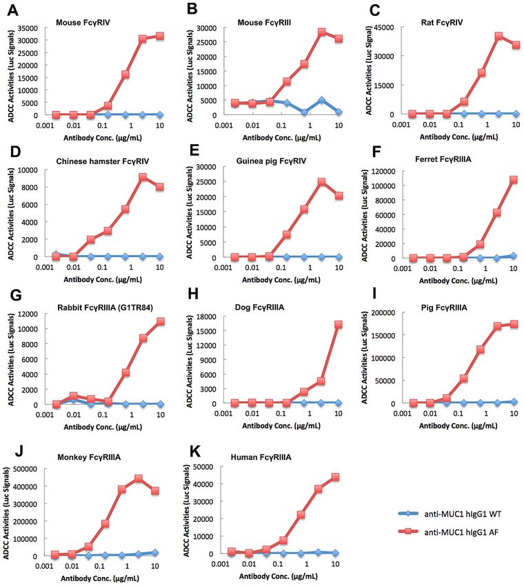
162 Antibody Therapeutics, 2021

                                                                                                                                                           Downloaded from https://academic.oup.com/abt/article/4/3/159/6354750 by guest on 21 November 2021
Figure 1. Comparison of the amino acid sequences of Fcγ RIIIA or Fcγ RIV from different mammalian species. (A) Alignment of the full-length sequences
of Fcγ RIIIA or Fcγ RIV with ClustalW via the server at http://www.phylogeny.fr. Similar residues are colored in cyan as the most conserved ones
(according to BLOSUM62 score). Average BLOSUM62 score: Max: 3.0 (cyan), Low: 0.5 (grey). YEEP motifs in the intracellular region of rodent Fcγ RIV
are boxed in red. (B) Phylogenetic tree of the relationships among members of the greater Fcγ RIIIA/Fcγ RIV family. The tree topology was estimated with
“neighbor joining” in which branch values (numbers above lines) represent percent “bootstrap” support (values below 50% are not included). Scale bar,
genetic distance. Amino acid sequences were derived from the following (UNIPROT or NCBI identifiers in parentheses): mouse Fcγ RIII (P08508), mouse
Fcγ RIV (A0A0B4J1G0), rat Fcγ RIII (P27645), rat Fcγ RIV (A0A0B4J2J1), guinea pig Fcγ RIII (H0V371), guinea pig Fcγ RIV (H0VDZ8), golden
hamster Fcγ RIII (A0A3Q0CHI5), golden hamster Fcγ RIV (A0A1U7Q211), Chinese hamster Fcγ RIII (XP_007645600), Chinese hamster Fcγ RIV
(XP_035301700), ferret Fcγ RIIIA (M3XWH1), rabbit Fcγ RIIIA (G1TR84), rabbit Fcγ RIIIA (G1T7E7), cat Fcγ RIIIA (Q9N2I5), dog Fcγ RIIIA
(E2RP87), pig Fcγ RIIIA (Q28942), monkey Fcγ RIIIA (A3RFZ7) and human Fcγ RIIIA (P08637).

XP_002715293) as rabbit low affinity immunoglobulin                             We compared the amino acid sequences of Fcγ RIII
gamma Fc region receptor III-B-like.                                          and Fcγ RIV from various mammals using BLAST search
  Similarly, we blasted gpFcγ RIV against the UNIPROT                         results from the UNIPROT and NCBI databases (Fig. 1A).
database and found a homologous entry A0A1U7Q211,                             Phylogenetic comparison of the full-length amino acid
which was marked as Fcγ RIV from golden hamster                               sequences for this collection of mammalian Fcγ RIII
(Mesocricetus auratus). The very same sequence has been                       and Fcγ RIV receptors indicated that rodent Fcγ RIV
de novo cloned from the cDNA of the golden Syrian                             receptors are more closely related to non-rodent Fcγ RIIIA
hamster liver and was named as hamster Fcγ RIV [34].                          orthologues than they are to the rodent Fcγ RIII receptors
However, the NCBI database annotates it as golden                             (Fig. 1B). Interestingly, the phylogenetic relationships of
hamster Fcγ RIII-like (NP_001268264), and it only shows                       rodent Fcγ RIV receptors perfectly mirror those of rodent
87% identities with Chinese hamster (Cricetulus griseus)                      Fcγ RIII receptors, i.e., the two hamster genes are as close
Fcγ RIV (XP_035314579). Thus, there is a certain degree                       as the mouse and rat genes, but the four only remotely
of confusion whether such homologues from other mam-                          resemble the guinea pig gene (Supplementary Fig. S2). This
malian species should be named as Fcγ RIII or Fcγ RIV,                        suggests that rodent Fcγ RIV genes separated from rodent
as at the protein sequence level they are all highly                          Fcγ RIII genes at an early time point during evolution,
homologous.                                                                   supporting that they have distinct functions.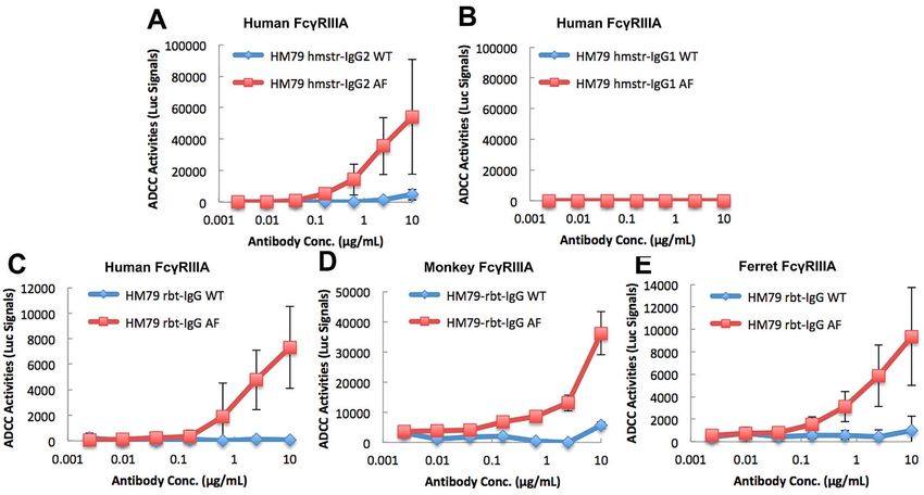
Antibody Therapeutics, 2021 163

Afucosylated mouse IgG2c but not IgG1 exhibits enhanced        from ferret, rabbit, cat, dog, pig, monkey and human
ADCC via Fcγ RIV orthologues                                   (Fig. 2C–N). That is, afucosylation of mIgG2c enhances
                                                               ADCC over WT mIgG2c for at least 10-fold (in pig
Given the drastic difference in effector functions of
                                                               and guinea pig) and usually >20–100-fold in most other
the activating murine Fcγ RIV vs. Fcγ RIII receptors
                                                               mammalian species. Not only the effective concentrations
[33], it is important to functionally characterize the
                                                               of AF mIgG2c antibody are greatly reduced, but also
Fcγ RIIIA/Fcγ RIV receptors from different mammalian
                                                               the magnitudes of ADCC response are dramatically
species, and give them the right nomenclature to guide
                                                               increased. For golden hamster Fcγ RIV, rabbit and cat
in vivo studies. Because mouse Fcγ RIV only binds to
                                                               Fcγ RIIIA, simple afucosylation transforms an otherwise
mIgG2a/b/c isotypes, whereas mouse Fcγ RIII also binds
                                                               ineffective WT mIgG2c into a highly effective antibody
to mIgG1 in addition [32, 35], we used a panel of WT
                                                               with potent ADCC (Fig. 2E, H–J). On the contrary,
and afucosylated (AF) mIgG2c and mIgG1 to perform the
                                                               AF mIgG1 does not trigger any ADCC response by
ADCC assay. Note that, mouse IgG2a and IgG2c genes are
                                                               Fcγ RIV from mouse and hamster, or by Fcγ RIIIA from
not linked but allelic [36]. Although BALB/c mice express

                                                                                                                               Downloaded from https://academic.oup.com/abt/article/4/3/159/6354750 by guest on 21 November 2021
                                                               ferret, rabbit (G1T7E7), cat, dog, monkey and human
IgG2a, some other inbred mouse strains such as C57BL/6,
                                                               (Fig. 2A, D, E, G, I–K, M, N), confirming that they are all
C57BL/10 and NOD do not have the gene for IgG2a but
                                                               orthologues of the murine Fcγ RIV. Interestingly, perhaps
instead express the IgG2c isotype [36].
                                                               due to cross-species binding, gpFcγ RIV and pig Fcγ RIIIA
   We determined that a mouse hybridoma (5F2) of
                                                               do respond to WT mIgG1, and the responses are further
C57BL/6 background against mouse CD39 is of mIgG1
                                                               enhanced by afucosylation of mIgG1 (Fig. 2F, L). Similar
isotype, and also cloned its spontaneously class-switched
                                                               enhanced responses to AF mIgG1 are also seen with
mIgG2c subline derived from a rare event during a culture
                                                               rat Fcγ RIV and rabbit Fcγ RIIIA (G1TR84), although
crash incident. DNA sequencing of the RT-PCR-amplified
                                                               at much lower levels (Fig. 2C, H). Clearly, these Fcγ Rs
heavy chain genes confirmed that this was an authentic
                                                               are different from mFcγ RIII in response to AF mIgG1
class-switching event (not shown). The Fut8−/− 5F2 mIgG1
                                                               (Fig. 2B).
and mIgG2c sublines were subsequently established after
CRISPR-mediated knocking out of the murine Fut8 gene,
and confirmed by negative staining with fucose-binding
LCA (not shown). Thus, we have set up a perfect panel of       Afucosylated hamster IgG2 but not IgG1 also exhibits
                                                               enhanced ADCC via Fcγ RIIIA/Fcγ RIV orthologues
four mouse hybridoma antibodies with the identical Fab
portion for the same antigen recognition, but with mIgG1       To rule out the possibilities that the observed effects
vs. mIgG2c isotypes and WT vs. AF glycan profiles.             were specific for mouse hybridoma-derived antibodies
   In ADCC assays using mCD39-expressing CHO cells as          or for the recognized mCD39 antigen only, we used
target cells and luciferase-equipped Jurkat cells expressing   WT and Fut8−/− CHO cells to recombinantly express an
mFcγ RIV as effector cells, AF mIgG2c is 50–100-fold           Armenian hamster (Cricetulus migratorius) anti-mouse
more potent than WT mIgG2c in mediating ADCC,                  CD79b antibody (clone HM79-16 or HM79b; [37]). The
whereas mIgG1 antibodies, regardless of WT or AF,              original HM79–16 hamster antibody has IgG2 heavy chain
are completely ineffective (Fig. 2A). In the same system       (GenBank: AGH06135) and lambda light chain (GenBank:
but with Jurkat reporter cells expressing mFcγ RIII, AF        AGH06134). We also generated a hamster IgG1 version
mIgG2c also exhibits enhanced ADCC compared with               of HM79-16 by replacing its Fc domain with the Fc of
WT, although their EC50 concentrations are ∼10-fold            Armenian hamster IgG1 antibody HL4E10 (GenBank:
higher than those with mFcγ RIV (Fig. 2B). Interestingly,      AEG80191). Alignment analyses indicate that hamster
mIgG1 stimulates mFcγ RIII for ADCC at even lower              IgG2 (hmstr-IgG2) and hmstr-IgG1 have high homologies
concentrations, but mFcγ RIII does not differentiate the       with mouse IgG2a and mIgG1, respectively (not shown).
content of fucose on mIgG1, and the two ADCC curves of         Similarly, we engineered two additional chimeric HM79-16
WT and AF mIgG1 are completely overlapped (Fig. 2B).           antibodies with the same original Fab in fusion with Fc
These results are in line with the reports that mFcγ RIII      domains from either rabbit or pig IgG. All four HM79-16
but not mFcγ RIV also binds to mIgG1 [32, 35]. Thus,           antibodies were produced by WT or Fut8−/− CHO cells to
our 5F2 mIgG1/mIgG2c combo set and ADCC assay                  generate matched antibody pairs with WT or AF glycan
can functionally differentiate whether a rodent Fcγ R          profiles.
homologue should be classified as Fcγ RIII or Fcγ RIV,            In ADCC assays using mCD79b-expressing CHO cells
i.e., a receptor that is hyperstimulated by the afucosylated   as target cells and luciferase-equipped Jurkat cells express-
form of mIgG2c but not by mIgG1 should be an Fcγ RIV           ing hFcγ RIIIA as effector cells, AF HM79-hmstr-IgG2
orthologue. Conversely, a receptor that cannot differentiate   shows more enhanced ADCC than WT HM79-hmstr-IgG2
the WT vs. AF forms of mIgG1 should be an Fcγ RIII             (Fig. 3A), whereas HM79-hmstr-IgG1 antibodies, regard-
orthologue.                                                    less of WT or AF, are completely ineffective (Fig. 3B). The
   With this in mind, we tested 5F2 mIgG1/mIgG2c               same pattern is also true for Jurkat cells expressing mon-
combo set in ADCC assays with luciferase-equipped              key or ferret Fcγ RIIIA and mouse or hamster Fcγ RIV
Jurkat cells expressing species-specific Fcγ RIII/Fcγ RIV.     (Supplementary Fig. S3). The enhanced ADCC in vitro by
The patterns of ADCC response are overall very similar,        afucosylated HM79-hmstr-IgG2 also translates into more
with a few exceptions, among Fcγ RIV receptors from            efficient depletion of mCD79b+ B cells in vivo in C57BL/6
mouse, rat, hamster, guinea pig, and Fcγ RIIIA receptors       mice (Supplementary Fig. S4).
164 Antibody Therapeutics, 2021
Downloaded from https://academic.oup.com/abt/article/4/3/159/6354750 by guest on 21 November 2021
Figure 2. Afucosylated mouse IgG2c but not IgG1 exhibits enhanced ADCC via Fcγ RIV orthologues. The original mouse anti-mCD39 hybridoma (clone
5F2, mIgG1) was screened to select a spontaneously class-switched subline with mIgG2c isotype. Both clonal 5F2-mIgG1 and 5F2-mIgG2c hybridomas
were rendered Fut8−/− by CRISPR technology to produce afucosylated (AF) antibodies. Purified WT and AF 5F2 antibodies with different isotypes
and fucose content all have the exact same Fab, recognizing mCD39 with equal strength (not shown), but demonstrated drastic differences in ADCC
assays with various Fcγ receptors. Compared with WT 5F2-mIgG2c, AF 5F2-mIgG2c exhibits greatly enhanced ADCC via mouse Fcγ RIV (A), mouse
Fcγ RIII (B), rat Fcγ RIV (C), Chinese hamster Fcγ RIV (D), golden hamster Fcγ RIV (E), guinea pig Fcγ RIV (F), ferret Fcγ RIIIA (G), rabbit Fcγ RIIIA
(G1TR84) (H), rabbit Fcγ RIIIA (G1T7E7) (I), cat Fcγ RIIIA (J), dog Fcγ RIIIA (K), pig Fcγ RIIIA (L), monkey Fcγ RIIIA (M) and human Fcγ RIIIA
(N). Note that mouse Fcγ RIII mediates equal ADCC responses regardless of the fucose content on mIgG1 (B), a feature that is not seen among all the
other Fcγ RIV orthologues. Data are summarized from at least two independent experiments with Mean ± standard deviation (SD) calculated from repeat
experiments when luciferase assay readouts are close, or otherwise a representative experiment of the two is presented.Antibody Therapeutics, 2021 165
Downloaded from https://academic.oup.com/abt/article/4/3/159/6354750 by guest on 21 November 2021
Figure 3. Afucosylated hmstr-IgG2 and rabbit IgG exhibit enhanced ADCC via Fcγ RIIIA orthologues. Recombinant HM79 antibodies with Fc domains
from hmstr-IgG2, hamster IgG1, or rabbit IgG, were produced by WT or Fut8−/− CHO-K1 cells. Purified WT and AF HM79 antibodies with different
isotypes and fucose content all have the exact same mCD79b-recognizing Fab, but demonstrated drastic differences in ADCC assays with various Fcγ
receptors. Compared with WT HM79-hmstr-IgG2, AF HM79-hmstr-IgG2 exhibits greatly enhanced ADCC via human Fcγ RIIIA (A), but HM79-
hmstr-IgG1 antibodies had no activities (B). AF MH79-rbt-IgG is more stimulatory than WT MH79-rbt-IgG towards human Fcγ RIIIA (C), monkey
Fcγ RIIIA (D) and ferret Fcγ RIIIA (E) in ADCC assays. Data are summarized from two independent experiments with Mean ± SD calculated from
repeat experiments.
Interestingly, perhaps due to species difference, AF an attractive target for immune intervention in cancer [39,
HM79-hmstr-IgG2 does not show any binding to rat 40]. We have generated a humanized 5T4 IgG1 antibody
Fcγ RIV (not shown). Also, compared with AF HM79- with high affinity towards 5T4 antigen (Kd = 4.1 nM). In
hmstr-IgG2, chimeric AF rabbit HM79-16 antibody ADCC assays using h5T4-expressing CHO cells as target
(HM79-rbt-IgG) exhibits much weaker ADCC activities cells and luciferase-equipped Jurkat cells expressing various
under exactly the same conditions. But still, afucosylation species-specific Fcγ RIIIA or Fcγ RIV as effector cells,
of rabbit IgG turns an otherwise ineffective WT antibody AF anti-5T4 hIgG1 universally exhibits stronger ADCC
into a highly effective one for human, monkey and than WT anti-5T4 hIgG1 (Fig. 4). Perhaps because our
ferret Fcγ RIIIA (Fig. 3C–E). The chimeric pig HM79- anti-5T4 antibody is already highly effective, i.e., even the
16 antibody (HM79-pig-IgG), however, does not show any WT version has EC50 numbers between 1 and 10 ng/mL
ADCC activities by human, monkey or ferret Fcγ RIIIA for Fcγ RIIIA or Fcγ RIV from most tested species except
and mouse Fcγ RIV, except for pig Fcγ RIIIA itself where for golden hamster, guinea pig, rabbit, cat and pig (EC50
the AF pig IgG outperforms the WT counterpart (not numbers between 10 and 100 ng/mL), afucosylation of
shown). This is in line with the report that pig IgG only anti-5T4 enhances ADCC by only ∼10-fold.
binds to its own Fcγ RIIIA but not to human or rabbit As different antibodies have different binding charac-
Fcγ RIIIA [38]. teristics, to address the same question with a different
antibody, we then turned to an anti-MUC1-C hIgG1
similarly produced by WT or Fut8−/− CHO cells. In
Afucosylated human IgG1 exhibits enhanced ADCC with ADCC assays using MUC1-expressing human colon
Fcγ RIIIA/Fcγ RIV from multiple mammalian species cancer cell line HCT116 as target cells and luciferase-
Having established the testing systems to functionally equipped Jurkat cells expressing various species-specific
assign the right nomenclature to orthologues of Fcγ RIIIA Fcγ RIIIA or Fcγ RIV as effector cells, AF anti-MUC1-
or Fcγ RIV in multiple species, we now address the question C hIgG1 is highly effective whereas WT anti-MUC1-
whether an afucosylated therapeutic human IgG1 antibody C hIgG1 does not show any ADCC activity even at
can be directly evaluated for higher efficacy in various the highest doses (Fig. 5), although both have the same
animal models. For that, we first used WT and AF anti- binding profiles towards hMUC-1+ HCT116 by FACS
5T4 hIgG1, produced by WT and Fut8−/− CHO cells. The (Supplementary Fig. S5). The enhanced ADCC in vitro
oncofetal antigen 5T4 (trophoblast glycoprotein, TPBG) by afucosylated anti-MUC1-C hIgG1 also translates into
is expressed on a wide range of human solid tumors with effective growth control of hMUC-1+ HCT116 solid tumor
expression level correlated with disease progression, and is in vivo in C57BL/6 mice, whereas the WT antibody is166 Antibody Therapeutics, 2021
Downloaded from https://academic.oup.com/abt/article/4/3/159/6354750 by guest on 21 November 2021
Figure 4. Afucosylation of an already highly potent human IgG1 (anti-h5T4) can still boost its ADCC through Fcγ RIIIA/Fcγ RIV from multiple
mammalian species. Recombinant human anti-h5T4 IgG1 antibodies were produced by WT or Fut8−/− CHO-K1 cells. Compared with WT anti-5T4
hIgG1, AF anti-5T4 hIgG1 exhibits greatly enhanced ADCC via mouse Fcγ RIV (A), mouse Fcγ RIII (B), rat Fcγ RIV (C), Chinese hamster Fcγ RIV (D),
golden hamster Fcγ RIV (E), guinea pig Fcγ RIV (F), ferret Fcγ RIIIA (G), rabbit Fcγ RIIIA (G1TR84) (H), rabbit Fcγ RIIIA (G1T7E7) (I), cat Fcγ RIIIA
(J), dog Fcγ RIIIA (K), pig Fcγ RIIIA (L), monkey Fcγ RIIIA (M) and human Fcγ RIIIA (N). Data are representative of two repeat experiments.
ineffective (unpublished observations). Our data strongly ADCP. Producing afucosylated IgG by Fut8−/− CHO
suggest that cross-species IgG:Fcγ RIIIA/Fcγ RIV binding cells can render greater ADCC activities, and potentially
profile dictates the effectiveness of the therapeutic antibody allows the therapeutic effects of the antibody to be directly
when the MOA of the antibody is through ADCC and/or assessed across a wide range of animal species.Antibody Therapeutics, 2021 167
Downloaded from https://academic.oup.com/abt/article/4/3/159/6354750 by guest on 21 November 2021
Figure 5. Afucosylation of an intrinsically weak human IgG1 (anti-MUC1-C) endows it with strong ADCC through Fcγ RIIIA/Fcγ RIV from multiple
mammalian species. Recombinant human anti-hMUC1-C IgG1 antibodies were produced by WT or Fut8−/− CHO-K1 cells. WT anti-hMUC1-C IgG1
does not show ADCC even at the highest doses, whereas AF anti-hMUC1-C IgG1 exhibits greatly enhanced ADCC via mouse Fcγ RIV (A), mouse
Fcγ RIII (B), rat Fcγ RIV (C), Chinese hamster Fcγ RIV (D), guinea pig Fcγ RIV (E), ferret Fcγ RIIIA (F), rabbit Fcγ RIIIA (G1TR84) (G), dog
Fcγ RIIIA (H), pig Fcγ RIIIA (I), monkey Fcγ RIIIA (J) and human Fcγ RIIIA (K). Golden hamster Fcγ RIV and cat Fcγ RIIIA do not respond to
this particular hIgG1 even in AF form (not shown). Data are representative of two repeat experiments.
DISCUSSION animals (usually mice) used at young ages have T and
B cell repertoires very different from those in a human
One of the biggest hurdles in biomedical research is being where antigen-experienced memory lymphocytes
the translatability of animal studies in the context of also constitute and regulate immune responses in many
human disease settings. At the cellular level, naïve lab unique ways [41]. At the molecular level, the species168 Antibody Therapeutics, 2021
differences in the interacting Ag/Ab and ligand/receptor With the sensitive luciferase-based ADCC assay, we are
pairs make result interpretation not entirely without now confident to conclude that IgG afucosylation can
reservation. Transgenic or humanized knockin animals be a universal method to enhance ADCC/ADCP medi-
provide only part of the solution, as the challenges posed ated by the orthologues of human Fcγ RIIIA or mouse
by the convoluted networks of ligand:receptor interactions, Fcγ RIV across multiple mammalian species, most likely
esp. within the immune system, are beyond the current not limited to those in our current study. For research
technologies of making genetically-modified animals can and development of antibody therapeutics, IgG afucosy-
offer to overcome. Generating hematopoietic chimeric lation can be achieved via two major routes. First, if one
animals with human immune cells is a viable strategy, but has generated a hybridoma-derived MAb, which has an
these animals are extremely expensive, and the technology isotype of ADCC potential, such as mouse IgG2a/b from
is not widely applicable other than in our little furry BALB/c or mIgG2b/c from C57BL/6, then one can use
friends called Mus musculus. Yet, many underrepresented CRISPR technology to directly knock out the Fut8 gene
mammalian species, such as hamster, guinea pig, ferret, in the existing hybridoma cell line to produce the afu-
Downloaded from https://academic.oup.com/abt/article/4/3/159/6354750 by guest on 21 November 2021
cotton rat, pig (including minipigs), rabbit and marmoset cosylated antibody (Fig. 2). If the hybridoma is of IgG1
are increasingly being used as human disease models, isotype, although one can perform in vitro class switching
especially in the infectious disease area. The technologies to IgG2a/b/c, the conditions are tricky and the efficiency
suitable for evaluating antibody therapeutics and studying could be very low [43]. Second, and most appealing from
their MOAs have not been adequately applied in these the pharmaceutical industry perspective, one can clone the
underrepresented species. MAb’s VH/VL genes from the hybridoma mRNA by RT-
This study was prompted during our attempt of inves- PCR, and recombinantly express the original or humanized
tigating a therapeutic human IgG1 fusion protein in the antibody with the desired isotype and afucosyl form in
hamster model of SARS-CoV-2 infection, where we were Fut8−/− CHO cells (Figs 3–5).
puzzled whether afucosylated hIgG1 could still exhibit To our surprise, in the ADCC assays, simple afucosyla-
enhanced ADCC and achieve better therapeutic effects tion can transform an otherwise ineffective IgG antibody
in hamster. In fact, throughout the literature, there lacks into a highly active one with potent ADCC activities in
not only a systematic functional study to address this certain species. For instance, WT mIgG2c barely triggers
crucial question across many species, but also the careful any ADCC by hamster Fcγ RIV, or by rabbit and cat
characterization of orthologues of human Fcγ RIIIA in Fcγ RIIIA, whereas AF mIgG2c is highly effective (Fig. 2).
these species in the first place. We performed exhaustive The same is also true for a hmstr-IgG2 and a rabbit IgG
BLAST searches and used bioinformatics tools to narrow (Fig. 3), as well as a hIgG1 (Fig. 5). Therefore, as an eco-
down the potential orthologues of human Fcγ RIIIA or nomic way to tap into the vast pool of hybridoma-derived
mouse Fcγ RIV from rat, hamster, guinea pig, ferret, rabbit, antibodies from academia labs or biotech companies for
cat, dog, pig and monkey, some of which are only newly pre-clinical studies of candidates with therapeutic poten-
characterized in this paper. For instance, prior to this study, tial, one should always bear in mind to: 1) select or switch
the existence of ferret Fcγ RIIIA was merely predicted by to an ADCC-capable isotype, and 2) generate its afucosy-
automated computational analysis, but the receptor itself lated form.
has not been functionally studied. We found that ferret A good example is when we were approached by our
Fcγ RIIIA is most closely related to dog Fcγ RIIIA in the academic collaborators on how to evaluate a mouse CD39
phylogenetic tree (Fig. 1B), and confirmed that these two antibody (clone 5F2) for any anti-tumor effect. Its mIgG1
receptors exhibit very similar response patterns towards the form had been extensively examined by the collaborators
5F2 mIgG2c/mIgG1 combo set (Fig. 2G and K). Likewise, in B16F10 mouse melanoma model, and was found to
even though the two Fcγ RIV receptors from Chinese be completely ineffective. We first tried to recombinantly
hamster and golden hamster are as disparate as Fcγ RIV express AF 5F2-mIgG2c in Fut8−/− CHO cells, but 5F2
receptors from mouse and rat in the phylogenetic tree antibody genes belong to that “difficult-to-express” cate-
(Fig. 1B), they are still orthologues to each other, not gory, and this approach was not successful. Fortuitously,
different genes. Chinese hamster and golden hamster using ISO-M8c mouse immunoglobulin isotyping kit spe-
do have a different set of Fcγ RIII receptors, similar to cially formatted for MAbs of C57BL/6 origin, we spotted in
those found in mouse and rat (Supplementary Fig. S2). the culture supernatant the presence of 5F2’s spontaneously
Interestingly, Fcγ RIII and Fcγ RIV receptors from guinea class-switched mIgG2c form and subcloned this hybridoma
pig are only remotely related to the other rodent family subline. This was followed by knocking out the murine Fut8
members (Supplementary Fig. S2). Although the Fcγ RIV gene in the WT 5F2 mIgG1 and mIgG2c hybridomas, with
receptors from mouse, rat and hamster all have in their the ultimate ensemble of WT and AF 5F2 mIgG1/mIgG2c
intracellular domains the YEEP motif, whose tyrosine combo to test our hypothesis. In both B16F10 melanoma
residue becomes phosphorylated after receptor ligation, and MC38 colon cancer models, and reminiscent of their
gpFcγ RIV has the KEEY sequence and all the Fcγ RIIIA dramatic differences in ADCC capacities (Fig. 2A), AF
receptors from non-rodent species do not have this YEEP 5F2-mIgG2c demonstrated most striking anti-tumor effect,
signature (Fig. 1A). Thus, tyrosine phosphorylation at followed by WT 5F2-mIgG2c, whereas 5F2-mIgG1 anti-
this position is not absolutely required, as signaling of bodies were completely ineffective (manuscript in prepara-
Fcγ RIIIA and Fcγ RIV is mediated by the Fc receptor tion, US provisional patent application 62/685 176).
common gamma chain [42], which is co-expressed in all With such dramatic in vivo outcomes directly correlated
our Jurkat reporter cell lines. with in vitro ADCC activities, we are confident that theAntibody Therapeutics, 2021 169
observations in our study can provide valuable insights DATA AVAILABILITY STATEMENT
for researchers to break the boundary and explore possi- The data underlying this article are available in the article
bilities not ever imagined before. For example, as global and in its online supplementary material.
concerns arise as the emerged and rapidly spreading SARS-
CoV-2 variants might escape host immunity induced by
vaccination, researchers have isolated rabbit MAbs with CONFLICTS OF INTEREST STATEMENT
potent neutralization capability against not only SARS-
C.M. and W.G. are employees of Antagen Pharmaceuticals,
CoV-2 WT strain but also emergent variants [44]. The
Inc., which owns a proprietary Fut8−/− CHO cell expression
longer CDR3 of rabbit IgG permits these antibodies to
system.
bind inside clefts and grooves that are generally inaccessible
to conventional antibodies and make them great candi-
dates for binding within enzyme active sites and other
“hard to reach” protein surfaces. Based on our results, FUNDING
This study was supported by National Institutes of
Downloaded from https://academic.oup.com/abt/article/4/3/159/6354750 by guest on 21 November 2021
afucosylated rabbit IgG exhibits much potent ADCC via
ferret Fcγ RIIIA than WT rabbit IgG, which is ineffective Health (NIH) Small Business Innovative Research grants
(Fig. 3E), it is now possible to test the hypothesis in the (HHSN272201800017C, 75N93019C00014 and 75N93020
ferret model of SARS-CoV-2 infection whether afucosy- C00042) awarded to Antagen.
lated rabbit IgG can be more protective not only through
neutralization but also through ADCC-mediated clearing
of virus-infected cells. Conversely, by comparing adoptively REFERENCES
transferred WT and AF anti-Spike/RBD IgG side-by-side 1. Mimoto, F, Igawa, T, Kuramochi, T et al. Novel asymmetrically
in the ferret model of SARS-CoV-2 infection, scientists engineered antibody Fc variant with superior Fcγ R binding affinity
may acquire valuable insights whether disease is aggravated and specificity compared with afucosylated Fc variant. MAbs 2013;
5: 229–36. 10.4161/mabs.23452.
via enhanced stimulation through Fcγ RIIIA/Fcγ RIV by 2. Mimoto, F, Kadono, S, Katada, H et al. Crystal structure of a novel
afucosylated IgG that may trigger host cytokine storm [16, asymmetrically engineered fc variant with improved affinity for
17]. If that is the case, counter measures can be developed Fcγ Rs. Mol Immunol 2014; 58: 132–8.
to treat severe illness in COVID-19. 10.1016/j.molimm.2013.11.017.
3. Hanson, QM, Barb, AW. A perspective on the structure and
In summary, our findings indicate that increased sen- receptor binding properties of immunoglobulin G Fc. Biochemistry
sitivities of Fcγ RIIIA/Fcγ RIV to afucosylated IgG with 2015; 54: 2931–42. 10.1021/acs.biochem.5b00299.
enhanced ADCC is a universal cross-species phenomenon, 4. Yamane-Ohnuki, N, Satoh, M. Production of therapeutic antibodies
underlining the rationale of testing afucosylated therapeu- with controlled fucosylation. MAbs 2009; 1: 230–6.
tic antibodies directly in various animal models for efficacy 10.4161/mabs.1.3.8328.
5. Listinsky, JJ, Siegal, GP, Listinsky, CM. Glycoengineering in cancer
and toxicity evaluation. Not only this strategy could speed therapeutics: a review with fucose-depleted trastuzumab as the
up antibody discovery and help save tremendous resources model. Anticancer Drugs 2013; 24: 219–27.
in animal studies, perhaps it could also turn someone’s 10.1097/CAD.0b013e328359e3f4.
trash into another one’s treasure, and nurture the next gen- 6. Shields, RL, Lai, J, Keck, R et al. Lack of fucose on human IgG1
N-linked oligosaccharide improves binding to human Fcgamma
eration glycoengineered therapeutic antibodies with novel RIII and antibody-dependent cellular toxicity. J Biol Chem 2002;
mechanisms of disease-fighting actions. 277: 26733–40. 10.1074/jbc.M202069200.
7. Yamane-Ohnuki, N, Kinoshita, S, Inoue-Urakubo, M et al.
Establishment of FUT8 knockout Chinese hamster ovary cells: an
AUTHORS’ CONTRIBUTIONS ideal host cell line for producing completely defucosylated antibodies
with enhanced antibody-dependent cellular cytotoxicity. Biotechnol
C.M. performed most experiments, including the ADCC Bioeng 2004; 87: 614–22. 10.1002/bit.20151.
8. Okazaki, A, Shoji-Hosaka, E, Nakamura, K et al. Fucose depletion
assay, FACS and the animal studies and analyzed the data. from human IgG1 oligosaccharide enhances binding enthalpy and
R.N. made lentiviruses and established Jurkat cell lines association rate between IgG1 and FcgammaRIIIa. J Mol Biol 2004;
for the ADCC assay. X.Z. and W.G. designed the experi- 336: 1239–49. 10.1016/j.jmb.2004.01.007.
ments, wrote the animal protocols, monitored and finan- 9. Kanda, Y, Yamada, T, Mori, K et al. Comparison of biological
cially supported the study. W.G. wrote the manuscript. All activity among nonfucosylated therapeutic IgG1 antibodies with
three different N-linked Fc oligosaccharides: the high-mannose,
authors critically reviewed the drafts of the manuscript and hybrid, and complex types. Glycobiology 2007; 17: 104–18.
approved the final version. 10.1093/glycob/cwl057.
10. Golay, J, Da Roit, F, Bologna, L et al. Glycoengineered CD20
antibody obinutuzumab activates neutrophils and mediates
phagocytosis through CD16B more efficiently than rituximab. Blood
SUPPLEMENTARY DATA 2013; 122: 3482–91. 10.1182/blood-2013-05-504043.
Supplementary data are available at ABT online. 11. Herter, S, Birk, MC, Klein, C et al. Glycoengineering of therapeutic
antibodies enhances monocyte/macrophage-mediated phagocytosis
and cytotoxicity. J Immunol 2014; 192: 2252–60.
10.4049/jimmunol.1301249.
ACKNOWLEDGEMENTS 12. Mori, K, Iida, S, Yamane-Ohnuki, N et al. Non-fucosylated
The authors would like to thank Dr Simon C. Robson from therapeutic antibodies: the next generation of therapeutic antibodies.
Cytotechnology 2007; 55: 109–14. 10.1007/s10616-007-9103-2.
Beth Israel Deaconess Medical Center for the original anti- 13. Kapur, R, Kustiawan, I, Vestrheim, A et al. A prominent lack of
mCD39 hybridoma 5F2, and Genus Oncology, LLC. for IgG1-Fc fucosylation of platelet alloantibodies in pregnancy. Blood
anti-MUC1-C antibody. 2014; 123: 471–80. 10.1182/blood-2013-09-527978.170 Antibody Therapeutics, 2021
14. Wang, TT, Sewatanon, J, Memoli, MJ et al. IgG antibodies to 29. Mao, C, Near, R, Gao, W. Identification of a Guinea pig Fcγ
dengue enhanced for Fcγ RIIIA binding determine disease severity. receptor that exhibits enhanced binding to afucosylated human and
Science 2017; 355: 395–8. 10.1126/science.aai8128. mouse IgG. J Infect Dis Med 2016; 1: 1–3.
15. Bournazos, S, Vo, HTM, Duong, V et al. Antibody fucosylation 10.4172/2576-1420.1000102.
predicts disease severity in secondary dengue infection. Science 2021; 30. Cong, L, Ran, FA, Cox, D et al. Multiplex genome engineering using
372: 1102–5. 10.1126/science.abc7303. CRISPR/Cas systems. Science 2013; 339: 819–23.
16. Larsen, MD, de Graaf, EL, Sonneveld, ME et al. Afucosylated IgG 10.1126/science.1231143.
characterizes enveloped viral responses and correlates with COVID 31. Sommer, CA, Stadtfeld, M, Murphy, GJ et al. Induced pluripotent
-19 severity. Science 2021; 371: eabc8378. 10.1126/science.abc8378. stem cell generation using a single lentiviral stem cell cassette. Stem
17. Hoepel, W, Chen, H-J, Geyer, CE et al. High titers and low Cells 2009; 27: 543–9. 10.1634/stemcells.2008-1075.
fucosylation of early human anti-SARS-CoV-2 IgG promote 32. Nimmerjahn, F, Bruhns, P, Horiuchi, K et al. Fcγ RIV: a novel FcR
inflammation by alveolar macrophages. Sci Transl Med 2021; 13: with distinct IgG subclass specificity. Immunity 2005; 23: 41–51.
eabf8654. 10.1126/scitranslmed.abf8654. 10.1016/j.immuni.2005.05.010.
18. Zeitlin, L, Pettitt, J, Scully, C et al. Enhanced potency of a 33. Nimmerjahn, F, Lux, A, Albert, H et al. Fcγ RIV deletion reveals its
fucose-free monoclonal antibody being developed as an Ebola virus central role for IgG2a and IgG2b activity in vivo. Proc Natl Acad Sci
immunoprotectant. PNAS 2011; 108: 20690–4. U S A 2010; 107: 19396–401. 10.1073/pnas.1014515107.
Downloaded from https://academic.oup.com/abt/article/4/3/159/6354750 by guest on 21 November 2021
10.1073/pnas.1108360108. 34. Hirano, M, Davis, RS, Fine, WD et al. IgEb immune complexes
19. Wec, AZ, Bornholdt, ZA, He, S et al. Development of a human activate macrophages through FcgammaRIV binding. Nat Immunol
antibody cocktail that deploys multiple functions to confer 2007; 8: 762–71. 10.1038/ni1477.
pan-ebolavirus protection. Cell Host Microbe 2019; 25: 39–48.e5. 35. Bruhns, P. Properties of mouse and human IgG receptors and their
10.1016/j.chom.2018.12.004. contribution to disease models. Blood 2012; 119: 5640–9.
20. Termini, JM, Martinez-Navio, JM, Gao, G et al. Glycoengineering 10.1182/blood-2012-01-380121.
of AAV-delivered monoclonal antibodies yields increased ADCC 36. Zhang, Z, Goldschmidt, T, Salter, H. Possible allelic structure of
activity. Mol Ther Methods Clin Dev 2021; 20: 204–17. IgG2a and IgG2c in mice. Mol Immunol 2012; 50: 169–71.
10.1016/j.omtm.2020.11.001. 10.1016/j.molimm.2011.11.006.
21. Moldt, B, Shibata-Koyama, M, Rakasz, EG et al. A nonfucosylated 37. Haggart, R, Perera, J, Huang, H. Cloning of a hamster anti-mouse
variant of the anti-HIV-1 monoclonal antibody b12 has enhanced CD79B antibody sequences and identification of a new hamster
Fcγ RIIIa-mediated antiviral activity in vitro but does not improve immunoglobulin lambda constant IGLC gene region.
protection against mucosal SHIV challenge in macaques. J Virol Immunogenetics 2013; 65: 473–8. 10.1007/s00251-013-0698-5.
2012; 86: 6189–96. 10.1128/JVI.00491-12. 38. Bhatti, MM, Cai, AG, Theunissen, J-W. Binding affinities of human
22. Albrecht, RA, Liu, W-C, Sant, AJ et al. Moving forward: recent IgG1 and chimerized pig and rabbit derivatives to human, pig and
developments for the ferret biomedical research model. MBio 2018; rabbit Fc gamma receptor IIIA. PLoS One 2019; 14: e0219999.
9: e01113–8. 10.1128/mBio.01113-18. 10.1371/journal.pone.0219999.
23. Wong, J, Layton, D, Wheatley, AK et al. Improving immunological 39. Cappuccini, F, Pollock, E, Stribbling, S et al. 5T4 oncofoetal
insights into the ferret model of human viral infectious disease. glycoprotein: an old target for a novel prostate cancer
Influenza Other Respi Viruses 2019; 13: 535–46. 10.1111/irv.12687. immunotherapy. Oncotarget 2017; 8: 47474–89.
24. Kim, Y-I, Kim, S-G, Kim, S-M et al. Infection and rapid 10.18632/oncotarget.17666.
transmission of SARS-CoV-2 in ferrets. Cell Host Microbe 2020; 27: 40. Stern, PL, Harrop, R. 5T4 oncofoetal antigen: an attractive target
704–709.e2. 10.1016/j.chom.2020.03.023. for immune intervention in cancer. Cancer Immunol Immunother
25. Beale, DJ, Shah, R, Karpe, AV et al. Metabolic profiling from an 2017; 66: 415–26. 10.1007/s00262-016-1917-3.
asymptomatic ferret model of SARS-CoV-2 infection. Metabolites 41. Lakkis, FG, Sayegh, MH. Memory T cells: a hurdle to immunologic
2021; 11: 327. 10.3390/metabo11050327. tolerance. J Am Soc Nephrol 2003; 14: 2402–10.
26. Sia, SF, Yan, L-M, Chin, AWH et al. Pathogenesis and transmission 10.1097/01.asn.0000085020.78117.70.
of SARS-CoV-2 in golden hamsters. Nature 2020; 583: 834–8. 42. Brandsma, AM, Hogarth, PM, Nimmerjahn, F et al. Clarifying the
10.1038/s41586-020-2342-5. confusion between cytokine and Fc receptor ‘common gamma
27. Imai, M, Iwatsuki-Horimoto, K, Hatta, M et al. Syrian hamsters as a chain’. Immunity 2016; 45: 225–6. 10.1016/j.immuni.2016.07.006.
small animal model for SARS-CoV-2 infection and countermeasure 43. Deenick, EK, Hasbold, J, Hodgkin, PD. Decision criteria for
development. PNAS 2020; 117: 16587–95. 10.1073/pnas.200979 resolving isotype switching conflicts by B cells. Eur J Immunol 2005;
9117. 35: 2949–55. 10.1002/eji.200425719.
28. Trimpert, J, Vladimirova, D, Dietert, K et al. The Roborovski dwarf 44. Chen, Y, Zhu, L, Huang, W et al. Potent RBD-specific neutralizing
hamster is a highly susceptible model for a rapid and fatal course of rabbit monoclonal antibodies recognize emerging SARS-CoV-2
SARS-CoV-2 infection. Cell Rep 2020; 33: 108488. variants elicited by DNA prime-protein boost vaccination. Emerg
10.1016/j.celrep.2020.108488. Microbes Infect 2021; 10: 1390–403. 10.1080/22221751.2021.1942227.You can also read