Polygonum multiflorum extract support hair growth by elongating anagen phase and abrogating the effect of androgen in cultured human dermal ...
←
→
Page content transcription
If your browser does not render page correctly, please read the page content below
Shin et al. BMC Complementary Medicine and Therapies               (2020) 20:144
https://doi.org/10.1186/s12906-020-02940-5
                                                                                                                           BMC Complementary
                                                                                                                           Medicine and Therapies

 RESEARCH ARTICLE                                                                                                                                 Open Access

Polygonum multiflorum extract support hair
growth by elongating anagen phase and
abrogating the effect of androgen in
cultured human dermal papilla cells
Jae Young Shin1, Yun-Ho Choi1, Jaeyoon Kim1, Se Young Park1, You Jin Nam2, So Young Lee1, Jeong Hoon Jeon1,
Mu Hyun Jin1 and Sanghwa Lee1*

  Abstract
  Background: Dermal papilla cells (DPCs) play a key role in hair growth among the various cell types in hair follicles.
  Especially, DPCs determine the fate of hair follicle such as anagen to telogen transition and play a pivotal role in
  androgenic alopecia (AGA). This study was performed to elucidate the hair growth promoting effects of Polygonum
  multiflorum extract (PM extract) in cultured human DPCs and its underlying mechanisms.
  Methods: The effects of PM extract on cultured DPCs were investigated. Cell viability and mitochondrial activity
  were measured by CCK-8 and JC-1 analysis, respectively. Western blotting, dot blotting, ELISA analysis,
  immunocytochemistry and real-time PCR analysis were also performed to elucidate the changes in protein and
  mRNA levels induced by PM extract. 3D cultured DPC spheroids were constructed for mimicking the in vivo DPs.
  The hair growth stimulatory effect of PM extract was evaluated using human hair follicle organ culture model.
  Results: PM extract increased the viability and mitochondrial activity in cultured human DPCs in a dose dependent
  manner. The expression of Bcl2, an anti-apoptotic protein expressed dominantly in anagen was significantly
  increased and that of BAD, a pro-apoptotic protein expressed in early catagen was decreased by PM extract in
  cultured DPCs and/or 3D DPC spheroid culture. PM extract also decreased the expression of catagen inducing
  protein, Dkk-1. Growth factors including IGFBP2, PDGF and VEGF were increased by PM extract, revealed by dot blot
  protein analysis. We also have found that PM extract could reverse the androgenic effects of dihydrotestosterone
  (DHT), the most potent androgen. Finally, PM extract prolonged the anagen of human hair follicles by inhibiting
  catagen entry in human hair follicle organ culture model.
  Conclusion: Our data strongly suggest that PM extract could promote hair growth by elongating the anagen and/
  or delaying the catagen induction of hair follicles through activation of DPCs.
  Keywords: Dermal papilla, 3D DPC spheroid, Growth factor, Anagen elongation, Dkk1, Dihydrotestosterone,
  Androgen receptor, Polygonum multiflorum

* Correspondence: shleek@lghnh.com
1
 Research Park, LG Household & Healthcare Ltd, 70, Magokjoongang 10-ro,
Gangseo-gu, Seoul 07795, South Korea
Full list of author information is available at the end of the article

                                      © The Author(s). 2020 Open Access This article is licensed under a Creative Commons Attribution 4.0 International License,
                                      which permits use, sharing, adaptation, distribution and reproduction in any medium or format, as long as you give
                                      appropriate credit to the original author(s) and the source, provide a link to the Creative Commons licence, and indicate if
                                      changes were made. The images or other third party material in this article are included in the article's Creative Commons
                                      licence, unless indicated otherwise in a credit line to the material. If material is not included in the article's Creative Commons
                                      licence and your intended use is not permitted by statutory regulation or exceeds the permitted use, you will need to obtain
                                      permission directly from the copyright holder. To view a copy of this licence, visit http://creativecommons.org/licenses/by/4.0/.
                                      The Creative Commons Public Domain Dedication waiver (http://creativecommons.org/publicdomain/zero/1.0/) applies to the
                                      data made available in this article, unless otherwise stated in a credit line to the data.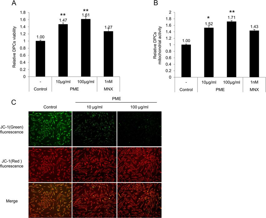
Shin et al. BMC Complementary Medicine and Therapies   (2020) 20:144                                                   Page 2 of 12

Background                                                             production [15]. Especially, 2,3,5,4′-Tetrahydroxystilbene-
Hair follicles undergo three stages of the hair growth                 2-O-β-D-glucoside (TSG) and emodin, single compounds
cycle called anagen, catagen, and telogen, which corre-                identified in PM extract, were reported to show hair
sponds to the growing, regressing, and resting phases,                 growth properties. TSG exerted anti-apoptotic effect in
respectively. Each stage of the hair cycle is classified ac-           C57BL/6 murine follicles [16] and pharmacological effects
cording to the morphological, functional, and compos-                  on age related diseases, resulting in cardio-protective,
itional characteristics of hair follicles. Among various               neuro-protective and anti-hair loss [17]. Concretely, TSG
cell types that organize the hair follicle, dermal papilla             acts as a protector of dopaminergic neurons by regulating
cells (DPCs) play key roles in proliferation and differen-             Akt, GSK3β and Bcl2/BAD expressions as well as a
tiation of hair follicles and controlling hair cycle in each           hypotensive agent in vascular endothelial cells like min-
phase [1]. Especially, growth factors, including insulin-              oxidil [18, 19]. Emodin was reported to strongly inhibit 5-
like growth factor-1 (IGF-1), hepatocyte growth factor                 α reductase activity in benign prostatic hyperplasia [20]
(HGF), vascular endothelial growth factor (VEGF) and                   and promote topical hair growth in C57BL/6 [21]. Al-
keratinocyte growth factor (KGF), from the follicular                  though hair growth stimulating effects of PM extract were
dermal papilla (DP) stimulate keratinocytes to proliferate             reported in several studies using mouse models, detailed
and differentiate into the hair shaft during anagen [2, 3].            biological mechanism for anti-hair loss effects of PM ex-
Transition of hair cycles is also controlled by DPCs. Se-              tract has not been elucidated in the human system, espe-
creted proteins such as transforming growth factors                    cially focused on DPCs.
(TGF) β1, TGF β2, and dickkopf1 (DKK-1) are known to                      In this study, we investigated the hair growth promot-
induce transition from anagen to telogen [4]. When telo-               ing effects of PM extract in cultured human DPCs and
gen is induced, apoptosis and follicular regression occur              the underlying molecular and cellular mechanisms. It
throughout the hair follicle [5]. During anagen or at the              was found that treatment of PM extract stimulated pro-
end of telogen, Bcl2 is highly expressed in DPCs, com-                 liferation and mitochondrial activity in cultured human
pared to other types of hair follicle cells [6]. Among Bcl2            DPCs. PM extract increased the expression of BCl2, an
family proteins, Bcl2 is regarded as one of the most im-               anti-apoptotic protein and decreased the expression of
portant anti-apoptotic agents. On the other hand, BAD                  BAD, a pro-apoptotic protein in cultured DPCs and/or
protein, another member of bcl2 family, is known to in-                3D DPC spheroid culture. Also, PM extract decreased
duce apoptosis [7].                                                    the expression of catagen inducing protein, Dkk-1. In
   Androgens, steroid hormones that regulate the devel-                addition, the expression of growth factors like PDGF-aa
opment and maintenance of male characteristics in ver-                 and VEGF, known to be crucial for hair growth, was in-
tebrates, also indirectly control hair growth by affecting             creased by PM extract treatment. These results clearly
the activities of the DP [8]. Dihydrotestosterone (DHT)                demonstrate the potential role of PM extract in promot-
is derived from testosterone by the action of 5α-                      ing hair growth by elongating anagen and/or delaying
reductase and is considered to be more potent in trig-                 catagen entry. PM extract was found to prolong the ana-
gering hair loss. DHT causes the miniaturization of DP                 gen of human hair follicles by inhibiting catagen entry in
that leads to hair shaft thinning and stimulate the ex-                human hair follicle organ culture model. We observed
pression of TGF β1 in DP resulting in growth inhibition                anti- androgenic effects of PM extract, different from
of epithelial cells [9]. Consequently, prolonged exposure              previously reported mechanism related to inhibition of
of DHT on DP causes androgenic alopecia (AGA) or                       5α-reductase. It was revealed that PM extract signifi-
hair shaft weakening [10, 11].                                         cantly reduced the expression of androgen receptor (AR)
   Polygonum multiflorum (PM) is a species of flowering                induced by DHT and recovered the reduced size of DPC
plant in the buckwheat family polygonaceae. It is one of               spheroid by DHT treatment which mimicked the hair
the most popular perennial traditional Chinese medi-                   follicle miniaturization observed in AGA.
cines called He Shou Wu in China and East Asia. PM                        In conclusion, our data strongly suggest that PM ex-
has long been used as a component for anti-hair loss                   tract could support hair growth by extending anagen
and anti-hair greying treatment prescriptions [12]. Sev-               duration and delaying catagen progression and could
eral reports demonstrated hair growth effects of PM ex-                possibly prevent hair loss by abrogating the effects of an-
tract. Histological analysis of C57BL/6 mouse cases                    drogen which result in hair follicle DP miniaturization,
showed that PM extract increased the size and the num-                 suspected to be a main cause of AGA.
ber of hair follicles via upregulating β-catenin and sonic
hedgehog expressions by both topical and oral applications             Methods
[13, 14]. Also, anti-androgenic effects of PM extract were             Polygonum multiflorum (PM) extract preparation
reported in several studies with prostate cancer cells, by in-         The dried roots of Polygonum multiflorum Thunberg
hibition of 5-α reductase, a key enzyme for DHT                        were purchased from Humanherb (product no.
Shin et al. BMC Complementary Medicine and Therapies   (2020) 20:144                                                  Page 3 of 12

G152150411, Daegu, Korea) in August 2016 and identi-                     For immunocytochemistry, 3D DP spheroids were em-
fied by Prof. Seok-Seon Roh in the College of Korean                   bedded in OCT compound for cryo section. Then OCT
Medicine, Daejeon University. The voucher specimen                     compound embedded 3D DP spheroids were frozen at −
was stored in LG households and healthcare Natural                     80 °C for more than 1 h. The frozen 3D DP spheroids
Plant Center (LG008462). The dried roots of PM (40 g)                  were sliced in 15 μm thick using pre-chilled Thermo
were extracted with 50% aqueous ethanol for 2 days at                  Shandon Cryotome (Thermofisher scientific, MA, USA).
room temperature, and then filtered through Whatman
No. 4 filter paper. The filtrate was concentrated by ro-               DKK-1 ELISA
tary evaporator under reduced pressure to give 50%                     DPCs (2x10⁵cells/well) were seeded in 24 well plates and
aqueous ethanol extract (11.69 g, 29% yields).                         cultured for 24 h. Cells were treated with various con-
                                                                       centrations of PM extract for 24 h. Then, culture super-
Cell culture                                                           natants were collected and the amounts of DKK-1 were
Human Dermal Papilla cells (DPCs) were purchased from                  measured using HUMAN DKK-1 DuoSet ELISA (R&D
Promocell (Promocell, Heidelberg, Germany). DPCs were                  systems) kit, according to the manufacturer’s instruction.
cultured in a basal medium supplemented with Supple-                   Briefly, anti-human Dkk-1 capture antibody was plated
ment Mix which contains 4% fetal calf serum, 0.4% bovine               to 96well plate for 12 h at room temperature. Wells were
pituitary extract, 1 ng/ml basic fibroblast growth factor,             blocked with blocking buffer (1% BSA in PBS, pH 7.2–
and 5 μg/ml insulin. Cells were maintained in humidified               7.4), and 100 μl of supernatant samples were added to
incubator at 37 °C with 5% CO2.                                        wells in triplicate and incubated for 2 h at room
                                                                       temperature. After anti-human DKK-1 detection anti-
Cell viability assay                                                   body was treated for 12 h at 4 °C, 100 μl of the working
The effects of PM extract on DPCs cell viability were ex-              dilution of Streptavidin-HRP was added to each well and
amined using CCK-8 assay (Dojindo, MA, USA) and JC-                    incubated for 20 min. Wash steps were included between
1 mitochondrial membrane potential assay (Abcam,                       each step. After incubation with 100 μl of substrate solu-
Cambridge, UK) following the manufacturer’s protocols.                 tion for 20 min, absorbance at 450 nm was measured
Briefly, DPCs (3 × 10³ cells/well) were seeded in 96-well              using microplate reader. Background wavelength correc-
plates and cultured for 24 h. Triplicate cultures of DPCs              tion was performed at 540 nm.
were treated with various concentrations of PM extract
and cultured for another 24 h. The NADH and NADPH                      Bcl2, androgen receptor (AR) immunocytochemistry
generation was determined by CCK-8 assay which indi-                   3D cultured DP spheroids (three pre-made spheroids/
cates the cell viability. The absorbance at 450 nm was                 well) were seeded in 96well plates and cultured over-
measured using Epoch micro plate spectrophotometer                     night. After PBS wash, DP spheroids were fixed with 4%
(BioTek, VT, USA). The mitochondrial membrane po-                      paraformaldehyde at room temperature for 10 min. Cells
tential was measured by JC-1 staining. DPCs were                       were then permeabilized with PBS containing 0.1% triton
stained with 1 μM JC-1 solution. JC-1 aggregate form                   x-100 and blocked with PBS containing 5% FBS and 1%
was measured at 590 nm for emission (535 nm for exci-                  BSA. After consecutive incubation with primary anti-
tation) and monomer form at 530 nm for emission (475                   bodies (200:1 dilution, Abcam, Cambridge, UK) at 4 °C
nm for excitation). Distribution of JC-1 aggregate and                 for 12 h and alexa 488 nm or alexa 594 nm conjugated
monomer was detected by immunofluorescence micros-                     secondary antibodies (1000:1 dilution, Thermofisher sci-
copy using EVOS™ FL Auto2 Imaging System (Thermo-                      entific, MA, USA) at room temperature for 1 h, nucleus
fisher scientific, MA, USA).                                           were stained with DAPI (2000:1 dilution, Thermofisher
                                                                       scientific, MA, USA) in the dark for 10 min. High reso-
3D DP spheroid construction & cryo section                             lution fluorescence images were taken using EVOS™ FL
For assessing the size of DPC spheroids, DPCs                          Auto2 Imaging System (Thermofisher scientific, MA,
(2x10⁵cells/well) were seeded in 24well Hydrocell plates               USA).
(Nunc, Roskilde, Denmark), which have chemical coated
surface for preventing cell attachment, consequently                   Dot blot array of growth factors
promoting spheroid formation. For immunocytochem-                      Human growth factor antibody array membrane kit was
ical analysis, DPCs (1.5x10⁴cells/well) were seeded in                 used (Abcam, Cambridge, UK) to evaluate the changes
96well u-bottom hydrocell plate (Nunc, Roskilde,                       in growth factor profiles of DPCs by PM extract treat-
Denmark). Cells were incubated for 72 h to generate                    ment. Total of 41types of human growth factors could
spheroids with / without PM extract. Then the size of                  be analyzed at once. Briefly, DPCs (1x10⁵cells/well) were
spheroids was measured by microscopic photography                      seeded in 24well plates and cultured overnight. Cells
(Leica, Wetzlar, Germany) and sorted by diameter.                      were treated with 20 μg/ml of PM extract for 24 h, and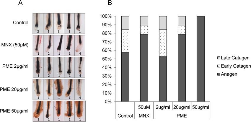
Shin et al. BMC Complementary Medicine and Therapies   (2020) 20:144                                                   Page 4 of 12

then culture supernatants were collected for growth fac-               Institutional Review Board of the CHA Bundang Medical
tor analysis. Fresh medium and culture supernatant from                Center (IRB No. 2018–09-009).
non-treated cells were used as blank and control, re-                     Anagen human hair follicles were isolated by micro-
spectively. Conventional immunoblot process was per-                   dissection and maintained in William’s E medium (Wel-
formed following the manufacturer’s guide. Biotin-                     GENE, Kyungsan, Korea) supplemented with 10 μg/ml insu-
conjugated anti-cytokines antibodies were used as pri-                 lin (Sigma-Aldrich, MO, USA), 10 ng/ml hydrocortisone
mary antibody and HRP-conjugated streptavidin was                      (Sigma-Aldrich, MO, USA), 20 mM HEPES (Invitrogen-
used for chemiluminescence detection. The resulting                    Gibco-BRL, NY, USA), and 1x antibiotic-antimycotic (Invi-
blots were analyzed under identical condition using                    trogen-Gibco-BRL, NY, USA) for 1 day.
chemiluminescence detector Fusion FX5 (Vilber Lour-                       Each group of 20 isolated hHFs was cultured in medium
mat, France).                                                          containing PM extract at concentrations of 2, 20 and
                                                                       50 μg/ml. On every third days, medium was replaced and
Western blot                                                           hHFs were photo-documented. Hair cycle stages of cul-
DPCs (1x10⁶cells/dish) were seeded in 100 mm culture                   tured human hair follicles were determined on day 0 and
dishes and cultured for 24 h. PM extract were treated at               6, according to hair cycle guideline [22, 23].
concentrations of 10 and 100 μg/ml for 24 h. Cells were
then washed with ice-cold PBS and lysed on ice in M-                   Statistical analysis
PER buffer (Thermofisher scientific, MA, USA) supple-                  All experimental data were presented as the mean ±
mented with Complete™ protease inhibitor cocktail and                  standard deviation (S.D.) of at least three independent
phosphatase inhibitor (Roche, Indianapolis, IN, USA).                  experiments. Experimental results were analyzed using
  40 μg of protein was analyzed by western blotting with               the SigmaPlot (Systat Software Inc., IL, USA). The statis-
appropriate antibodies to evaluate protein expression;                 tical significance of the difference was determined using
DKK-1 (1000:1 dilution, Cambridge, MA, USA), AR                        Student’s t-test. The value of p < 0.05 was considered
(1000:1 dilution, Santa Cruz, CA, USA), Erk (p44/42)                   statistically significant.
(1000:1 dilution, Santa Cruz, CA, USA), GAPDH (2000:1
dilution, Santa Cruz, CA, USA). Western blot was ana-                  Results
lyzed by chemiluminescence detector (Vilber Lourmat,                   PM extract increased cell viability and mitochondrial
France).                                                               activity of cultured human DPCs
                                                                       The effects of PM extract on cell viability and mitochon-
mRNA analysis                                                          drial activity in cultured human DPCs were investigated
DPCs (1x10⁶cells/well) were seeded in 6 well plates and                using CCK-8 assay and JC-1 mitochondrial staining, re-
cultured for 24 h. Then PM extract was treated at concen-              spectively. CCK-8 assay represents viability of cells via
trations of 20 μg/ml and 50 μg/ml for 24 h. Total RNA                  measuring dehydrogenase activity. JC-1 staining labels
was isolated using RNA isolation kit (Qiagen, RNeasy mini              mitochondria according to membrane potential, high
kit) according to the manufacturer’s guide. After RNA iso-             with red, low with green. As shown in Fig. 1a, treatment
lation, cDNA was synthesized by reverse transcription                  of PM extract increased cell viability in a dose
using eCube cDNA synthesis kit (philekorea, Korea) with                dependent manner, resulting in 47 and 61% increase at
PCR thermocycler (R&D systems, MN, USA), according                     concentrations of 10 μg/ml and 100 μg/ml, respectively.
to the manufacturer’s protocol. cDNA obtained from con-                Minoxidil, most well-known topical hair growth stimu-
trol cells and PM extract treated cells were subject to real-          lating agent, used as a positive control, also increased
time PCR analysis. TaqMan probes used in this study were               cell viability by 27% at 1 nM (Fig. 1 a). We hypothesized
as follows: GAPDH assay id 4352934E; BAD assay id                      that increased cell viability might be a consequence of
Hs00188930_m1; Bcl2 assay id Hs00608023_m1. TaqMan                     stimulated mitochondrial activity in cultured DPCs.
One-Step RT-PCR Master Mix Reagents (Life Technolo-                       To verify this hypothesis, the mitochondrial activity of
gies, CA, USA) was used. The PCR reactions were per-                   DPCs was evaluated by JC-1 staining, which measures
formed on ABI7500 Real Time PCR system following the                   mitochondrial membrane potential. As shown in Fig. 1b,
manufacturer’s protocol. The resulting data were analyzed              PM extract also increased mitochondrial membrane po-
with ABI software.                                                     tential in a dose dependent manner. The mitochondrial
                                                                       membrane potential of cultured human DPCs was in-
Human hair follicle organ culture and hair cycle scoring               creased by 52 and 71% with treatment of PM extract at
Human scalp skin specimens were obtained from pa-                      concentrations of 10 μg/ml and 100 μg/ml, respectively.
tients undergoing reconstructive plastic surgery after                 Minoxidil increased the mitochondrial potential by 43%
obtaining informed consent, following Declaration of                   when treated at 1 nM concentration (Fig. 1b). Highly ac-
Helsinki principles. The study was approved by the                     tivated mitochondrial membrane potential could be
Shin et al. BMC Complementary Medicine and Therapies (2020) 20:144 Page 5 of 12
Fig. 1 Effect of PM extract on DPCs’ viability and mitochondrial activity. DPCs were treated with 10 μg/ml and 100 μg/ml of PM extract for 24 h. a
Cell viability of DPCs treated with PM extract was measured using CCK8 assay. b JC-1 aggregates (A590)/monomer (A530) ratio of DPCs treated
with PM extract. c Distribution of JC-1 aggregates (590 nm, red) and monomer (530 nm, green) by immunofluorescence photography. Data are
presented as mean ± SD. n > 3 for each group. *p < 0.05, **p < 0.01 compared with control group
visualized by fluorescence microscopy where red dots protein levels were significantly decreased by PM extract
were increased and green dots were decreased by PM both in whole lysate and culture supernatant of cultured
extract in cultured human DPCs (Fig. 1c). Our data sug- human DPCs in dose dependent manners, revealed by
gest that PM extract enhanced the viability of DPCs by western blot and ELISA analysis, respectively (Fig. 2a, b).
stimulating the mitochondrial activity, especially mito- Because DKK-1 plays pivotal role in inducing catagen in
chondrial membrane potential. murine and human hair follicles, it could be conjectured
that PM extract could delay catagen entry by down-
PM extract decreased the expression of DKK-1 and BAD regulating DKK-1 expression in DPCs.
while increased the expression of Bcl2 Different from other cell types in hair follicles which
Hair follicle enters a regression phase, catagen, in response undergo apoptosis during catagen, DPCs are considered
to various micro-environmental stimuli. DKK-1, one of to be resistant to proapoptotic environment throughout
the most important proteins that induce catagen in hair hair cycles. This anti-apoptotic property is thought to be
follicles, is involved in apoptosis in various cell types [24, conferred by highly expressed Bcl2 level in DPCs [27].
25]. When hair follicles enter catagen, hair follicle cells Bcl2 family proteins play an important role in promoting
undergo apoptosis, resulting in follicular regression and cell survival and inhibiting the actions of pro-apoptotic
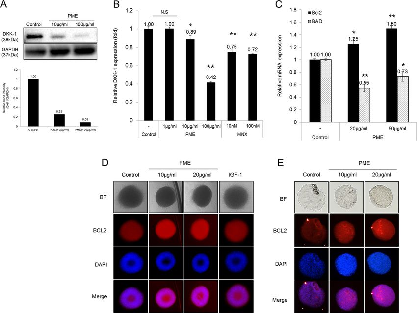
eventually hair loss [26]. As shown in Fig. 2, DKK-1 proteins. They also activate DNA repair mechanism toShin et al. BMC Complementary Medicine and Therapies (2020) 20:144 Page 6 of 12 Fig. 2 Effect of PM extract on DPCs’ protein expression related to hair cycle. DPCs (1 × 10⁶ cells/100 mm dish) were treated with 1 μg/ml, 10 μg/ ml and 100 μg/ml of PM extract for 24 h. For DP spheroids, 10 μg/ml and 20 μg/ml of PM extract were treated at the start of spheroid formation. a The protein level of DKK-1 in whole cell lysate of 2D cultured human DPCs and (b) supernatant. c Relative mRNA expression of Bcl2 and BAD in 2D cultured DPCs. d Bcl2 expression in 3D DP spheroids. e Bcl2 expression in cryo-sectioned 3D DP spheroids. Data are presented as mean ± SD. n > 3 for each group. *p < 0.05, **p < 0.01 compared with control group prevent cell death in follicular stem cells [28]. The treat- 65% increase of Bcl2 expression in whole spheroids, re- ment of PM extract had shown to increase Bcl2 and de- spectively, as measured by fluorescence intensity. The crease BAD mRNA expression in cultured human DPCs Bcl2 expression in cryo sectioned spheroids was mark- (Fig. 2c). It was suggested that the Bcl2/BAD ratio could edly increased by 67 and 105% with 10 μg/ml and 20 μg/ represent a parameter for anti-apoptotic status since ml of PM extract, respectively. Our data suggest that BAD induces apoptosis [29]. PM extract at concentra- PM extract could prolong the anagenand put off catagen tions of 20 μg/ml and 50 μg/ml increased the Bcl2/BAD entry by increasing the expression of Bcl2, and by de- ratio to 2.27 and 2.05 folds (calculated based on Fig. 2c), creasing the expression of DKK-1. suggesting possible anti-apoptotic effects of PM extract in cultured DPCs. To confirm the Bcl2 inductive effect PM extract increased the expression of growth factors in of PM extract in mimicking the DP in hair follicles, 3D cultured DPCs spheroid cultures of DPCs were constructed in the pres- DPCs are known to interact with adjacent cells, such as ence of PM extract. The expression of Bcl2 protein was hair germ cells, matrix progenitor cells, and outer root assessed by immunocytochemistry in whole and 15 μm sheath cells. These interactions lead to hair follicle re- thick cryo-sectioned 3D spheroids. As shown in Fig. 2d generation, hair shaft differentiation and hair cycle deci- and e, the expression of Bcl2 protein was significantly in- sion [30]. To investigate the changes in growth factors creased by PM extract. Treatment of PM extract at con- secreted in cultured DPCs by PM extract, growth factor centrations of 10 μg/ml and 20 μg/ml resulted in 67 and analysis was performed using dot blot assay. The
Shin et al. BMC Complementary Medicine and Therapies (2020) 20:144 Page 7 of 12
expression of IGFBP2, which controls the transcription BAD, IGFBP2, EGF, VEGF and PDGF. To verify the ef-
of VEGF by regulating the binding of IGF family pro- fects of PM extract on human hair cycle, human hair fol-
teins to their receptors [31, 32], was markedly increased licle (hHF) organ culture model was adopted. During 6-
by PM extract (PM extract, 3rd row in Fig. 3). The ex- day incubation period, 57.8% of hHF showed anagen hair
pression of PDGF-AA, EGF, VEGF were also signifi- follicle morphology in non-treated control group. In PM
cantly increased by PM extract compared with control extract treated groups, on the other hand, the number of
group (Fig. 3). IGFBP2, as mentioned above, regulates hair follicles in anagen was increased in a dose
the binding affinity between IGF and IGFR by binding dependent manner. As shown in Fig. 4, treatment of PM
with IGF family. When existed as a singular form in extract at concentrations of 20 μg/ml and 50 μg/ml in-
extracellular matrix, IGFBP2 translocates to the cyto- creased the ratio of hair follicles in anagen morphology
plasm and acts as a transcription factor for VEGF. VEGF to 78.9, 100%, respectively. Minoxidil, on the other hand,
is known to play an important role in mediating angio- showed a comparable result with 20 μg/ml of PM extract
genesis during hair growth cycle [33]. Stimulation of treated group (Fig. 4a, b). Our data clearly demonstrates
VEGF production by DPCs could explain the hair that PM extract could promote hair growth by extending
growth promoting potential of herbal extracts [34]. EGF anagen and delaying catagen entry in human hair
is known to play an important role in anagen elongation follicles.
or telogen escape, especially for telogen to anagen transi-
tion by stimulating EGFR located in the outer root Anti-androgenic effects of PM extract in cultured human
sheath [35]. PDGF-AA also induces and maintains ana- DPCs
gen of hair follicles. Studies have shown that injection of AGA is caused by hair follicle miniaturization resulted
anti-PDGF antibody to mouse skin immediately trig- from repeated hair cycles with shortened anagen that
gered catagen and resulted in hair loss [36]. Our data over time produces shorter and thinner hair [37, 38]. In
strongly suggest that PM extract could support hair DPCs, testosterone could be converted to more potent
growth by stimulating the expression of growth factors androgen dihydrotestosterone (DHT) by 5α-reductase in
essential for anagen induction and maintenance, e.g. cytoplasm. Transformed androgen, DHT, binds to an-
IGFBP2, VEGF, EGF, and PDGF-AA. drogen receptor (AR) and then stimulate the expression
of androgenic proteins which cause AGA [39]. In this
PM extract elongated anagen in human hair follicle organ context, anti-androgenic effects of PM extract in pros-
culture tate cancer cell lines, 22RV1 and LNCap were investi-
As mentioned previously, PM extract could possibly gated. Treatment of PM extract in 22RV1 cells
elongate anagen by modulating the expression of several transiently expressing pGL4.36 [luc2P/MMTV/Hygro]
markers in cultured human DPCs, e.g. DKK-1, Bcl2, reporter vector markedly decreased the DHT induced
Fig. 3 Effect of PM extract on growth factors secreted from DPCs. 41 types of growth factor analysis were performed with 20 μg/ml of PM extract
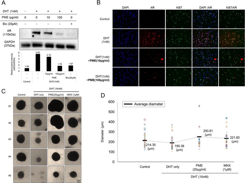
treatment. PM extract treated DPCs’ supernatant was analyzed compared with DPCs growth media and culture supernatant of non-treated controlShin et al. BMC Complementary Medicine and Therapies (2020) 20:144 Page 8 of 12 Fig. 4 Effect of PM extract on anagen elongation and catagen entry in human hair follicle organ culture model. Human hair follicles (20 hair follicles/ group) were treated with 2 μg/ml, 20 μg/ml and 50 μg/ml of PM extract and 50 μM of minoxidil. After 6 days of incubation, hair follicle morphology was assessed following hair cycle scoring criteria. a Representative images of hair follicles for each experimental group. b Calculated ratio of anagen, early categen, late catagen AR response by 40% compared with DHT only treated size of DHT affected spheroids but the mean diameter group (data not shown). Moreover, prostate specific was far less than that of PM extract treated group (Fig. antigen (PSA) ELISA in LNCap cells showed that DHT- 5d). The spheroid forming ability and the size of spher- induced PSA level was significantly decreased by PM ex- oids formed by cultured hair follicle DPCs are generally tract (data not shown). To investigate the possible anti- regarded as the hair inductive capacity and hair thick- androgenic role of PM extract in preventing hair loss, ness, respectively [40, 41]. Also, because the expression the androgenic phenotypes in cultured human DPCs of AR in balding hair is much higher than that in non- were examined. In cultured human DPCs, treatment of balding hair [42], our study suggests that PM extract 1 nM DHT increased AR protein level by 562% and this could prevent androgen induced hair loss by reducing increment was abrogated by PM extract in a dose AR expression and preventing hair follicle dependent manner (Fig. 5a). Immunocytochemistry also miniaturization mediated by DHT which are thought to showed that AR expression in the nucleus of DPCs was be the main cause of AGA. induced by DHT but significantly reversed by PM ex- tract (Fig. 5b), suggesting that the previous anti- Discussion androgenic results obtained from prostate cancer cell Hair follicles are developmental organs. After neonatal lines were exerted by regulation of AR gene expression. follicle development has progressed, each hair follicle To confirm anti-androgenic effect of PM extract, 3D enters a regular cycle called anagen, catagen and telogen, DPC spheroids were constructed in the presence of results in repeating follicle formations and regressions. DHT with/without PM extract. As shown in Fig. 5c, During catagen of the cycle, most of the cell types in treatment of DHT at 10 nM concentration resulted in hair follicles undergo apoptotic progression caused by formation of spheroid-like small cell aggregates mostly activation of apoptotic ligands or mitochondrial dysfunc- under 100 μm in diameter, in contrast to the control tions and lead follicles to be phagocytized while DP re- group without DHT. Treatment of 20 μg/ml of PM ex- mains intact [43]. This perpetuating property of DP is tract, however, fully abrogated the inhibitory effect of conferred by anti-apoptotic molecules such as Bcl-2 DHT on spheroid formation and recovered the size of expressed in DP. AGA, also referred to as male pattern spheroid (Fig. 5c). PM extract increased the mean diam- hair loss, is characterized by thinning of hair resulted eter (μm) of spheroids by 17 and 31% compared with from hair follicle miniaturization, leading to gradual re- non-treated and DHT treated control, respectively (cal- placement of large pigmented hairs (terminal hairs) by culated based on Fig. 5d). Minoxidil also recovered the barely visible, unpigmented hairs (vellus hairs) in
Shin et al. BMC Complementary Medicine and Therapies      (2020) 20:144                                                          Page 9 of 12

 Fig. 5 Anti-androgenic effect of PM extract on both 2D cultured DPCs and 3D DP spheroids. 2D cultured DPCs were co-treated with 1 nM of
 DHT and 10 μg/ml or 100 μg/ml of PM extract. Then, AR protein expression was analyzed by western blot or immunocytochemistry. For spheroids
 cultures, 10 nM of DHT was co-treated with 20 μg/ml of PM extract from the start of 3D DP spheroid formation. a AR protein was decreased by
 PM extract in dose dependent manner. b Nuclear AR was decreased by PM extract. c, d The size of spheroids was reduced by DHT treatment
 and recovered by PM extract as shown in representative images and calculated size distribution

genetically predisposed individuals [44, 45]. Androgen                    Bax, etc. [4, 48]. It is expected that PM extract might
plays a crucial role in accelerating hair loss by shorten-                prolong anagen by delaying catagen entry, and this idea
ing of hair cycle period resulted from premature catagen                  was further supported by the fact that PM extract in-
induction with reduced anagen duration [46, 47].                          creased the expression of Bcl-2 and decreased the ex-
  The present report demonstrates the hair growth pro-                    pression of BAD (Fig. 2b, d and e).
moting effects of PM extract, especially focused on hu-                     It is well-documented in many reports that some
man DPCs, a key regulator of the hair cycle. We have                      growth factors are essential for hair growth. In this
found that PM extract enhanced cell viability and mito-                   study, treatment of PM extract in cultured human DPCs
chondrial membrane potential in cultured human DPCs                       stimulated the production of IGFBP2, VEGF, PDGF, and
(Fig. 1a, b, c). This upregulated mitochondrial potential                 EGF, suggesting a possible underlying mechanism of hair
means that DPCs get harnessed with opposing force to                      growth promoting activity of PM extract (Fig. 3). The
apoptotic regression. We have found that the expression                   VEGF and VEGFR are essential proteins that support
of DKK-1, which promotes catagen entry, was signifi-                      proliferation of DPCs and angiogenesis around hair folli-
cantly decreased both in protein and mRNA levels by                       cles during hair cycle [33, 49]. IGFBP2 is also of import-
PM extract (Fig. 2a, c). Treatment of DKK-1 is reported                   ance because the transcription level of VEGF family is
to induce apoptosis in cultured outer root sheath kera-                   controlled by binding affinity of IGFBP2 to its receptor
tinocyte cells through changing the expression of anti-                   [31, 32]. PDGF, which mainly supplied from adipocyte
apoptotic and pro-apoptotic proteins like Bcl-2, Bad,                     near DP, is also expressed in DP and affects the DP itself
Shin et al. BMC Complementary Medicine and Therapies   (2020) 20:144                                                                    Page 10 of 12

[50, 51]. In terms of maintaining the size of DP, PDGF-                found that PM extract prolonged the anagen stage of
AA increases the DP size by modulating stem cells in                   hHF in the human hair follicle organ culture model, that
hair follicle [52]. EGF is well known growth factor for                supports the possible therapeutic potential of PM extract
maturation of hair follicles, especially promoting prolif-             for anti-hair loss treatment.
eration of DP via notch signaling pathway [53].
   By integrating all the results obtained from in vitro ex-           Conclusions
periments, we raised a hypothesis that PM extract could                In conclusion, our data suggest that PM extract could
support hair growth by stimulating growth factors and es-              promote hair growth possibly through prolongation of
pecially by elongating anagen. To verify our hypothesis,               anagen by preventing catagen entry with increased pro-
we performed an ex-vivo organ culture experiment for                   duction of growth factors and abrogating the effects of
evaluating hair growth. As a result, PM extract extended               androgen, DHT.
anagen and prevented catagen induction of hair follicles in
human hair follicle organ culture model. Our results                   Supplementary information
strongly demonstrate the possible potential of PM extract              Supplementary information accompanies this paper at https://doi.org/10.
as a new therapeutic agent for treating hair loss.                     1186/s12906-020-02940-5.

   Because androgen plays a pivotal role in the process of
                                                                        Additional file 1.
AGA, searching for topically applicable anti-androgenic
                                                                        Additional file 2.
materials could be a promising therapeutic regimen for
androgenetic hair loss. Anti-androgenic therapies are
                                                                       Abbreviations
commonly performed in various methods like blocking                    22RV1: The cell line expresses prostate specific antigen; BAD: Bcl2 associated
androgen receptor, reducing the production of androgen,                agonist of cell death; Bcl2: B-cell lymphoma2; BSA: Bovine serum albumin;
inhibiting 5-alpha-reductase activity etc. Also, pathogen-             CCK8: Cell counting kit8; DAPI: 4′,6-diamidino-2-phenylindole;
                                                                       DHT: Dihydrotestosterone; DKK1: Dickkopf-related protein 1; EGF: Epidermal
esis of androgenetic alopecia patients displayed                       growth factor; EGFR: Epidermal growth factor receptor; ELISA: Enzyme-linked
miniaturization of DP size via constant exposure to an-                immunosorbent assay; HGF: Heptocyte growth factor; hHF: Human hair
drogens [44]. In this context, PM extract was found to                 follicle; IGF1: Insulin-like growth factor1; IGFBP2: Insulin-like growth factor
                                                                       binding protein 2; JC1: Mitochondrial membrane potential kit;
abrogate the DHT- induced AR expression almost com-                    KGF: Keratinocyte growth factor; LNcap: The cells which are responsive to 5-
pletely and phenotypically recover DHT- induced DP                     alpha-dihydrotestosterone; MMTV: Mouse mammary tumor virus;
miniaturization by enhancing spheroid forming capacity                 NADH: Reduction form of nicotinamide adenine dinucleotide;
                                                                       NADPH: Reduction form of nicotinamide adenine dinucleotide phosphate;
in 3D cultured human DPCs. Our data strongly suggest                   PDGF: Platelet-derived growth factor; PSA: Prostate specific antigen;
an anti-androgenic therapeutic potential of PM extract,                TGF: Transforming growth factor; VEGF: Vascular endothelial growth factor
possibly exerted by blocking of androgen receptor
                                                                       Acknowledgements
expression.
                                                                       Not applicable.
   Natural product extract (NPE) is generally composed
of thousands of chemicals with various biological activ-               Authors’ contributions
ities, and this is why PM extract showed pleiotropic ef-               SL designed and guaranteed the whole experiment studies, revised
                                                                       manuscript; JS wrote the paper and carried ELISA, 3D DPC spheroids
fects on cultured human DPCs. Even though we could                     formation, immunocytochemistry, dot blot assay, all statistical data analysis.
not completely exclude the possibility that one chemical               JK carried out experiments about DPCs’ cell viability and mRNA analysis; YC
component of PM extract exerted all the hair growth                    carried out western blot; SP, JJ carried out HPLC analysis of PM extract; YN
                                                                       carried out ex-vivo test for hair growth; SYL, MJ prepared PM extract. All au-
promoting effects we have found, it seems plausible that               thors have read and approved the manuscript.
several chemicals worked separately and/or cooperatively
and this question is open for further investigation.                   Funding
                                                                       Not applicable.
   Although several reports demonstrated hair growth
promoting effects of PM extract, the experiments were                  Availability of data and materials
performed in mice and mouse cell lines [14, 54].                       The datasets used and/or analyzed during the current study available from
   In this report, we have found that treatment of PM ex-              the corresponding author on reasonable request.

tract to cultured human DPCs showed hair growth sup-                   Ethics approval and consent to participate
porting activities by stimulating cell proliferation and               Human scalp skin specimens were obtained from patients undergoing
mitochondrial activity, decreasing the gene expression of              reconstructive plastic surgery after obtaining informed consent, following
                                                                       Declaration of Helsinki principles. The study was approved by the Institutional
DKK-1, and modulating the expression of Bcl-2 and                      Review Board of the CHA Bundang Medical Center (IRB No. 2018–09-009).
BAD. In addition, PM extract stimulated the secretion of
growth factors essential for hair growth, prominently ab-              Consent for publication
                                                                       Not applicable.
rogating the DHT-induced stimulation of androgen re-
ceptor (AR) expression, and inhibiting the increment of                Competing interests
DP 3D spheroids size by DHT treatment. We also have                    The authors report no conflicts of interest to declare.
Shin et al. BMC Complementary Medicine and Therapies (2020) 20:144 Page 11 of 12
Author details 22. Peters EM, Hansen MG, Overall RW, Nakamura M, Pertile P, Klapp BF, Arck
1
Research Park, LG Household & Healthcare Ltd, 70, Magokjoongang 10-ro, PC, Paus R. Control of human hair growth by neurotrophins: brain-derived
Gangseo-gu, Seoul 07795, South Korea. 2Department of biotechnology, CHA neurotrophic factor inhibits hair shaft elongation, induces catagen, and
University, 335, Pangyo-ro, Bundang-gu, Seongnam-si, Gyeonggi-do 13488, stimulates follicular transforming growth factor β2 expression. J Investig
South Korea. Dermatol. 2005;124(4):675–85.
23. Oh JW, Kloepper J, Langan EA, Kim Y, Yeo J, Kim MJ, Hsi TC, Rose C, Yoon
Received: 1 August 2019 Accepted: 27 April 2020 GS, Lee SJ, et al. A guide to studying human hair follicle cycling in vivo. J
Invest Dermatol. 2016;136(1):34–44.
24. Hirata H, Hinoda Y, Nakajima K, Kawamoto K, Kikuno N, Ueno K, Yamamura
S, Zaman MS, Khatri G, Chen Y. Wnt antagonist DKK1 acts as a tumor
References suppressor gene that induces apoptosis and inhibits proliferation in human
1. Yang CC, Cotsarelis G. Review of hair follicle dermal cells. J Dermatol Sci. renal cell carcinoma. Int J Cancer. 2011;128(8):1793–803.
2010;57(1):2–11. 25. Di M, Wang L, Li M, Zhang Y, Liu X, Zeng R, Wang H, Chen Y, Chen W,
2. Kubanov A, Gallyamova YA, Korableva O, Kalinina P. The role of the VEGF, Zhang Y. Dickkopf1 destabilizes atherosclerotic plaques and promotes
KGF, EGF, and TGF-Β1Growth factors in the pathogenesis of Telogen plaque formation by inducing apoptosis of endothelial cells through
effluvium in women. Biomed Pharmacol J. 2017;10(1):191–8. activation of ER stress. Cell Death Dis. 2017;8(7):e2917.
3. Madaan A, Verma R, Singh AT, Jaggi M. Review of hair follicle dermal papilla 26. Morgan MB, Rose P. An investigation of apoptosis in androgenetic alopecia.
cells as in vitro screening model for hair growth. Int J Cosmet Sci. 2018; Ann Clin Lab Sci. 2003;33(1):107–12.
40(5):429–50. 27. Soma T, Hibino T. Dominant Bcl-2 expression during telogen–anagen
4. Kwack MH, Kim MK, Kim JC, Sung YK. Dickkopf 1 promotes regression of transition phase in human hair. J Dermatol Sci. 2004;36(3):183–5.
hair follicles. J Investig Dermatol. 2012;132(6):1554–60. 28. Sotiropoulou PA, Candi A, Mascre G, De Clercq S, Youssef KK, Lapouge G,
5. Alonso L, Fuchs E. The hair cycle. J Cell Sci. 2006;119(Pt 3):391–3. Dahl E, Semeraro C, Denecker G, Marine JC, et al. Bcl-2 and accelerated DNA
6. Muller-Rover S, Rossiter H, Lindner G, Peters EM, Kupper TS, Paus R. Hair repair mediates resistance of hair follicle bulge stem cells to DNA-damage-
follicle apoptosis and Bcl-2. J Invest Dermatol Symp Proc. 1999;4(3):272–7. induced cell death. Nat Cell Biol. 2010;12(6):572–82.
7. Botchkareva NV, Ahluwalia G, Shander D. Apoptosis in the hair follicle. J 29. Ruvolo P, Deng X, May W. Phosphorylation of Bcl2 and regulation of
Investig Dermatol. 2006;126(2):258–64. apoptosis. Leukemia. 2001;15(4):515.
8. Inui S, Fukuzato Y, Nakajima T, Yoshikawa K, Itami S. Androgen-inducible 30. Oshima H, Rochat A, Kedzia C, Kobayashi K, Barrandon Y. Morphogenesis
TGF-β1 from balding dermal papilla cells inhibits epithelial cell growth: a and renewal of hair follicles from adult multipotent stem cells. Cell. 2001;
clue to understand paradoxical effects of androgen on human hair growth. 104(2):233–45.
FASEB J. 2002;16(14):1967–9. 31. Azar WJ, Azar SH, Higgins S, Hu J-F, Hoffman AR, Newgreen DF, Werther GA,
9. Hibino T, Nishiyama T. Role of TGF-β2 in the human hair cycle. J Dermatol Russo VC. IGFBP-2 enhances VEGF gene promoter activity and consequent
Sci. 2004;35(1):9–18. promotion of angiogenesis by neuroblastoma cells. Endocrinology. 2011;
10. Kwack MH, Sung YK, Chung EJ, Im SU, Ahn JS, Kim MK, Kim JC. 152(9):3332–42.
Dihydrotestosterone-inducible dickkopf 1 from balding dermal papilla cells 32. Russo V, Azar W, Yau S, Sabin M, Werther G. IGFBP-2: the dark horse in
causes apoptosis in follicular keratinocytes. J Investig Dermatol. 2008;128(2): metabolism and cancer. Cytokine Growth Factor Rev. 2015;26(3):329–46.
262–9. 33. Yano K, Brown LF, Detmar M. Control of hair growth and follicle size by
11. Song S-H, Lim JH, Son SK, Choi J, Kang N-G, Lee S-M. Prevention of lipid VEGF-mediated angiogenesis. J Clin Invest. 2001;107(4):409–17.
loss from hair by surface and internal modification. Sci Rep. 2019;9(1):9834. 34. Shin H, Cho A-R, Kim DY, Munkhbayer S, Choi S-J, Jang S, Kim SH, Shin H-C,
12. Bounda G-A, Feng Y. Review of clinical studies of Polygonum multiflorum Kwon O. Enhancement of human hair growth using Ecklonia cava
Thunb. and its isolated bioactive compounds. Pharm Res. 2015;7(3):225. polyphenols. Ann Dermatol. 2016;28(1):15–21.
13. Park HJ, Zhang N, Park DK. Topical application of Polygonum multiflorum 35. Mak KK, Chan SY. Epidermal growth factor as a biologic switch in hair
extract induces hair growth of resting hair follicles through upregulating growth cycle. J Biol Chem. 2003;278(28):26120–6.
Shh and beta-catenin expression in C57BL/6 mice. J Ethnopharmacol. 2011; 36. Tomita Y, Akiyama M, Shimizu H. PDGF isoforms induce and maintain
135(2):369–75. anagen phase of murine hair follicles. J Dermatol Sci. 2006;43(2):105–15.
14. Li Y, Han M, Lin P, He Y, Yu J, Zhao R. Hair growth promotion activity and 37. Rathnayake D, Sinclair R. Male androgenetic alopecia. Expert Opin
its mechanism of Polygonum multiflorum. Evid Based Complement Alternat Pharmacother. 2010;11(8):1295–304.
Med. 2015;2015:517901. 38. Kaliyadan F, Nambiar A, Vijayaraghavan S. Androgenetic alopecia: an update.
15. Chen H-S, Liu Y, Lin L-Q, Zhao J-L, Zhang C-P, Jin J-C, Wang L, Bai M-H, Indian J Dermatol Venereol leprol. 2013;79(5):613–25.
Wang Y-C, Liu M. Anti-proliferative effect of an extract of the root of 39. Yang YC, Fu HC, Wu CY, Wei KT, Huang KE, Kang HY. Androgen receptor
Polygonum multiflorum Thunb. On MCF-7 human breast cancer cells and accelerates premature senescence of human dermal papilla cells in
the possible mechanisms. Mol Med Rep. 2011;4(6):1313–9. association with DNA damage. PLoS One. 2013;8(11):e79434.
16. Chen L, Duan H, Xie F, Gao Z, Wu X, Chen F, Wu W. Tetrahydroxystilbene 40. Chi W, Wu E, Morgan BA: Dermal papilla cell number specifies hair size,
Glucoside effectively prevents apoptosis induced hair loss. Biomed Res Int. shape and cycling and its reduction causes follicular decline. Development.
2018;2018:1380146. 2013;140(8):1676–83.
17. Ling S, Xu JW. Biological activities of 2,3,5,4′-Tetrahydroxystilbene-2-O-beta- 41. Randall VA, Hibberts NA, Thornton MJ, Hamada K, Merrick AE, Kato S, Jenner
D-Glucoside in Antiaging and Antiaging-related disease treatments. TJ, De Oliveira I, Messenger AG. The hair follicle: a paradoxical androgen
Oxidative Med Cell Longev. 2016;2016:4973239. target organ. Horm Res Paediatr. 2000;54(5–6):243–50.
18. Dong Q, Xing W, Fu F, Liu Z, Wang J, Liang X, Zhou X, Yang Q, Zhang W, Gao F, 42. Hibberts N, Howell A, Randall V. Balding hair follicle dermal papilla cells
et al. Tetrahydroxystilbene Glucoside inhibits excessive autophagy and improves contain higher levels of androgen receptors than those from non-balding
microvascular endothelial dysfunction in Prehypertensive spontaneously scalp. J Endocrinol. 1998;156(1):59–65.
hypertensive rats. American J Chinese Med. 2016;44(7):1393–412. 43. Paus R, Nickoloff BJ, Ito T. A 'hairy' privilege. Trends Immunol. 2005;26(1):32–40.
19. Xu S, Liu J, Shi J, Wang Z, Ji L. 2,3,4′,5-tetrahydroxystilbene-2-O-beta-D- 44. Ellis JA, Sinclair R, Harrap SB. Androgenetic alopecia: pathogenesis and
glucoside exacerbates acetaminophen-induced hepatotoxicity by inducing potential for therapy. Expert Rev Mol Med. 2002;4(22):1–11.
hepatic expression of CYP2E1, CYP3A4 and CYP1A2. Sci Rep. 2017;7(1): 45. Whiting DA. Possible mechanisms of miniaturization during androgenetic
16511. alopecia or pattern hair loss. J Am Acad Dermatol. 2001;45(3 Suppl):S81–6.
20. Cho CH, Bae JS, Kim YU. 5alpha-reductase inhibitory components as 46. Rebora A. Pathogenesis of androgenetic alopecia. J Am Acad Dermatol.
antiandrogens from herbal medicine. J Acupunct Meridian Stud. 2010;3(2): 2004;50(5):777–9.
116–8. 47. Lolli F, Pallotti F, Rossi A, Fortuna MC, Caro G, Lenzi A, Sansone A,
21. Yon J, Park S, Lin C, Gwon L, Lee J-G, Baek I-J, Lee B, Yun Y, Nam S-Y. Hair Lombardo F. Androgenetic alopecia: a review. Endocrine. 2017;57(1):9–17.
growth promoting effects of emodin in telogenic C57BL/6 mice. Korean J
Vet Res. 2016;56:97–101.Shin et al. BMC Complementary Medicine and Therapies (2020) 20:144 Page 12 of 12
48. Grotewold L, Ruther U. The Wnt antagonist Dickkopf-1 is regulated by bmp
signaling and c-Jun and modulates programmed cell death. EMBO J. 2002;
21(5):966–75.
49. Li W, Man XY, Li CM, Chen JQ, Zhou J, Cai SQ, Lu ZF, Zheng M. VEGF
induces proliferation of human hair follicle dermal papilla cells through
VEGFR-2-mediated activation of ERK. Exp Cell Res. 2012;318(14):1633–40.
50. Kamp H, Geilen CC, Sommer C, Blume-Peytavi U. Regulation of PDGF and
PDGF receptor in cultured dermal papilla cells and follicular keratinocytes of
the human hair follicle. Exp Dermatol. 2003;12(5):662–72.
51. Heldin CH. Autocrine PDGF stimulation in malignancies. Ups J Med Sci.
2012;117(2):83–91.
52. Gonzalez R, Moffatt G, Hagner A, Sinha S, Shin W, Rahmani W, Chojnacki A,
Biernaskie J. Platelet-derived growth factor signaling modulates adult hair
follicle dermal stem cell maintenance and self-renewal. NPJ Regen Med.
2017;2:11.
53. Zhang H, Nan W, Wang S, Zhang T, Si H, Wang D, Yang F, Li G. Epidermal
growth factor promotes proliferation of dermal papilla cells via notch
signaling pathway. Biochimie. 2016;127:10–8.
54. Sun YN, Cui L, Li W, Yan XT, Yang SY, Kang JI, Kang HK, Kim YH. Promotion
effect of constituents from the root of Polygonum multiflorum on hair
growth. Bioorg Med Chem Lett. 2013;23(17):4801–5.
Publisher’s Note
Springer Nature remains neutral with regard to jurisdictional claims in
published maps and institutional affiliations.You can also read