Low cytotoxicity, and antiproliferative activity on cancer cells, of the plant Senna alata (Fabaceae) - SciELO
←
→
Page content transcription
If your browser does not render page correctly, please read the page content below
ISSN Printed: 0034-7744           ISSN digital: 2215-2075

                                                 DOI 10.15517/rbt.v69i1.42144

           Low cytotoxicity, and antiproliferative activity on cancer cells,
                        of the plant Senna alata (Fabaceae)
Amir Modarresi Chahardehi1, Hasni Arsad1*, Noor Zafirah Ismail1 & Vuanghao Lim1
1.   Integrative Medicine Cluster, Advanced Medical and Dental Institute, Universiti Sains Malaysia, 13200, Bertam,
     Penang, Malaysia; amirmch@gmail.com, hasniarsad@usm.my, piecesnzi@gmail.com, vlim@usm.my

                        Received 04-VI-2020.               Corrected 23-XI-2020.         Accepted 14-XII-2020.

     ABSTRACT. Introduction: The leaves of Senna alata from the Fabaceae family have been used in folk
     medicine for the cure of skin disease. In this study, we tested the extract and fractions on brine shrimp lethal-
     ity test and antiproliferative activity on cancer and normal cell lines. Objective: In this study, we assessed the
     cytotoxicity of S. alata using brine shrimp test and two cell lines. Methods: The 80 % ethanolic leaf extract
     and its fractions were examined for possible cytotoxic effect using sulforhodamine B (SRB) cytotoxicity assay
     towards breast cancer (MCF-7), normal (MCF10A) cell lines, and brine shrimp lethality test (BSLT). Results:
     The brine shrimp lethality bioassay exhibits no cytotoxicity even at high concentration (5 000 µg/mL). The LC50
     for dichloromethane, chloroform, butanol, and aqueous were > 1 000 µg/mL (non-toxic). The IC50 for in vitro
     SRB cytotoxicity against MCF-7 for n-hexane was 0.013 µg/mL, which was considered highly toxic, while
     dichloromethane and chloroform recorded at 47.11 and 57.61 µg/mL, respectively after 72 hours exposure time
     although there was no cytotoxicity found on the normal cell line. Conclusion: This study shows that S. alata
     crude ethanolic leaf extract and its fractions potentially contain significant bioactive compounds that are safe
     from adverse effects, which proves the therapeutic application of S. alata in traditional remedy.

     Key words: cytotoxicity; brine shrimp; Senna alata; breast cancer.

                 Modarresi Chahardehi, A., Arsad, H., Zafirah Ismail, N., & Lim, V. (2021). Low
                     cytotoxicity, and antiproliferative activity on cancer cells, of the plant Senna alata
                     (Fabaceae). Revista de Biología Tropical, 69(1), 317-330. DOI 10.15517/rbt.
                     v69i1.42144

     Natural products have been recognized as                                 stimulating enzymes such as glutathione trans-
the source of medicinal substances and struc-                                 ferase or preventing cell proliferation (Shareef,
tural sustainability for several years (Beutler,                              Ashraf, & Sarfraz, 2016).
2019). The natural resources of medicinal                                          Over a million women with breast can-
plants are precious phytochemicals that are                                   cer are identified per year around the globe
often employed for the treatment of different                                 (Shareef et. al., 2016); therefore, breast cancer
diseases (Al-Ansari et al., 2019), especially for                             has been the second most common cause of
cancer treatment. Plants as natural resources                                 death for women (Azamjah, Soltan-Zadeh, &
are used for several years provide poten-                                     Zayeri, 2019; Levitsky & Dembitsky, 2014).
tial chemical therapeutics in cancer treatment                                Since mammography is not available for rou-
and interest in nature (Akindele et al., 2015).                               tine screening, late stages of breast cancer are
Hence, phytochemicals cover a wide range                                      usually investigated (Shareef et al., 2016). The
of chemical spaces for the discovery of drugs                                 function of flavonoids in cancer prevention
(Mohanraj et al., 2018). Phytochemicals have                                  has been documented (Elsyana, Bintang, &
various pharmacology mechanisms, including                                    Priosoeryanto, 2016). Their ability and healing

           Rev. Biol. Trop. (Int. J. Trop. Biol.) • Vol. 69(1): 317-330, March 2021                                        317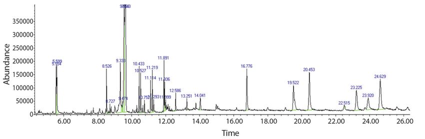
potential have been separately documented                Tatong, & Tchuente, 2017). Research on plant
worldwide, indicating that plants could become           chemistry showed that the leaves of S. alata
a prospective source of new medicines (Idris,            include saponins, anthraquinones, tannins,
Wintola, & Afolayan, 2019).                              terpenes, alkaloids, and steroids (Prasenjit,
     Cassia alata L. (also recognized as Senna           Tanaya, Sumanta, Basudeb, & Kumar, 2016).
alata) is a shrub that belongs to the Fabaceae           This significant worldwide herbal medicine has
family (sub-family Caesalpinioideae), which              been used historically as an anti-helminthic,
is distributed in the intertropical region (Saito        anti-inflammatory, uterus illness (Heyde, 1990)
et al., 2012). This plant is popularly known as          and bacterial infection (Igoli, Igwue, & Igoli,
the candle bush and also ringworm tree due               2004; Panda, Padhi, & Mohanty, 2011; Prom-
to its folk medicine, which is referenced in             gool, Pancharoen, & Deachatai, 2014; Prasenjit
the complete flower head (Hennebelle, Weni-              et al., 2016).
ger, Joseph, Sahpaz, & Bailleul, 2009). It is                 Meyer et al. (1982) identified the brine
originally from Central America, primarily               shrimp lethality bioassay (BSLA) as a par-
found in the Caribbean region, and has also              ticular test that was able to detect screening the
been spread to several tropical climates on all          range of crude plant extracts in herbal medicine
continents (Hennebelle et al., 2009). Senna              for cytotoxicity in a simple, quick and exten-
alata has been utilized primarily for traditional        sive bioassay for bioactive compounds of natu-
medicine against skin infection, and constipa-           ral product (Meyer et al., 1982; Karchesy et al.,
tion (Elsyana et al., 2016; Hennebelle et al.,           2016; Henry, 2017). The brine shrimp lethality
2009) and lately has been suggested for the              test (BSLT) is the primary anticancer test pro-
cosmetic industry as a natural product (Elsyana          cess (Prasetyo, Sidharta, Hartini, & Mursyanti,
et al., 2016). Extracts of S. alata are consid-          2019). However, there is a significant correla-
ered to possess antibacterial activity; however,         tion between BSLT toxicity and cytotoxicity
some other antibacterial effects such as preven-         in certain cell lines, but this approach is not
tion bacterial adhesion and biofilm formation            unique to anticancer activity (Asnaashari et al.,
besides specific compounds and mechanisms                2017). The previous result showed that LC50
of action are not discovered properly (Saito             value on brine shrimp larvae for ethanol extract
et al., 2012). This plant possesses potential            of S. alata was 7.7 µg/mL (Logarto, Silva,
insecticidal, fungicidal (Iyengar, Rama, & Rao,          Guerra, & Iglesias, 2001). The previous study
1995; Palanichamy & Nagarajan, 1990), anti-              showed that ethanol extract brought more reli-
inflammatory (Abatan, 1990), antimicrobial               able activity than other extracts (Panda et al.,
(Ibrahim & Osman, 1995; Khan, Kihara, &                  2011). In recent years, GC-MC has developed
Omoloso, 2001), wound healing (Palanichamy,              as a primary technical tool for the secondary
Bhaskar, Bakthavathsalam, & Nagarajan, 1991)             profiling of metabolites in both plant and non-
and antitumor activity (Olarte, Herrera, Villase-        plant organisms (Kanthal, Dey, Satyavathi,
nor, & Jacinto, 2013; Pamulaparthi & Nanna,              & Bhojaraju, 2014). Thus, the aim of analys-
2015; Karchesy, Kelsey, Constantine, & Karch-            ing 80 % ethanolic extract of this study was,
esy, 2016). S. alata leaf extract is traditionally       therefore, to detect potential chemicals and to
used for treating any type of diseases (Olarte           separate the compounds and to identify them
et al., 2013), which is rich in polyphenols and          by GC-MS application (Kanthal et al., 2014).
anthraquinones (Fernand et al., 2008). The               Also, the ethanolic extract of S. alata can cause
extensive use of S. alata has been encouraged            significant toxic effects on rats due to the pres-
to look for its pharmaceutically significant             ence of some compounds like emodin, aloe-
compounds in traditional medicine in several             emodin, kaempferol and rhein (Yagi, Tigani, &
research studies (Saito et al., 2012). Tradition-        Adam, 1998). Fernand and colleagues assessed
ally, this plant used for treatment of cancer in         the range of phenolic compounds for S. alata
Cameroon (mostly breast cancer) (Tene, Tala,             between 81.2 to 106.0 % (Fernand et al., 2008).

318                                           Rev. Biol. Trop. (Int. J. Trop. Biol.) • Vol. 69(1): 317-330, March 2021
Therefore, the present study focused on the The yield of each extract was measured and
antioxidant, antiproliferative, and cytotoxicity kept until further use. Fifty milligrams of dried
induced by extract/fractions in breast cancer samples from maceration, including crude 80
(MCF-7) cells and normal human mammary % ethanol extract, n-hexane, dichloromethane
epithelial (MCF10A) cells. (DCM), chloroform, butanol and aqueous were
dissolved with 100 % DMSO in 1 mL tube,
then sonicated to dissolve the dried samples.
MATERIALS AND METHODS
Plant materials: The leaves of S. alata ABTS scavenging activity: The antioxi-
were obtained from Penang Golf Resort dant activity of various concentration (10,
(5°31’13.8” N & 100°26’35.4” E) in Bertam 5, 2.5, 1.25, 0.625, 1563, 0.078 mg/mL) of
(North of Penang State) in November 2019. S. alata 80 % ethanol extract and Trolox was
The plant was identified by a botanist, and a determined by using ABTS assay. ABTS free
voucher specimen kept at the Herbarium Unit, radical scavenging was carried out as previous-
School of Biological Sciences, Universiti Sains ly explained (Re et al., 1999). Firstly, the stock
Malaysia, Penang Island, Malaysia. solutions of 7 mM ABTS solution and 2.45
mM of potassium persulfate solution was pre-
Extraction Procedure: The leaves (3 Kg) pared and combined to make the working solu-
of S. alata were thoroughly washed with double tion ABTS•+ at an 8:12 (v/v) ratio. Then was
distilled water, dried at room temperature, and maintained in the dark at room temperature for
pulverized using a mechanical blender (Retsch, 16 to 18 h. The solution was then blended by
ZM200, Germany) at Cluster of Integrative mixing 4.0-4.5 mL ABTS radical solution with
Medicine Laboratory, Advanced Medical and 250 mL distilled water to give an absorbance
Dental Institute, Universiti Sains Malaysia. of 0.70 ± 0.02 at 734 nm. Next, 100 µL extract
Then the powder plants weighed 100 g in each (0.078 to 10 mg/mL) in absolute ethanol was
Erlenmeyer flask. Each flask was macerated applied to 180 µL of ABTS•+ working reagent
with hydroalcoholic (80 % ethanol) containing in a 96-well plate. At room temperature, for 45
400 mL of solvent. Maceration was done for minutes the 96-well plate was incubated, and
three days with mechanical stirring (Bioteck, the absorbance was recorded at 734 nm. Tripli-
Elx808) with a constant speed of 150 rpm. The cate tests have been performed. The scavenging
solvent changed daily with a new hydroalco- capacity was analyzed as a scavenging activity.
holic solvent, and residues were macerated in Scavenging activity (%)
the respective solvent for the next day to reach
exhaustive extraction (up to 3 days). After
maceration, filtration was performed using
Whatman filter paper (150 mm). The rotavapor
(Eyela, Japan) was used to concentrate the total The percentage of ABTS extract scaveng-
filtrate of alcoholic extract to dryness. The ing activity was compared with the percentage
concentrated extract was removed from the of Trolox. A graph of percent inhibition against
round bottom flask into a weighed small glass concentration was used to establish IC50.
bottle as crude 80 % ethanolic extract. This
crude extract was then fractioned by liquid- Gas chromatographic-mass spectrom-
liquid extraction using separation funnel and etry (GC-MS) analysis for crude extract:
resulted in n-hexane, dichloromethane, chloro- Elmer Clarus Mass Spectrometer together with
form, butanol, and aqueous fractions. Vacuum the Agilent Gas Chromatography (Santa Clara,
evaporator was used to evaporate each of the CA, U.S.A.) was performed for GC-MS to ana-
extract and fractions. The concentrated extracts lyze the 80 % ethanol extract. A 10 µL syringe
were frozen at -2 °C until further application. was used to inject one microliter (1 µL) into
Rev. Biol. Trop. (Int. J. Trop. Biol.) • Vol. 69(1): 317-330, March 2021 319the chromatogram system. The Helium gas of Pasteur pipette to each well; for each well a
transported the analyte in the column at a flow volume of 2 mL has been retained in order to
rate of 1.2 mL/min. During the examination, a achieve the required concentration for extract.
split ratio of 5:1 was performed. Temperature The experiment performed nine concentration
of the injector has been scheduled at 220 °C. of samples (5000, 2500, 1250, 625, 312, 156,
The analytes are extracted from capillary col- 78.1, 39.06, 19.50 µg/mL). Each concentration
umn model Agilent 19091S-433 with HP-5MS, was conducted in three replicates. When larvae
0.25 mm × 30 m × 0.25 film width. Initially the did not show any motion for 10 seconds of
temperature of the oven had been adjusted at 70 monitoring, they were supposedly dead (Meyer
°C for 2.00 min, heating up to 280 °C at 10 °C/ et al., 1982). Samples of LC50 (lethal concen-
min. It took 32.5 min overall. The energy used tration 50 %) higher than 1000 µg/mL is found
for ionisation was 60.922eV. Mass measure- to be toxic to brine shrimp. The surviving
ment was conducted at 300 °C. Identification larvae were recorded after 6 and 24 hours of
of compounds was achieved by contrasting the sample exposure. Statistical analysis was used
mess spectra with the MS library. to determine the mortality rate and the lethal
concentrations of S. alata extract resulting to
Brine shrimp lethality test (BSLT): The 50 % mortality of the brine shrimp (LC50).
cytotoxicity activity of extract and fractions
was used using BSLT method. This test was Mortality rate (%)
performed following the mentioned protocol
by Meyer et al. (1982) and McLaughlin, Rog-
ers, & Anderson (1998) with a bit modifica-
tion. The larvae of brine shrimp were used as
research specimen. Cysts were put and hatched Cytotoxicity using Sulforhodamine B
at room temperature for 48 hours with a con- (SRB) assay: Sulphorhodamine B (SRB) assay
tinuous supply of oxygen, and there is a lamp has been used to determine the cytotoxicity
above the tank’s open side which attracts the activity of S. alata extracts using breast car-
hatched shrimps near the wall of the tank, and cinoma cell line (MCF-7) and normal human
then incubate for 25-27 °C. The shrimp became mammary epithelial cells (MCF10A) from
matured as nauplii after 48 hours and ready for American Type Culture Collection (ATCC,
the experiment. The artificial seawater has been Manassas, VA, USA). MCF-7 and MCF10A
prepared to produce a 38 g/L concentration cell lines were cultured in RPMI 1640 and
by dissolving the sea salt, then the unwanted Dulbecco’s Modified Eagle Medium (DMEM)
particles were extracted to eliminate them. The medium, respectively, containing 10 % (v/v)
number of dead and surviving brine shrimp fetal bovine serum (FBS) and 1 % (v/v) peni-
nauplii was calculated in every well after 6 cillin-streptomycin (PS) (Invitrogen Co., Carls-
and 24 hours of incubation under light. Potas- bad, CA, USA). Briefly, 1×10⁴ cells/well of
sium dichromate was dissolved in artificial MCF-7 and MCF10A were separately seeded
seawater as a positive control, functioned like in 96-well plates in a triplicate row and loaded
a positive control between 0.01 to 3.00 µg/mL 100 µL culture medium (RPMI 1640 and
concentrations. Larvae from the first day were DMEM, for MCF-7 and MCF10A cell lines,
transferred to the 24-well plates (10 per each respectively). Microplates were incubated at 37
well). All the extract and fractions dissolved in °C, 5 % CO2, 95 % air, and humidity about 100
saline water and dimethyl sulfoxide (DMSO). %. On the following day, the cells were treated
As a negative control, a saline media contain- with seven concentrations of extracts (0, 9.38,
ing DMSO (1 %) were used. Ten nauplii are 18.75, 37.50, 75.00, 150.00, and 300.00 µg/mL)
counted under a dissecting microscope (Meiji for 24, 48, and 72 hrs. Following these hours,
Techno, 10X) and then transferred with the aid the plate containing extract concentration was
320 Rev. Biol. Trop. (Int. J. Trop. Biol.) • Vol. 69(1): 317-330, March 2021incubated, and finally, the test ended by adding RESULTS
cold TCA. 50 µL of cold 30 % (w/v) TCA (at
final concentration, 10 % TCA) was applied for The residue of the plant was then extracted
in-situ cell fixation with incubation at 4 °C for with n-hexane, dichloromethane (DCM), chlo-
30 minutes. Then, the supernatant solution has roform, butanol and water (aqueous) subse-
been discarded, microplates were rinsed with quently in the same way to give 80 % EtOH
(10.9 %, yield: 43.9 g), n-hexane (0.22 %,
tap water five times and kept for air-dried. Sul-
yield: 0.89 g), DCM (0.05 %, yield: 0.18 g),
forhodamine B (SRB) (50 µL) at 0.4 % (w/v)
chloroform (0.02 %, yield: 0.09 g), butanol
in 1 % acetic acid was loaded and incubated for
(0.13 %, yield: 0.51 g) and aqueous (0.16 %,
30 minutes at room temperature. Once staining
yield: 0.65 g) for using 400 g powder leaves
is finished, loose dyes have been retrieved, and
of S. alata. This study demonstrates that the
the remaining dyes have been removed using ABTS assay IC50 values of Trolox as positive
five times washing with 1 % acetic acid. After control and 80 % ethanol extract were 0.092
the plates were air-dried at room temperature, ± 0.02 and 5.59 ± 1.50 mg/mL, respectively
and then bounded stain with a 10 mM Tris base. (Table 1).
The optical density (OD) of the plate wells
has been measured with a microplate reader TABLE 1
(Biotek, Elx808) at 570 nm, and the data were Antioxidant activity of 80 % ethanolic extract
held. The percentage survival (viability) of of S. alata using ABTS assay
treated cells over the control cells ×100 (T/C)
was calculated as cell viability. ABTS (Radical scavenging assay) mg/mL
80 % Ethanol extract Trolox
IC50 value 5.59 ± 1.50 0.092 ± 0.02
% Cell viability
The findings of the GC-MS study of
Senna alata ethanolic extract contribute to
many compounds being identified. The mass
A linear regression of absorbance against spectrometry attached to the GC classifies
the examined concentrations was calculated these substances. The GC-MS spectrum and the
the concentration at which cell proliferation is potential cytotoxicity of 80 % ethanol extract
inhibited by 50 % (IC50). (crude extract) to evaluate the biomass chemi-
cal groups revealed the existence of various
Cell imaging: The high-resolution cell compounds with different retention time, as
microscopes were demonstrated after 72 hours shown in Fig. 1 and Table 2.
of incubation of cancer cell line and 24 hours The big fragments of the compound into
for normal cell line, capturing and tracking small compounds lead to peaks with varying
images using an inverted phase-contrast micro- ratios of m/z. These mass spectra are the com-
scope (Olympus, CKX41) of each concentra- pound fingerprint detectable in the data library.
tion for clearly visible cell viability and cell In this analysis, the formula and struc-
morphology evaluation. ture of 20 biomolecules can be predicted.
Further study can proceed to the isolation
Statistical analysis: Statistical analysis of bioactive compounds, and their structural
was performed using GraphPad Prism Ver.8 clarification and evaluation and screening of
(GraphPad Software, 1996). The means of pharmaceutical activity will be useful for fur-
three replicates are shown in all analytical data ther drug research. GC-MS investigated ste-
(mean ± standard deviation). P ≤ 0.05 was con- roids (ɣ-sitosterol), linear alkanes (undecane,
sidered statistically significant. octadecane, eicosane), esters (ethylparaben,
Rev. Biol. Trop. (Int. J. Trop. Biol.) • Vol. 69(1): 317-330, March 2021 321Fig. 1. GC-MC chromatogram of 80 % ethanolic extract of Senna alata.
TABLE 2
Compound investigated in the 80 % ethanol extract of Senna alata in GC-MS
Molecular Molecular Compound
RT Name of compound Percentage
formula weight (g/mol) nature
5.32 1,3,5-Triazine-2,4,6,-triamine C3H6N6 126.11 80 Cyanamide
5.59 Undecane C11H24 156.31 94 Alkane
7.73 Phenol,2-propyl- C9H12O 136.19 87 Phenylpropanes
8.52 Cycloheptasiloxane, tetradecamethyl- C14H42O7Si7 519.07 91 Cyclomethicone
8.72 Ethylparaben C8H8O3 152.15 93 Ester
8.79 Benzoic acid, 4-ethoxy, ethyl ester C11H14O3 194.23 87 Ester
9.00 Beta-D-Glucopyranoside, methyl C7H14O6 194.18 80 Glucoside
10.43 Cyclononasiloxane, octadecamethyl- C18H54O9Si9 667.40 91 Polysiloxane
10.96 Hexadecanoic acid, methyl ester C17H34O2 270.45 93 Ester
11.11 n-Hexadecanoic acid C16H32O2 256.42 97 Saturated fatty acid
11.28 Hexadecanoic acid, ethyl ester C18H36O2 284.47 93 Ester
11.88 Phytol C20H40O 296.50 90 Alcohol
12.00 9,12,5-octadecatrienoic acid, methyl ester C19H32O2 292.50 83 Ester
12.06 Octadecanoic acid C18H36O2 284.48 95 Saturated fatty acid
16.76 Octadecane C18H38 254.50 96 Alkane
16.77 Eicosane C20H42 282.50 98 Alkane
19.04 Beta-tocophenol C28H48O2 416.70 83 Tocopherol
19.50 Eicosane C20H42 282.50 91 Alkane
20.44 Vitamin E C29H50O2 430.71 97 Tocopherol
24.62 Gamma.sitosterol C29H52O2 432.70 98 Steroid
benzoic acid, hexadecanoic acid, ethyl ester, 43.76 μg/mL for the corresponding regression
hexadecanoic acid, methyl ester), tocopherol line and showed toxic signs (LC50 against
(vitamin E and β-tocopherol) as well as fatty the brine shrimp was less than 1 000 µg/mL).
acid such as n-hexadecanoic acid and octadeca- Due to high toxicity on A. salina cysts, potas-
noic acid in the 80 % EtOH extract. sium dichromate has shown limited hatching
Only two hours after an interaction with success. The median lethal concentration of
the higher potassium dichromate concentration, the brine shrimp lethality assay (LC50) for
there was a fatal effect in the brine shrimp. Senna alata leaf extract/fractions are shown
The LC50 value for potassium dichromate was in Table 3.
322 Rev. Biol. Trop. (Int. J. Trop. Biol.) • Vol. 69(1): 317-330, March 2021TABLE 3 In the present study, the cytotoxic effect
Cytotoxicity activity of various extracts (IC50) of the crude ethanol and fractioned
of Senna alata on brine shrimp
extracts (hexane, dichloromethane, chloroform,
LD50 (µg/mL)
butanol and aqueous) were identified on one
Extract/fraction 6 hrs 24 hrs human cancer cells (MCF-7) and one normal
(acute) (chronic) non-cancer cells (MCF10A) using the SRB
80 % EtOH (Crude extract) ND ND assay. 80 % EtOH extract did not show toxicity
Hexane ND ND on both cell lines (Fig. 2A). Hexane fraction
Dichloromethane ND 1 432 of S. alata exhibited an excellent inhibition
Chloroform 2 520 1 214 towards MCF-7 cells with IC50 of 0.013 µg/mL
Butanol 1 447 1 034 at 72 h, in comparison to IC50 values of 48 h
Aqueous 5 053 2 428 (Fig. 2B). It is interesting to note that this frac-
ND = not determined; LD50 value for potassium dichromate tion did not show cytotoxicity against MCF-7
was 43.76 µg/mL. cells at 24 hours. Others, such as dichlorometh-
Note: The brine shrimp mortality percentage were ane and butanol against MCF-7 cell line after
measured as mean ± SD. 72 hours with IC50 values of 47.11 and 57.61
µg/mL, respectively, have been shown to have
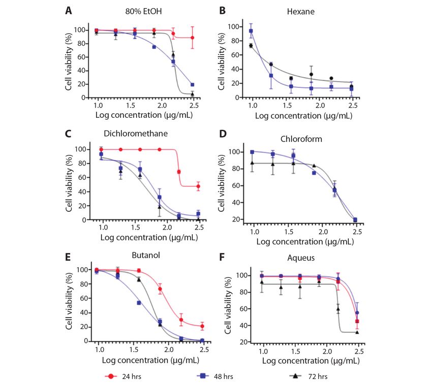
Of the six extracts tested, 2 exhibited no significant cytotoxic activity (Table 2; Fig. 2B,
toxicity to the brine shrimps. These included 80 Fig. 2C, Fig. 2E). Generally, the 80 % EtOH,
% ethanolic crude extract and n-hexane frac- chloroform, and aqueous exhibited weaker
tion, in which no mortality was observed dur- cytotoxicity profile against the MCF-7 cell line
ing screening. Dichloromethane, chloroform, (IC50 > 100 µg/mL). The viability of untreated
butanol, and aqueous fractions showed an LC50 control cells corresponds to 100 % because all
value higher than 1 000 µg/mL. There was no extracts had no cytotoxic effect on the normal
cytotoxic effect on any of the concentrations of cell, though we did not analyze the selectivity
the candle bush 80 % ethanol extract and hex- index (SI). Although, most importantly, all the
ane fraction using the BSLT method and, brine extracts did not show the cytotoxic effect on
shrimp were still moved vigorously. However, MCF10A as normal human mammary epithe-
the other four extracts showed practically non- lial cells. In addition, IC50 values were deter-
toxic (LC50 > 1 000 µg/mL) to brine shrimps. mined for SRB assay, extracts and the results
These extracts were aqueous, dichloromethane, are tabulated (Table 4), and also in Fig. 2.
chloroform, and butanol with LC50 values Treated cells were observed for the
between 1 034-2 428 µg/mL (after 24 hrs). morphological feature using a bright-field
TABLE 4
Inhibition concentration (IC50) of various extracts of Senna alata against breast cancer (MCF-7)
and normal human mammary epithelial cells (MCF10A)
MCF-7 MCF10A
Extract/fraction IC50 (µg/mL)
24 hrs 48 hrs 72 hrs 24 hrs
80 % EtOH > 100 > 100 > 100 ND
Hexane ND 9.626 0.013 ND
Dichloromethane > 100 60.03 47.11 ND
Chloroform > 100 > 100 > 100 ND
Butanol 89.30 41.98 57.61 ND
Aqueous > 100 > 100 > 100 ND
ND = not detected.
Rev. Biol. Trop. (Int. J. Trop. Biol.) • Vol. 69(1): 317-330, March 2021 323Fig. 2. In vitro cytotoxic activity of various extracts in MCF-7 cells (Human breast cancer cells) by SRB assay at different
times of exposure (24, 48 and 72 hours). All the values are mean ± SD of three samples. A. 80 % Ethanolic extract, B.
Hexane, C. Dichloromethane, D. Chloroform, E. Butanol, F. Aqueous fraction.
microscope (Olympus, CKX41) at 4X and started to decrease and round shape in contrast
10X magnification. MCF-7 and MCF10A cells to the control of the MCF-7 cells treated with
treated with various extract/fractions and then Tamoxifen which were a simple function of
observed after 72 h incubation (Fig. 3). The apoptosis (figure not shown). After 48 and 72
results only showed for hexane (Fig. 3A, Fig. hours, cells became cluster together, exhibited
3D, Fig. 3G), DCM (Fig. 3B, Fig. 3E, Fig. 3H) membrane blebbing (Fig. 3D, Fig. 3E, Fig. 3F,
and butanol (Fig. 3C, Fig. 3F, Fig. 3I) fractions. 48 hrs), and began to detach from the dish (Fig.
Significant phenotypic differences were 3H, Fig. 3I, 72 hrs). Normal MCF10A cells,
observed in the presence of Senna alata by contrast, have not shown those significant
extracts as cancer cell line was incubated morphologic changes (data not shown). This
(Fig. 3). From cell photographs at first day (24 indicates that S. alata is effective and reason-
hours) that the cells treated with fractions in ably non-toxic for folk/conventional drugs and
Fig. 3B and Fig. 3C the cells and their volume appropriate for cancer treatment.
324 Rev. Biol. Trop. (Int. J. Trop. Biol.) • Vol. 69(1): 317-330, March 2021Fig. 3. Morphological changes of MCF-7 and MCF10A cells treated with extract/fractions of Senna alata L. during 24,
48 and 72 h. IC50 calculated with the SRB assay evaluating dose-responsive curves. Various cell forms shown on MCF-7
and MCF10A, treated with S. alata during 72 h. Vehicle DMSO is used to treat control cells. A. MCF-7 cells with hexane
treatment at 24 hrs; B. Cells with dichloromethane treatment at 24 hrs; C. MCF-7 cells with butanol treatment at 24 hrs;
D. MCF-7 cells with hexane treatment at 48 hrs; E. MCF-7 cells with dichloromethane treatment at 48 hrs; F. MCF-7 cells
with butanol treatment at 48 hrs; G. MCF-7 cells with hexane treatment at 72 hrs; H. MCF-7 cells with dichloromethane
treatment at 72 hrs; I. MCF-7 cells with butanol treatment at 72 hrs; J. MCF10A cells untreated as control after 72 hrs; and
K. MCF-7 cells untreated as a control after 72 hrs.
DISCUSSION from adopting a hydrogen ion from the antioxi-
dant, decolorizing its blue colors, as ABTS free
Breast cancer is the world’s second most radicals become steady (Lee, Oh, Cho, & Ma,
fatal illness for women (Kamalanathan & Nata- 2015). The ABTS assay seems to be more sen-
rajan, 2018). Several other findings have shown
sitive than DPPH assay in detecting antioxidant
that numerous medicinal plants can be used to
activity due to extreme faster reaction kinetics,
prevent the growth of human breast cancer
and its reaction to antioxidants is stronger (Lee
(Kamalanathan & Natarajan, 2018). However,
et al., 2015), and The ABTS radical is signifi-
a collection of antioxidant compounds exists
in herbs, fruits and plants have already shown cantly more water-soluble than DPPH (He et
that breast cancer cells are destroyed by them al., 2010). Although the antioxidant activity of
without no toxic effect on normal cells (Raj, leaf extract from S. alata fractionation obtained
Ireland, Ouhtit, Gaur, & Abdraboh, 2015). Both a new indole alkaloid, 1-(4′-hydroxyphenyl)-
BSLT and ABTS (antioxidant assay) are easy 2,4,6-trihydroxy-indole-3-carboxylic acid that
to handle, low cost, and use small quantities exhibited strong antioxidant potential with an
of test equipment (Peteros & Uy, 2010; Asna- IC50 of 0.0311 μM ± 0.002 (Olarte, Her-
ashari et al., 2017). The ABTS radical-scav- rera, Villasenor, & Jacinto, 2010). In other
enging measuring technique, a popular method study, ethanol extract from leaves of this plant
utilized to test the antioxidant activity, gains showed 67% of the antioxidant activity (Sagnia
Rev. Biol. Trop. (Int. J. Trop. Biol.) • Vol. 69(1): 317-330, March 2021 325et al., 2014). Also, the hexane extract of S. butanol, and aqueous fractions. However, the
alata showed no free radical scavenging activ- percentage of deaths as time and concentra-
ity (Jacinto, Olarte, Galvez, Villasenor, & Pez- tion was increased for these fractions and the
zuto, 2005). To identify bioactive compounds existence of toxic compounds in the fractions,
from 80 % ethanolic extract of S. alata, our which requires further examination, may lead
GC-MS result confirmed the study by Ali et to that effect. Several studies showed a strong
al. (2017), which found the same compounds correlation with different tumor cell lines in the
mostly, fatty acids composition from leaves of BSLT (Elsyana et al., 2016). In BSLT, the cyto-
Senna alata (Ali et al., 2017). toxicity activity of the extract is determined by
It indicated that the brine shrimp lethality a 50 % death response to brine shrimp (LC50)
test was helpful in assessing the toxicity of the (Elsyana et al., 2016). Based on our hexane
plant extract (Sahgal et al., 2010). This proce- fraction results from MCF-7 cell line, and
dure involves exposure of brine shrimp larvae according to Elsyana et al., compared this frac-
to plant extract in saline media, and the death tion with other extracts and fractions contain-
of larvae is measured after one day (Mayilsamy ing flavonoids and triterpenoids, the maximum
& Geetharamanan, 2016). Logarto has shown cytotoxic activity was reported by hexane
that a strong link was found between the LC50 fraction (Elsyana et al., 2016). Also, Olarte
of the brine shrimp lethality test and LD50 in and colleagues (Olarte et al., 2013) found out
the acute oral toxicity test in mice (r = 0.85; P that the hexane extract from S. alata showed
< 0.05) (Logarto et al., 2001). Upon 24 hours the highest growth inhibition against MCF-7
of treatment, Artemia salina larvae with LC50; cell line among three other extracts with IC50
if the sample extract is LC50 < 1 000 μg/mL, its value 16 µg/mL which confirm our present
toxicity is high, and the cytotoxicity is expect- study with IC50 values 9.63 and 0.01 µg/mL for
ed to occur. The level of toxicity would have 48 and 72 hrs, respectively. However, based
an anticancer effect on extracts (Prasetyo et al., on the National Cancer Institute guideline
2019). Evaluating the efficiency of hatching (NCI, USA) that 30 µg/mL is the higher IC50
cysts concerning the time of exposure showed ranges assumed reasonable for purification
that extracts had notable hatching success of an extract (Akindele et al., 2015). Another
after 36-48 hours, which would be the greatest study revealed that hexane fraction of S. alata
hatching time for brine shrimp (Meyer et al., possessed cytotoxic effect against lung cancer
1982; Braguini, Pires, & Alves, 2018). cell (A549) and ovarian cancer cells (OV2008)
The method of Meyer et al., graded as (Levy & Carley, 2012). Also, ethyl acetate
toxic (LC50 value < 1 000 μg/mL) and non-toxic extract of S. alata by other studies showed 50
(LC50 value > 1 000 μg/mL) for crude extracts % inhibition (GI50) value at 5.90 µg/mL against
and pure materials (Meyer et al., 1982; Naher the MCF-7 cell line (Onyegeme-Okerenta,
et al., 2019). Another study revealed that seed 2018). On the other hand, the chloroform frac-
extract showed more toxic than leaf extract of tion showed anticancer activity against MCF-7
S. alata showed LC50 value at 4.31 and 5.29 with IC50 value 37.4 µg/ mL (Ali et al., 2017).
ppm, respectively, from the result of the brine According to other studies, chloroform extract
shrimp lethality test (Rahman, 2004). Also, from the stem of three species from Cassia
the LC50 value of the C. alata seed oil extract sp., namely, C. glauca, C. obtusifolia and
was at 250 µg/mL (Mannan et al., 2011), C. sophera showed high cytotoxicity against
and 7.74 µg/mL (Parra, Yhebra, Sardiñas, & MCF-7 cell line (Shankar & Surekha, 2017).
Buela, 2001). This suggests that these fractions Emodin was previously separated from S. alata
may contain no cytotoxic compound. Brine leaves (Prasenjit et al., 2016; Ali et al., 2017)
shrimp mortality was predicted to be related and showed anticancer activity (Hsu & Chung,
to bioactive compounds and not malnutrition 2012). These findings revealed that there is
after exposure to dichloromethane, chloroform, a direct connection between the brine shrimp
326 Rev. Biol. Trop. (Int. J. Trop. Biol.) • Vol. 69(1): 317-330, March 2021lethality test and in vitro cytotoxicity towards bromoflavone as a chemo preventive agent
the S. alata extracts. In the present study, we (Jacinto et al., 2005). More pharmacological
displayed that hexane and butanol fractions and phytochemical tests are worthwhile in this
induce apoptosis in MCF-7 human breast cells research to establish the exact principal cyto-
in a time- and concentration-dependent basis, toxicity compound reaction.
which is similar with previous studies using The result of this study shows S. alata
different extracts of S. alata (Olarte et al., could be an outstanding lead in the progress of
2013; Onyegeme-Okerenta, 2018). Our find- breast cancer anticancer agents (IC50 < 100 µg/
ing indicates that S. alata extracts cytotoxicity mL), which did not exhibit toxicity on normal
is performed through apoptotic cell death in cell line as well. Interestingly, in contrast to
tumor cells. In a study by Olarte and colleagues SRB assay results, the S. alata extract/fractions
that they treated hexane fraction with MCF-7 exhibited non-toxic activity (LC50 > 1 000
cell line. The MCF-7 cells rounded up and µg/mL) was assessing using the brine shrimp
missed contact with adjacent cells between lethality test as a primary assay for anticancer
12-24 hrs (Olarte et al., 2013). activity. The source of organic antioxidants
The anticancer function of flavonoids and is available and provides significant medical
triterpenoids, according to their antioxidant benefits. It could be inferred from GC-MS
characteristics, is consistent with their capacity findings that S. alata contains many bioactive
to scavenge free radicals, to suppress radical compounds. Our laboratory is also investigat-
oxygen species (ROS), enzymes and to prevent ing further research to clarify the mechanism of
cells and extracellular compound oxidation action of apoptosis in breast cancer and bioac-
(Elsyana et al., 2016). Flavonoids and triter- tive compounds, which will be published in a
penoids were concentration-dependent toxic future manuscript.
to the brine shrimp and, therefore, could have
resulted in the death of brine shrimp (Elsyana Ethical statement: authors declare that
et al., 2016). Several studies have shown that they all agree with this publication and made
flavonoids can prevent the proliferation and significant contributions; that there is no con-
delay of tumor cells (Razak et al., 2019). flict of interest of any kind; and that we fol-
Assessment of bioactive compounds such as lowed all pertinent ethical and legal procedures
flavonoids, alkaloids, glycosides, carbohy- and requirements. All financial sources are
drates, protein, saponins, triterpenoids, and fully and clearly stated in the acknowledge-
amino acids indicated the existence of most of ments section. A signed document has been
the component in polar extracts such as etha- filed in the journal archives.
nol, methanol and aqueous extracts comparison
with nonpolar extracts such as petroleum ether
and chloroform. Though, all extracts possessed ACKNOWLEDGMENTS
flavonoids, phenols, and tannins (Panda et
This study was supported in part by the
al., 2011). Because of its perfect fundamental
FRGS grant (203.CIPPT.6711684) from Min-
chemistry to free radical scavenging activities,
istry of Education (MOE) Government of
phenols are a significant class of antioxidants
Malaysia.
(Chaudhary et al., 2015). However, S. alata
extracts showed potential cancer cell inhibition
and reduced the risk of further proliferation RESUMEN
based on the results of the SRB assay. Jacinto
Baja citotoxicidad, y actividad antiproliferativa
et al. (2005) identified a high cancer chemo
sobre las células cancerosas, de la planta Senna alata
preventive ability while S. alata hexane leaf (Fabaceae). Introducción: Las hojas de Senna alata de la
extract was found to cause a particular activ- familia Fabaceae se han utilizado en la medicina popular
ity of the quinone reductase similar to the para la cura de enfermedades de la piel. En este estudio,
Rev. Biol. Trop. (Int. J. Trop. Biol.) • Vol. 69(1): 317-330, March 2021 327probamos el extracto de la planta en líneas celulares norma- from rhizomes of Eremostachys azerbaijanica rech.
les y cancerosas. Objetivo: Evaluamos la citotoxicidad de f. growing in Iran. Iranian Journal of Pharmaceutical
S. alata usando una prueba del camarón Artemia y la activi- Research, 16(1), 306-314.
dad antiproliferativa. Métodos: El extracto de hoja etanóli-
co al 80 % y sus fracciones se examinaron en busca de un Azamjah, N., Soltan-Zadeh, Y., & Zayeri, F. (2019). Global
posible efecto citotóxico utilizando un ensayo de citotoxi- trend of breast cancer mortality rate: A 25-year study.
cidad de sulforrodamina B (SRB) frente a líneas celulares Asian Pacific Journal of Cancer Prevention, 20(7),
de cáncer de mama (MCF-7), normales (MCF10A) y prue- 2015-2020. DOI: 10.31557/APJCP.2019.20.7.2015
ba de letalidad del camarón Artemia (BSLT). Resultados:
Beutler, J.A. (2019). Natural products as a foundation for
El bioensayo de letalidad del camarón Artemia no presenta
drug discovery. Current Protocols in Pharmacology,
citotoxicidad incluso en alta concentración (5 000 µg/mL).
86(1), e67. DOI: 10.1002/cpph.67
La CL50 para diclorometano, cloroformo, butanol y acuoso
fue > 1000 µg/mL (no tóxico). La CI50 para la citotoxici- Braguini, W.L., Pires, N.V., & Alves, B.B. (2018). Phyto-
dad in vitro de SRB contra MCF-7 para n-hexano fue de chemical analysis, antioxidant properties and brine
0.013 µg/mL, que se consideró altamente tóxica, mientras shrimp lethality of unripe fruits of Solanum viarum.
que el diclorometano y el cloroformo registraron 47.11 y Journal of Young Pharmacists, 10(2), 159-163.
57.61 µg/mL, respectivamente, después de 72 horas de
tiempo de exposición, aunque no hubo citotoxicidad encon- Chaudhary, S., Chandrashekar, K.S., Pai, K.S.R., Setty,
trada en la línea celular normal. Conclusión: Este estudio M.M., Devkar, R.A., Reddy, N.D., & Shoja, M.H.
muestra que el extracto de hoja etanólico crudo de S. alata (2015). Evaluation of antioxidant and anticancer
y sus fracciones contienen potencialmente compuestos bio- activity of extract and fractions of Nardostachys jata-
activos significativos que están a salvo de efectos adversos, mansi DC in breast carcinoma. BMC Complementary
lo que demuestra la aplicación terapéutica de S. alata como and Alternative Medicine, 15(1), 50. DOI: 10.1186/
remedio tradicional. s12906-015-0563-1
Palabras clave: citotoxicidad; Artemia; Senna alata; cán- Elsyana, V., Bintang, M., & Priosoeryanto, B.P. (2016).
Cytotoxicity and antiproliferative activity assay of
cer de mama.
Clove Mistletoe (Dendrophthoe pentandra (L.) Miq.)
Leaves Extracts. Advances in Pharmacological Scien-
ces, 2016, 3242698. DOI: 10.1155/2016/3242698
REFERENCES
Fernand, V.E., Dinh, D.T., Washington, S.J., Fakayode,
Abatan, M.O. (1990). A note on the anti-inflammatory
S.O., Losso, J.N., van Ravenswaay, R.O., & Warner,
action of plants of some Cassia species. Fitoterapia,
61, 336-338. I.M. (2008). Determination of pharmacologically
active compounds in root extracts of Cassia alata
Akindele, A.J., Wani, Z.A., Sharma, S., Mahajan, G., Satti, L. by use of high-performance liquid chromato-
N.K., Adeyemi, O.O., . . . Saxena, A.K. (2015). In graphy. Talanta, 74(4), 896-902. DOI: 10.1016/j.
vitro and In vivo anticancer activity of root extracts of talanta.2007.07.033
Sansevieria liberica gerome and labroy (Agavaceae).
Evidence-Based Complementary and Alternative GraphPad Software. (1996). GraphPad Prism version 8.00
Medicine, 2015, 560404. DOI: 10.1155/2015/560404 for Windows. Retrieved from https://www.graphpad.
com/scientific-software/prism
Al-Ansari, M., Al-Humaid, L.A., Vijayaraghavan, P.,
Ravindran, B., Chang, S.W., Agastian, P., . . . Balamu- He, W., Liu, X., Xu, H., Gong, Y., Yuan, F., & Gao, Y.
ralikrishnan, B. (2019). Identification of phytochemi- (2010). On-line HPLC-ABTS screening and HPLC-
cal components from Aerva lanata (Linn.) medicinal DAD-MS/MS identification of free radical scaven-
plants and its in-vitro inhibitory activity against drug gers in Gardenia (Gardenia jasminoides Ellis) fruit
resistant microbial pathogens and antioxidant pro- extracts. Food Chemistry, 123(2), 521-528.
perties. Saudi Journal of Biological Sciences, 26(6),
1129-1133. DOI: 10.1016/j.sjbs.2019.02.010
Hennebelle, T., Weniger, B., Joseph, H., Sahpaz, S., &
Bailleul, F. (2009). Senna alata. Fitoterapia, 80(7),
Ali, M.I., Aboul-Enein, A.M., Mohamed, S.M., Elella,
F.M.A., Mohammed, M.M.D., & Hamed, A.R. 385-393. DOI: 10.1016/j.fitote.2009.05.008
(2017). Phytochemical, cytotoxicity and antioxidant
investigation of Cassia alata leaves growing in Henry, Y.W. (2017). Brine Shrimp Cytotoxic Activity of
Egypt. Journal of Innovations in Pharmaceutical and Morinda elliptica Leaves and Root Crude Extracts.
Biological Sciences (JIPBS), 4(4), 97-105. Borneo Journal of Resource Science and Technology,
7(1), 43-46. DOI: 10.33736/bjrst.390.2017
Asnaashari, S., Delazar, A., Asgharian, P., Lotfipour, F.,
Moghaddam, S.B., & Afshar, F.H. (2017). In-vitro Heyde, H. (1990). Medicinal plants in Suriname. Parama-
bioactivity and phytochemical screening of extracts ribo, Suriname: Mungra & Madarie.
328 Rev. Biol. Trop. (Int. J. Trop. Biol.) • Vol. 69(1): 317-330, March 2021Hsu, S.C., & Chung, J.G. (2012). Anticancer potential of and Bioprospecting, 5(1), 1-16. DOI: 10.1007/
emodin. BioMedicine, 2(3), 108-116. DOI: 10.1016/j. s13659-014-0048-9
biomed.2012.03.003
Levy, A.S., & Carley, S. (2012). Cytotoxic activity of
Ibrahim, D., & Osman, H. (1995). Antimicrobial acti- hexane extracts of Psidium guajava L (Myrtaceae)
vity of Cassia alata from Malaysia. Journal of and Cassia alata L (Caesalpineaceae) in Kasumi-1
Ethnopharmacology, 45(3), 151-156. DOI: and OV2008 cancer cell lines. Tropical Journal of
10.1016/0378-8741(94)01200-J Pharmaceutical Research, 11(2), 201-207.
Idris, O.A., Wintola, O.A., & Afolayan, A.J. (2019). Eva- Logarto, A., Silva, R., Guerra, I., & Iglesias, L. (2001).
luation of the bioactivities of Rumex crispus L. leaves Comparative study of the assay of Artemia salina L.
and root extracts using toxicity, antimicrobial, and and the estimate of the medium lethal dose (LD50
antiparasitic assays. Evidence-Based Complementary value) in mice, to determine oral acute toxicity of
and Alternative Medicine, 2019, 6825297. DOI: plant extracts. Phytomedicine, 8(5), 395-400. DOI:
10.1155/2019/6825297 10.1078/0944-7113-00044
Igoli, J.O., Igwue, I.C., & Igoli, N.P. (2004). Traditional Mannan, A., Kawser, M.J., Ahmed, A.A., Islam, N.N.,
medicinal practices among the Igede people of Nige- Alam, S.M., Khan, M.A.E., & Gupta, S.D. (2011).
ria. Journal of Herbs, Spices & Medicinal Plants, Assessment of antibacterial, thrombolytic and cyto-
10(4), 1-10. DOI: 10.1300/J044v10n04_01 toxic potential of Cassia alata seed oil. Journal of
Applied Pharmaceutical Science, 1(9), 56.
Iyengar, M.A., Rama, M.P., & Rao, G. (1995). Antifungal
activity of Cassia alata, leaf extract. Indian Drugs, Mayilsamy, M., & Geetharamanan, K. (2016). Cytotoxic
32(5), 230-231. activity of certain medicinal plants extracts against
sea monkey: Artemia salina. Journal of Medicinal
Jacinto, S.D., Olarte, E.I., Galvez, M., Villasenor, I.M., & Herbs and Ethnomedicine, 2, 19-25.
Pezzuto, J.M. (2005). Leaf extracts from Cassia alata
L. (“Akapulko”) induces quinone reductase and com- McLaughlin, J.L., Rogers, L.L., & Anderson, J.E. (1998).
The use of biological assays to evaluate botanicals.
petes for estrogen receptor binding indicating cancer
Drug Information Journal, 32(2), 513-524. DOI:
chemopreventive property. Philippine Agricultural
10.1177/009286159803200223
Scientist, 88(2), 175-178.
Meyer, B.N., Ferrigni, N.R., Putnam, J.E., Jacobsen, L.B.,
Kamalanathan, D., & Natarajan, D. (2018). Anticancer
Nichols, D.E., & McLaughlin, J.L. (1982). Brine
potential of leaf and leaf-derived callus extracts of
shrimp: a convenient general bioassay for active
Aerva javanica against MCF-7 breast cancer cell line.
plant constituents. Planta Medica, 45(5), 31-34. DOI:
Journal of Cancer Research and Therapeutics, 14(2), 10.1055/s-2007-971236
321-327. DOI: 10.4103/0973-1482.171210
Mohanraj, K., Karthikeyan, B.S., Vivek-Ananth, R.P.,
Kanthal, L.K., Dey, A., Satyavathi, K., & Bhojaraju, Chand, R.P.B., Aparna, S.R., Mangalapandi, P., &
P. (2014). GC-MS analysis of bio-active com- Samal, A. (2018). IMPPAT: A curated database of
pounds in methanolic extract of Lactuca runcinata Indian medicinal plants, phytochemistry and thera-
DC. Pharmacognosy Research, 6(1), 58-61. DOI: peutics. Scientific Reports, 8(1), 4329. DOI: 10.1038/
10.4103/0974-8490.122919 s41598-018-22631-z
Karchesy, Y.M., Kelsey, R.G., Constantine, G., & Kar- Naher, S., Aziz, M.A., Akter, M.I., Rahman, S.M.M.,
chesy, J.J. (2016). Biological screening of selected Sajon, S.R., & Mazumder, K. (2019). Anti-diarrheal
Pacific Northwest forest plants using the brine shrimp activity and brine shrimp lethality bioassay of metha-
(Artemia salina) toxicity bioassay. Springerplus, 5, nolic extract of Cordyline fruticosa (L.) A. Chev. lea-
510. DOI: 10.1186/s40064-016-2145-1 ves. Clinical Phytoscience, 5(1), 15. DOI: 10.1186/
s40816-019-0109-z
Khan, M.R., Kihara, M., & Omoloso, A.D. (2001). Anti-
microbial activity of Cassia alata. Fitoterapia, 72(5), Olarte, E.I., Herrera, A.A., Villasenor, I.M., & Jacinto, S.D.
561-564. DOI: 10.1016/S0367-326X(00)00335-X (2010). Antioxidant activity of a new indole alkaloid
from Cassia alata L. Philippine Agricultural Scien-
Lee, K.J., Oh, Y.C., Cho, W.K., & Ma, J.Y. (2015). Antioxi- tist, 93(3), 250-254.
dant and anti-Inflammatory activity determination of
one hundred kinds of pure chemical compounds using Olarte, E.I., Herrera, A.A., Villasenor, I.M., & Jacinto, S.D.
offline and online screening HPLC assay. Evidence- (2013). In vitro antitumor properties of an isolate
Based Complementary and Alternative Medicine, from leaves of Cassia alata L. Asian Pacific Journal
2015, 165457. DOI: 10.1155/2015/165457 of Cancer Prevention, 14(5), 3191-3196.
Levitsky, D.O., & Dembitsky, V.M. (2014). Anti-breast Onyegeme-Okerenta, B. (2018). Ethyl acetate extract of
cancer agents derived from plants. Natural Products Senna alata (L) Roxb increases cytotoxicity in the
Rev. Biol. Trop. (Int. J. Trop. Biol.) • Vol. 69(1): 317-330, March 2021 329human breast, prostate and colorectal cancer cells. Razak, N.A., Abu, N., Ho, W.Y., Zamberi, N.R., Tan, S.W.,
Journal of Cancer Treatment and Research, 6(3), Alitheen, N.B., ... Yeap, S.K. (2019). Cytotoxicity
44-53. of eupatorin in MCF-7 and MDA-MB-231 human
breast cancer cells via cell cycle arrest, anti-angioge-
Palanichamy, S., Bhaskar, E.A., Bakthavathsalam, R., & nesis and induction of apoptosis. Scientific Reports,
Nagarajan, S. (1991). Wound healing activity of Cas- 9(1), 1514. DOI: 10.1038/s41598-018-37796-w
sia alata. Fitoterapia, 62(1), 153-156.
Re, R., Pellegrini, N., Proteggente, A., Pannala, A., Yang,
Palanichamy, S., & Nagarajan, S. (1990). Antifungal M., & Rice-Evans, C. (1999). Antioxidant acti-
activity of Cassia alata leaf extract. Journal of vity applying an improved ABTS radical cation
Ethnopharmacology, 29(3), 337-340. DOI: decolorization assay. Free Radical Biology and
10.1016/0378-8741(90)90043-S Medicine, 26(9-10), 1231-1237. DOI: 10.1016/
s0891-5849(98)00315-3
Pamulaparthi, A., & Nanna, R.S. (2015). Determination of
anticancer activity of aqueous leaf extracts of Senna Sagnia, B., Fedeli, D., Casetti, R., Montesano, C., Falcioni,
alata using MTT assay. Journal of Cancer Science G., & Colizzi, V. (2014). Antioxidant and anti-inflam-
and Therapy, 7, 10. DOI: 10.4172/1948-5956.C1.055 matory activities of extracts from Cassia alata, Eleu-
sine indica, Eremomastax speciosa, Carica papaya
Panda, S.K., Padhi, L.P., & Mohanty, G. (2011). Antibac- and Polyscias fulva medicinal plants collected in
terial activities and phytochemical analysis of Cassia Cameroon. PLoS One, 9(8), e103999. DOI: 10.1371/
fistula (Linn.) leaf. Journal of Advanced Pharma- journal.pone.0103999
ceutical Technology & Research, 2(1), 62-67. DOI:
10.4103/2231-4040.79814 Sahgal, G., Ramanathan, S., Sasidharan, S., Mordi, M.N.,
Ismail, S., & Mansor, S.M. (2010). Brine shrimp
Parra, A.L., Yhebra, R.S., Sardiñas, I.G., & Buela, L.I. lethality and acute oral toxicity studies on Swietenia
(2001). Comparative study of the assay of Artemia mahagoni (Linn.) Jacq. seed methanolic extract.
salina L. and the estimate of the medium lethal dose Pharmacognosy Research, 2(4), 215-220. DOI:
(LD50 value) in mice, to determine oral acute toxicity 10.4103/0974-8490.69107
of plant extracts. Phytomedicine, 8(5), 395-400.
Saito, S.T., Trentin, S., Macedo, A.J., Pungartnik, C.,
Peteros, N.P., & Uy, M.M. (2010). Antioxidant and cyto- Gosmann, G., Silveira, D., . . . Brendel, M. (2012).
toxic activities and phytochemical screening of four Bioguided fractionation shows Cassia alata Extract
Philippine medicinal plants. Journal of Medicinal to inhibit Staphylococcus epidermidis and Pseudo-
Plants Research, 4(5), 407-414. monas aeruginosa growth and biofilm formation.
Evidence-Based Complementary and Alternative
Prasenjit, M., Tanaya, G., Sumanta, G., Basudeb, B., & Medicine, 2012, 867103. DOI: 10.1155/2012/867103
Kumar, M.P. (2016). Isolation and characterization
of a compound from the leaves of Cassia alata Linn. Shankar, B.N., & Surekha, R.D. (2017). Cytotoxicity of
ECronicon Chemistry, 2(2), 138-144. stem extracts of selected Cassia species against Hela
and breast cancer cell lines in vitro. Asian Journal of
Prasetyo, A., Sidharta, B.R., Hartini, Y.S., & Mursyanti, Pharmaceutical and Clinical Research, 10(3), 80-82.
E. (2019). Toxicity of bioactivity compound from DOI: 10.22159/ajpcr.2017.v10i3.11991
endophytic fungi isolated from red ginger (Zingiber
officinale var. rubrum) utilizing brine shrimp lethality Shareef, M., Ashraf, M.A., & Sarfraz, M. (2016). Natural
assay. Biogenesis: Jurnal Ilmiah Biologi, 7(1), 30-37. cures for breast cancer treatment. Saudi Pharma-
DOI: 10.24252/bio.v7i1.6000 ceutical Journal, 24(3), 233-240. DOI: 10.1016/j.
jsps.2016.04.018
Promgool, T., Pancharoen, O., & Deachatai, S. (2014).
Antibacterial and antioxidative compounds from Tene, O., Tala, V.R.S., Tatong, N., & Tchuente, K. (2017).
Cassia alata Linn. Songklanakarin Journal of Scien- Ethnobotanical uses, phytochemical and pharma-
ce and Technology, 36(4), 459-463. cological profiles, and toxicity of Cassia alata L.
An overview. Medicine and Medical Sciences, 4(2),
Rahman, Ì. (2004). Brine shrimp toxicity of leaf and seed 16-24.
extracts of Cassia alata Linn, and their antibacterial
potency. Journal of Medical Sciences, 4(3), 188-193. Yagi, S.M., Tigani, S.E., & Adam, S.E.I. (1998).
Toxicity of Senna obtusifolia fresh and fermen-
Raj, M., Ireland, S., Ouhtit, A., Gaur, R., & Abdraboh, M. ted leaves (kawal), Senna alata leaves and some
(2015). Complementary/alternative medicine strate- products from Senna alata on rats. Phytotherapy
gies for prevention and or cure of breast cancer: a Research, 12(5), 324-330. DOI: 10.1002/(sici)1099-
review. Women’s Health International, 1(2), 111. 1573(199808)12:53.0.Co;2-2
330 Rev. Biol. Trop. (Int. J. Trop. Biol.) • Vol. 69(1): 317-330, March 2021You can also read