IDH1 gene mutation activates Smad signaling molecules to regulate the expression levels of cell cycle and biological rhythm genes in human glioma ...
←
→
Page content transcription
If your browser does not render page correctly, please read the page content below
MOLECULAR MEDICINE REPORTS 23: 354, 2021

            IDH1 gene mutation activates Smad signaling molecules
           to regulate the expression levels of cell cycle and biological
                 rhythm genes in human glioma U87‑MG cells
                YONGYING GAO1,2*, YANWEI WU1*, NINGMEI ZHANG3*, HONGMEI YUAN4, FEI WANG5,
                    HUI XU1, JIAXIANG YU1, JIE MA1, SHAOZHANG HOU1 and XIANGMEI CAO1

           1
            Department of Pathology, School of Basic Medicine, Ningxia Medical University, Yinchuan, Ningxia 750004;
            2
              Department of Neurology, Ningxia Hui Autonomous Region People's Hospital, Yinchuan, Ningxia 750021;
      3
        Department of Pathology, Tumor Hospital, General Hospital of Ningxia Medical University, Yinchuan, Ningxia 750004;
              4
               Functional Department, Ningxia Hui Autonomous Region People's Hospital, Yinchuan, Ningxia 750021;
             5
              Department of Pathology, The First People's Hospital of Yinchuan, Yinchuan, Ningxia 750001, P.R. China

                                    Received September 4, 2020; Accepted February 9, 2021

                                                  DOI: 10.3892/mmr.2021.11993

Abstract. Isocitrate dehydrogenase1 (IDH1) mutation is the          and western blotting was used to detect the expression levels of
most important genetic change in glioma. The most common            wild‑type and mutant IDH1, cyclins, biological rhythm genes
IDH1 mutation results in the amino acid substitution of argi‑       and Smad signaling pathway‑associated genes in U87‑MG cells.
nine 132 (Arg/R132), which is located at the active site of the     TCGA database results suggested that BMAL1 and CLOCK
enzyme. IDH1 Arg132His (R132H) mutation can reduce the              were abnormally expressed in glioma. Cells were success‑
proliferative rate of glioma cells. Numerous diseases follow        fully infected with wild‑type and mutant IDH1 lentiviruses.
circadian rhythms, and there is growing evidence that circa‑        Colony formation assay revealed decreased cell proliferation
dian disruption may be a risk factor for cancer in humans.          in the IDH1 R132H mutant group. The cell cycle distribution
Dysregulation of the circadian clock serves an important role       detected by flow cytometry indicated that IDH1 gene mutation
in the development of malignant tumors, including glioma.           increased the G1 phase ratio and decreased the S phase ratio
Brain‑Muscle Arnt‑Like protein 1 (BMAL1) and Circadian              in U87‑MG cells. The western blotting results demonstrated
Locomotor Output Cycles Kaput (CLOCK) are the main                  that IDH1 R132H mutation decreased the expression levels
biological rhythm genes. The present study aimed to further         of the S phase‑associated proteins Cyclin A and CDK2, and
study whether there is an association between IDH1 R132H            increased the expression levels of the G1 phase‑associated
mutation and biological rhythm in glioma, and whether this          proteins Cyclin D3 and CDK4, but did not significantly change
affects the occurrence of glioma. The Cancer Genome Atlas           the expression levels of the G2/M phase‑associated protein
(TCGA) database was used to detect the expression levels of         Cyclin B1. The expression levels of the positive and negative
the biological rhythm genes BMAL1 and CLOCK in various              rhythm regulation genes BMAL1, CLOCK, period (PER s
types of tumor. Additionally, U87‑MG cells were infected            (PER1, 2 and 3) and cryptochrom (CRY)s (CRY1 and 2)
with wild‑type and mutant IDH1 lentiviruses. Colony forma‑          were significantly decreased, those of the Smad signaling
tion experiments were used to detect cell proliferation in each     pathway‑associated genes Smad2, Smad3 and Smad2‑3 were
group, cell cycle distribution was detected by flow cytometry       decreased, and those of phosphorylated (p)‑Smad2, p‑Smad3
                                                                    and Smad4 were increased. Therefore, the present results
                                                                    suggested that the IDH1 R132H mutation may alter the cell
                                                                    cycle and biological rhythm genes in U87‑MG cells through
                                                                    the TGF‑β/Smad signaling pathway.
Correspondence to: Professor Xiangmei Cao or Professor
Shaozhang Hou, Department of Pathology, School of Basic Medicine,
                                                                    Introduction
Ningxia Medical University, 1160 Shengli Road, Yinchuan,
Ningxia 750004, P.R. China
E‑mail: caoxm.nxmu@163.com                                          Isocitrate dehydrogenase (IDH) genes are mutated in multiple
E‑mail: houshzh@nxmu.edu.cn                                         types of tumor, including glioma, chondrogenic tumors,
                                                                    leukemia and other bone marrow proliferative tumors. In
*
    Contributed equally                                             glioma and leukemia, IDH1 and IDH2 mutations occur in
                                                                    >70% of low‑grade tumors (level II and III) (1,2). IDH mutation
Key words: isocitrate dehydrogenase 1 mutation, biorhythm,          is the most important genetic change in glioma. The muta‑
glioma, cyclins, Smad                                               tion is located at the isocitric acid binding site (Arg/R132 of
                                                                    IDH1 and R172 of IDH2) of a single amino acid (3,4). IDH1/2
                                                                    mutations are common in World Health Organization (WHO)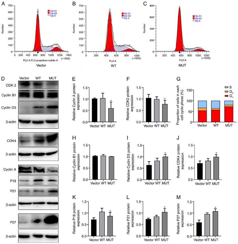
2           GAO et al: IDH1 GENE MUTATION REGULATES CELL CYCLE AND BIOLOGICAL RHYTHM GENES IN U87 CELLS

grade II and III gliomas and secondary glioblastoma (GBM;            IDH1 R132H mutant gene was introduced into human glioma
70‑80%), while primary GBM (WHO grade IV) IDH1/2 muta‑               U87‑MG cells to observe the effect of IDH1 on biological
tions are rare (90% of       rhythm genes and to analyze its relevant mechanism, laying a
glioma IDH1/2 mutations (7). IDH1 Arg132His (R132H) muta‑            theoretical foundation for the study of the effect of biological
tion can affect the proliferation of glioma cells, which is slower   rhythm on the biological function of malignant tumors.
than the corresponding wild‑type IDH1 cells (8,9). Clinical
studies (10,11) have shown that mutations in IDH1 were found         Materials and methods
to be associated with younger age, secondary GBMs (grade IV
tumors that arise from biopsy‑proven lower‑grade predecessors),      Cell line and culture. The full name of the cell line used is
and increased overall survival (OS) (12). Further studies (13,14)    U87‑MG human brain astroblastoma, which is a GBM of
have revealed that IDH1/2 mutations as good prognostic markers       unknown origin. Cells were cultured in Minimum Essential
are universally present in grade II and III glioma and secondary     Medium (MEM; Shanghai Zhongqiao Xinzhou Biotechnology
glioblastoma, and serve an important role in the occurrence,         Co., Ltd.) containing 10% fetal bovine serum (FBS; HyClone;
development and evolution of glioma (15). Therefore, studying        Cytiva) at 37˚C with 5% CO2. The cell line was purchased
the role of the IDH1 R132H mutation in the occurrence of             from Shanghai Zhongqiao Xinzhou Biotechnology Co., Ltd.
glioma may provide new ideas for clinical treatment.
    There is growing evidence that dysregulation of the circa‑       Reagents and instruments. pLVX‑IDH1‑mCMV‑ZsGreen-
dian clock serves an important role in the development of            PGK‑Puro and pLVX‑IDH1(MUT)‑mCMV‑ZsGreen‑PG
malignant tumors, including glioma (16,17). Circadian timing is      K‑Puro lentiviruses were purchased from Beijing Xibei
a basic biological process that affects most aspects of eukaryotic   Hongcheng Biotechnology Co., Ltd. (http://www.xbhcbio.
and prokaryotic physiology. Circadian dysrhythmia may lead           com/). Trypsin (0.25%)‑EDTA was purchased from
to an increased risk of cancer, as well as affect the response of    Invitrogen (Thermo Fisher Scientific, Inc.) and 0.45‑µm
patients with cancer to treatment (18). In a circadian rhythm,       PVDF membranes and a chemiluminescence kit (Immobilon
the oscillator is coordinated by a set of interlocked transcrip‑     Western Chemiluminescent HRP Substrate, eCl@ss
tion‑translation feedback loops. Brain‑Muscle Arnt‑Like              cat. no. 42029053) were purchased from EMD Millipore.
protein 1 (BMAL1) and Circadian Locomotor Output Cycles              A total protein extraction kit (Whole protein extraction kit,
Kaput (CLOCK) are the main biological rhythm genes (19).             cat. no. KGP250), a BCA assay protein content detection
CLOCK and BMAL1 heterogeneous dimers combined with                   kit (BCA Protein Quantitation Assay, cat. no. KGPBCA),
Period (PER) and Cryptochrom (CRY) proteins in E‑box device          an SDS‑PAGE gel preparation kit (KGI SDS‑PAGE Gel
drive rhythmic transcription (20‑22). The PER and CRY proteins       Preparation kit, cat. no. KGP113) and a flow cytometry cell
inhibit CLOCK‑BMAL1 complexes, which inhibit PER and                 cycle analysis kit (Cell Cycle Detection kit, cat. no. KGA511)
CRY protein degradation after release (23‑25). Upon epigenetic       were purchased from Jiangsu Kaiji Biotechnology Co., Ltd.
modification and increase post‑translational modifications,          (http://keygentec.com.cn/index.php?cid=1). The BSA (BSA‑V,
the core CLOCK proteins in the suprachiasmatic nucleus can           cat. no. A8020) used to dilute the antibody was purchased
maintain peripheral CLOCK oscillation and rhythmic expres‑           from Beijing Solarbio Science & Technology Co., Ltd. Mouse
sion (26), and the downstream targets are similar to those in        anti‑IDH1 (R132H; cat. no. SAB4200548) was purchased
the suprachiasmatic nucleus; the molecular biological clock of       from Sigma‑Aldrich (Merck KGaA). Rabbit anti‑IDH1
the peripheral tissues and organs of the body is also composed       (cat. no. 8137S), rabbit anti‑BMAL1 (cat. no. 14020S),
of a transcription‑translation feedback loop regulated by clock      rabbit anti‑CLOCK (cat. no. 5157S), rabbit anti‑phosphor‑
genes (27‑32). Disruption of circadian rhythms may negatively        ylated (p)‑Smad2 (cat. no. 18338S), rabbit anti‑p‑Smad3
affect normal cellular function and may lead to an increased inci‑   (cat. no. 9520S), rabbit anti‑Smad2 (cat. no. 5339S),
dence of multiple types of cancer, such as colorectal cancer (33),   rabbit anti‑Smad2‑3 (cat. no. 86855S), rabbit anti‑Smad3
breast cancer (34), prostate cancer (35), pancreatic cancer (36),    (cat. no. 9513S), rabbit anti‑Smad4 (cat. no. 46535S), mouse
osteosarcoma (37) and others (38,39). Therefore, it is very          anti‑ β ‑actin (cat. no. 3700S) and mouse anti‑GAPDH
important to study the role of the circadian clock in the develop‑   (cat. no. 51332S) primary antibodies, as well as HRP‑labelled
ment and progression of cancer. Mutations in circadian clock         goat anti‑rabbit IgG (cat. no. 7074S) and goat anti‑mouse
components can increase the proliferation rate of cells through      IgG (cat. no. 7076S) secondary antibodies, were purchased
the general dysregulation of the cell cycle, thereby causing the     from Cell Signaling Technology, Inc. Rabbit anti‑Cyclin A
cell to become cancerous (40). Several studies have revealed         (cat. no. AF0142), rabbit anti‑CyclinB1 (cat. no. DF6786),
an interaction between the biological clock and the cell cycle.      rabbit anti‑CyclinD3 (cat. no. DF6229), rabbit anti‑CDK2
For example, a previous study has indicated that light‑induced       (cat. no. AF6237), rabbit anti‑CDK4 (cat. no. DF6102), rabbit
phase shifts in mouse behavior lead to corresponding changes         anti‑P18 (cat. no. AF0620), rabbit anti‑P21 (cat. no. AF6290),
in the time of intestinal cell proliferation (41). Another study     rabbit anti‑P27 (cat. no. AF6324), rabbit anti‑PER1
has suggested that clock genes are involved in the regulation of     (cat. no. DF9080), rabbit anti‑PER2 (cat. no. DF12304), rabbit
important cell cycle checkpoints (42).                               anti‑PER3 (cat. no. DF7349), rabbit anti‑Cry1 (cat. no. DF8932)
    Understanding the mechanism of circadian clock changes           and rabbit anti‑Cry2 (cat. no. DF12919) primary antibodies
in tumors is of great importance for the improvement of tumor        were purchased from Affinity Biosciences. An IX73 inverted
therapy. However, to the best of our knowledge, the mechanism        fluorescence microscope was purchased from Olympus
of biorhythm change in glioma with IDH1 R132H mutation               Corporation, and an Amersham Imager 600 gel imaging
has not been previously reported. In the present study, the          analysis system was purchased from General Electric.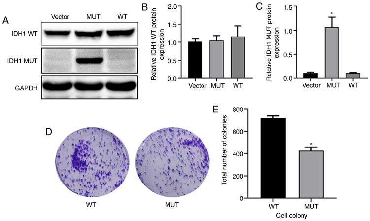
MOLECULAR MEDICINE REPORTS 23: 354, 2021 3
The cancer genome atlas (TCGA) database analysis. BMAL1 measurement. The protein expression level was semi‑quantified
and CLOCK expression data of all types of tumor analyzed using Image Pro Plus 6.0 (Media Cybernetics, Inc.).
in the present study were derived from TCGA database
(https://portal.gdc.cancer.gov/) and the online analysis Colony formation experiments. After seeding 1,000 lenti‑
software GEPIA2 (http://gepia2.cancer‑pku.cn/#index). virus‑infected U87‑MG cells into 100‑mm culture dishes, the
cells were divided into the WT and MUT groups. After 5 days
U87‑MG human glioma cell culture and lentiviral infection. of culture, the medium was changed once, and whether the
Construction of recombinant lentiviral vector: 1 µg Plasmid and cells formed clumps or sheets was observed under a 10x light
pLVX/IDH1, 1 µl EcoRI, 1 µl BamHI, 2 µl pLVX, 6 µl target microscope. The medium was then discarded. The cells were
gene, 1 µl 10X DNA Ligase Buffer and 1 µl T4 DNA Ligase, were fixed in 4% paraformaldehyde at room temperature for 20 min.
used to construct lentiviral vectors. The 239T cell line (Beijing Methylene violet solution (4 ml) was added to each dish, and
Xibei Hongcheng Biotechnology Co., Ltd.) was used for virus the cells were stained at room temperature for 30 min before
purification. After culturing for 12 to 16 h, the medium was rinsing with pure water. After drying at room temperature,
discarded (500 µl HET Buffer A + 10 µl recombinant lentiviral images were captured, and the colonies, which are clumps or
vector + 15 µl lentiviral packaging vector + HET Buffer B 50 µl + flakes formed by cells, were counted using Image‑Pro Plus 6.0
ddH2O 425 µl), and replaced with 10 ml fresh complete medium (Media Cybernetics, Inc.) for statistical analysis.
solution (DMEM + 10% FBS + P/S). The cells were placed in a
37˚C, 5% CO2 incubator for 48 h, the supernatant was collected Cell cycle distribution detection by flow cytometry. A cell cycle
after centrifugation at 500 x g and room temperature for 5 min analysis kit (Cell Cycle Detection kit, cat. no. KGA511) was used
and the cell debris was discarded. The supernatant was filtered for cell cycle detection. The cells in each group were digested
with a 45 µm PVDF filter into a 50 ml round bottom centrifuge with trypsin at 37˚C for 1 min, collected by centrifugation
tube. This was centrifugated at a high speed of 6,000 x g at 4˚C at 1,500 x g for 5 min at normal temperature and rinsed with
for 2 h, and purified recombinant lentivirus was obtained after PBS to form a cell suspension. The concentration was adjusted to
centrifugation. Lentivirus can be introduced into U87‑MG cells 1x10⁶ cells/ml. After removing the supernatant by centrifugation
after 48 h of infection with MOI value (20:1) (43). U87‑MG cells at 1,500 x g for 5 min at normal temperature, 500 µl precooled
were inoculated in MEM containing 10% FBS at a density of 75% ethanol was used to fix the cells overnight at 4˚C. The fixa‑
2x10⁵ cells/100‑mm culture dish, incubated at 37˚C and then tion solution was removed by centrifugation at 1,500 x g for 5 min
subjected to lentivirus infection 6‑8 h after becoming adherent. at 4˚C, 100 µl RNase A was added and the cells were incubated
The titre of the treatment and control lentiviruses was set to an in a water bath at 37˚C for 30 min. A 400 µm mesh screen was
MOI of 20:1, and MEM with 2% FBS was used to prepare viral used for filtration. Then, 400 µl PI dye was added and the solu‑
titre gradient solutions for subsequent experiments. The experi‑ tion was gently mixed and incubated at 4˚C for 1 h in the dark.
mental groups were the IDH1 wild‑type (WT) and the IDH1 Analysis was performed using a flow cytometer (FACSCalibur,
R132H mutant (MUT) groups, and the corresponding lentiviral BD Biosciences; FlowJo, Version 10, FlowJo LLC).
vectors were pLVX‑IDH1‑mCMV‑ZsGreen‑PGK‑Puro and
pLVX‑IDH1(MUT)-mCMV‑ZsGreen‑PGK‑Puro, respectively. Statistical analysis. SPSS 21.0 (IBM Corp.) statistical soft‑
Subsequent experiments were performed 72 h after lentiviral ware was used for statistical analysis. The data are expressed
infection. as the mean ± SD. Each independent experiment was repeated
three times. Independent sample t‑test was used to analyze
Western blot analysis. Following lentiviral transfection, differences between two groups of continuous data. One‑way
U87‑MG cells in each group were collected for extraction of ANOVA was used for the comparison among multiple groups
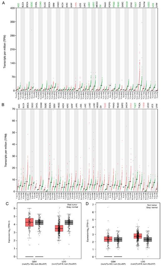
total protein using the aforementioned whole protein extrac‑ followed by Tukey's post hoc test. P4 GAO et al: IDH1 GENE MUTATION REGULATES CELL CYCLE AND BIOLOGICAL RHYTHM GENES IN U87 CELLS Figure 1. Expression of BMAL1 and CLOCK in different tumors in TCGA database. (A) BMAL1 and (B) CLOCK expression in 33 types of tumor from The Cancer Genome Atlas database. Red represents tumor samples, while green represents normal tissue samples. (C) BMAL1 and (D) CLOCK expression in GBM and LGG tumor and normal tissues. The red box represents tumor samples, while the grey box represents normal tissue samples. T, tumor; N, normal; TPM, transcripts per million; BMAL1, Brain‑Muscle Arnt‑Like protein 1; CLOCK, Circadian Locomotor Output Cycles Kaput; ACC, Adrenocortical carcinoma; BLCA, Bladder Urothelial Carcinoma; BRCA, Breast invasive carcinoma; CESC, Cervical squamous cell carcinoma and endocervical adenocarcinoma; CHOL, Cholangiocarcinoma; COAD, Colon adenocarcinoma; DLBC, Lymphoid Neoplasm Diffuse Large B‑cell Lymphoma; ESCA, Esophageal carcinoma; GBM, Glioblastoma multiforme; HNSC, Head and Neck squamous cell carcinoma; KICH, Kidney Chromophobe; KIRC, Kidney renal clear cell carcinoma; KIRP, Kidney renal papillary cell carcinoma; LAML, Acute Myeloid Leukemia; LGG, Brain Lower Grade Glioma; LIHC, Liver hepatocellular carcinoma; LUAD, Lung adenocarcinoma; LUSC, Lung squamous cell carcinoma; MESO, Mesothelioma; OV, Ovarian serous cystadenocarcinoma; PAAD, Pancreatic adenocarcinoma; PCPG, Pheochromocytoma and Paraganglioma; PRAD, Prostate adenocarcinoma; READ, Rectum adenocarcinoma; SARC, Sarcoma; SKCM, Skin Cutaneous Melanoma; STAD, Stomach adenocarcinoma; TGCT, Testicular Germ Cell Tumors; THCA, Thyroid carcinoma; THYM, Thymoma; UCEC, Uterine Corpus Endometrial Carcinoma; UCS, Uterine Carcinosarcoma; UVM, Uveal Melanoma.
MOLECULAR MEDICINE REPORTS 23: 354, 2021                                                                 5

Figure 2. Establishment of a cell model transfected with IDH1 lentivirus, and detection of the IDH1 R132H mutation affects the proliferation of glioma cells.
(A) Detection of lentiviral transfection in U87‑MG cells. The protein expression levels of IDH1 WT and MUT in each group of U87‑MG cells was detected
by western blot analysis. (B) Relative IDH1 WT protein expression. (C) Relative IDH1 MUT protein expression. (D and E) Colony formation experiment was
used to detect the number of colonies in the two groups. *P
6             GAO et al: IDH1 GENE MUTATION REGULATES CELL CYCLE AND BIOLOGICAL RHYTHM GENES IN U87 CELLS

Figure 3. Effect of IDH1 gene mutation on cell cycle. Changes in (A) vector, (B) WT and (C) MUT U87‑MG cells after transfection with lentivirus. The cell
cycle changes in each group were detected by flow cytometry. (D) Protein expression levels of CyclinA, CyclinB1, CyclinD3, CDK2, CDK4, P18, P21 and
P27 in each group of U87‑MG cells were detected by western blot analysis. Quantification of relative protein expression levels of (E) CyclinA and (F) CDK2.
(G) Proportion of cells in each cell cycle phase in each group. Semi‑quantification of relative protein expression levels of (H) CyclinB1, (I) CyclinD3, (J) CDK4,
(K) P18, (L) P21 and (M) P27. *P
MOLECULAR MEDICINE REPORTS 23: 354, 2021                                                                 7

Figure 4. Effects of IDH1 gene mutation on biological rhythm genes. (A) Protein expression levels of BMAL1 and CLOCK in the WT, MUT and vector groups.
Quantification of relative protein expression levels of (B) BMAL1 and (C) CLOCK. (D) Protein expression levels of PER1, PER2, PER3, CRY1 and CRY2 in
the WT, MUT and vector groups. Quantification of relative protein expression levels of (E) PER1, (F) PER2, (G) PER3, (H) Cry1 and (I) Cry2. *P
8            GAO et al: IDH1 GENE MUTATION REGULATES CELL CYCLE AND BIOLOGICAL RHYTHM GENES IN U87 CELLS

are transferred to the nucleus to interact with multiple tran‑     group, as well as the expression levels of the negative regulation
scription factors in order to induce cellular responses (54,55).   factors PER1, PER2, PER3, Cry1 and Cry2. A previous study
Thus, it was hypothesized that the IDH1 R132H mutation may         has demonstrated that the disturbance of the circadian clock
affect biological rhythm genes of glioma cells through the         has a strong influence on tumor transformation and growth
TGF‑β/Smad signaling pathway.                                      by affecting various cancer regulatory signaling pathways,
                                                                   including cell cycle, apoptosis and metabolism (40). The detec‑
Discussion                                                         tion of biological rhythm genes in the present study revealed
                                                                   that IDH1 R132H mutation inhibited the protein expression
Glioma arises from glial cells and most often occurs in the        levels of CLOCK and BMAL1, and further inhibited PER and
brain (56). Glioma accounts for ~30% of central nervous system     CRY proteins. Therefore, it was shown that the IDH1 R132H
tumors and 80% of malignant brain tumors (57). It is charac‑       mutation has an effect on the expression of biological rhythm
terized with highly infiltrative growth and a poor prognosis.      genes and has been confirmed in in vitro experiments.
The most common genetic changes in glioma include IDH1/2               Furthermore, how the IDH1 R132H mutation may affect
mutations, TP53 mutations and 1p/19q heterozygous deletion,        biological rhythm genes was further investigated. Previous
which are present in most cases (>90%) of glioma (58,59).          studies have shown that the TGF‑ β signaling pathway is
The most common and early genetic change in glioma is the          closely associated with the occurrence of glioma (66,67).
IDH1/2 mutation. The most common IDH1 mutation results             TGF‑β is a factor that strongly inhibits the proliferation of
in the amino acid substitution of R132, which is located at the    epithelial, astrocyte and immune cells, and is considered
active site of the enzyme (60). IDH1 mutated glioma cells have     a tumor suppressor (68). Some tumors acquire mutations in
a reduced proliferative rate (61). Numerous diseases follow        components of the TGF‑ β signaling pathway to evade the
circadian rhythms, and there is growing evidence that circa‑       TGF‑β cellular inhibitory response (69). On the other hand,
dian disruption may be a risk factor for cancer in humans (62).    in some malignant tumors, including glioma, the ability of
A previous study has reported that the BMAL1 gene serves a         TGF‑β to inhibit cell proliferation and maintain the integ‑
role as a potential tumor suppressor gene in pancreatic cancer     rity of the TGF‑β signaling pathway is selectively lost (70).
by activating the P53 tumor suppressor signaling pathway (63).     TGF‑β signaling decreases differences in IDH1 expression
Circadian rhythm genes activate certain pathways in tumors or      by normalizing the Smad signaling pathway, and inhibition
are activated by certain pathways to affect the development of     of Cav1 expression by IDH1 is regulated by α‑KG epigenetic
tumors. β‑catenin showed an increased expression in NIH‑3T3        regulation. Finally, downregulation of Cav1 expression inter‑
cells after BMAL1 overexpression, indicating that activation of    rupts the degradation of TGFBR and enhances the Smad
the canonical Wnt pathway may be the mechanism underlying          signal. IDH1 regulates the TGF‑β signaling feedback loop
the effect of the circadian clock gene BMAL on promoting cell      through α‑KG and TGFBR‑IDH1‑Cav1 to enhance TGF‑ β
proliferation (64). BMAL1 suppresses cancer cell invasion by       signaling in a regulatory network between cellular signaling
blocking the PI3K‑Akt‑MMP‑2 signaling pathway (65).                and cell metabolism (51). In the present study, it was observed
    However, to the best of our knowledge, there are no            that the IDH1 R132H mutation significantly decreased the
studies on the mechanism between IDH1 R132H mutation and           protein expression levels of Smad2, Smad3 and Smad2‑3, and
biological rhythm genes. Therefore, the present study aimed        significantly upregulated the levels of p‑Smad2, p‑Smad3 and
to describe the role of biological rhythm genes in glioma cells    Smad4. A previous study has suggested that TGF‑β is a multi‑
with IDH1 R132H mutation to determine how the IDH1 R132H           potent cytokine that controls tissue homeostasis and embryonic
mutation affects biological rhythm genes and the proliferation     development. TGF‑β binds and activates a membrane receptor
of glioma cells.                                                   serine threonine kinase complex to phosphorylate Smad2 and
    The current study revealed that the IDH1 R132H mutation        Smad3; after phosphorylation, Smad proteins accumulate in
in U87‑MG cells decreased the number of new colonies formed        the nucleus and form complexes with transcription factors,
by tumor cells. The effect of the IDH1 R132H mutation on           such as Smad4, to regulate transcription (54). The present
the cell cycle was further investigated, leading to increased      study revealed that the IDH1 R132H mutation affected the
cells in G1 phase and decreased cells in S phase. The effect       TGF‑β/Smad signaling pathway. Additionally, TGF‑β is an
of the mutation on cyclins was also examined. The expres‑          important regulator of the physiological clock. A previous
sion levels of various cyclically related proteins was altered,    study has demonstrated that TGF‑β, by regulating the expres‑
including the S phase‑associated proteins Cyclin A and             sion of positive and negative regulators of circadian rhythm
CDK2. No change in Cyclin B1 expression in G2/M phase was          oscillation, serves a vital role in regulating circadian rhythm.
observed. The expression levels of the G1 phase‑associated         Adenovirus‑mediated TGF‑ β expression can significantly
proteins Cyclin D3 and CDK4 were increased in the MUT              induce BMAL1 and NPAS2 expression (71). It has been
group compared with in the WT group. The protein expres‑           demonstrated that the expression levels of the TGF‑β‑activated
sion levels of P18 were decreased, while those of P21 and P27      transcription factor Smad3 display similar expression patterns
were increased in the MUT group compared with in the WT            with BMAL1, and that Smad3 functions as an upstream
group. Subsequently, the effect of the IDH1 R132H mutation         molecule of BMAL1, explaining how TGF‑β induces BMAL1
was examined on biological rhythm genes. To the best of our        expression (53). However, TGF‑ β strongly inhibits the
knowledge, the current study was the first to analyze the effect   expression levels of PER1, PER2, PER3, Rev‑erbα, retinoic
of the IDH1 R132H mutation on biological rhythm. The IDH1          acid receptor‑related orphan receptor α and D‑site albumin
R132H mutation significantly decreased the protein expression      promoter‑binding protein (71). Therefore, increased expres‑
levels of both BMAL1 and CLOCK compared with in the WT             sion levels of TGF‑β and negative regulators of the circadian
MOLECULAR MEDICINE REPORTS 23: 354, 2021 9
clock prolong the arousal cycle (52). The impact of CLOCK Ethics approval and consent to participate
and BMAL1 on cancer pathogenesis is highly context‑ and
disease‑dependent (50). For instance, CLOCK or BMAL1 Not applicable.
provide tumor suppressor‑like functions in prostate, breast,
ovarian and pancreatic cancer, but exhibit tumor‑promoting Patient consent for publication
roles in colorectal cancer and acute myeloid leukemia (50,72).
In glioma, CLOCK or BMAL1 are tumor‑promoting factors Not applicable.
that regulate glioma cell proliferation and migration via
regulating the NF‑κ B signaling pathway (73), and can support Competing interests
glioma stem cell function via regulation of anabolic metabo‑
lism (74). Therefore, the current study hypothesized that the The authors declare that they have no competing interests.
IDH1 R132H mutation may affect biological rhythm genes
through the TGF‑β/Smad signaling pathway, thus affecting the References
proliferation of glioma cells.
In summary, the IDH1 gene serves crucial roles in the 1. Sjoblom T, Jones S, Wood LD, Parsons DW, Lin J, Barber TD,
occurrence and development of numerous types of tumor (75), Mandelker D, Leary RJ, Ptak J, Silliman N, et al: The consensus
coding sequences of human breast and colorectal cancers.
and imbalance in the circadian clock plays an important role in Science 314: 268‑274, 2006.
the development of malignant tumors, including glioma (76). 2. Mardis ER, Ding L, Dooling DJ, Larson DE, McLellan MD, Chen K,
The present study revealed that the IDH1 R132H mutation Koboldt DC, Fulton RS, Delehaunty KD, McGrath SD, et al:
Recurring mutations found by sequencing an acute myeloid
affected the expression levels of cyclin and biological rhythm leukemia genome. N Engl J Med 361: 1058‑1066, 2009.
genes, and thus may affect the occurrence of glioma. Based 3. Cardaci S and Ciriolo MR: TCA cycle defects and cancer: When
on the current results, it was hypothesized that IDH1 muta‑ metabolism tunes redox state. Int J Cell Biol 2012: 161837, 2012.
4. Pastore M, Lori G, Gentilini A, Taddei ML, Di Maira G,
tion may affect the expression levels of biological rhythm Campani C, Recalcati S, Invernizzi P, Marra F and Raggi C:
genes of cells through the TGF‑β/Smad signaling pathway. Multifaceted aspects of metabolic plasticity in human cholangio‑
To the best of our knowledge, the present results are the first carcinoma: An overview of current perspectives. Cells 9: 596,
2020.
to report the association between IDH1 R132H mutation 5. Bosnyák E, Michelhaugh SK, Klinger NV, Kamson DO,
and biological rhythm genes, as well as the effect on glioma Barger GR, Mittal S and Juhász C: Prognostic molecular and
cell proliferation and the possible underlying mechanisms. imaging biomarkers in primary glioblastoma. Clin Nucl Med 42:
341‑347, 2017.
However, further studies are required to confirm the current 6. Krell D, Assoku M, Galloway M, Mulholland P, Tomlinson I
results. and Bardella C: Screen for IDH1, IDH2, IDH3, D2HGDH and
L2HGDH mutations in glioblastoma. PLoS One 6: e19868,
2011.
Acknowledgements 7. Yang H, Ye D, Guan KL and Xiong Y: IDH1 and IDH2 mutations
in tumorigenesis: Mechanistic insights and clinical perspectives.
Not applicable. Clin Cancer Res 18: 5562‑5571, 2012.
8. Zhang Y, Lv W, Li Q, Wang Q, Ru Y, Xiong X, Yan F, Pan T,
Lin W and Li X: IDH2 compensates for IDH1 mutation to
Funding maintain cell survival under hypoxic conditions in IDH1‑mutant
tumor cells. Mol Med Rep 20: 1893‑1900, 2019.
9. Chittaranjan S, Chan S, Yang C, Yang KC, Chen V, Moradian A,
The present study was funded by the National Natural Science Firme M, Song J, Go NE, Blough MD, et al: Mutations in CIC
Foundation of China (grant no. 81560501), the Ningxia and IDH1 cooperatively regulate 2‑hydroxyglutarate levels and
Natural Science Foundation (grant no. 2020AAC03351) cell clonogenicity. Oncotarget 5: 7960‑7979, 2014.
10. Hersh DS, Peng S, Dancy JG, Galisteo R, Eschbacher JM,
and the Ningxia Innovation Team of the foundation and Castellani RJ, Heath JE, Legesse T, Kim AJ, Woodworth GF, et al:
clinical researches of diabetes and its complications (grant Differential expression of the TWEAK receptor Fn14 in IDH1
no. NXKJT2019010). wild‑type and mutant gliomas. J Neurooncol 138: 241‑250,
2018.
11. Sakai Y, Yang C, Kihira S, Tsankova N, Khan F, Hormigo A,
Availability of data and materials Lai A, Cloughesy T and Nael K: MRI radiomic features to predict
IDH1 mutation status in gliomas: A machine learning approach
using gradient tree boosting. Int J Mol Sci 21: 8004, 2020.
The datasets used and/or analyzed during the current study are 12. Kim W and Liau LM: IDH mutations in human glioma.
available from the corresponding author on reasonable request. Neurosurg Clin N Am 23: 471‑480, 2012.
13. Ducray F, El Hallani S and Idbaih A: Diagnostic and prognostic
markers in gliomas. Curr Opin Oncol 21: 537‑542, 2009.
Authors' contributions 14. Bergo E, Lombardi G, Pambuku A, Della Puppa A, Bellu L,
D'Avella D and Zagonel V: Cognitive rehabilitation in patients
XC and SH designed the experiments, guided the study and with gliomas and other brain tumors: State of the Art. Biomed
Res Int 2016: 3041824, 2016.
revised the draft. YW performed the experiments and wrote 15. Lu C, Ward PS, Kapoor GS, Rohle D, Turcan S, Abdel‑Wahab O,
the manuscript with support from all the other authors. YG and Edwards CR, Khanin R, Figueroa ME, Melnick A, et al: IDH
NZ performed the data analysis and contributed to the writing mutation impairs histone demethylation and results in a block to
cell differentiation. Nature 483: 474‑478, 2012.
of the manuscript. HY, JY and JM participated in some of the 16. Chen Z, Liu P, Li C, Luo Y, Chen I, Liang W, Chen X, Feng Y,
experiments and in data collection. FW and HX participated Xia H and Wang F: Deregulated expression of the clock genes in
in some of the data analyses. XC, YW and YG confirmed the gliomas. Technol Cancer Res Treat 12: 91‑97, 2013.
17. Luo Y, Wang F, Chen LA, Chen XW, Chen ZJ, Liu PF, li FF,
authenticity of the raw data. All authors read and approved the Li CY and Liang W: Deregulated expression of cry1 and cry2 in
final manuscript. human gliomas. Asian Pac J Cancer Prev 13: 5725‑5728, 2012.10 GAO et al: IDH1 GENE MUTATION REGULATES CELL CYCLE AND BIOLOGICAL RHYTHM GENES IN U87 CELLS
18. Chang WH and Lai AG: Timing gone awry: Distinct tumour 42. Farshadi E, Yan J, Leclere P, Goldbeter A, Chaves I and
suppressive and oncogenic roles of the circadian clock and cross‑ van der Horst GTJ: The positive circadian regulators CLOCK and
talk with hypoxia signalling in diverse malignancies. J Transl BMAL1 control G2/M cell cycle transition through Cyclin B1.
Med 17: 132, 2019. Cell Cycle 18: 16‑33, 2019.
19. Chan AB, Huber AL and Lamia KA: Cryptochromes modulate 43. Shan DZ, Zhang C and Cao XM: Preparation and expression of
E2F family transcription factors. Sci Rep 10: 4077, 2020. isocitrate dehydrogenase 1 and mutant recombinant lentivirus.
20. Young MW and Kay SA: Time zones: A comparative genetics of Chin J Neuroanatomy 36: 200‑206, 2020.
circadian clocks. Nat Rev Genet 2: 702‑715, 2001. 44. Yu H, Yuan Y, Shen H and Cheng T: Hematopoietic stem cell
21. Lowrey PL and Takahashi JS: Genetics of the mammalian exhaustion impacted by p18 INK4C and p21 Cip1/Waf1 in oppo‑
circadian system: Photic entrainment, circadian pacemaker site manners. Blood 107: 1200‑1206, 2006.
mechanisms, and posttranslational regulation. Ann Rev Genet 34: 45. Yuan Y, Shen H, Franklin DS, Scadden DT and Cheng T:
533‑562, 2000. In vivo self‑renewing divisions of haematopoietic stem cells
22. Dunlap JC: Molecular bases for circadian clocks. Cell 96: are increased in the absence of the early G1‑phase inhibitor,
271‑290, 1999. p18INK4C. Nat Cell Biol 6: 436‑442, 2004.
23. Kume K, Zylka MJ, Sriram S, Shearman LP, Weaver DR, Jin X, 46. Sherr CJ: Cancer cell cycles. Science 274: 1672‑1677, 1996.
Maywood ES, Hastings MH and Reppert SM: mCRY1 and 47. Mandal AS, Biswas N, Karim KM, Guha A, Chatterjee S,
mCRY2 are essential components of the negative limb of the Behera M and Kuotsu K: Drug delivery system based on chrono‑
circadian clock feedback loop. Cell 98: 193‑205, 1999. biology‑A review. J Control Release 147: 314‑325, 2010.
24. van der Horst GT, Muijtjens M, Kobayashi K, Takano R, Kanno S, 48. Zhao Q, Zheng G, Yang K, Ao YR, Su XL, Li Y and Lv XQ: The
Takao M, de Wit J, Verkerk A, Eker AP, van Leenen D, et al: clock gene PER1 plays an important role in regulating the clock
Mammalian Cry1 and Cry2 are essential for maintenance of gene network in human oral squamous cell carcinoma cells.
circadian rhythms. Nature 398: 627‑630, 1999. Oncotarget 7: 70290‑70302, 2016.
25. Vitaterna MH, Selby CP, Todo T, Niwa H, Thompson C, 49. Qu M, Duffy T, Hirota T and Kay SA: Nuclear receptor HNF4A
Fr uechte EM, Hitom i K, T h resher R J, Ish i kawa T, transrepresses CLOCK:BMAL1 and modulates tissue‑specific
Miyazaki J, et al: Differential regulation of mammalian period circadian networks. Proc Natl Acad Sci USA 115: E12305‑E12312,
genes and circadian rhythmicity by cryptochromes 1 and 2. Proc 2018.
Natl Acad Sci USA 96: 12114‑12119, 1999. 50. Shafi AA and Knudsen KE: Cancer and the circadian clock.
26. Dibner C, Schibler U and Albrecht U: The mammalian circadian Cancer Res 79: 3806‑3814, 2019.
timing system: Organization and coordination of central and 51. Hou X, Zhang J, Wang Y, Xiong W and Mi J: TGFBR‑IDH1‑Cav1
peripheral clocks. Ann Rev Physiol 72: 517‑549, 2010. axis promotes TGF‑beta signalling in cancer‑associated fibro‑
27. Harmer SL, Panda S and Kay SA: Molecular bases of circadian blast. Oncotarget 8: 83962‑83974, 2017.
rhythms. Ann Rev Cell Dev Biol 17: 215‑253, 2001. 52. Lopez M, Meier D, Muller A, Franken P, Fujita J and Fontana A:
28. Oster H: The genetic basis of circadian behavior. Genes Brain Tumor necrosis factor and transforming growth factor beta
Behav 5 (Suppl 2): S73‑S79, 2006. regulate clock genes by controlling the expression of the cold
29. Balsalobre A, Brown SA, Marcacci L, Tronche F, Kellendonk C, inducible RNA‑binding protein (CIRBP). J Biol Chem 289:
Reichardt HM, Schütz G and Schibler U: Resetting of circa‑ 2736‑2744, 2014.
dian time in peripheral tissues by glucocorticoid signaling. 53. Sato F, Sato H, Jin D, Bhawal UK, Wu Y, Noshiro M, Kawamoto T,
Science 289: 2344‑2347, 2000. Fujimoto K, Seino H, Morohashi S, et al: Smad3 and Snail show
30. Damiola F, Le Minh N, Preitner N, Kornmann B, Fleury‑Olela F circadian expression in human gingival fibroblasts, human
and Schibler U: Restricted feeding uncouples circadian oscil‑ mesenchymal stem cell, and in mouse liver. Biochem Biophys
lators in peripheral tissues from the central pacemaker in the Res Commun 419: 441‑446, 2012.
suprachiasmatic nucleus. Genes Dev 14: 2950‑2961, 2000. 54. Kang Y, He W, Tulley S, Gupta GP, Serganova I, Chen CR,
31. Yagita K, Tamanini F, van Der Horst GT and Okamura H: Manova‑Todorova K, Blasberg R, Gerald WL and Massagué J:
Molecular mechanisms of the biological clock in cultured Breast cancer bone metastasis mediated by the Smad tumor
fibroblasts. Science 292: 278‑281, 2001. suppressor pathway. Proc Natl Acad Sci USA 102: 13909‑13914,
32. Shearman LP, Sriram S, Weaver DR, Maywood ES, Chaves I, 2005.
Zheng B, Kume K, Lee CC, van der Horst GT, Hastings MH 55. Dou C, Lee J, Liu B, Liu F, Massague J, Xuan S and Lai E: BF‑1
and Reppert SM: Interacting molecular loops in the mammalian interferes with transforming growth factor beta signaling by
circadian clock. Science 288: 1013‑1019, 2000. associating with Smad partners. Mol Cell Biol 20: 6201‑6211,
33. Mazzoccoli G, Vinciguerra M, Papa G and Piepoli A: Circadian 2000.
clock circuitry in colorectal cancer. World J Gastroenterol 20: 56. Parisot S, Wells W III, Chemouny S, Duffau H and Paragios N:
4197‑4207, 2014. Concurrent tumor segmentation and registration with uncer‑
34. Blakeman V, Williams JL, Meng QJ and Streuli CH: Circadian tainty‑based sparse non‑uniform graphs. Med Image Anal 18:
clocks and breast cancer. Br Cancer Res 18: 89, 2016. 647‑659, 2014.
35. Cao Q, Gery S, Dashti A, Yin D, Zhou Y, Gu J and Koeffler HP: 57. de Robles P, Fiest KM, Frolkis AD, Pringsheim T, Atta C,
A role for the clock gene per1 in prostate cancer. Cancer Res 69: St Germaine‑Smith C, Day L, Lam D and Jette N: The worldwide
7619‑7625, 2009. incidence and prevalence of primary brain tumors: A systematic
36. Oda A, Katayose Y, Yabuuchi S, Yamamoto K, Mizuma M, review and meta‑analysis. Neurooncology 17: 776‑783, 2015.
Shirasou S, Onogawa T, Ohtsuka H, Yoshida H, Hayashi H, et al: 58. Eckel‑Passow JE, Lachance DH, Molinaro AM, Walsh KM,
Clock gene mouse period2 overexpression inhibits growth of Decker PA, Sicotte H, Pekmezci M, Rice T, Kosel ML,
human pancreatic cancer cells and has synergistic effect with Smirnov IV, et al: Glioma Groups Based on 1p/19q, IDH, and TERT
cisplatin. Anticancer Res 29: 1201‑1209, 2009. promoter mutations in tumors. N Engl J Med 372: 2499‑2508, 2015.
37. Zhang S, Zhang J, Deng Z, Liu H, Mao W, Jiang F, Xia Z and 59. Miller JJ, Shih HA, Andronesi OC and Cahill DP: Isocitrate
Li JD: Circadian clock components RORα and Bmal1 mediate dehydrogenase‑mutant glioma: Evolving clinical and therapeutic
the anti‑proliferative effect of MLN4924 in osteosarcoma cells. implications. Cancer 123: 4535‑4546, 2017.
Oncotarget 7: 66087‑66099, 2016. 60. Ferreira MSV, Sørensen MD, Pusch S, Beier D, Bouillon AS,
38. Papagiannakopoulos T, Bauer MR, Davidson SM, Heimann M, Kristensen BW, Brümmendorf TH, Beier CP and Beier F:
Subbaraj L, Bhutkar A, Bartlebaugh J, Vander Heiden MG and Alternative lengthening of telomeres is the major telomere
Jacks T: Circadian rhythm disruption promotes lung tumorigen‑ maintenance mechanism in astrocytoma with isocitrate
esis. Cell Metab 24: 324‑331, 2016. dehydrogenase 1 mutation. J Neurooncol 147: 1‑14, 2020.
39. Yang MY, Lin PM, Hsiao HH, Hsu JF, Lin HY, Hsu CM, Chen IY, 61. Karpel‑Massler G, Nguyen TTT, Shang E and Siegelin MD: Novel
Su SW, Liu YC and Lin SF: Up‑regulation of PER3 expression IDH1‑targeted glioma therapies. CNS Drugs 33: 1155‑1166, 2019.
is correlated with better clinical outcome in acute leukemia. 62. Salavaty A, Mohammadi N, Shahmoradi M and Naderi Soorki M:
Anticancer Res 35: 6615‑6622, 2015. Bioinformatic analysis of circadian expression of oncogenes
40. Farshadi E, van der Horst GTJ and Chaves I: Molecular links and tumor suppressor genes. Bioinform Biol Insights 11:
between the circadian clock and the cell cycle. J Mol Biol 432: 1177932217746991, 2017.
3515‑3524, 2020. 63. Jiang W, Zhao S, Jiang X, Zhang E, Hu G, Hu B, Zheng P, Xiao J,
41. Scheving LE, Tsai TH and Scheving LA: Chronobiology of Lu Z, Lu Y, et al: The circadian clock gene Bmal1 acts as a
the intestinal tract of the mouse. Am J Anatomy 168: 433‑465, potential anti‑oncogene in pancreatic cancer by activating the
1983. p53 tumor suppressor pathway. Cancer Lett 371: 314‑325, 2016.MOLECULAR MEDICINE REPORTS 23: 354, 2021 11
64. Lin F, Chen Y, Li X, Zhao Q and Tan Z: Over‑expression of circa‑ 71. Dong C, Gongora R, Sosulski ML, Luo F and Sanchez CG:
dian clock gene Bmal1 affects proliferation and the canonical Regulation of transforming growth factor‑beta1 (TGF‑ β1)-
Wnt pathway in NIH‑3T3 cells. Cell Biochem Funct 31: 166‑172, induced pro‑fibrotic activities by circadian clock gene BMAL1.
2013. Respir Res 17: 4, 2016.
65. Jung CH, Kim EM, Park JK, Hwang SG, Moon SK, Kim WJ 72. Puram RV, Kowalczyk MS, de Boer CG, Schneider RK,
and Um HD: Bmal1 suppresses cancer cell invasion by blocking Miller PG, McConkey M, Tothova Z, Tejero H, Heckl D,
the phosphoinositide 3‑kinase‑Akt‑MMP‑2 signaling pathway. Järås M, et al: Core circadian clock genes regulate leukemia stem
Oncol Rep 29: 2109‑2113, 2013. cells in AML. Cell 165: 303‑316, 2016.
66. Guan F, Kang Z, Wang L, Wang K, Mao BB, Peng WC, Zhang BL, 73. Li A, Lin X, Tan X, Yin B, Han W, Zhao J, Yuan J, Qiang B
Lin ZY, Zhang JT and Hu ZQ: Retinol dehydrogenase 10 and Peng X: Circadian gene Clock contributes to cell prolif‑
promotes metastasis of glioma cells via the transforming growth eration and migration of glioma and is directly regulated by
factor‑ β/SMAD signaling pathway. Chin Med J (Engl) 132: tumor‑suppressive miR‑124. FEBS Lett 587: 2455‑2460, 2013.
2430‑2437, 2019. 74. Dong Z, Zhang G, Qu M, Gimple RC, Wu Q, Qiu Z, Prager BC,
67. Derynck R and Zhang YE: Smad‑dependent and Smad- Wang X, Kim LJY, Morton AR, et al: Targeting glioblastoma
independent pathways in TGF‑beta family signalling. Nature 425: stem cells through disruption of the circadian clock. Cancer
577‑584, 2003. Discov 9: 1556‑1573, 2019.
68. Frei K, Gramatzki D, Tritschler I, Schroeder JJ, Espinoza L, 75. Liu WS, Chan SH, Chang HT, Li GC, Tu YT, Tseng HH, Fu TY,
Rushing EJ and Weller M: Transforming growth factor‑ β Chang HY, Liou HH, Ger LP and Tsai KW: Isocitrate dehy‑
pathway activity in glioblastoma. Oncotarget 6: 5963‑5977, 2015. drogenase 1‑snail axis dysfunction significantly correlates with
69. Huse K, Bakkebø M, Wälchli S, Oksvold MP, Hilden VI, breast cancer prognosis and regulates cell invasion ability. Breast
Forfang L, Bredahl ML, Liestøl K, Alizadeh AA, Smeland EB Cancer Res 20: 25, 2018.
and Myklebust JH: Role of Smad proteins in resistance to 76. Xia HC, Niu ZF, Ma H, Cao SZ, Hao SC, Liu ZT and Wang F:
BMP‑induced growth inhibition in B‑cell lymphoma. PLoS Deregulated expression of the Per1 and Per2 in human gliomas.
One 7: e46117, 2012. Can J Neurol Sci 37: 365‑370, 2010.
70. Sadeghi Y, Tabatabaei Irani P, Rafiee L, Tajadini M,
Amouheidari A and Javanmard SH: Evaluation of rs1982073 This work is licensed under a Creative Commons
polymorphism of transforming growth factor‑β1 in glioblastoma. Attribution-NonCommercial-NoDerivatives 4.0
J Res Med Sci 24: 40, 2019. International (CC BY-NC-ND 4.0) License.You can also read


