PS2 transfection of murine adenocarcinoma cell line 410.4 enhances dispersed growth pattern in a 3-D collagen gel
←
→
Page content transcription
If your browser does not render page correctly, please read the page content below
Journal of Cell Science 109, 63-71 (1996)                                                                                         63
Printed in Great Britain © The Company of Biologists Limited 1996
JCS8940

pS2 transfection of murine adenocarcinoma cell line 410.4 enhances
dispersed growth pattern in a 3-D collagen gel

R. Williams1, G. W. H. Stamp1, C. Gilbert2, M. Pignatelli1 and E.-N. Lalani1,*
1Department of Histopathology, Royal Postgraduate Medical School, Hammersmith Hospital, Du Cane Road, London W12 0NN,
UK
2Imperial Cancer Research Fund, Histopathology Unit, Lincoln’s Inn Fields, London WC2A 3PX, UK

*Author for correspondence (e-mail: elalani@rpms.ac.uk)

SUMMARY

We describe the first model system employing human pS2              or no matrix deposition, radiating long cords composed of
gene transfer and expression in a non-pS2-expressing cell           single elongated cells, an effect previously observed in other
line, mouse mammary adenocarcinoma 410.4, in order to               cell lines with hepatocyte growth factor. pS2 transfection
analyse the potential effect of human trefoil peptide pS2 in        had no demonstrable effect on proliferation and this is not
glandular epithelium. Two selected clones, AA4 and AD4,             a morphogenetic phenomenon, as tubulogenesis is not seen.
were established and shown to have incorporated the pS2             Motility assays suggest that the pS2 ‘dispersant’ effect in
cDNA sequence into the genome, express pS2 containing               collagen gels is due to an increase in cell motility. There
transcript and produce the pS2 peptide. When grown in 3-            were no measurable alterations in either E-cadherin
D collagen gels both transfectants show striking morpho-            expression or E-cadherin-dependent cell-cell aggregation.
logical changes compared to the vector control clone (VA5).         pS2 may play a role in maintenance and restitution of
VA5 forms large cohesive spherical aggregates with rare             mucosal integrity by accelerating migration/dispersion.
coarse spicular outgrowths, accompanied by prominent
hyalinised extracellular matrix deposition. pS2 transfec-
tants form poorly cohesive, stellate colonies with very little      Key words: pS2, Trefoil peptide, Migration, Cell motility

INTRODUCTION                                                        unique super-secondary structure distinguishing them from
                                                                    other highly disulphide cross-linked domains such as EGF
In 1982 the human pS2 gene was cloned from an expression            (Gregory and Preston, 1977) and insulin-like growth factor 1
library of the oestrogen-dependent human breast adenocarci-         (Blundell and Humbel, 1980; Warne and Laskowski, 1990). To
noma cell line MCF-7 (Masiakowski et al., 1982). The                date three trefoil peptides have been identified in humans.
oestrogen-upregulated product of this gene is a 60 amino acid       These are the single-trefoil domain-containing pS2, intestinal
mature peptide, formed from an 84 amino acid precursor              trefoil factor (hITF), and human spasmolytic polypeptide
(Jackowlew et al., 1984; Rio et al., 1988; Mori et al., 1988).      (hSP), a peptide with a double-trefoil motif (Podolsky et al.,
Analysis of the 5′ flanking region of the pS2 gene revealed a       1993; Hauser et al., 1993; Tomasetto et al., 1990).
complex enhancer region responsive to oestrogen, epidermal             pS2, hSP and hITF are highly expressed in regional-specific
growth factor (EGF), tumour promoter 12-tetradecanoyl-              patterns throughout the gastrointestinal tract (Rio et al., 1988;
phorbol 13-acetate, c-Ha-ras and c-jun proteins (Nunez et al.,      Tomasetto et al., 1990; Podolsky et al., 1993). Thus, although
1989). Several groups have isolated what is now known to be         the pS2 gene was found originally in a human breast cancer
the same peptide, which has been variously termed BCEI,             cell line it is expressed constitutively in antrum of the normal
pNR-2/-105, Md2 and EGF-immunoreactive factor                       stomach and the peptide is secreted into the gastric juice (Rio
(Prud’homme et al., 1990; May and Westley, 1986, 1988; Mori         et al., 1988). hSP is widely expressed in gastric foveolar epi-
et al., 1988; Skilton et al., 1989). The homology of pS2 with       thelium and gastric glands (Hanby et al., 1993), while hITF is
porcine pancreatic spasmolytic polypeptide (PSP) and a              confined largely to small intestinal and colonic goblet cells
protein from the skin of Xenopus laevis led to the proposal of      (Podolsky et al., 1993). Mouse spasmolytic polypetide and pS2
a new peptide motif, termed the trefoil motif or P-domain           as well as rat spasmolytic polypetide and intestinal trefoil
(Thim, 1989; Tomasetto et al., 1990), composed of 38-39             factor have the same distribution as is found in humans
amino acids in which six cysteine residues are disulphide-          (Lefebvre et al., 1993; Suemori et al., 1991; Tomasetto et al.,
linked to form a distinct three-loop structure. From NMR spec-      1990). Indeed there is a higher degree of homology among the
troscopic and X-ray diffraction studies on PSP (Carr, 1992;         same class of trefoils between species than there is between
Gajhede et al., 1993) it is clear that trefoil peptides have a      different trefoils from the same species. Thus there is 67%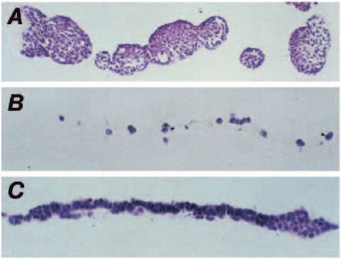
64      R. Williams and others

homology between human and mouse pS2 and only a 37%                    Transfection
homology between human pS2 and hITF (Lefebvre et al.,                  Prior to transfection the 410.4 cell line was grown in increasing con-
1993; Podolsky et al., 1993). The degree of evolutionary con-          centrations of puromycin (1-10 µg/ml) to ascertain the level of resis-
servation in both trefoil structure and distribution clearly           tance and death. At a concentration of 2.5 µg/ml no viable cells were
indicates that they have an important function, but their real         observed after 7 days in culture. The transfection vector pBppS2 was
physiological role is unclear.                                         constructed by insertion of the 490 bp BamHI/EcoRI fragment con-
   Early studies with purified PSP indicated that it may have          taining pS2 cDNA into the retroviral expression vector pBabe Puro
                                                                       (Morgenstern and Land, 1990) (see Fig. 1, below), kindly provided
weak physiological effects, including inhibition of intestinal         by H. Land (ICRF, London, UK). Transfection was carried out by a
muscular contraction and gastric acid secretion (Jørgensen et          modification of the CaPO4 method (Chen and Okayama, 1987) using
al., 1982), and a weak proliferative effect on HCT 116 and             the Stratagene mammalian transfection kit (Stratagene, Cambridge,
MCF-7 cell lines in vitro (Hoosein et al., 1989). A possible           UK). Duplicate 90 mm tissue culture dishes were seeded with 2×10⁴
clue to trefoil function emerged with the discovery of increased       410.4 cells per dish and at 20% confluency 10 µg of either pBpps2
expression in a number of physiological and pathological con-          (for pS2 transfection) or pBabe Puro (for vector control transfection),
ditions. Of particular interest is the association of pS2 and hSP      as a CaPO4-DNA precipitate, was added. After 18 hours the medium
expression with mucosal injury and ulceration (Wright et al.,          was removed and the culture was washed with Dulbecco’s PBSA, pH
1990a,b, 1993). Wright et al. (1990b) demonstrated increased           7.2, and fresh medium was applied. After a further 24 hours of culture
                                                                       the cells were split and seeded into 10 Petri dishes at 1000 cells per
pS2 and hSP expression in the ulcer-associated cell lineage            dish. Cells were allowed to settle for 24 hours and then subjected to
(UACL) formed in Crohn’s disease. It has been proposed that            puromycin selection (2.5 µg/ml). Puromycin-resistant clones were
trefoil peptides may be involved in the maintenance of mucosal         isolated by ring cloning.
integrity and may have a role in ulcer healing, tentatively based
on topological evidence. Some evidence for this presumed role          Southern blotting
has recently come from the observation that hSP and ITF can            Genomic DNA was prepared from cells grown to confluency in 15
apparently accelerate restitution in an in vitro ‘wounding’            cm Petri dishes. Cells were washed with ice-cold Dulbecco’s PBSA,
model (Dignass et al., 1994). In addition, it has been shown in        pH 7.2, before addition of 4 ml of lysis buffer (10 mM Tris buffer,
an experimental in vivo model of ulceration in rat stomach that        pH 8.0, containing 100 mM NaCl, 10 mM EDTA, 0.5% SDS and pro-
                                                                       teinase K 50 µg/ml). Lysis was carried out at 37°C for 5 hours. The
it is rSP and rITF that are upregulated in response to injury
                                                                       cell lysates were then extracted twice with phenol followed by cholo-
(Alison et al., 1995). Indeed, the trefoil expression is apparent      roform/isoamyl alcohol (24:1, v/v), and the genomic DNA was
before EGF and TGFα peptides, which are more typically                 ethanol precipitated. For Southern analysis 20 µg of genomic DNA
associated with ulcer healing.                                         was digested with KpnI and then run out on a 0.6% (w/v) agarose gel
   pS2 is expressed in adenocarcinomas of the colon, breast,           in TBE buffer (45 mM Tris base, 45 mM boric acid, 1 mM EDTA,
pancreas, stomach, gall bladder and ovary (Henry et al., 1991;         pH 8, buffer). The DNA was then transferred and fixed to Genescreen
Seitz et al., 1991), and in cell lines derived from these tissues.     Plus hybridisation Transfer membrane (Du Pont Ltd, Herts, UK) by
Thus pS2 expression has been demonstrated in human cell                the salt transfer protocol (Sambrook et al., 1989).
lines of the breast: MCF-7, T47D and ZR75; stomach: Kato3,             Northern blotting
MKN45 and MKN28; and pancreas: CAPAN2 and BXPC3
                                                                       Total cellular RNA was isolated by the acid guanidium isothio-
(Mori et al., 1988; Takahashi et al., 1990; our unpublished            cyanate-phenol-chloroform extraction method (Chomczynski and
observations). In view of this, to examine the effect of pS2 in        Sacchi, 1987) as modified by Cinna/Biotecx (Biogenesis Ltd, Poole,
isolation, we selected the murine mammary cell line 410.4              UK). For northern analysis 20 µg of the RNA samples were denatured
because it does not express pS2 (there is no cross-hybridisa-          at 65°C for 5 minutes in a solution containing 50% formamide and
tion with human pS2 cDNA probe at low stringency). In this             2.2 M formaldehyde. The samples were run out on a 0.8% agarose
paper we show how the transfection of human pS2 cDNA into              gel using formaldehyde-containing buffer (20 mM MOPS, 5 mM
the mouse 410.4 cell line causes changes in growth patterns in         NaOAc, 1 mM EDTA, 4.75% formaldehyde). RNA quality and
collagen matrix that could be consistent with its putative phys-       loading were assessed by ethidium bromide staining. Samples were
iological role.                                                        transferred to Genescreen Plus hybridisation Transfer membrane in
                                                                       the presence of 10× SSC (1.5 M NaCl, 0.15 M sodium citrate) and
                                                                       crosslinked in a UV Stratalinker 2400 (Stratagene, Cambridge, UK).
MATERIALS AND METHODS
                                                                       Hybridisation
Materials                                                              Membranes from northern and Southern blots were probed with the
410.4, a murine mammary adenocarcinoma cell line (Dexter et al.,       490 bp BamHI/EcoRI insert containing the full-length pS2 cDNA.
1978), screened for mycoplasma infection, was obtained from our cell   The probe was labelled with [α-32P]dCTP (Amersham Life Sciences,
culture stock. Full-length pS2 cDNA as a BamHI/EcoRI insert in the     Bucks, UK) by random priming (Sambrook et al., 1989). Hybridisa-
pGem1 vector was provided by R. Playford (ICRF, London, UK). All       tion was carried out at 42°C overnight in 50% formamide, 10%
reagents were purchased from Sigma (Dorset, UK) unless otherwise       dextran sulphate, 1% SDS, 1 M NaCl and 20 µg/ml denatured salmon
stated. The mBCEI1 antibody (Prud’homme et al., 1990) was a gift       sperm DNA. The membranes were washed in 2× SSC for 5 minutes
from Prof. E. Milgrom (Faculty of Medicine Paris-Sud, Cedex,           at room temperature, 2× SSC, 1% SDS for 30 minutes at room tem-
France) and the GE1 antibody (Elia et al., 1995) was kindly provided   perature, 2× SSC, 1% SDS for 30 minutes at 65°C, 0.2× SSC, 1%
by G. Elia (ICRF, London, UK).                                         SDS for 30 minutes at 65°C and finally in 0.1× SSC, 1% SDS for 30
                                                                       minutes at 65°C. The membranes were visualised after exposure to
Cell culture                                                           Hyperfilm-MP film (Amersham Life Sciences, Bucks, UK).
Unless otherwise indicated, all cells were grown in Dulbecco’s            The level of E-cadherin expression was assessed by probing
modified Eagle’s medium (DMEM) supplemented with 10% foetal            northern blots of total RNA of cells grown in collagen gel with the
bovine serum (FBS) supplied by Gibco BRL Ltd, Paisley, UK.             2.5 kb EcoRI fragment of mouse E-cadherin cDNA from the
pS2 causes epithelial cell dispersion 65
pBatEM2 vector (Nose et al., 1988) kindly provided by Dr M. and developed using the ABC method. All of the antibodies cross-
Takeichi (Institute of Basic Biology, Kyoto University, Japan). react with mouse proteins.
Immunoprecipitation Quantification of colony type
Cells for immunoprecipitation (IP) were grown to confluence in 15 A total of 300 colonies for each clone were counted from H and E-
cm dishes. Cells were washed in ice-cold Dulbecco’s PBSA, pH 7.2, stained en face sections of collagen gels after 14 days growth (two
and then lysed for 30 minutes on ice with 1 ml of 50 mM Tris-HCl, separate sets of four collagen gels). The colonies were assigned to one
pH 7.5, buffer containing 100 mM NaCl, 0.5% Nonidet P40, 0.2 mM of three categories: circumscribed, branched or dispersed (for a
Na3VO4, 50 mM NaF, 2 µg/ml leupeptin, 2 µg/ml aprotinin, 100 description see Results).
µg/ml N-tosyl-L-phenylalanine chloromethyl ketone and 100 µg/ml
4-(2-aminoethyl)-benzenesulphonyl fluoride. Lysates were trans- Cell aggregation assay
ferred to Eppendorf tubes and centrifuged at 13,000 g for 5 minutes. For the aggregation assay cells were grown in T75 cm3 flasks to 70-
A 50 µl sample of pre-clearing complex, Protein G-Sepharose 80% confluency. The cells were washed with PBSA prewarmed to
(Pharmacia Biotech Ltd, Herts, UK), incubated with pooled normal 37°C and then incubated with 5 ml of a non-enzymic cell-dissocia-
mouse immunoglobulins for 1 hour at 4°C, was added to the super- tion medium (Sigma, Dorset, UK) for 10 minutes at 37°C. Cells were
natants and incubated for 1 hour at 4°C on a rotator. A 1 µg sample washed with Ca2+- and Mg2+-free DMEM to obtain a single cell sus-
of GE1 antibody was added to the recovered supernatant. Following pension. A total of 3×10⁶ cells were suspended in 3 ml of DMEM
a 1 hour incubation at 4°C, 50 µl of Protein G-Sepharose was added containing 0.8% FBS and incubated in a gyration shaker rotating at
and a further 1 hour incubation was carried out. The beads were then 100 rpm at 37°C. Duplicate 10 µl samples were withdrawn after 0,
washed 5 times with TBST (25 mM Tris-HCl, 150 mM NaCl, pH 7.2, 15, 30, 45 and 60 minutes of incubation and the number of single cells
containing 0.5% Tween 20) and finally resuspended in 50 ml of lysis was counted. The assay was repeated a total of 5 times and the data
buffer (0.5 M Tris-HCl, pH 6.8, buffer containing 10% SDS, 500 mM presented as (Nt/No)×100, where Nt and No refer to the number of
EDTA, β-mercaptoethanol and 5% glycerol). The lysates were boiled single cells at time t and 0, respectively.
for 5 minutes, centrifuged at 13,000 g for 5 minutes and supernatants
run out on a 15% SDS-PAGE gel and electroblotted onto nylon Motility assay using time-lapse videomicroscopy
membranes (Bio-Rad Laboratories Ltd, Herts, UK). The membranes Time-lapse videomicroscopy experiments were performed using
were washed in TBST, blocked with 5% dried skimmed milk in TBS Olympus IMT 1 or 2 microscopes that were enclosed within environ-
(25 mM Tris-HCl, 150 mM NaCl, pH 7.2) for 1 hour at room tem- mental chambers. High resolution monochrome CCD cameras (Sony
perature. The membranes were incubated with mBCEI1 (2.5 µg/ml) M370CE) were attached to each microscope. Images were recorded
in TBST overnight, then washed in TBST (3× 10 minutes). The using broadcast quality videorecorders (Sony Betacam PVW 2800)
membranes were incubated for 1 hour at room temperature with that were driven externally by animation controllers (BAC 900 from
biotinylated rabbit anti-mouse IgG antibodies (Dako Ltd, Bucks, UK) EOS Electronics AV Ltd, Barry, S. Wales).
and then developed using the ABC method (Dako Ltd, Bucks, UK) For the motility assay cells were plated on 35 mm dishes at 104
followed by an ECL chemiluminescence assay (Amersham Life cells per dish, 24 hours before the start of the assay. A field contain-
Sciences, Bucks, UK) and visualised by exposure to Hyperfilm-MP ing between 30 and 40 cells was chosen for each cell line. Images
film (Amersham Life Sciences, Bucks, UK). were recorded at one frame every minute for 24 hours. The record-
ings were downloaded onto an S-VHS videorecorder (Mitsubishi B-
Collagen gels 82), which was linked to an Apple MacIntosh 6100 computer, and the
Quadruplicate collagen type 1 gels using Vitrogen 100 (Celtrix Phar- cells that remained within the observed field for the duration of the
maceuticals, Santa Clara, CA) and bovine dermal collagen were made film were tracked using Cell Motility software (EOS Electronics). A
up according to the manufacturer’s instructions. Briefly, 8 vol. of printout of the track of each cell was obtained together with calcula-
collagen stock solution was mixed with 1 vol. of 10× DMEM, neu- tion off the mean distance and speed for each cell line.
tralised with 0.1 M NaOH and made up to 10 vol. with sterile distilled
water in a sterile flask on ice. A 0.5 ml sample of appropriate cells
was added to 5 vol. of the gel solution to give a final concentration
1×10⁴ cells per ml, and 1 ml aliquots were dispensed into 35 mm RESULTS
tissue culture dishes and allowed to gel for about 15 minutes at 37°C
before addition of culture medium. For surface collagen gel growth Characterisation of transfected clones
studies gels were set up as above except that the cells (1×10⁴ cells per For transfection the pS2 cDNA, a 490 bp BamHI/EcoRI
gel) were added after the collagen gels had set. All collagen gels were
restriction fragment, was inserted into the pBabe Puro retrovi-
fed with DMEM/10% FBS containing 2.5 mg/ml puromycin every 72
hours. Growth was assessed every 24 hours by examination under an ral expression vector as is shown in Fig. 1. In this vector the
Olympus IMT-2 phase-contrast microscope and followed for a promoter in the 5′ Mo MuLV long terminal repeat (LTR) is
maximum of 21 days. Collagen gels were fixed at weekly intervals in used to transcribe the pS2 inserted gene and the internal SV40
10% formol saline overnight, processed and subsequently paraffin- early promoter expresses puromycin drug resistance marker,
embedded and serially sectioned (5 µm sections). both transcripts sharing a common termination and poly-
adenylation signal in the 3′ LTR. Transfectants were selected
Histology and immunocytochemistry by their ability to grow in media supplemented with puromycin
Sections were routinely stained with haematoxylin and eosin (H and at 2.5 µg/ml, a concentration that was shown to kill 410.4 cells.
E). For fibronectin, type IV collagen and laminin detection sections Two pS2 cDNA transfected clones (AA4, AD4) and one clone
were digested with protease for 20 minutes at 37°C and then stained
(VA5) from transfection with pBabe Puro were isolated by ring
immunohistochemically using a diaminobenzidine peroxidase-
antiperoxidase technique using a 1 in 100 dilution of either anti- cloning. Genomic DNA isolated from all three clones (AA4,
human laminin rabbit polyclonal antibody (EURO-PATH, Bude, UK) AD4 and VA5) and the parental cell line was digested with
or anti-human fibronectin rabbit polyclonal antibody (Dako Ltd, KpnI, Southern blotted and hybridised with a pS2 cDNA probe.
Bucks, UK). The anti-human collagen type IV rabbit polyclonal The resulting blot (Fig. 2A) shows that both AA4 and AD4
antibody (EURO-PATH, Bude, UK) was used at a 1 in 1000 dilution display a single band of 3 kb; by contrast, neither the parental66 R. Williams and others
Kpn 1 3420
Kpn 1 365
BamH 1
EcoR 1
SV40
R U5 gag pS2 early puro r U3 R U5
S-D promoter
-
ATG
Long Terminal Repeat (LTR) Long Terminal Repeat (LTR)
amp r pUC Ori Fig. 1. Map of pBppS2
transfection vector.
cell line nor the vector control VA5 clone shows any bands.
The 3 kb band is in keeping with the expected size of the KpnI
fragment of the pBppS2, since there is one KpnI site in each
of the LTRs (see Fig. 1), and indicates no gross rearrangement
or truncation of integrated DNA copy. This clearly demon-
strates that both pS2 transfected clones have incorporated the
pS2 cDNA into the 410.4 genome.
The presence of pS2 transcripts in the clones was determined
by northern analysis of total RNA. The northern blot analysis
for pS2 containing transcripts (Fig. 2B) shows clear 3.5 kb
bands in both the AA4 and AD4 clones that correspond to the
expected full-length transcript from 5′ viral LTR (see Fig. 1).
The presence of an extra band at 2 kb in the AD4 clone tract
may be due to a splicing event from the cryptic termination or
a poly(A) site at the 3′ end of the pS2 cDNA insert (Dai et al.,
1993; Kirschmeier et al., 1988). This band is also observed in
the AA4 clone on prolonged exposure of the northern blot (data
not shown). Levels 6-fold higher, as determined by laser den-
sitometry (Molecular Dynamics, Bucks, UK), of the transcript
are present in the AD4 clone. No bands were evident with VA5
or 410.4 RNA samples.
To ascertain whether the transcripts were appropriately
translated immunoprecipitation was carried out on the cell
lysates using the GE1 anti-pS2 antibody. Fig. 2C shows a
western blot of the immunoprecipitates developed using the
anti-pS2 mBCEI1 antibody. Bands at approximately 7 kDa
were shown by AD4 and AA4 but not 410.4 and VA5. The Fig. 2. Characterisation of transfectants. (A) Southern blot of
genomic DNA from transfectants and parental cell line. A 20 µg
band size matches that obtained for pS2 immunoprecipitation sample of genomic DNA was digested with KpnI and run out on a
from MCF-7 cells (Elia et al., 1995). It is also evident that the 0.6% agarose gel in TBE. The DNA was transferred to a GeneScreen
AD4 clone expresses greater amounts of pS2 peptide than the nylon membrane and probed with 32P-labelled 490 bp pS2 cDNA.
AA4 clone. From these results we can say that both AD4 and (B) Northern blot of total RNA from transfectants and parental cell
AA4 contain pS2 cDNA at the genomic level, produce pS2- lines. A 20 µg sample of total RNA was run out on 0.8% agarose
containing transcripts and produce the correct translation formaldehyde gel and transferred to a GeneScreen nylon membrane.
product. The membrane was probed with 32P-labelled 490 bp pS2 cDNA.
(C) Immunoprecipitation of pS2 peptide from transfected clones.
Growth on plastic and in type 1 collagen gels GE1 immunoprecipitates from cell lysates were run out on a 15%
When grown on plastic the pS2 transfected clones AD4 and SDS-PAGE gel and transferred to nitrocellulose membrane. The
membrane was developed with the anti-pS2 antibody mBCEI1.
AA4 showed the same morphology as the vector control trans- Throughout, lane 1 refers to AA4 clone, lane 2 to AD4 clone, lane 3
fectant (VA5) and the parental cell line. No difference in cyto- to the vector control (VA5) and lane 4 to the parental cell line.
logical characteristics or growth rate measured by DNA flu-
orimetry (Rao and Otto, 1992) was evident (data not shown).
ences in growth pattern the clones were propagated in type 1
Phase contrast microscopy (7-14 days) collagen matrix. Phase-contrast microscopy showed marked
To establish whether expression of pS2 would lead to differ- differences in the growth pattern of the transfectants as earlypS2 causes epithelial cell dispersion 67
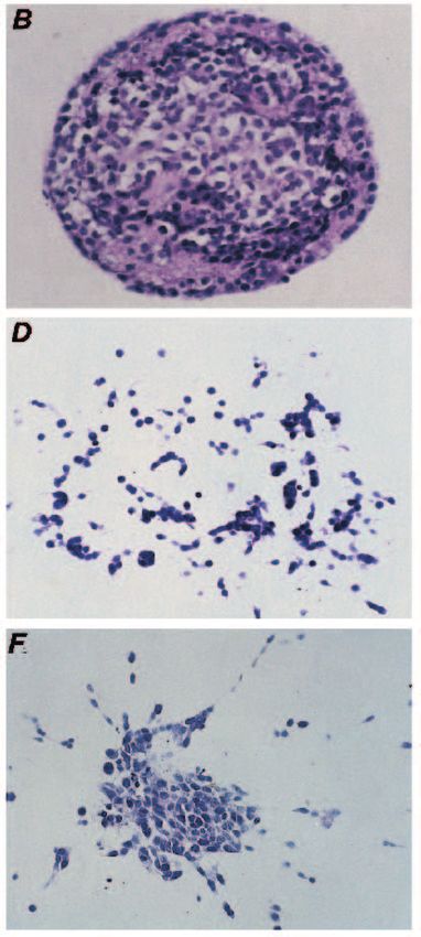
Fig. 3. Growth of transfectants in
collagen type 1 gels. (A) A phase-
contrast photograph of VA5
colonies formed after 14 days
growth in collagen gel. (B) H and
E-stained en face section of a
typical VA5 colony. (C) A phase-
contrast photograph of a typical
AD4 colony after 14 days growth
in collagen gel. (D) H and E-
stained en face section of a typical
AD4 colony. (E) A phase-contrast
photograph of AA4 colonies
formed after 14 days growth in
collagen gel. (F) H and E-stained
en face section of typical AA4
colony. Bars, 150 µm.
as after 7 days of growth. Both the parental and vector control were observed and thus this process is not regarded as a mor-
(VA5) cell lines formed small tight compact spherical colonies phogenetic phenomenon. No matrix deposition was evident.
(Fig. 3A), while the pS2 transfectants formed dispersed stellate Confirmation that pS2 was expressed in cells grown in collagen
structures with peripheral thin extensions (Fig. 3C,E). These was obtained from northern analysis of pS2 RNA extracted
differences became even more pronounced after 14 days in from cells grown in collagen gel. A similar level of pS2-con-
culture. taining transcript was produced as was observed with cells
grown on plastic (data not shown).
Histology of cells grown in collagen (7-14 days)
H and E-stained sections of the collagen gels (Fig. 3B) show Quantitative analysis of structures
that VA5 forms tight compact colonies with circumscribed Three types of colonies were quantified on the gel sections.
margins, composed of large cells with abundant cytoplasm. In Circumscribed colonies were typified by VA5. Branching
addition there is prominent extracellular matrix deposition in colonies had outgrowths into the gel, which are short, often
the colony shown up as hyaline eosinophilic aggregates. Occa- clubbed and composed of rounded or polygonal cells, which
sional less circumscribed colonies with broad short projections are often over 2 cells in width. Dispersed colonies had radiating
into the gel were observed. The parental cell line had a similar cords of cells, usually elongated, with no more than 1-2 cells
appearance. Sections of the transfectants show that the at the extremities. Fig. 4 shows a quantitative analysis of the
dispersed structures with radiating cords are essentially colony type formed by the transfectants (day 14). The vector
composed of extensive cords of elongated single cells control VA5 showed predominantly circumscribed structures
migrating out into the gel (Fig. 3D,F). No tubular formations (94.5%) with some branching (4.2%), and very little in the way68 R. Williams and others
AA Table 1. Motility assay
100
AA
AD4
AA4
Cell line Cell motility (µm/h)
80 VA5 VA5 12.64±1.13
AA
AD4 29.87±1.75
Percentage of Colonies
60
AA The results represent the mean speed (µm/h) determined for 40 cells of
AA
each cell line ± s.e.m.
AA AA
AA
40
frequently down-regulated in highly invasive, poorly differen-
AA AA tiated carcinomas (Frixen et al., 1991; Behrens et al., 1991;
AA AA
20 Pignatelli et al., 1992). The level of E-cadherin expression of
AA AA AA
the transfectants was assessed by northern analysis of total
RNA extracted from cells grown in collagen gel using a mouse
0 E-cadherin cDNA probe (Nose et al., 1988). A single band of
Circumscribed Branching Dispersed
4.5 kb was observed in all RNA samples and there was no dif-
Colony Type ference in the level of expression in the pS2 transfectants
compared to the VA5 and parental cell lines as determined by
Fig. 4. Quantification of colony types formed by transfectants in
densitometric analysis (data not shown). To determine whether
collagen gels. The type of colonies formed by the pS2 transfectants
AD4 and AA4 and the vector control VA5 were counted in H and E- the observed changes were due to changes in E-cadherin-
stained en face sections from two separate sets of four collagen gels mediated cell adhesion, the calcium-dependent cell-cell aggre-
after 14 days growth (300 colonies were counted). The results gation of the pS2 transfectants was compared with that of the
represent the mean ± s.e.m. vector control. From Fig. 6 it is evident that pS2 expression
does not result in any changes in the calcium-dependent cell-
of dispersed colonies (1.3%) was evident. Some of these may cell aggregation. It is therefore unlikely that the observed dis-
even have arisen by intracolony cell death and regeneration, as persant growth pattern of the pS2 transfectants changes are due
they appeared to contain cellular debris. Only 3% of AD4 and to alteration in E-cadherin function.
5% of AA4 colonies were circumscribed, with a majority being It is possible that the changes in growth pattern of 410.4 cells
dispersed, 80% in the case of AD4 and 68% for AA4. There as a result of pS2 expression could be explained by pS2 causing
would appear to be a correlation between the extent of disper- an increase in cell motility. To test this a motility assay was
sion and the level of pS2 expression. The higher pS2 express- carried out on AD4 pS2 transfectant and the vector control
ing clone AD4 showed a greater tendency to form dispersed VA5. The results show that the pS2 transfectant has a 2.4-fold
colonies than the lower expressor AA4. increase in motility over that of the vector control cell line
(Table 1).
Growth on type 1 collagen gels
When the clones were grown on the surface of collagen gels a
similar difference was observed in the growth pattern of the DISCUSSION
pS2 transfectants compared to the vector control. Under phase-
contrast the parental and VA5 cell lines (vector control) formed We describe the first model system employing human pS2 gene
large circumscribed spherical colonies showing very little transfer and expression in a non-pS2-expressing cell line.
lateral migration, whereas the transfectants spread over the gel There is a high degree of conservation between human and
surface. Each gel was longitudinally serially sectioned and mouse pS2 (Lefebvre et al., 1993), and we could not show any
stained. Sections of the parental gel showed large spherical cross-hybridisation of human pS2 probes in the parental cell
colonies protruding above the surface of the gel, with very line at low stringency. Our findings were that both the AD4
prominent deposits of ECM forming hyaline eosinophilic and AA4 clones had integrated the pS2 cDNA into the 410.4
aggregates (Fig. 5A). The pS2 transfected clones spread out genome, and produced pS2-containing transcripts and the
over the surface of the gel with no nodular growths or ECM mature pS2 peptide. The AA4 transfectant showed lower levels
deposition (Fig. 5B,C). Significantly, we found no evidence of of pS2 transcript and peptide compared to the AD4 clone.
invasion into the collagen gel by VA5, AD4 or AA4 clones. From the growth in collagen type 1 gels it is clear that pS2
Immunocytochemistry (data not shown) on sections did not expression in the two transfectants resulted in a more dispersed
reveal any significant difference in the amount of laminin, pattern of growth. The vector control clone (VA5), like the
fibronectin and collagen type IV staining between the pS2 parental cell line, typically formed cohesive islands of cells
transfected clones and the vector control, although these com- showing no invasion into the collagen matrix, with prominent
ponents tended to be more spread out in close approximation ECM deposition. The pS2 transfectants tended to form poorly
to the cells in the transfectants. However, the majority of the cohesive stellate colonies from which radiate thin cords of
eosinophilic ECM did not show specific immunoreactity for elongated cells into the collagen matrix, with no visible ECM
specific matrix components, which is predictable from studies deposition. The extent to which the transfectants formed these
on hyalinised ECM in human tissues. dispersed structures appeared to correlate with the level of pS2
It has been reported that dispersant growth patterns of cells expression, with the lower pS2 expressor, AA4, showing a
are mediated by changes in their cadherin-based cell adhesion smaller degree of dispersion.
ability (Behrens et al., 1993) and that E-cadherin expression is Assay of cellular motility using videomicroscopy clearlypS2 causes epithelial cell dispersion 69 showed a marked increase in the speed and distance travelled component of the gastrointestinal mucosa. The expression of by the AD4 transfectant. Since dispersion might be a conse- trefoil peptides in response to mucosal injury has led to the quence of reduced intercellular cohesion, which is primarily suggestion that they participate in the ulcer-healing response. mediated by E cadherin (Takeichi, 1991), we performed cell- Thus pS2 and hSP expression is found in the ulcer-associated cell aggregation assays, which showed no differences between cell lineage (UACL) in chronic ulcerative conditions such as vector controls and transfectants. Moreover, there were no dif- Crohn’s disease (Wright et al., 1990a,b, 1993). This differen- ferences in E cadherin expression as determined by northern tiating cell lineage buds from the bases of intestinal crypts analysis. adjacent to the ulcer, ramifying and anastomosing in the Thus pS2 appears to exert a motogenic effect on 410.4 cells, submucosa and finally fusing with the villous epithelial surface but not a mitogenic or a morphogenetic effect. A similar via terminal ducts (Wright et al., 1990a). UACL development motogenic or ‘scatter’ effect is induced by hepatocyte growth with concomitant pS2/hSP expression has also been observed factor (HGF), which has been shown to dissociate layers of at other sites of chronic endodermal injuries such as ducts in epithelial cells, increasing their motility and invasiveness chronic pancreatitis or biliary obstruction, and in nasal polyps (Stoker et al., 1987; Weidner et al., 1990). However, HGF is (Wright et al., 1990b; Seitz et al., 1991). also a mitogen for kidney tubular epithelium, keratinocytes, Healing of ulcerative lesions occurs throughout the gastroin- endothelial cells and melanocytes (Rubin et al., 1991; testinal tract and is characterised by the ability to reconstitute Bussolino et al., 1992), and can function as a morphogen stim- the mucosa followed by the re-formation of specialised struc- ulating the three-dimensional organisation of Madin-Darby tures. When a mucosal defect occurs, it is initially covered by canine kidney cells (MDCK) (Montesano et al., 1991). It has a layer of necrotic cells, fibrin and mucus termed the ‘mucoid also been shown to promote the progression of carcinoma cells cap’. This mucoid cap may be of particular importance in the towards a malignant invasive phenotypes (Weidner et al., stomach in re-establishing a pH gradient so that the cells of the 1990). surviving mucosa are maintained at a neutral pH rather than that Early studies with PSP suggested that this trefoil had a of the acidic gastric juice (Wallace and Whittle, 1986). Epi- mitogenic activity towards MCF-7 and HCT 116 cell lines thelial cells at the margin of the defect migrate to re-establish (Hoosein et al., 1989). However, the levels of PSP required to a continuous epithelial layer. This process is termed ‘epithelial elicit a response were 10 times higher than the expected range restitution’. Restitution occurs within the first hour following for most growth factors. We have been unable to reproduce injury and over the next 48 hours cellular proliferation and this effect and have found no mitogenic activity for hSP in a differentiation in adjacent glands occur, re-establishing the number of cell lines (unpublished observations). Recently, normal architecture. In ulcer healing it is the re-epithelialisation Dignass et al. (1994) also failed to observe any mitogenic of the damaged area that is the important first response and this activity for hSP, rITF and hITF on a panel of cell lines. In this is achieved by epithelial cells from the wound edges migrating study, expression of pS2 had no effect on the proliferation of into the area, and does not involve a proliferative response (at the 410.4 cell line. It therefore seems unlikely that trefoils are least initially). Indeed it has been shown that healing of mucosal mitogens. Since we did not demonstrate any invasive action of erosions (injury not involving the full thickness of the mucosa) pS2-expressing cells from the surface into the collagen matrix, in rats is very rapid. Silen and Ito (1985) have demonstrated it may be that pS2, unlike HGF, induces a spreading rather than complete re-epithelialisation of totally desquamated surfaces an invasive phenotype. Indeed, we have demonstrated that pS2 within 1 hour by migration of cells from neighbouring pits expression results in increased cell motility. before cell proliferation and inflammation. The changes in growth pattern of the 410.4 cell line brought Our observation that pS2 has the potential to induce a about by pS2 expression were associated with an alteration in migratory response, taken together with the topographical ECM deposition. Prominent deposits of ECM were shown by expression of pS2 in response to ulcerative damage, supports the vector control. Both the pS2 transfectants showed very little a role for trefoil peptides in ulcer healing. Our results are com- in the way of visible ECM when propagated in or on the surface patible with the recent observation that showed that two trefoil of collagen gels. No quantitative differences in type IV peptides, intestinal trefoil factor (ITF) and human spasmolytic collagen, fibronectin or laminin were apparent apart from redis- polypetide (hSP), accelerated restitution of wounded mono- tribution commensurate with the alteration in growth pattern. It layers in an in vitro restitution assay (Dignass et al., 1994). may be that other components of the ECM associated with cell This study also demonstrated that the effect is not necessarily migration and spreading, such as tenascin, vitronectin or species-specific, since hITF was as effective as its rat equiv- laminin fragments, possibly in the non-immunoreactive parts of alent in the restitution assays carried out on rat intestinal epi- the ECM, could be responsible for these effects. thelial cell lines. Further support for the proposed role comes It has been demonstrated that the motogenic and mor- from the observation of an up-regulation of rSP and rITF at phogenic activity in epithelial cells induced by HGF is associ- ulcer margins in experimentally induced gastric ulcers in rat ated with degradation of ECM. Thus the HGF-induced mor- (Alison et al., 1995). This expression of the trefoils in phogenesis of MDCK cells when grown in collagen type 1 is response to injury occurs prior to the expression of the EGF repressed by protease inhibitors (Montesano et al., 1991). It is and TGFα molecules traditionally associated with ulcer conceivable that pS2 expression leads to an increased healing. It is conceivable that pS2, as well as other trefoils, expression of proteases, resulting in a degradation in ECM, initiates the healing process by induction of a migratory which leads to a loosening of cell interaction, an area that we response in the epithelial population around the margins of the are currently investigating. ulcer. This would explain the high levels of pS2 and hSP Is there any relationship between these in vitro effects of pS2 expression in the gastric mucosa, since a high level of resti- and its physiological role? Trefoil peptides are a normal tutive peptides would be necessary to enable a rapid re-estab-
70                 R. Williams and others

                                                                            The authors thank Jonathan Wilding for his contribution to this
                                                                         work. This work is supported by the Wellcome Trust and the Crohn’s
                                                                         in Childhood Research Association.

                                                                         REFERENCES
                                                                         Alison, M. R., Chinnery, R., Poulsom, R., Ashwood, P., Longcroft, J. M.
                                                                           and Wright, N. A. (1995). Experimental ulceration leads to sequential
                                                                           expression of spasmolytic polypeptide, intestinal trefoil factor, epidermal
                                                                           growth factor and transforming growth factor alpha mRNAs in rat stomach.
                                                                           J. Pathol. 175, 405-414.
                                                                         Behrens, J., Weidner, K. M., Frixen, U. H., Schipper, J. H., Sachs, M.,
                                                                           Arakaki, N., Daikuhara Y. and Birchmeier, W. (1991). The role of E-
                                                                           cadherin and scatter factor in tumor invasion and cell motility. EXS. 59,
                                                                           109-26.
                                                                         Behrens, J. L., Vakaet, R., Friis, E., Winterhager, R. F., Van Mareel, M. M.
                                                                           and Birchmeier, W. (1993). Loss of epithelial differentiation and gain of
                                                                           invasiveness correlates with tyrosine phosphorylation of the E-
Fig. 5. Growth on collagen type 1 gels. (A) H and E-stained section        cadherin/beta-catenin complex in cells transformed with a temperature-
of parental cells grown on collagen gel. (B) H and E-stained section       sensitive v-SRC gene. J. Cell Biol. 120, 757-766.
of AD4 clone grown on collagen gel. (C) H and E-stained section of       Blundell, T. L. and Humbel, R. E. (1980). Hormone families: pancreatic
AA4 clone grown on collagen gel. (In all figures the collagen gels         hormones and homologous growth factors. Nature 287, 781-787.
were embedded on edge after 14 days in culture and the sections are      Bussolino, F., Di Renzo, M. F., Ziche, M., Bocchietto, E., Olivero, M.,
oriented with the collagen gel at the bottom of the field.)                Naldini, L., Gaudino, G., Tamagnone, L., Coffer, A. and Comoglio, P. M.
                                                                           (1992). Hepatocyte growth factor is a potent angigenic factor which
                                                                           stimulates endothelial cell motility and growth. J. Cell Biol. 119, 629-641.
                   100                                                   Carr, M. D. (1992). 1H NMR-based determination of the secondary structure
                                                                           of porcine pancreatic spasmolytic polypeptide: one of a new family of
                                                                           ‘trefoil’ motif containing cell growth factors. Biochemistry 31, 1998-2004.
                   90                                                    Chen, C. and Okayama, H. (1987). High efficiency transformation of
                                                                           mammalian cells by plasmid DNA. Mol. Cell. Biol. 7, 2745-2752.
                                                             AA4         Chomczynski, P. and Sacchi, N. (1987). Single-step method of RNA isolation
                   80                                                      by acid guanidium thiocyanate-phenol-chloroform extraction. Anal.
                                                             VA5           Biochem. 162, 156-159.
                                                                         Dai, Y., Rashba-Step, J. and Cederbaum, A. I. (1993). Stable expression of
                   70                                        AD4
                                                                           human cytochrome P4502E1 in HepG2 cells: Characterization of catalytic
                                                                           activities and production of reactive oxygen intermediates. Biochemistry 32,
                   60                                                      6928-6937.
                                                                         Dexter, D. L., Kowalski, H. M., Blazar, B. A., Fligiel, Z., Vogel, R. and
     Nt/No x 100

                                                                           Heppner, G. H. (1978). Heterogeneity of tumour cells from a single mouse
                   50                                                      mammary tumour. Cancer Res. 10, 3174-3181.
                                                                         Dignass, A., Lynch-Devaney, K., Kindon, H., Thim, L. and Podolsky, D. K.
                                                                           (1994). Trefoil peptides promote epithelial migration through a transforming
                   40                                                      growth factor b-independent pathway. J. Clin. Invest. 94, 376-383.
                                                                         Elia, G., Williams, R., Oates, T., Stamp, G. W. H., Wright, N. A., Pignatelli,
                                                                           M. and Lalani, E.-N. (1995). Characterisation of monoclonal antibodies
                   30                                                      raised to C-terminal peptides of pS2. J. Pathol. Suppl. 175, 115A.
                                                                         Frixen, U. H., Behrens, J., Sachs, M., Eberle, G., Voss, B., Warda, A.,
                                                                           Lochner, D. and Birchmeier W. (1991). E-cadherin-mediated cell-cell
                   20                                                      adhesion prevents invasiveness of human carcinoma cells. J. Cell Biol. 113,
                                                                           173-85.
                                                                         Gajhede, M., Petersen, T. N., Henriksen, A., Petersen, J. F., Dauter, Z.,
                   10
                                                                           Wilson, K. S. and Thim, L. (1993). Pancreatic spasmolytic polypetide: First
                                                                           three-dimensional structure of a member of the mammalian trefoil family of
                    0                                                      peptides. Structure 1, 253-262
                          0      15         30          45         60    Gregory, H. and Preson, B. M. (1977). The primary structure of human
                                                                           urogastrone. Int. J. Pept. Protein Res. 9, 107-118.
                                      Time in minutes                    Hanby, A. M., Poulsom, R., Elia, G., Singh, S., Longcroft, J. M. and Wright,
                                                                           N. A. (1993). The expression of the trefoil peptides pS2 and human spasmolytic
Fig. 6. Cell aggregation assay. Nt and N0 refer to the number of           polypeptide (hSP) in ‘gastric metaplasia’ of the proximal duodenum:
single cells at time t and time 0, respectively. The results represent     implications for the nature of ‘gastric metaplasia’. J. Pathol. 169, 355-360.
the mean values of five separate experiments ± s.e.m.                    Hauser, F., Poulsom, R., Chinery, R., Rogers, L. A., Hanby, A. M., Wright,
                                                                           N. A. and Hoffmann, W. (1993). hP1 B, a human P-domain peptide
                                                                           homologous with rat intestinal trefoil factor, is expressed also in the ulcer-
lishment of mucosal integrity in a particularly hostile envi-              associated cell lineage and the uterus. Proc. Nat. Acad. Sci. USA 90, 6961-
ronment. Presumably, the formation of UACL at sites of more                6965.
extensive damage provides a large pool of trefoils to facilitate         Henry, J. A., Bennett, M. K., Piggott N. H,. Levett, D. L., May, F. E. and
restitution.                                                               Westley, B. R. (1991). Expression of the pNR-2/pS2 protein in diverse
                                                                           human epithelial tumours. Br. J. Cancer 64, 677-82,
   Our data are therefore consistent with a role for trefoil             Hoosein, N. M., Thim, L., Jørgensen, K. H. and Brattain, M. G. (1989).
peptides in the maintenance and restoration of mucosal                     Growth stimulatory effect of pancreatic spasmolytic polypeptide on cultured
integrity following mucosal injury and ulceration.                         colon and breast tumor cells. FEBS Lett. 247, 303-6.
pS2 causes epithelial cell dispersion 71 Jackowlew, S. B., Breathnach, R., Jeltsch, J. M., Masiakowski, P. and Rubin, J. S., Chan, A. M.-L., Bottaro, D. P., Burgess, W. H., Taylor, W. G., Chambon, P. (1984). Sequence of the pS2 mRNA induced by estrogen in the Cech, A. C., Hirschfield, D. W., Wong, J., Miki, T., Finch, P. W. and human breast cancer cell line MCF-7. Nucl. Acids Res. 12, 2861-78. Aaronson, S. A. (1991). A broad-spectrum human long fibroblast-derived Jørgensen, K. D., Diamant, B., Jørgensen, K. H. and Thim, L. (1982). mitogen is a variant of hepatocyte growth factor. Proc. Nat. Acad. Sci. USA Pancreatic spasmolytic peptide (PSP): III. Pharmacology of a new porcine 88, 415-419. pancreatic polypeptide with spasmolytic and gastric acid secretion inhibitory Sambrook, J., Fritsch, E. F. and Maniatis, T. (1989). Molecular Cloning: a effects. Regul. Pept. 3, 231-243. Laboratory Manual, 2nd edn. Cold Spring Harbor, NY: Cold Spring Harbor Kirschmeier, P. T., Housey, G. D., Johnson, M. D., Perkins, A. S. and Laboratory Press. Weinsttein, I. B. (1988). Construction of a retroviral vector demonstrating Seitz, G., Thelsinger, B., Tomasetto, G., Rio, M.-C., Chambon, P., Blin, N. efficient expression of cloned cDNA sequences. DNA 7, 219-225. and Welter, G. (1991). Breast cancer-associated protein pS2 expression in Lefebvre, O., Wolf, C., Kedinger, M., Chernard, M. P., Tomasetto, C., tumours of the biliary tract. Am. J. Gastroenterol. 86, 1491-1494. Chambon, P. and Rio, M. C. (1993). The mouse one P-domain (pS2) and Silen, W. and Ito, S. (1985). Mechanisms for rapid re-epithilialization of the the two P-domain (mSP) genes exhibit distinct patterns of expression. J. Cell gastric mucosal surface. Annu. Rev. Physiol. 47, 217-229. Biol. 122, 191-198. Skilton, R. A., Luqmani, Y. A., McClelland, R. A. and Coombes, R. C. Masiakowski, P., Breathnach, R., Bloch, J., Gannon, F., Krut, A. and (1989). Characterisation of a messenger RNA selectively expressed in Chambon, P. (1982). Cloning of cDNA sequences of hormone-regulated human breast cancer. Br. J. Cancer 60, 168-175. genes from the MCF-7 human breast-cancer cell line. Nucl. Acids Res. 10, Stoker, M., Gherardi, E., Perryman, M. and Gray, J. (1987). Scatter factor 7895-7903. is a fibroblast-derived modulator of epithelial cell mobility. Nature 327, 239- May, F. E. B. and Westley, B. R. (1986). Cloning of estrogen-regulated 242. messenger RNA sequences from human breast cancer cells. Cancer Res. 46, Suemori, S., Lynch-Devaney, K. and Podolsky, D. K. (1991). Identification 6034-6040. and characterization of rat intestinal trefoil factor: tissue- and cell-specific May, F. E. B. and Westley, B. R. (1988). Identification and characterization of member of the trefoil protein family. Proc. Nat. Acad. Sci USA 88, 11017- estrogen-regulated RNAs in human breast cancer cells. J. Biol. Chem. 263, 11021. 12901-12908. Takahashi, H., Kida, N., Fujii, R., Tanaka, K., Ohta, M., Mori, K. and Montesano, R., Schaller, G. and Orci, L. (1991). Induction of epithelial Hayashi, K. (1990). Expression of the pS2 gene in human gastric cancer tubular morphogenesis in vitro by fibroblast-derived soluble factors. Cell 66, cells derived from poorly differentiated adenocarcinoma. FEBS Lett. 261, 697-711. 283-286. Morgenstern, J. P. and Land, H. (1990). Advanced mammalian gene transfer: Takeichi, M. (1991). Cadherin cell adhesion receptors as a morphogenetic high titre retroviral vectors with multiple drug selection markers and a regulator. Science 251, 1451-1455. complementary helper-free packaging cell line. Nucl. Acids Res. 18, 3587- Thim, L. (1989). A new family of growth factor-like peptides. ‘Trefoil’ 3596. disulphide loop structures as a common feature in breast cancer associated Mori, K., Fujii, R., Kida, N., Ohta, M. and Hayashi, K. (1988). Identification peptide (pS2), pancreatic spasmolytic polypetide (PSP), and frog skin of a polypeptide secreted by human breast cancer cells (MCF-7) as the peptides (spasmolysins). FEBS Lett. 250, 85-90. human estrogen-responsive gene (pS2) product. Biochem. Biophys. Res. Tomasetto, C., Rio, M.-C., Gautier, C., Wolf, C., Hareuveni, M., Chambon, Commun. 155, 366-372. C. and Lather, R. (1990). hSP, the domain-duplicated homolog of pS2 Nose, A., Nagafuchi, A. and Takeichi, M. (1988). Expressed recombinant protein, is co-expressed with pS2 in the stomach but not in breast carcinoma. cadherins mediate cell sorting in model systems. Cell 54, 993-1001. EMBO J. 9, 407-414. Nunez, A. M., Berry, M., Imler, J. L. and Chambon, P. (1989). The 5′ Wallace, J. L. and Whittle, B. J. R. (1986). Role of mucus in the repair of flanking region of the pS2 gene contains a complex enhancer region gastric epithelial damage in the rat. Gastroenterology 91, 603-611. responsive to oestrogens, epidermal growth factor, a tumour promoter Warne, N. W. and Laskowski, M., Jr (1990). All fifteen possible (TPA), the c-Ha-ras oncoprotein and the c-jun protein. EMBO J. 8, 823-829. arrangements of three disulphide bridges in proteins are known. Biochem. Pignatelli, M., Liu, D., Nasim, M. M., Stamp, G. W. H, Hirano, S. and Biophys. Res. Commun. 172, 1364-1370. Takeichi M. (1992). Morphoregulatory activities of E-cadherin and beta-1 Weidner, K. M., Behrens, J., Vandekerckhove, J. and Birchmeier, W. integrins in colorectal tumour cells. Br. J. Cancer 66, 629-634. (1990). Scatter factor: molecular characteristics and effect on the Podolsky, D. K., Lynch-Devaney, K., Stow, J. L., Oates, P., Murgue, B., De invasiveness of epithelial cells. J. Cell Biol. 111, 2097-2108. Bauemont, M., Sands, B. E. and Mahidi, Y. R. (1993). Identification of Wright, N. A., Pike, C. and Elia, G. (1990a). Induction of a novel epidermal human intestinal trefoil factor: goblet cell specific expression of a peptide growth factor-secreting cell lineage by mucosal ulceration in gastrointestinal targeted for apical secretion. J. Biol. Chem. 268, 6694-6702. stem cells. Nature 343, 82-85. Prud’homme, J.-F, Jolivet, A., Pichon, M.-F., Savouret, J.-F. and Milgrom, Wright, N. A., Poulsom, R., Stamp, G. W. H., Hall, P. A., Jeffery, R. E, E. (1990). Monoclonal antibodies against native and denatured forms of Longcroft, J. M., Rio, M.-C., Tomasetto, C. and Chambon, P. (1990b). estrogen-induced breast cancer protein (BCEI/pS2) obtained by expression Epidermal growth factor (EGF/URO) induces expression of regulatory in Escherichia coli. Cancer Res. 50, 2390-2396. peptides in damaged human gastrointestinal tissues. J. Pathol. 162, 279-284. Rao, J. and Otto, W. R. (1992). Fluorimetric DNA assay for cell growth Wright, N. A., Poulsom, R., Stamp, G. W. H., Vannorden, S., Sarraf, C., estimation. Anal. Biochem. 207, 186-192. Elia, G., Ahnen, D., Jeffery, R. E., Longcroft, J. M., Pike, C. and Rio, M.- Rio, M.-C., Bellocq, J. P., Daniel, J. Y., Tomasetto, C., Lathe, R., Chenard, C. (1993). Trefoil peptide gene expression in gastrointestinal epithelial cells M. P., Batzenschlager, A. and Chambon, P. (1988). Breast cancer- in inflammatory bowel disease. Gastroenterology 104, 12-20. associated pS2 protein: synthesis and secretion by normal stomach mucosa. Science 241, 705-708. (Received 12 April 1995 - Accepted 30 October 1995)
You can also read