Dynamic substrates for cell biology Pradeep Bugga and Milan Mrksich
←
→
Page content transcription
If your browser does not render page correctly, please read the page content below
Available online at www.sciencedirect.com
                                                                                              Current Opinion in
                                              ScienceDirect                                   Colloid & Interface Science

Dynamic substrates for cell biology
Pradeep Bugga and Milan Mrksich

Abstract                                                            immobilized ligands is not intended to change during
The interactions of adherent cells with their insoluble extra-      cell culturedyet biological matrices are highly dynamic
cellular matrices are complex and challenging to study in the       and modulate the display of ligands and mechanical
laboratory. Approaches from interface science have been             properties in ways that are important to regulating cell
important to preparing models of the biological matrix wherein      function. In this Opinion, we review recent work in
discrete ligands are immobilized and interact with cellular re-     developing dynamic substrates that can alter adhesion as
ceptors. A recent theme has been to develop dynamic sub-            well as the ligand-receptor interactions with adherent
strates, where the activities of immobilized ligands can be         cells, and we discuss recent applications for these
modulated in real-time during cell culture. This short opinion      exciting bio-interfaces (see Figure 1).
reviews the strategies to manipulate ligand activity, highlights
recent work that has advanced the field and discusses the           In tissue, the extracellular matrix is an aggregate of large
applications that have been enabled. This work suggests that        proteins and glycansdfor example, fibronectin, the
dynamic substrates will continue to find important uses in basic    collagens, and laminindthat present peptide and car-
and applied biointerfaces.                                          bohydrate ligands, that have relevant mechanical prop-
                                                                    erties, and that bind soluble proteins for presentation to
Addresses                                                           the cell [1]. Cells use membrane-bound protein re-
Department of Chemistry and Biomedical Engineering, Northwestern
                                                                    ceptors, primarily of the integrin family, to bind peptide
University, Evanston, IL, 60208, United States
                                                                    motifs in the matrix [8]. The Arg-Gly-Asp (RGD)
Corresponding author: Mrksich, Milan (milan.mrksich@northwestern.   tripeptide from fibronectin is the best characterized of
edu)                                                                these ligands and binds to approximately one half of the
                                                                    integrin family of receptors [9]. When bound to the
                                                                    matrix, the receptors organize into clusters known as
Current Opinion in Colloid & Interface Science 2018, 38:80–87
                                                                    focal adhesions and which both regulate signaling
This review comes from a themed issue on Biological Colloids and    pathways and integrate the cellular cytoskeleton with
Interfaces
                                                                    the matrix. This peptide has also been the most
Edited by Martin Malmsten and Stefan Zauscher                       important for modifying materials to permit cell adhe-
For a complete overview see the Issue and the Editorial             sion, both for fundamental studies of cell adhesion and
https://doi.org/10.1016/j.cocis.2018.09.003                         signaling and also for promoting tissue interactions in
1359-0294/© 2018 Elsevier Ltd. All rights reserved.                 clinically relevant biomaterials [10,11].

                                                                    A critical complication in the design and use of any
Keywords
Biointerfaces, Self-assembled monolayer, Cell patterning, Cell
                                                                    model substrate for cell adhesion is that proteins adsorb
migration.                                                          non-specifically to essentially all synthetic materials,
                                                                    and this adsorption can block interactions with immo-
                                                                    bilized ligands and can introduce unintended ligands
Introduction                                                        that facilitate cell adhesion. Hence, it is important to
Most cells in the body are adherent, and must attach to             use materials that are modified so that they prevent the
and interact with a protein matrix in order to survive,             non-specific adsorption of protein and attachment of
proliferate and maintain their normal functions [1e3].              cells, so that all interactions with an adherent cell
The interactions between the cell and insoluble matrix              involve ligands that are immobilized to the substrate.
are mediated by molecular recognition between re-                   Materials having this property include substrates
ceptors on the cell-surface and ligands within the                  modified with hydrogels, self-assembled monolayers,
matrix. A major theme in bio-interface science has been             and plasma-deposited films that present short oligomers
the design and assembly of model substrates that mimic              of the ethylene glycol group [12e14]. Many chemistries
the protein matrix by presenting ligands that can                   are available for immobilizing ligands to these sub-
mediate cell adhesion and isolate signaling activities              strates. Further, a large effort has developed a suite of
that are present in vivo [4e7]. The first and still majority        methods for patterning ligands to the substratesdwith
of model substrates used in cell adhesion are static                feature sizes ranging from nano to microdand that can
structuresdin that the composition and arrangement of               be used to control the shapes, sizes and positions of cells

Current Opinion in Colloid & Interface Science 2018, 38:80–87                                                  www.sciencedirect.com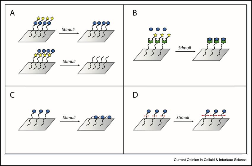
Dynamic substrates Bugga and Mrksich         81

Figure 1

Distinct strategies for designing dynamic substrates that modulate cell-substrate interactions: A) Uncaging or cleavage of a ligand with a
photo/electro-labile group; B) uncaging of a receptor for ligand binding; C) conformational switching of ligand chains; D) modulation of lateral
ligand mobility.

as well as the sub-cellular location of cellular attach-                  corresponding benzoquinone groups, which could then
ments to the matrix [15e17].                                              undergo a DielseAlder reaction with a RGD-
                                                                          cyclopentadiene conjugate to immobilize the peptide.
The first examples of dynamic substrates were reported                    This process effectively removed the pattern and
nearly twenty years ago. Wong, Langer and Ingber                          allowed the cells to migrate [20]. We also reported stra-
cultured aortic endothelial cells on conducting poly-                     tegies for electrochemically releasing ligands from the
pyrrole films that could be electrochemically oxidized or                 monolayer and uncaging immobilized ligands [21,22].
reduced, and they showed that cell growth and spreading
were different on the two states of the surface [18].                     These early examples motivated a substantial effort in
Okano       and      co-workers    developed      poly(N-                 biointerface science and engineering to develop and
isopropylacrylamide) substrates for cell culture and                      apply dynamic substrates in cell biology. Most examples
harnessed the thermal phase transition of this material                   have used applied potentials and light to effect changes
between states that promoted or prevented cell attach-                    in the properties of the substrate, but examples with
ment. They showed that cells could be cultured on the                     mechanical and magnetic forces, with enzymes and with
material at incubator temperatures, but released when                     solution additives have also been used. In this Opinion,
the substrates were cooled to room temperature [19].                      we review these basic strategies, with particular atten-
They developed this approach for culturing cells into                     tion to advances reported in the past three years. We end
sheets for tissue engineering applications. To realize                    with a discussion of the applications for these dynamic
molecular-level control over ligandereceptor in-                          substrates.
teractions, our early work developed electroactive self-
assembled monolayers of alkanethiolates on gold that                      Electrochemical modulation
allowed the activities of immobilized RGD ligands to be                   Electrochemical strategies were first used by our group
turned on or off in response to applied potentials. In one                to modulate the structure of bio-interfaces with well-
example, monolayers were patterned with the RGD                           defined molecular control. These strategies were
adhesion ligand, and where the surrounding areas                          motivated by the use of self-assembled monolayers of
presented a hydroquinone group against an inert back-                     alkanethiolates on gold for fundamental studies of
ground of tri(ethylene glycol) groups. Hence, 3T3                         electron transfer and showed that redox-active groups
fibroblast cells attached and spread within the patterned                 that were attached to the monolayers could be effi-
regions. Application of an oxidizing potential to the                     ciently oxidized and reduced through applied poten-
substrate converted the hydroquinone groups to the                        tials [23]. By designing molecular groups that are

www.sciencedirect.com                                                          Current Opinion in Colloid & Interface Science 2018, 38:80–87
82 Biological Colloids and Interfaces
stable in one oxidation state but undergo reaction in a conformation but can sample many conformations.
second oxidation state, we demonstrated how the ac- When the chain is terminated in a charged group,
tivities of immobilized ligands can be modulated. In application of an applied repulsive potential can either
one example, we prepared monolayers that had the force it into an ‘extended’ conformation or with the
RGD peptide immobilized by way of a benzoquinone attractive potential, enforce a ‘bent’ conformation. The
ester linker. This linker was stable, but upon reduc- wettability of the surface depends on the conformation,
tion, the phenolic group of the hydroquinone reacted with the ‘extended’ surfaces having a hydrophilic prop-
with the ester to give a lactone with release of the erty and the ‘bent’ having a hydrophobic character. It was
RGD peptide. We demonstrated that these monolayers also found that the attachment of bacteria to monolayer
could release adherent cells in response to an applied was dependent on the conformation of alkanethiolates
reducing potential [21]. In another example, we [28,29]. One concern with the low density monolayers is
exploited the reactivity of aldehyde groups on a surface whether the lack of packing of chains gives a less stable
[24]. We designed an electrolabile protecting group for layer, or whether lateral mobility of the chains can lead to
the aldehyde functionality by forming an acetal with an a surface having patches of well-packed alkanethiolates,
ortho-hydroxymethyl hydroquinone. Application of a which are then unable to undergo electrostatically-
positive potential resulted in uncaging of the aldehyde driven changes in conformation.
and allowed the non-specific adhesion of cells. In this
way, we could create surfaces that were patterned with Photochemical modulation
RGD and where the alternate regions had the caged Early photochemical strategies for manipulating the ac-
aldehyde. Cells would only attach to the former regions tivities of immobilized ligands were based on photo-
and remain there during culture. When an oxidative protected ligands that are inactive, but that can be
potential was applied to the substrate cells initiated uncaged with light to yield the active ligand. For example,
migration from their initial positions. an early report by Del Campo and coworkers prepared
alkylsiloxane monolayers that presented a RGD peptide
Yousaf and co-workers introduced another reaction of that was blocked at the aspartate residue with a nitro-
benzoquinone groups, termed oxime click chemistry. Li- phenyl protecting group [30]. Without illumination, 3T3
gands functionalized with a hydroxylamine group could be fibroblasts were unable to attach to the substrate. How-
conjugated to the benzoquinone group of a monolayer, but ever, irradiation of the substrate with 350 nm light
were then released following electrochemical reduction at through a mask resulted in spatial deprotection of the
physiological pH with regeneration of the hydroquinone. RGD peptides and subsequently directed cell attach-
Using this strategy, monolayers were prepared that ment to the patterned regions. Nakanishi and coworkers
presented both the RGD peptide and a hydroquinone first applied this strategy to demonstrate a dynamic
group [25]. In this way, a second ligand could be immo- substrate, wherein activation could be performed while
bilizeddhere, the ‘synergy’ ligand PHSRN from fibro- cells were already present on the substrate [31]. In this
nectindto increase the complexity of the substrate, and case, however, rather than uncaging RGD, removal of the
still allow release of the ligand at a later time in culture. photoprotecting group resulted in loss of adsorbed albu-
These surfaces should prove useful for addressing the min followed by adsorption of exogenously added fibro-
synergistic/antagonistic effects of the roles multiple li- nectin. Thus, surfaces were UV irradiated through a
gands play in cellular processes. In another example, these photomask to initially pattern the cells followed by a
electrically responsive surfaces were used to compare cell second irradiation in culture to pattern a second cell type.
migration between static and stimulated conditions These approaches are general and recently have been
[26]. The observation that cell behavior depended on applied towards activation of the laminin-based IKVAV
whether cells were initially confined to a patterned region peptide and an a5b1 integrin-specific ligand [32,33].
revealed that cell migration is dependent on history and,
in a broader sense, demonstrated the value of these dy- A limitation in the use of light-controlled strategies for
namic surfaces for studying how cell-matrix-mediated modulating cell adhesion is the potential for cytotoxic
processes are spatiotemporally regulated. effects of UV and near-UV light sources. This concern
can be mitigated by using short pulses of light and
Langer and coworkers developed incompletely formed protecting groups that respond to visible light (i.e.
monolayers, or low-density SAMs (LDSAMs) that pre- BODIPY-based) or those that undergo two-photon
sent charged tail groups [27]. The low density is excitation. However, an alternative strategy employs
achieved by assembly of alkanethiols that have bulky end lanthanide-doped upconversion nanoparticles
groups (i.e. trityl groups). These groups are sterically (UCNPs), wherein multiple photons at near-IR wave-
demanding and will limit the density of alkanethiolates lengths can be absorbed and re-emitted locally at shorter
within the monolayerdwith larger groups giving lower wavelength UV light, hence enabling the uses of tradi-
densitiesdbut the groups can then be removed to give tional photoprotecting groups with milder light treat-
monolayers that lack close packing of the alkane chains, ments [34,35]. The Qu lab first demonstrated the use
and therefore are not constrained to a trans-extended of these strategy for controlling cell adhesion by
Current Opinion in Colloid & Interface Science 2018, 38:80–87 www.sciencedirect.comDynamic substrates Bugga and Mrksich 83
immobilizing nanoparticles on quartz substrates and manipulating interfacial charge to control cell adhesion.
covalently attaching photocleavable linkers terminated The anti-fouling property of these surfaces is based on
in RGD. Light with a wavelength of 980 nm could be having strong solvation about each ionic center, but
used to uncage the peptides; the greater tissue pene- maintaining an overall net neutral charge. Hence,
tration with longer wavelengths allowed in vivo applica- strategies that screen the charges or remove the zwit-
tions of this method. terionic character (through changes in pH) have been
recently used to modulate cell adhesion. In one
Additionally, though photocleavable methods are irre- example, the Yang group developed polyzwitterionic
versible and allow one-time switching of adhesion states, brushes with imidazolium and sulfonate components
photoisomerization approaches enable dynamic surfaces (polyVBIPS) that were anti-fouling at high ionic
that can be reversibly modulated multiple times. In these strengths and adhesive at low ionic strengths [42]. With
approaches, the structural changes triggered in a chro- this “anti-polyelectrolyte effect,” the presence of salt
mophore lead to an alteration of the accessibility of a disrupts the inter/intra-chain electrostatic interactions
terminal ligand (i.e. RGD) for cell attachment. The two of the polymer brushes in the collapsed conformation
most commonly used isomerization groups are the to yield a more extended (and inert) state. In another
azobenzenes, which undergo isomeric changes in double example, the Jiang lab employed a zwitterionic polymer
bond configuration in the presence of UV light, and the brush containing a tertiary amine and carboxylic acid
spiropyrans, which undergo a ring-opening at 350 nm to groups and could cycle between charge-neutral states
the merocyanine form and ring-closing at 560 nm. The that are cell adhesive (pH 4e8) and charged states that
spiropyran group can be directly used as a cell adhesion promote adhesion (pH < 4 or pH > 8) [43].
“ligand” as the spiropyran form displays a stronger Importantly, the cationic and anionic adhesive regimes
interaction with fibronectin than does the merocyanine could support adsorption of both oppositely charged
form [35]. With the azobenzenes, the group can either proteins and be cycled for sequential adsorption and
be used as a linker terminated in RGD [36] or as a ter- release. We note that polyelectrolyte gels also exhibit
minal group itself for host-guest interactions with func- pH and ionic strength-dependent properties but are
tionalized cyclodextrins [37e40]. In the former case, less commonly employed for dynamically controlling
Selhuber-Unkel and co-workers altered the photo- cell adhesion.
physical properties of the azobenzene via “pushepull”
substitution with an electron-withdrawing and electron- Magnetic modulation
donating group [41]. The generated species displayed The dynamic strategies described above give examples
a rapid cis -trans back-reaction (102e105 Hz) that enabled of the modulation of ligand activity. However, interfacial
studies of integrin-mediated cellular responses to oscil- interactions in biology between cellular receptors and
latory “tickling” forces at previously inaccessible time surfaces are often multivalent and can be dependent on
scales, though the incomplete photoisomerization of the the lateral organization or clustering of a ligand. Indeed,
azobenzene chromophore is a limitation. extensive studies with supported lipid bilayer substrates
(SLBs) that are functionalized with adhesion ligands
The photochemical strategies for modulating the ac- have allowed the modulation of lateral organization of
tivities of immobilized ligands offer many benefits over ligands for cell adhesion. Recently, Bian and coworkers
other strategies. First, the literature on photocaged pioneered the development of RGD-presenting surfaces
molecules is extensivedwith a significant amount whose two-dimensional mobility could be tuned in
directed towards uncaging ligands in cell culturedand response to an applied magnetic field [44,45]. The
offers a wide choice of protecting groups for different ligands were conjugated to magnetic nanoparticles,
functional groups. Second, the photodeprotection re- which in turn were bound to a silica surface by way of a
actions often proceed rapidlydin seconds when using flexible linker. The ligand mobility could then be
laser sources on a microscopedand in high yield. Third, modulated with a magnetic field oscillating at varying
these strategies give excellent spatio-temporal control frequencies. Importantly, this work found that stem cell
in activating immobilized ligands, making it straight- adhesion, spreading, and differentiation were depen-
forward to control activation at mm length scales. The dent on the frequency of the field oscillation, with
potential concerns are that the photoprotecting groups greater spreading observed at lower frequencies. This
can sometimes cause significant non-specific adsorption observation aligns with our current understanding of
of protein and therefore cell attachment, and care must integrin-mediated events that require sufficient time
be taken to avoid unintentional exposure to light before and pre-organization for focal adhesion maturation and
or during the cell culture. traction force sensing. In addition, considering magnetic
fields are routinely used for patient imaging and can
Solution additives for modulation penetrate tissue, this stimulus bears a real potential for
The development of zwitterionic surfaces as a leading the modulation of cell adhesion in vivo. Indeed, the Bian
class of inert substrates has led to the strategy of group reported the successful modulation of adhesion
www.sciencedirect.com Current Opinion in Colloid & Interface Science 2018, 38:80–8784 Biological Colloids and Interfaces
and polarization with magnetically responsive substrates strategies require a conductive surface to transduce
implanted in mice [45,46]. applied electrical potentials and thus are best suited for
manipulating ligand activities present on SAMs. Ther-
Enzymatic modulation mal methods are based on phase transitions in the sur-
The in vivo properties of extracellular matrix are often face layer and therefore are most relevant to polymeric
modulated through the action of enzymes on the protein and hydrogel layers. The use of light and solution ad-
scaffold. For example, metalloproteases digest the ditives (including pH, metal ions, enzymes and chemi-
insoluble matrix and allow for its remodeling and they cal reagents) are applicable to the broad range of
are also harnessed by cancer cells to permit their chemistries discussed here. Magnetic strategies, in
migration and metastasis. Lysine 6-oxidase converts contrast, necessitate paramagnetic species e iron
lysine side chain amines in collagen and elastin to nanoparticles, for example e and this requirement will
reactive aldehydes that facilitate cross-linking and fibril limit the types of chemistries that can be used. Finally,
stabilization. Recently, citrullination (with protein we note that there has been relatively little work in
arginine deaminase enzymes) has been found to modify extending these strategies to three-dimensional culture
cell adhesion by way of modification of arginine residues systems, though the most important strategies have
in integrin binding sites [47]. In pioneering work, relied on photochemical manipulation [52].
Hubbell and coworkers developed hydrogel matrices
that included a RGD peptide for cell adhesion and a Applications
second peptide cross-linker that was a substrate for the One notable application for the dynamic substrates is
metalloprotease MMP-1 [48]. In this way, the bio- directed towards methods for single cell analysis. Here,
mimetic matrix could be used to study the protease- surfaces are designed to initially allow the adhesion and
mediated migration of cells. Ulijn and coworkers re- culture of cells, but that can then be stimulated to
ported a related strategy, wherein an immobilized RGD release cells. As noted earlier, early work by Okano’s
peptide was inactive towards cell adhesion because it group had developed poly(N-isopropylacrylamide) gels
was terminated in a bulky FMOC-Phe group [49]. as such a substrate [19]. When the gel composition is
Treatment with a protease released this group and tuned to undergo the phase transition just below 37 C
afforded a functional RGD ligand that could then (which is typical in growth incubators), then cells can be
mediate cell adhesion. Ulijn and Dalby went on to apply cultured but will detach when the substrate is removed
this strategy to control the differentiation of mesen- from the incubator and allowed to cool to room tem-
chymal stem cells [50]. They could switch on perature. These materials have been developed for
enhanced adhesion and spreading by proteolytically tissue engineering applications, for growing and recov-
uncaging RGD peptides to increase the affinity of the ering interconnected sheets of cells. Wang and co-
peptides. The resulting cells had a more contractile workers have extended this approach by modifying
cytoskeleton and an increased preference for silicon nanopillar substrates with this polymeric coating
osteogenesis. but that is further modified with an antibody that binds
epithelial cell adhesion molecule, a receptor that is
These enzyme-directed strategies are akin to the use of overexpressed on certain cancer cells [53]. In this way,
light to remove photoprotecting groups, but have the the surfaces are more selective in allowing a target cell
benefits that they do not require light sources and they population to attach. As before, the cultured cells can be
also use specific enzyme-substrate pairs that can give released from the substrate upon cooling to 20 C, and
multiple orthogonally controlled routes to modulate the the surface undergoes reversible phase transitions to
biological properties of a synthetic matrix, though they allow repeated attachment and release of cells. In
do not offer the spatial resolution that is possible with another example, these authors prepared acrylamide
focused light. Finally, another exciting example showed gels that were functionalized with phenylboronic acid
that cells could be engineered with designed receptors groups for engagement of carbohydrates on the cell
that presented an enzyme that could act on an immo- surface, and demonstrated reversible cell attachment
bilized substrate. We reported an example wherein the that was regulated by pH and soluble glucose [54].
enzyme cutinase could hydrolyze a hydroquinone ester,
and yield the hydroquinone, which could then be One limitation of the thermally responsive gels is that it
detected using cyclic voltammetry, demonstrating a bio- is difficult to selectively release an individual cell from a
electronic interface [51]. culture, since thermal conductivity of the substrate does
not allow spatial localization of the phase transition at
We have organized the discussion above by the modu- small (w100 mm) length scales. Photochemically-active
lation strategy, but were not explicit as to which stra- substrates can be used for this purpose, where focused
tegies are most relevant for the different surface light can trigger the modification of a substrate and
chemistries (principally, those based on self-assembled release an individual cell, but carries the limitation that
monolayers and on polymers). Electrochemical the cultures cannot be analyzed by fluorescent
Current Opinion in Colloid & Interface Science 2018, 38:80–87 www.sciencedirect.comDynamic substrates Bugga and Mrksich 85
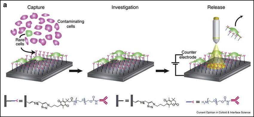
Figure 2
A design for using a combination of applied potential and light to stimulate the release of cells from the irradiated regions [55].
microscopy prior to release of the cells, since the sur- cationicdin response to applied potentials [56].
faces (by design) are not stable to the light. Hence, Interestingly, each state had a distinct property; the
these strategies would make it difficult to monitor cells zwitterionic state resisted bacterial attachment; the
in culture using microscopy, where cells having a rele- anionic state maintained attachment; and the cationic
vant phenotype can be identified and selected for state presented quaternary ammonium salts that have
release. Gooding and coworkers recently described an bactericidal properties.
innovative light-activated electrochemical method for
releasing individual cells [55] (See Figure 2). This These examples have demonstrated the enabling ap-
work used p-type silicon surfaces that presented an anti- plications of dynamic substrates, particularly for con-
EpCAM antibody by way of a benzoquinone-based trolling cell adhesion, proliferation and growth. These
linker that undergoes cleavage when reduced to the properties can be quite useful in bioanalytical systems to
corresponding hydroquinone. Cells having the EpCAM analyze and manipulate cells. Yet, the field still has not
receptor on their surfaces could attach and spread on the addressed problems in signaling, where the introduction
substrate. Application of an electrical potential does not or removal of a ligand at the cell-ECM interface results
lead to reduction of the benzoquinone because of poor in activation of specific signal transduction pathways.
conductivity of the silicon substrate, but illumination of We believe the prospects for such applications are
the substrate promotes electrons to the conducting exciting and that the dynamic substrates can emerge as a
band and allows for redox reactions at the interface, and powerful approach in cell biology. But these studies are
release of cells only in the illuminated region. This more challenging and will require the collaboration of
paper demonstrated that a population of lung tumor cell biologists in designing studies and interpreting data.
cells could be treated with the drug doxorubicin and
that only those cells that displayed efficient uptake of The past decade has seen a substantial increase in the
the drug could be identified and released to permit gene development and application of strategies to dynami-
expression profiling of those cells. This example repre- cally regulate ligand-receptor interactions at the cell-
sents a compelling application for dynamic substrates matrix interface. The range of methods, and the
and will likely find wide use. further opportunities they motivate for molecular engi-
neering of the interface, now represent a frontier area in
In another strategy, Yeo and coworkers prepared low biointerface science. Future work will certainly see
density monolayers that included three different chains growth in the application of these dynamic substrates to
terminated in acidic and basic functional groups as well a range of studies in cell signaling and as enabling
as an uncharged chain. This monolayer could be components of cell-based assays for drug discovery and
switched between three statesdzwitterionic, anionic or diagnostics.
www.sciencedirect.com Current Opinion in Colloid & Interface Science 2018, 38:80–8786 Biological Colloids and Interfaces
Conflict of interest statement 19. Okano T, Yamada N, Okuhara M, Sakai H, Sakurai Y: Mecha-
nism of cell detachment from temperature-modulated, hy-
Nothing declared. drophilic-hydrophobic polymer surfaces. Biomaterials 1995,
16:297–303.
Acknowledgement 20. Yousaf MN, Houseman BT, Mrksich M: Turning on cell migra-
We are grateful for support of our work by the National Cancer Institute of tion with electroactive substrates. Angew Chem Int Ed 2001,
the National Institutes of Health (U54CA199091) and the Air Force Office 40:1093–1096.
of Scientific Research (AFOSR FA9550-16-1-0150).
21. Yeo W-S, Mrksich M: Electroactive self-assembled mono-
layers that permit orthogonal control over the adhesion of
References cells to patterned substrates. Langmuir 2006, 22:
Papers of particular interest, published within the period of review, 10816–10820.
have been highlighted as: 22. Yeo W-S, Hodneland CD, Mrksich M: Electroactive monolayer
substrates that selectively release adherent cells. Chem-
of special interest BioChem 2001, 2:590–593.
of outstanding interest
23. Chidsey CED: Free energy and temperature dependence of
1. Frantz C, Stewart KM, Weaver VM: The extracellular matrix at a electron transfer at the metal-electrolyte interface. Science
glance. J Cell Sci 2010, 123:4195. 1991, 251:919.
2. Hynes RO: Extracellular matrix: not just pretty fibrils. Science 24. Yeo WS, Mrksich M: Electroactive substrates that reveal
2009, 326:1216–1219. aldehyde groups for bio-immobilization. Adv Mater 2004, 16:
1352–1356.
3. Bonnans C, Chou J, Werb Z: Remodelling the extracellular
matrix in development and disease. Nat Rev Mol Cell Biol 2014, 25. Pulsipher A, Park S, Dutta D, Luo W, Yousaf MN: In situ mod-
15:786. ulation of cell behavior via smart dual-ligand surfaces.
Langmuir 2014, 30:13656–13666.
4. Mrksich M: Using self-assembled monolayers to model the
extracellular matrix. Acta Biomater 2009, 5:832–841. 26. Lee E-j, Luo W, Chan EWL, Yousaf MN: A molecular smart
surface for spatio-temporal studies of cell mobility. PLoS One
5. Gooding JJ, Parker SG, Lu Y, Gaus K: Molecularly engineered 2015, 10:e0118126.
surfaces for cell biology: from static to dynamic surfaces. Hydroquinone-based dynamic surfaces were used to study
Langmuir 2014, 30:3290–3302. electrochemically-initiated cell migration as a function of initial adhe-
sion parameters.
6. Kyburz KA, Anseth KS: Synthetic mimics of the extracellular
matrix: how simple is complex enough? Ann Biomed Eng 27. Lahann J, Mitragotri S, Tran T-N, Kaido H, Sundaram J, Choi IS,
2015, 43:489–500. Hoffer S, Somorjai GA, Langer R: A reversibly switching sur-
face. Science 2003, 299:371.
7. Kocer G, Jonkheijm P: About chemical strategies to fabricate
cell-instructive biointerfaces with static and dynamic 28. Pranzetti A, Mieszkin S, Iqbal P, Rawson FJ, Callow ME,
complexity. Adv Healthc Mater 2018, 0:e1701192. Callow JA, Koelsch P, Preece JA, Mendes PM: An electrically
reversible switchable surface to control and study early
8. Campbell ID, Humphries MJ: Integrin structure, activation, and bacterial adhesion dynamics in real-time. Adv Mater 2013, 25:
interactions. Cold Spring Harb Perspect Biol 2011, 3:a004994. 2181–2185.
9. Ruoslahti E: RGD and other recognition sequences for integ- 29. Ng CC, Magenau A, Ngalim SH, Ciampi S, Chockalingham M,
rins. Annu Rev Cell Dev Biol 1996, 12:697–715. Harper JB, Gaus K, Gooding JJ: Using an electrical potential to
reversibly switch surfaces between two states for dynami-
10. Bellis SL: Advantages of RGD peptides for directing cell cally controlling cell adhesion. Angew Chem Int Ed 2012, 51:
association with biomaterials. Biomaterials 2011, 32: 7706–7710.
4205 –4210.
30. Petersen S, Alonso José M, Specht A, Duodu P, Goeldner M, del
11. Hersel U, Dahmen C, Kessler H: RGD modified polymers: Campo A: Phototriggering of cell adhesion by caged cyclic
biomaterials for stimulated cell adhesion and beyond. Bio- RGD peptides. Angew Chem Int Ed 2008, 47:3192–3195.
materials 2003, 24:4385–4415.
31. Kikuchi Y, Nakanishi J, Shimizu T, Nakayama H, Inoue S,
12. Chen S, Li L, Zhao C, Zheng J: Surface hydration: principles Yamaguchi K, Iwai H, Yoshida Y, Horiike Y, Takarada T, Maeda M:
and applications toward low-fouling/nonfouling biomaterials. Arraying heterotypic single cells on photoactivatable cell-
Polymer 2010, 51:5283–5293. culturing substrates. Langmuir 2008, 24:13084–13095.
13. Banerjee I, Pangule RC, Kane RS: Antifouling coatings: recent 32. Farrukh A, Fan W, Zhao S, Salierno M, Paez JI, Del Campo A:
developments in the design of surfaces that prevent fouling Photoactivatable adhesive ligands for light-guided neuronal
by proteins, bacteria, and marine organisms. Adv Mater 2010, growth. Chembiochem 2018, 19:1271–1279.
23:690–718. A leading example of the application of dynamic substrates in neuronal
14. Löpez GP, Ratner BD, Tidwell CD, Haycox CL, Rapoza RJ, cell biology.
Horbett TA: Glow discharge plasma deposition of tetra- 33. Nair Roshna V, Farrukh A, del Campo A: A photoactivatable
ethylene glycol dimethyl ether for fouling-resistant biomate- a5b1-specific integrin ligand. ChemBioChem 2018, 19:
rial surfaces. J Biomed Mater Res 1992, 26:415–439. 1280–1287.
15. Théry M: Micropatterning as a tool to decipher cell morpho- 34. Li W, Wang J, Ren J, Qu X: Near-infrared upconversion con-
genesis and functions. J Cell Sci 2010, 123:4201. trols photocaged cell adhesion. J Am Chem Soc 2014, 136:
16. Yap FL, Zhang Y: Protein and cell micropatterning and its 2248–2251.
integration with micro/nanoparticles assembly. Biosens Bio- 35. Li W, Chen Z, Zhou L, Li Z, Ren J, Qu X: Noninvasive and
electron 2007, 22:775–788. reversible cell adhesion and detachment via single-
17. Arnold M, Cavalcanti-Adam EA, Glass R, Blümmel J, Eck W, wavelength near-infrared laser mediated photoisomerization.
Kantlehner M, Kessler H, Spatz JP: Activation of integrin J Am Chem Soc 2015, 137:8199–8205.
function by nanopatterned adhesive interfaces. Chem- A spiropyran-functionalized UCNP (upconversion nanoparticle) surface
PhysChem 2004, 5:383–388. can be switched repeatedly using low energy photons that are not
cytotoxic.
18. Wong JY, Langer R, Ingber DE: Electrically conducting poly-
mers can noninvasively control the shape and growth of 36. Auernheimer J, Dahmen C, Hersel U, Bausch A, Kessler H:
mammalian cells. Proc Natl Acad Sci USA 1994, 91: Photoswitched cell adhesion on surfaces with RGD peptides.
3201–3204. J Am Chem Soc 2005, 127:16107–16110.
Current Opinion in Colloid & Interface Science 2018, 38:80–87 www.sciencedirect.comDynamic substrates Bugga and Mrksich 87
37. Ren T, Ni Y, Du W, Yu S, Mao Z, Gao C: Dual responsive 47. Sipilä KH, Ranga V, Rappu P, Mali M, Pirilä L, Heino I, Jokinen J,
surfaces based on host–guest interaction for dynamic Käpylä J, Johnson MS, Heino J: Joint inflammation related
mediation of cell–substrate interaction and cell migration. citrullination of functional arginines in extracellular proteins.
Adv Mater Interface 2016, 4:1500865. Sci Rep 2017, 7:8246.
38. Bian Q, Wang W, Wang S, Wang G: Light-triggered specific 48. Lutolf MP, Lauer-Fields JL, Schmoekel HG, Metters AT,
cancer cell release from cyclodextrin/azobenzene and Weber FE, Fields GB, Hubbell JA: Synthetic matrix
aptamer-modified substrate. ACS Appl Mater Interfaces 2016, metalloproteinase-sensitive hydrogels for the conduction of
8:27360–27367. tissue regeneration: engineering cell-invasion characteris-
tics. Proc Natl Acad Sci 2003, 100:5413.
39. Shen Q, Liu L, Zhang W: Fabrication of a photocontrolled
surface with switchable wettability based on host–guest in- 49. Todd SJ, Farrar D, Gough JE, Ulijn RV: Enzyme-triggered cell
clusion complexation and protein resistance. Langmuir 2014, attachment to hydrogel surfaces. Soft Matter 2007, 3:
30:9361–9369. 547–550.
40. Wei T, Zhan W, Yu Q, Chen H: Smart biointerface with 50. Roberts JN, Sahoo JK, McNamara LE, Burgess KV, Yang J,
photoswitched functions between bactericidal activity and Alakpa EV, Anderson HJ, Hay J, Turner L-A, Yarwood SJ,
bacteria-releasing ability. ACS Appl Mater Interfaces 2017, 9: Zelzer M, Oreffo ROC, Ulijn RV, Dalby MJ: Dynamic surfaces for
25767–25774. the study of mesenchymal stem cell growth through adhe-
sion regulation. ACS Nano 2016, 10:6667–6679.
41. Kadem Laith F, Suana KG, Holz M, Wang W, Westerhaus H, An important example of applying dynamic substrates in stem cell
Herges R, Selhuber-Unkel C: High-frequency mechanostimu- biology.
lation of cell adhesion. Angew Chem Int Ed 2016, 56:225–229.
An unprecedented demonstration of reversible modulation to rapidly 51. Collier JH, Mrksich M: Engineering a biospecific communica-
regulate engagement of adhesion receptors. tion pathway between cells and electrodes. Proc Natl Acad Sci
USA 2006, 103:2021.
42. Chen H, Yang J, Xiao S, Hu R, Bhaway SM, Vogt BD, Zhang M,
Chen Q, Ma J, Chang Y, Li L, Zheng J: Salt-responsive poly- 52. Kloxin AM, Kasko AM, Salinas CN, Anseth KS: Photodegradable
zwitterionic materials for surface regeneration between hydrogels for dynamic tuning of physical and chemical
switchable fouling and antifouling properties. Acta Biomater properties. Science 2009, 324:59–63.
2016, 40:62–69.
53. Liu H, Liu X, Meng J, Zhang P, Yang G, Su B, Sun K, Chen L,
43. Sundaram HS, Ella-Menye J-R, Brault ND, Shao Q, Jiang S: Han D, Wang S, Jiang L: Hydrophobic interaction-mediated
Reversibly switchable polymer with cationic/zwitterionic/ capture and release of cancer cells on thermoresponsive
anionic behavior through synergistic protonation and nanostructured surfaces. Adv Mater 2012, 25:922–927.
deprotonation. Chem Sci 2014, 5:200–205.
An innovative yet simple route to regulate protein/cell interactions with 54. Liu H, Li Y, Sun K, Fan J, Zhang P, Meng J, Wang S, Jiang L:
a polymeric material. Dual-responsive surfaces modified with phenylboronic acid-
containing polymer brush to reversibly capture and release
44. Wong DSH, Li J, Yan X, Wang B, Li R, Zhang L, Bian L: cancer cells. J Am Chem Soc 2013, 135:7603–7609.
Magnetically tuning tether mobility of integrin ligand regu-
lates adhesion, spreading, and differentiation of stem cells. 55. Parker SG, Yang Y, Ciampi S, Gupta B, Kimpton K, Mansfeld FM,
Nano Lett 2017, 17:1685–1695. Kavallaris M, Gaus K, Gooding JJ: A photoelectrochemical
platform for the capture and release of rare single cells. Nat
45. Kang H, Wong DSH, Yan X, Jung HJ, Kim S, Lin S, Wei K, Li G, Commun 2018, 9:2288.
Dravid VP, Bian L: Remote control of multimodal nanoscale An ingenious strategy to use light irradiation to direct applied potentials
ligand oscillations regulates stem cell adhesion and differ- for spatial modulation of a dynamic substrate at high resolution.
entiation. ACS Nano 2017, 11:9636–9649.
A surprising study that reveals the extent to which even subtle manipu- 56. Choi I, Lee J, Kim W, Kang H, Bae SW, Chang R, Kim S, Yeo W-
lation of ligand-receptor interactions can have on stem cell differentiation. S: On-demand modulation of bacterial cell fates on multi-
functional dynamic substrates. ACS Appl Mater Interfaces
46. Kang H, Kim S, Wong DSH, Jung HJ, Lin S, Zou K, Li R, Li G, 2018, 10:4324–4332.
Dravid VP, Bian L: Remote manipulation of ligand nano- An impressive demonstration of electrically responsive substrates
oscillations regulates adhesion and polarization of macro- based on low-density SAMs manipulating the adhesion of mycoplasma
phages in vivo. Nano Lett 2017, 17:6415–6427. contaminants from mammalian cell cultures.
www.sciencedirect.com Current Opinion in Colloid & Interface Science 2018, 38:80–87You can also read


