ROS-Sensitive Nanoparticles Co-delivering Dexamethasone and CDMP-1 for the Treatment of Osteoarthritis Through Chondrogenic Differentiation ...
←
→
Page content transcription
If your browser does not render page correctly, please read the page content below
ORIGINAL RESEARCH
                                                                                                                                         published: 28 January 2021
                                                                                                                                    doi: 10.3389/fbioe.2021.608150

                                            ROS-Sensitive Nanoparticles
                                            Co-delivering Dexamethasone and
                                            CDMP-1 for the Treatment of
                                            Osteoarthritis Through Chondrogenic
                                            Differentiation Induction and
                                            Inflammation Inhibition
                                            Xiaodong Wu 1,2 , Pengpeng Li 3 , Jian Cheng 1 , Qiang Xu 1 , Beiji Lu 1 , Conghui Han 1* and
                                            Weiling Huo 1*
                                            1
                                             Department of Orthopaedics, Xuzhou Central Hospital, Xuzhou, China, 2 Xuzhou Clinical School of Xuzhou Medical
                                            University, Xuzhou, China, 3 Bengbu Medical College, Bengbu, China
                        Edited by:
                     Jianxun Ding,
Chinese Academy of Sciences, China          Objective: Osteoarthritis (OA) is a common subtype of arthritis. To date, treatment of OA
                     Reviewed by:           focuses primarily on alleviating pain and improving joint function. The lack of a vascular
                        Zhipeng Gu,         system within synovial joints and the rapid removal of agents due to synovial exchange
            Sichuan University, China
                           Li Zheng,
                                            hinder continuous delivery of OA drugs. However, these obstacles are being addressed
    Guangxi Medical University, China       by promising nanoscale drugs.
                            Yixia Yin,
                 Wuhan University of        Methods: We synthesize and assemble a hydrogen peroxide [H2 O2 , belongs to
                  Technology, China         the category of active oxygen species (ROS)]-sensitive nanomicelle, which is loaded
                  *Correspondence:          with the anti-inflammation drug dexamethasone and chondrogenic differentiation
                        Conghui Han
                                            factor cartilage-derivedmor-phogeneticprotein-1. The micelle can induce bone marrow
         hanconghuidoc@hotmail.com
                         Weiling Huo        mesenchymal stem cells to repair cartilage while inhibiting joint inflammation.
           weilinghuo2019@sina.com
                                            Results: The prepared nanoparticles were of uniform size and displayed an obvious
                    Specialty section:      core-shell structure. Under H2 O2 stimulation, the shell layer could be removed gradually.
         This article was submitted to      The drug-loaded micelle effectively inhibited proliferation of activated macrophages,
                          Biomaterials,
                a section of the journal
                                            induced macrophage apoptosis with an anti-inflammatory effect, and caused the BMSCs
       Frontiers in Bioengineering and      to differentiate into chondrocytes.
                        Biotechnology
                                            Conclusion: This work provides an experimental and theoretical basis for further
      Received: 19 September 2020
        Accepted: 05 January 2021
                                            development of a drug-loaded micelle in the healing of osteoarthritis.
        Published: 28 January 2021
                                            Keywords: osteoarthritis (OA), reactive oxygen species (ROS), nanomicelle, dexamethasone, cartilage-
                               Citation:    derivedmor-phogeneticprotein-1 (CDMP-1)
     Wu X, Li P, Cheng J, Xu Q, Lu B,
              Han C and Huo W (2021)
         ROS-Sensitive Nanoparticles        INTRODUCTION
   Co-delivering Dexamethasone and
         CDMP-1 for the Treatment of
                                            Osteoarthritis (OA), a degenerative form of arthritis, causes articular cartilage damage and
 Osteoarthritis Through Chondrogenic
           Differentiation Induction and
                                            affects entire joints (Miller et al., 2019; Hunt et al., 2020). Because cartilage is a non-vascular,
                 Inflammation Inhibition.   non-lymphoid, and non-nerve tissue (Zhang et al., 2019a,b) and chondrocytes are highly
 Front. Bioeng. Biotechnol. 9:608150.       differentiated cells with little potential for proliferation and migration, self-repair and regeneration
     doi: 10.3389/fbioe.2021.608150         of cartilage is limited (Roseti et al., 2019; Zhu et al., 2020).

Frontiers in Bioengineering and Biotechnology | www.frontiersin.org              1                                          January 2021 | Volume 9 | Article 608150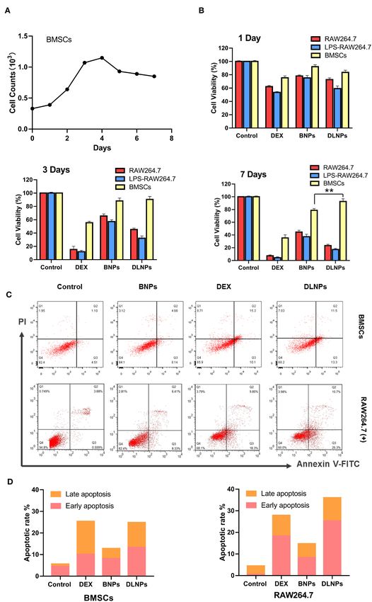
Wu et al.                                                                                                        ROS-Sensitive Nanoparticles for Osteoarthritis

    Several surgical procedures have been operated to repair                        prospect in cartilage repair and bone repair (Zhu et al., 2020). The
cartilage, including micro fractures (Bergink et al., 2019), mosaics                combination of bioactive factors and engineered 3D scaffolds can
(Jordan et al., 2017) and autologous chondrocytes (Stoop et al.,                    limit the decrease of biological activity, reduce the occurrence of
2007), but long-term success has proved elusive. To meet clinical                   complications and achieve targeted therapy (Wang et al., 2019;
needs and achieve long-term efficacy, non-invasive therapies                        Cui et al., 2020).
that promote cartilage regeneration and exert anti-inflammatory                        Cartilage-derive morphogenetic protein 1 (CDMP-1), as a
effects are being designed (Boulocher et al., 2007; Palmer                          cytokine which has the ability to induce the proliferation and
et al., 2013). At present, treatment of OA focuses primarily on                     differentiation of osteoblasts, promote the differentiation of
alleviating pain and improving joint function (Zhu et al., 2020).                   bone marrow mesenchymal stem cells into chondrocytes for
Glucocorticoids (Cooper et al., 2016; Zhou et al., 2019) are often                  repair(Dobbs et al., 2005).
used to relieve joint pain during inflammation, but they are only                      Here, we constructed a nanoparticle with low toxicity, ROS
effective in the short term. Hyaluronic acid drugs are able to                      response properties, and ability to eliminate joint inflammation
relieve the symptoms of various types of osteoarthritis, but they                   and induce cartilage repair, which was named as DLNPs. And
tend to rapidly exit the joint cavity and provide relief for no more                we used hydrogen peroxide (H2 O2 ) as a positive control. The
than 1–3 months.                                                                    nanoparticles use -SeSe- group as the ROS response component
    The lack of a vascular system within the synovial joint                         and DEX and CDMP-1 as the main pharmacophore. Drug-
(Baboolal et al., 2016; Watt et al., 2020) impedes the delivery of                  carrying nanoparticles were directly delivered to joint lesions
potent molecules to target sites during systemic administration.                    through joint cavity injection. By taking advantage of the high
The rapid removal of locally delivered therapeutic agents due to                    concentration of oxygen free radicals in arthritis lesions, the
synovial exchange poses another challenge (Jia et al., 2011). The                   fracture of -SeSe- and the slow release of DEX and CDMP-1
development of nanotechnology provides a promising alternative                      were observed. At the same time, PLGA hydrophobic groups
direction for the development of drug delivery systems for OA                       accumulated in the damaged cartilage due to hydrophobic effect,
(Yang et al., 2017; Zhao et al., 2021).                                             forming exogenous biological scaffold. The schematic diagram of
    Nanoparticle drugs offer several advantages over traditional                    the nanoparticle for the treatment of osteoarthritis was shown in
drugs, including increased drug hydrophilicity, reduced side                        Scheme 1.
effects and increased circulation time in vivo (Zhu et al., 2020).
Various functions can work through modification, such as
clearance of reactive oxygen species (ROS), lesion-site imaging,                    MATERIALS AND METHODS
photo-dynamic therapy, multi-drug-loading and targeting of                          Experimental Materials
lesion sites.
                                                                                    Reagents
    Recent studies have pointed out that the loss of cartilage
                                                                                    Dexamethasone, poly (lactic-co-glycolic acid (PLGA), mPEG5k -
matrix is closely related to oxidative stress. It is generally
                                                                                    COOH, selenocystamine, N-(3-dimethylaminopropyl)–n′ -ethyl
recognized that when reactive oxygen species (ROS) are
                                                                                    carbondiimine hydrochloride (EDC), n-hydroxysuccinimide
overexpressed, the function of articular cartilage is degraded
                                                                                    (NHS), and N, N-diisopropylamine (DIPEA) were obtained from
(Crivelli et al., 2019; Wegner et al., 2019; Shin et al., 2020). By
                                                                                    Energy Chemical. Chloroform, polyvinyl alcohol (PVA) and D-α-
introducing ROS responsive structural components (-SeSe -) into
                                                                                    tocopherol polyethylene glycol succinate (TPGS) were obtained
nanoparticles, it provides a basis for targeting the construction of
                                                                                    from Alladin.
nano-drugs for precision therapy.
                                                                                       Lipopolysaccharides (LPS), rhodamine-B (RhB), penicillin,
    Dexamethasone (DEX) is widely used in the treatment of
                                                                                    streptomycin, and 4′ , 6-diamidino-2-phenylindole (DAPI) were
various inflammatory diseases. It can inhibit the accumulation
                                                                                    also purchased from Aladdin. Trypsin and fetal bovine serum
of inflammatory cells, including macrophages and white blood
                                                                                    (FBS) were obtained from Gibco. CDMP-1 antibody (Product
cells, and inhibit the phagocytosis, the release of lysosomal
                                                                                    No. R30403) was obtained from Yuanye Bio-Technology.
enzymes and the synthesis and release of inflammatory chemical
intermediaries. DEX is mainly used in the treatment of
                                                                                    Instruments
OA to eliminate inflammatory response and relieve pain in
                                                                                    The techniques used in this study included high-performance
inflammatory sites (Said Ahmed et al., 2019; Zhao et al., 2019).
                                                                                    liquid chromatography (HPLC, Dionex UltiMate 3000,
    Osteoarthritis is fundamentally degenerative arthritis caused
                                                                                    ThermoFisher), transmission electron microscopy (TEM,
by the injury of articular cartilage (Brown et al., 2020). Therefore,
                                                                                    Hitachi, H-600), fluorescence microscopy (Zeiss 710, Jena,
the key to the treatment of OA lies in the repair of damaged
                                                                                    Germany), dynamic light scattering (DLS, Santa Barbara,
articular cartilage (Cui et al., 2019). With the progress of materials
                                                                                    Nicomp 380 ZLS), laser scanning confocal microscopy (Zeiss
and biological science, biomaterials have great application
                                                                                    710, Germany) and flow cytometry (BD Biosciences, BD FACS
                                                                                    Canto II).
Abbreviations: OA, Osteoarthritis; DEX, dexamethasone; CDMP-1, cartilage-
derivedmor-phogeneticprotein-1; Hydrogen peroxide, ROS, reactive oxygen             Cells and Animals
species; BMSCs, bone marrow mesenchymal stem cells; PLGA, poly(lactic-co-
glycolic acid; PEG, polyethylene glycol; DLS, dynamic light scattering; PBS,
                                                                                    Macrophages (RAW 264.7) were purchased from BeNa culture
phosphate buffer saline; ELISA, enzyme-linked immunosorbent assay; TNF, tumor       Collection. New Zealand rabbits were purchased from the Model
necrosis factor; IL, interleukin.                                                   Animal Research Center of Nanjing University.

Frontiers in Bioengineering and Biotechnology | www.frontiersin.org             2                                    January 2021 | Volume 9 | Article 608150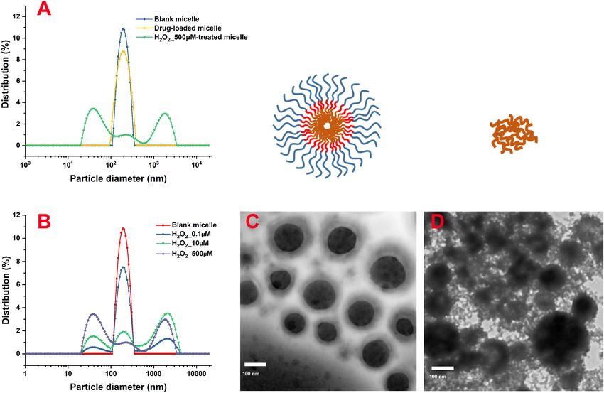
Wu et al.                                                                                                                 ROS-Sensitive Nanoparticles for Osteoarthritis

 SCHEME 1 | DLNPs are injected into the joint cavity of OA, and the high concentration of ROS in the articular cavity leads to the fracture of –SeSe-. The slow release
 of DEX reduces both pain and inflammation. The aggregated PLGA particles distributed on the surface of damaged cartilage serve as cartilage scaffoldings, and
 CDMP-1 biological factors in PLGA particles further induce BMSCs to transform into cartilage and repair damaged cartilage tissues. PLGA, poly(lactic-co-glycolic
 acid; DEX, dexamethasone; CDMP-1, cartilage-derivedmor-phogeneticprotein-1; ROS, reactive oxygen species; BMSCs, bone marrow mesenchymal stem cells.

  Animal experiments were performed according to protocols                             protection. The product was then precipitated by precooled
approved by the Ethical Committee of XuZhou Central Hospital.                          aether and the residue NHS and EDC were removed by washing
                                                                                       three times with a mixture of aether/methanol (50/50, v/v). The
Methods                                                                                resulting sediment was vacuum-dried and labeled PLGA-NHS.
The PLGA-SeSe-mPEG synthesis process was depicted in
Figure 1A. The nuclear magnetic resonance data of the PLGA-                            Preparation of the PLGA-SeSe-mPEG
SeSe-mPEG was present in Figure 1B.                                                    Briefly, PLGA-NHS (0.1 g), mPEG-SeSe-NH2 (0.2 g), and DIPEA
                                                                                       (6 uL) were dissolved in a dry solution of dimethyl sulfoxide.
Preparation of mPEG-SeSe-NH2
                                                                                       The solution was stirred under nitrogen protection for 48 h. The
The mPEG5k -COOH was dissolved in formamide with EDC
                                                                                       solution was dialysed (MWCO: 8,000–14,000) for 48 h, and the
and NHS (mPEG5k -COOH: EDC: NHS = 1:5:5). The mixture
                                                                                       final product PLGA-SeSe-mPEG was obtained by freeze-drying.
was stirred for 6 h to mix it thoroughly and then a mixture
of selenocystamine and formamide (V/V = 1:9) was dripped
at 0◦ C and allowed to react for another 5 h. Excess precooled                         Assembly of Blank Particles
acetone was added and allowed to crystallize into crystals, which                      PLGA-SeSe-mPEG (50 mg) was added to 1 mL of chloroform and
were filtered through a microporous membrane (0.22 mm). The                            mixed into 6 mL of a 1% PVA and TPGS mixture (PVA: TPGS
precipitate products were then dissolved in water and dialysed                         1:5). An 80 W ultrasound probe was used for 2 min in an ice bath
(MWCO: 3,500) for 48 h. The intermediate product, mPEG-SeSe-                           to form an oil-water emulsion.
NH2 , was obtained by freeze-drying.                                                       The emulsion was then added to 30 mL of 0.3% PVA
                                                                                       and stirred overnight to solidify the surface. Ultrafiltration
Synthesis of PLGA-NHS                                                                  concentration was carried out with a 100 KD ultrafiltration tube.
PLGA (0.65 g), EDC (80 mg), and NHS (48 mg) was dissolved in                           Finally, the volume of the mixture was fixed with pure water to
10 mL of dichloromethane, and reacted for 24 h under nitrogen                          5 mL, and the samples were collected and stored at 4◦ C.

Frontiers in Bioengineering and Biotechnology | www.frontiersin.org                3                                           January 2021 | Volume 9 | Article 608150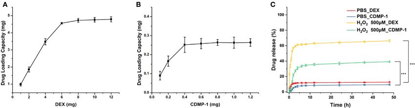
Wu et al.                                                                                                         ROS-Sensitive Nanoparticles for Osteoarthritis

 FIGURE 1 | Schematic diagram for synthesis (A) and Nuclear magnetic resonance spectroscopy (B) of PLGA-SeSe-Mpeg. PLGA, poly(lactic-co-glycolic acid); PEG,
 polyethylene glycol.

Assembly of Drugs-Loaded Particles Containing DEX                                 nanoparticles was characterized by TEM (120 keV,
and CDMP-1                                                                        5,000–30,000×).
DEX, CDMP-1, and PLGA-SeSe-mPEG were dissolved in                                    To prepare samples for morphological characterization, a total
trichloromethane, respectively. Next, 50 µL of 2 mg/mL DEX,                       of 5 µL of the formulation was deposited onto a carbon-disc
50 µL of 1 mg/mL CDMP-1, 50 µL of 1 mg/mL rhB, and                                ultrathin grid, followed by staining with 0.2% phosphotungstic
500 µg of the PLGA-SeSe-mPEG mixture were evenly and                              acid for 30 s.
ultrasonically prepared for use. The process for producing                           A total of 0.1 mM H2 O2 solution was used to simulate the ROS
drug-loaded particles was the same as that of blank particles                     environment at the inflammatory lesions.
described above.
                                                                                  Drug-Loading Coefficient and Drug
Characterization of Micellar Particles                                            Release
The size and potential of the nanoparticles were                                  Drug-loaded nanoparticles (5 mL) were transferred into a dialysis
measured by DLS, and the morphology of the                                        bag (MWCO, 12–14 K) and submerged into 45 mL of deionised

Frontiers in Bioengineering and Biotechnology | www.frontiersin.org           4                                        January 2021 | Volume 9 | Article 608150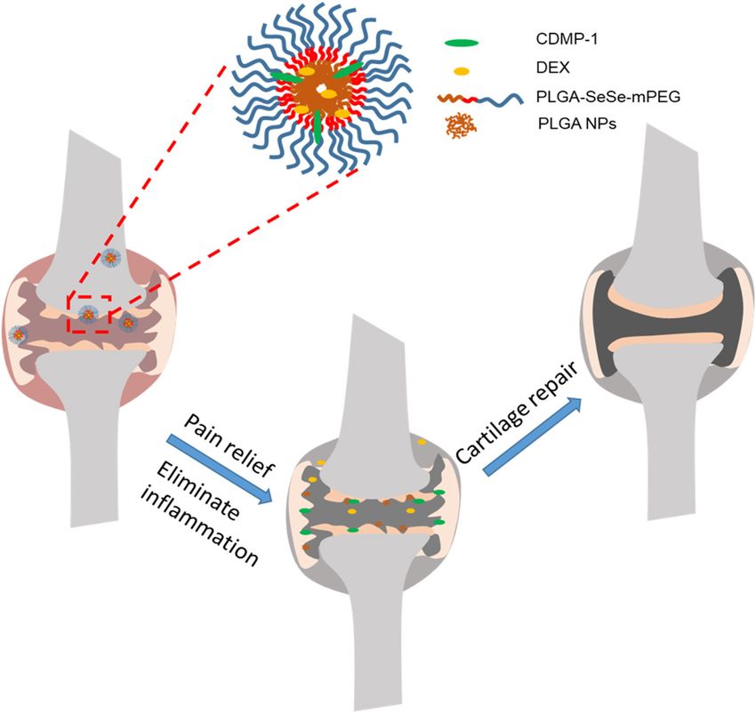
Wu et al.                                                                                                 ROS-Sensitive Nanoparticles for Osteoarthritis

(DI) water (37◦ C, pH 7.4). At set time points (10 min, 30 min,              Histochemical Detection of Collagen
1 h, 1.5 h, 2 h, 3 h, 4 h, 6 h, 8 h, 12 h, 24 h, 48 h, and 72 h), 1 mL       Type II
of dialysate was taken as a sample and 1 mL of fresh DI water                BMSCs induced for 21 days by CDMP-1 and drug-loaded
was refilled. After the samples were separated by HPLC, the                  micelles were digested into a cell suspension, and then fixed with
DEX contents were determined by mass spectrometry, and the                   4% paraformaldehyde at 4◦ C for 30 min. The membrane was
contents of CDMP-1 were determined by an enzyme-linked                       broken with 0.1% Triton solution for 30 min, sealed at room
immunosorbent assay (ELISA) kit.                                             temperature for 30 min, and then cleaned with PBS solution
   A total of 0.1 mM H2 O2 solution was used to replace the DI               3 times.
water to prepare dialysate to test the ROS sensitivity of drug-                  Polyclonal antibodies against type II collagen were diluted at a
loaded nanoparticles. The rest of the procedure was the same as              ratio of 1:100, incubated overnight at 4◦ C and an SABC immuno-
the control group.                                                           histochemical staining kit was used following the kit instructions.
                                                                             Finally, a DAB kit was used to detect the expression of type
                                                                             II collagen in the tissues, with a positive reaction producing a
Isolation and Culture of BMSCs
                                                                             brown-yellow color. PBS was used for negative control staining
BMSCs cells were isolated by density-gradient centrifugation.
                                                                             instead of an antibody.
    After anesthesia was successfully performed on healthy 1-
month-old New Zealand rabbits, the skin was prepared and lower               Alsin Blue Stain
limbs were disinfected with 75% alcohol and 3% iodine. Under                 BMSCs were inoculated in 12-well plates with 5,000 cells/well
aseptic conditions, 5 mL of bilateral iliac spine bone marrow was            and cultured for 21 days in an induction solution containing
rapidly extracted by a puncture with a 20 mL syringe. The same               100 ng/mL drug-loaded micelles. BMSCs were rinsed with PBS
amount of Dulbecco’s modified Eagle medium without FBS was                   for 3 min and fixed with 4% paraformaldehyde for 30 min. The
extracted with a sterile 5 mL syringe on an ultra-clean table, and           cells were then rinsed with 0.1 mol/L of HCl for 5 min. After the
the cells were resuspended. Then the cells were then placed into             pH was lowered to 1, a 0.1% alsin blue solution was used to stain
a screw centrifuge tube, centrifuged at 800 rpm for 6 min, and               the BMSCs overnight. The samples were then stained with a 0.1%
the supernatant was discarded to remove tissue cells, such as fat            neutral solid red solution for 5 min, rinsed with distilled water
and periosteum. The cells were resuspended in a complete cell                and dried, sealed with neutral resin and placed under an inverted
culture medium containing 10% FBS, and then slowly injected                  microscope for imaging.
into a centrifuge tube containing a 10 mL of lymphoid separation
fluid at a volume ratio of 1:1. After centrifuging at 1,500 rpm              Cellular Uptake In vitro
for 30 min, the annular pale-yellow monocyte cloud layer at the              The intracellular localization of drug-loaded nanoparticles was
junction of the liquid level was taken and resuspended in a                  further detected by confocal imaging. BMSCs and activated
complete cell culture solution to make a single-cell suspension,             macrophages were cultured in glass-bottom dishes at a density
which was then counted with a blood-count board under an                     of 2 × 10⁵ cells/well for 24 h. After 2 h of incubation with drug-
optical microscope. The suspension was then inoculated with a                loaded nanoparticles (10 µg/mL), the cells were washed with a
density of 2 × 10⁵ /mL in a 25 cm2 plastic culture bottle and                PBS buffer three times
placed in a constant-temperature incubator at 37◦ C in 5% CO2                   The cells were then fixed with 4% (v/v) paraformaldehyde and
at saturated humidity. After most of the cells grew adherent and             stained with Hoechst for 15 min before observation with a laser
slightly shook the bottle, the non-adherent cells were discarded             scanning confocal microscope.
and labeled as primary cells.                                                   Intracellular uptake of drug-loaded nanoparticles was
    After ∼12–14 days of primary cell culture, the cells were filled         detected by flow cytometry.
with culture bottles. At this time, they exhibited a fibroblast-
like appearance.                                                             Expression of Key Proteins Measured by
    Finally, cell passage cultivation of BMSCs were cultured at a            ELISA
1:3 ratio every 8 days, and the non-adherent cells were discarded            Activated macrophages and BMSCs were planted in 6-well plates
in each exchange to purify BMSCs. P3-generation BMSCs with                   at a density of 2 × 10⁵ cells/well and cultured for 24 h. The cells
good growth status were selected as experimental cells.                      were further treated by different concentrations of nanoparticles
                                                                             for 24 h.
Activation of Macrophages                                                       After the culture medium in the culture plate was removed, the
RAW 264.7 cells were activated by incubating them in a complete              cells were digested with trypsin, and an appropriate amount of
medium containing 0.5 µg/mL LPS for 12 h.                                    culture medium was added to wash the cells off the culture plate.
                                                                                The cell suspension was collected into a centrifuge tube,
                                                                             centrifuged at 1,000 g for 10 min, and then the culture medium
Cell Proliferation                                                           was removed and the cells were washed three times with
The BMSCs and the activated macrophages (1 × 10⁴ cells/well)                 precooled PBS.
were treated with various concentrations of PLGA-SeSe-mPEG                      Cells were resuspended by adding 150–250 mL of PBS to each
micelle and DEX&CDMP-1@PLGA-SeSe-mPEG micelle for 1, 3,                      well of the 6-well plates and then lysed by ultrasonic treatment of
and 7 days. Cell viability was determined using an MTT assay.                the suspension with an ultrasonic cell breaker.

Frontiers in Bioengineering and Biotechnology | www.frontiersin.org      5                                    January 2021 | Volume 9 | Article 608150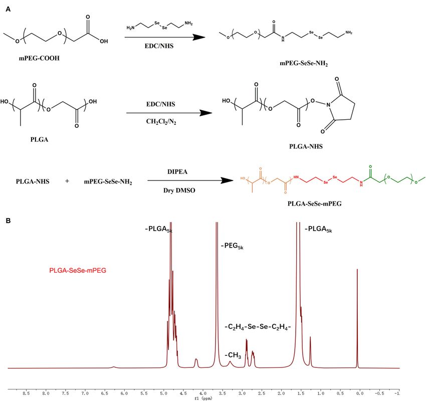
Wu et al.                                                                                                                  ROS-Sensitive Nanoparticles for Osteoarthritis

 FIGURE 2 | Size distribution of the nanoparticles after treatment with different concentrations of H2 O2 characterized by DLS. Morphological characterization of
 PLGA-SeSe-mPEG micelle (C) and uncoated nanoparticles (D). DLS, dynamic light scattering.

   At 4◦ C, the ultrasound-treated cell fluid was centrifuged at                       embraced a relatively uniform particle size with significant
10,000 g for 10 min to remove cell debris, and the supernatant                         particle spacing and clean particle backgrounds. However, after
was then collected and labeled.                                                        stimulation by H2 O2 , the hydrophilic shell of the nanoparticles
   The cell supernatants were further analyzed by ELISA to                             fell off, and accumulation of the hydrophobic nanoparticles was
determine the concentration tumor necrosis factor alpha (TNF-                          evident, resulting in different particle sizes. The detached mPEG
α), interleukin-1beta (IL-1β) and type II collagen.                                    segments were dispersed in the solution, contributing to a dirty
                                                                                       background for the nanoparticles.
Statistical Methods                                                                        Previous studies have shown that the most important factor
SPSS13.0 statistical software package was used for analysis. Data                      in determining drug loading is the compatibility between the
were expressed as mean ± standard deviation. One-way ANOVA                             hydrophobic segment and the drug molecule (Shi et al., 2016;
was used for comparison between groups with P-value & LT 0.05                          Wilkosz et al., 2018). Factors such as the amount of drug loaded,
was considered statistically significant.                                              particle size and particle-size distribution vary by drug-loading
                                                                                       method. Exploring suitable drug-loading methods that enable
RESULTS AND DISCUSSION                                                                 polymer nanoparticles to carry larger quantities of drugs is the
                                                                                       main content of this section (Zhang et al., 2017).
The synthesis path of the polymer PLGA-SeSe-mPEG was                                       Drug-loading performance tests of blank micelles were
prepared as shown in Figure 1A. The nuclear magnetic                                   conducted by assembling drugs of different concentrations with
resonance spectroscopy of PLGA-SeSe-mPEG (Figure 1B)                                   50 mg blank micelles. The prepared drug-loading nanoparticles
showed that in addition to the characteristic peaks of -PLGA and                       were filtered out of the unwrapped drugs by dialysis, and then
-mPEG, there was an obvious characteristic peak of -SeSe- at                           dissolved into the organic solvent again to detect the drug content
2.6–2.9 ppm.                                                                           in the organic solvent, and to predict the drug-loading amount.
   As shown in the DLS results (Figure 2A), the particle-size                              Drug-loading results (Figures 3A,B) showed that a 50 mg
distribution of blank and drug-loaded nanoparticles displayed                          blank micelle could load a maximum of 4.8 mg of DEX and
an obvious single-peak normal distribution, without trailing tail                      0.265 mg of CDMP-1, with drug-loading rates of 9.8 and
or double peak, which indicated that the sizes of the micelles                         0.53%, respectively. A PBS buffer (pH 7.4) was used as the
were relatively uniform. The drug load significantly increased                         release medium. The drug-loaded nanoparticles prepared by the
the size of nanoparticles from 260 to 295 nm with a wider                              physical-embedding method generally released the drug through
size distribution.                                                                     diffusion, which was faster than the drug-loaded nanoparticles
   After removing the mPEG shell by stimulation with H2 O2 ,                           prepared by chemical binding. Its release rate was related to
the size distribution of the nanoparticles showed a multi-                             factors such as the molecular weight of the nanoparticles and
peak distribution (Figure 2B). As the concentration of H2 O2                           the interaction between the drug and the nanoparticles. Strong
increased, the proportion of secondary peaks on both sides of the                      compatibility between the drug and the hydrophobic core can
main peak increased. When the concentration of H2 O2 reached                           delay the release of the drug, and a strong hydrogen bond
500 µM, the micelle particles were completely destroyed, and                           between the micellar core and the drug can also slow down the
the particle size presented an obvious bimorphic distribution,                         release of the drug.
indicating an obvious aggregation effect of nanoparticles                                  As shown in Figure 3C, DEX and CDMP-1 both had drug-
after uncoating.                                                                       release rates of
Wu et al. ROS-Sensitive Nanoparticles for Osteoarthritis
FIGURE 3 | The DEX (A) and CDMP-1 (B) loading performance of blank micelles, and accumulative DEX and CDMP-1 release with or without ROS500µm (C). DEX,
dexamethasone; CDMP-1, Cartilage-derivedmor-phogeneticprotein-1; ROS, reactive oxygen species. All error bars were presented as mean ± SD. *** p < 0.001.
to H2 O2 , the concentration of hydrogen peroxide can be reduced Measurement of flow-cell apoptosis (Figures 4C,D) showed
at the ischaemic site, limiting damage to the body. The H2 O2 - that DEX significantly affected the induction of apoptosis of
sensitive amphiphilic biomaterial of the present invention is used macrophages, with the cell survival rate dropping from 92.4 to
as a drug carrier to meet the needs of extended circulation and 65.9%. DEX had a relatively light effect on BMSCs, with the cell
targeted therapy (Shah et al., 2020). survival rate dropping from 87.2 to 77.5%. Blank nanoparticles
In order to verify the stem cell characteristics of extracted had little effect on apoptosis induction in the two groups;
BMSCs, BMSCs were incubated for a long time and the cells the proportion of normal cells was more than 80%, indicating
were counted. As shown in Figure 4A, BMSCs entered the that blank nanoparticles were safe and non-toxic. Drug-loaded
logarithmic growth phase at 2–4 days, and the number of nanoparticles showed a similar apoptotic effect with DEX on
cells decreased significantly and entered the plateau phase at activated macrophages, with the survival rate dropping to 68.2%.
5 days, indicating that BMSCs also had significant contact Drug-loaded nanoparticles had little effect on BMSCs, up to 75%
inhibition effect. of normal cells.
The cytotoxic results of drug-loaded micelles and DEX The fluorescent dye RhB was attached to the nanoparticles to
were shown in Figure 4B. The addition of DEX significantly indicate the location of the drug-loaded micelle inside the cell
reduced the activity of macrophages, but had little effect after being engulfed.
on the activity of BMSCs. Blank nanoparticles were less Cytophagocytosis diagrams (Figure 5A) showed that the
cytotoxic to all three groups of cells, with cell survival rates drug-loaded nanoparticles accumulated primarily in the
exceeding 80%, indicating the potential of blank nanoparticles cytoplasm and did not enter the nucleus. The differences in
to serve as drug carriers. Drug-loaded nanoparticles exhibited cell phagocytosis between cells were significant. The activated
cytotoxicity to certain macrophages. When treated with drug- macrophages were the brightest, followed by the inactive
loaded nanoparticles, macrophages exhibited a cell survival macrophages and the BMSC group.
rate of approximately 73%, and the survival rate of activated After incubating CDMP-1 and drug-loaded micelles with
macrophages was even lower, at 57%. Longer incubation resulted BMSCs for 14 days, BMSCs were fully induced to form
in a decrease in the relative number of treated macrophages. cartilage. Collagen and chondrogenic differentiation in cells was
However, blank micelles and drug-loaded micelles had little characterized by the histochemistry of alsin blue and type II
effect on BMSCs. After 7 days of culture, BMSCs treated collagen. As shown in Figure 5B, after 2-day differentiation
with DLNPs had higher activity than those treated with BNPs. induced by drug-loaded nanoparticles, a small number of BMSCs
Combined with the proliferation curve of BMSC, DLNPs changed from a spindle shape to polygons, and the black particles
treatment reduced the contact inhibition effect and restored the around the nucleus gradually increased with induction time.
vitality of BMSCs. After 14 days of differentiation induction, the cells began to
Frontiers in Bioengineering and Biotechnology | www.frontiersin.org 7 January 2021 | Volume 9 | Article 608150Wu et al. ROS-Sensitive Nanoparticles for Osteoarthritis FIGURE 4 | Cell counts of BMSCs Cells for 7 days (A).Cell viability of RAW 264.7 and BMSCs cell lines after 1 day, 3 days, and 7 days incubation in response to various treatments (B). Flow cytometric analyses (C) and quantitative analysis (D) of activated RAW264.7 and BMSCs after 4 h of incubation with various treatments, with PBS serving as the control. All error bars were presented as mean ± SD. ** p < 0.01. BMSCs, bone marrow mesenchymal stem cells; PBS, phosphate buffer saline. Frontiers in Bioengineering and Biotechnology | www.frontiersin.org 8 January 2021 | Volume 9 | Article 608150
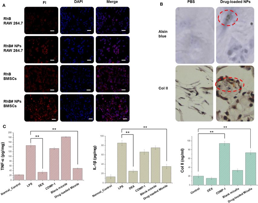
Wu et al.                                                                                                               ROS-Sensitive Nanoparticles for Osteoarthritis

 FIGURE 5 | (A) Cellular uptake of RhB and RhB#Nanoparticles in activated RAW 264.7 and BMSCs while the nucleus were stained with DAPI. (B) Results of
 chondrogenic differentiation of BMSCs were conducted by type II collagen immunohistochemistry staining and Alsin blue staining. (C) Expression of key factors
 (TNF-α, IL-1β, and type II collagen) were tested by an ELISA kit. All error bars were presented as mean ± SD. ** p < 0.01. ELISA, enzyme-linked immunosorbent
 assay; TNF, tumor necrosis factor; IL, interleukin.

grow in size, particles around the nucleus were visible, and the                     and drug-loaded nanoparticles had a significant inhibitory
cells gradually grew into clusters of cartilaginous nodules. Type                    effect on activated macrophages and reduced inflammatory
II collagen immunohistochemistry staining showed that some                           responses caused by LPS. Both CDMP-1 and drug-loaded
cells were dark brown and exhibited a strong positive reaction.                      nanoparticles significantly increased the expression level of
While the positive signals were located mainly in the cytoplasm                      type II collagen, indicating that CDMP-1 and drug-loaded
of the cells, a small number of positive signals occurred in the                     nanoparticles effectively induced chondrogenic differentiation of
extracellular matrix, indicating that the cells expressed type II                    BMSCs. All groups showed statistically significant differences
collagen after the induced differentiation.                                          (P < 0.05). This indicated that, at a higher concentration, the
   CDMP-1 and drug-loaded nanoparticles were incubated                               action time was longer, which caused drugs that entered the cell
with BMSCs for 24 h, and the supernatant contents of                                 through either of two ways to act fully on the cell and perform
TNF-α, IL-1β, and type II collagen were determined by                                its function.
ELISA, as shown in the Figure 5C. The expression of TNF-
α and IL-1β in activated macrophages was significantly                               CONCLUSION
increased, compared with normal macrophages. DEX and
drug-loaded nanoparticles therefore significantly reduced the                        OA is usually associated with cartilage damage and joint
expression level of the above factors, indicating that DEX                           inflammation (Chow and Chin, 2020). We designed,

Frontiers in Bioengineering and Biotechnology | www.frontiersin.org              9                                          January 2021 | Volume 9 | Article 608150
Wu et al.                                                                                                                           ROS-Sensitive Nanoparticles for Osteoarthritis

synthesized, and assembled a core-shell nanomicelle                                          AUTHOR CONTRIBUTIONS
loaded with the anti-inflammation drug DEX and CDMP-
1 to induce BMSCs to repair cartilage and inhibit joint                                      XW, WH, and CH: conception and design. CH, BL, and
inflammation. The nanometre-scale micelles were able                                         QX: administrative support. XW and WH: provision of study
to release drugs at lesion sites with high ROS, making                                       materials. JC, PL, and QX: collection and assembly of data.
them ROS-sensitive.                                                                          XW, PL, and CH: data analysis and interpretation. All authors:
   The function of the nanometre-scale micelle drug was                                      manuscript writing and final approval of manuscript.
verified through drug release, cytotoxicity, cartilage-induced
differentiation, and other experiments, providing a basis                                    FUNDING
for the further development of the drug in the treatment
of OA.                                                                                       This work was supported by the Xuzhou Science and Technology
                                                                                             Bureau Project (KC18039), Postdoctoral Funding Project of
DATA AVAILABILITY STATEMENT                                                                  Jiangsu Province (2018K244C), Double Innovation in Jiangsu
                                                                                             Province (2016SC04), Xuzhou Clinical Technical Backbone
The raw data supporting the conclusions of this article will be                              Research Programme (2018GG027), and Xuzhou social
made available by the authors, without undue reservation.                                    development project (KC15SH098).

REFERENCES                                                                                   Hunt, M. A., Charlton, J. M., and Esculier, J. F. (2020). Osteoarthritis
                                                                                                 year in review 2019: mechanics. Osteoarthr. Cartil. 28, 267–274.
Baboolal, T. G., Mastbergen, S. C., Jones, E., Calder, S. J., Lafeber, F. P., and                doi: 10.1016/j.joca.2019.12.003
   Mcgonagle, D. (2016). Synovial fluid hyaluronan mediates MSC attachment                   Jia, G., Takayama, Y., Flanigan, D. C., Kaeding, C. C., Zhou, J., Chaudhari,
   to cartilage, a potential novel mechanism contributing to cartilage repair in                 A., et al. (2011). Quantitative assessment of mobile protein levels in human
   osteoarthritis using knee joint distraction. Ann. Rheum. Dis. 75, 908–915.                    knee synovial fluid: feasibility of chemical exchange saturation transfer
   doi: 10.1136/annrheumdis-2014-206847                                                          (proteinCEST) MRI of osteoarthritis. Magn. Reson. Imaging 29, 335–341.
Bergink, A. P., Rivadeneira, F., Bierma-Zeinstra, S. M., Zillikens, M. C.,                       doi: 10.1016/j.mri.2010.10.006
   Ikram, M. A., Uitterlinden, A. G., et al. (2019). Are bone mineral density                Jordan, K. P., Edwards, J. J., Porcheret, M., Healey, E. L., Jinks, C.,
   and fractures related to the incidence and progression of radiographic                        Bedson, J., et al. (2017). Effect of a model consultation informed by
   osteoarthritis of the knee, hip, and hand in elderly men and women?                           guidelines on recorded quality of care of osteoarthritis (MOSAICS): a cluster
   The Rotterdam Study. Arthritis Rheumatol. 71, 361–369. doi: 10.1002/art.                      randomised controlled trial in primary care. Osteoarthr. Cartil. 25, 1588–1597.
   40735                                                                                         doi: 10.1016/j.joca.2017.05.017
Boulocher, C., Chereul, E., Langlois, J. B., Armenean, M., Duclos, M. E.,                    Miller, L. E., Bhattacharyya, S., Parrish, W. R., Fredericson, M., Bisson,
   Viguier, E., et al. (2007). Non-invasive in vivo quantification of the medial                 B., and Altman, R. D. (2019). Safety of intra-articular hyaluronic
   tibial cartilage thickness progression in an osteoarthritis rabbit model with                 acid for knee osteoarthritis: systematic review and meta-analysis of
   quantitative 3D high resolution micro-MRI. Osteoarthr. Cartil. 15, 1378–1387.                 randomized trials involving more than 8,000 patients. Cartilage 1–13.
   doi: 10.1016/j.joca.2007.04.012                                                               doi: 10.1177/1947603519888783
Brown, S. B., Wang, L., Jungels, R. R., and Sharma, B. (2020). Effects of cartilage-         Palmer, A. J., Brown, C. P., Mcnally, E. G., Price, A. J., Tracey, I., Jezzard, P., et al.
   targeting moieties on nanoparticle biodistribution in healthy and osteoarthritic              (2013). Non-invasive imaging of cartilage in early osteoarthritis. Bone Joint J.
   joints. Acta Biomater. 101, 469–483. doi: 10.1016/j.actbio.2019.10.003                        95-B, 738–746. doi: 10.1302/0301-620X.95B6.31414
Chow, Y. Y., and Chin, K. Y. (2020). The role of inflammation in the pathogenesis            Roseti, L., Desando, G., Cavallo, C., Petretta, M., and Grigolo, B. (2019). Articular
   of osteoarthritis. Mediators Inflamm. 2020:8293921. doi: 10.1155/2020/8293921                 cartilage regeneration in osteoarthritis. Cells 8:1305. doi: 10.3390/cells8111305
Cooper, C., Bardin, T., Brandi, M. L., Cacoub, P., Caminis, J., Civitelli, R., et al.        Said Ahmed, M. A., Saweeres, E. S. B., Abdelkader, N. A., Abdelmajeed, S. F., and
   (2016). Balancing benefits and risks of glucocorticoids in rheumatic diseases                 Fares, A. R. (2019). Improved pain and function in knee osteoarthritis with
   and other inflammatory joint disorders: new insights from emerging data. An                   dexamethasone phonophoresis: a randomized controlled trial. Indian J. Orthop.
   expert consensus paper from the European Society for Clinical and Economic                    53, 700–707. doi: 10.4103/ortho.IJOrtho_639_18
   Aspects of Osteoporosis and Osteoarthritis (ESCEO). Aging Clin. Exp. Res. 28,             Shah, S., Rangaraj, N., Laxmikeshav, K., and Sampathi, S. (2020). Nanogels as drug
   1–16. doi: 10.1007/s40520-015-0522-1                                                          carriers - introduction, chemical aspects, release mechanisms and potential
Crivelli, B., Bari, E., Perteghella, S., Catenacci, L., Sorrenti, M., Mocchi,                    applications. Int. J. Pharm. 581:119268. doi: 10.1016/j.ijpharm.2020.119268
   M., et al. (2019). Silk fibroin nanoparticles for celecoxib and curcumin                  Shi, C., Sun, Y., Wu, H., Zhu, C., Wei, G., Li, J., et al. (2016). Exploring the effect of
   delivery: ROS-scavenging and anti-inflammatory activities in an in                            hydrophilic and hydrophobic structure of grafted polymeric micelles on drug
   vitro model of osteoarthritis. Eur. J. Pharm. Biopharm. 137, 37–45.                           loading. Int. J. Pharm. 512, 282–291. doi: 10.1016/j.ijpharm.2016.08.054
   doi: 10.1016/j.ejpb.2019.02.008                                                           Shin, H. J., Park, H., Shin, N., Kwon, H. H., Yin, Y., Hwang, J. A., et al.
Cui, L., Zhang, J., Zou, J., Yang, X., Guo, H., Tian, H., et al. (2020).                         (2020). p47phox siRNA-Loaded PLGA nanoparticles suppress ROS/oxidative
   Electroactive composite scaffold with locally expressed osteoinductive factor for             stress-induced chondrocyte damage in osteoarthritis. Polymers 12:443.
   synergistic bone repair upon electrical stimulation. Biomaterials 230:119617.                 doi: 10.3390/polym12020443
   doi: 10.1016/j.biomaterials.2019.119617                                                   Stoop, R., Albrecht, D., Gaissmaier, C., Fritz, J., Felka, T., Rudert, M., et al. (2007).
Cui, Y., Zhu, T., Li, D., Li, Z., Leng, Y., Ji, X., et al. (2019). Bisphosphonate-               Comparison of marker gene expression in chondrocytes from patients receiving
   functionalized scaffolds for enhanced bone regeneration. Adv. Healthc. Mater.                 autologous chondrocyte transplantation versus osteoarthritis patients. Arthritis
   8:e1901073. doi: 10.1002/adhm.201901073                                                       Res. Ther. 9:R60. doi: 10.1186/ar2218
Dobbs, M. B., Gurnett, C. A., Robarge, J., Gordon, J. E., Morcuende, J. A.,                  Wang, C., Feng, N., Chang, F., Wang, J., Yuan, B., Cheng, Y., et al. (2019).
   and Bowcock, A. M. (2005). Variable hand and foot abnormalities in family                     Injectable cholesterol-enhanced stereocomplex polylactide thermogel loading
   with congenital vertical talus and CDMP-1 gene mutation. J. Orthop. Res. 23,                  chondrocytes for optimized cartilage regeneration. Adv. Healthc. Mater
   1490–1494. doi: 10.1016/j.orthres.2005.04.011.1100230636                                      8:e1900312. doi: 10.1002/adhm.201900312

Frontiers in Bioengineering and Biotechnology | www.frontiersin.org                     10                                               January 2021 | Volume 9 | Article 608150
Wu et al. ROS-Sensitive Nanoparticles for Osteoarthritis Watt, F. E., Hamid, B., Garriga, C., Judge, A., Hrusecka, R., Custers, R. J. H., et al. materials. Bioactive Mater. 6, 346–360. doi: 10.1016/j.bioactmat.2020. (2020). The molecular profile of synovial fluid changes upon joint distraction 08.016 and is associated with clinical response in knee osteoarthritis. Osteoarthr. Cartil. Zhao, Y., Wei, C., Chen, X., Liu, J., Yu, Q., Liu, Y., et al. (2019). Drug 28, 324–333. doi: 10.1016/j.joca.2019.12.005 delivery system based on near-infrared light-responsive molybdenum disulfide Wegner, A. M., Campos, N. R., Robbins, M. A., Haddad, A. F., Cunningham, H. nanosheets controls the high-efficiency release of dexamethasone to inhibit C., Yik, J. H., et al. (2019). Acute changes in NADPH oxidase 4 in early post- inflammation and treat osteoarthritis. ACS Appl. Mater. Interfaces 11, traumatic osteoarthritis. J. Orth. Res. 37, 2429–2436. doi: 10.1002/jor.24417 11587–11601. doi: 10.1021/acsami.8b20372 Wilkosz, N., Lazarski, G., Kovacik, L., Gargas, P., Nowakowska, M., Jamroz, Zhou, J., Liu, B., Li, C., Luo, Y., Liu, T., and Wang, W. (2019). Treatment D., et al. (2018). Molecular insight into drug-loading capacity of PEG- of hip osteoarthritis with glucocorticoids. Ann. Rheum. Dis. 78:e100. PLGA nanoparticles for itraconazole. J. Phys. Chem. B 122, 7080–7090. doi: 10.1136/annrheumdis-2018-213871 doi: 10.1021/acs.jpcb.8b03742 Zhu, T., Cui, Y., Zhang, M., Zhao, D., Liu, G., and Ding, J. (2020). Engineered Yang, M., Feng, X., Ding, J., Chang, F., and Chen, X. (2017). Nanotherapeutics three-dimensional scaffolds for enhanced bone regeneration in osteonecrosis. relieve rheumatoid arthritis. J. Control. Release 252, 108–124. Bioact. Mater. 5, 584–601. doi: 10.1016/j.bioactmat.2020.04.008 doi: 10.1016/j.jconrel.2017.02.032 Zhang, Y., Liu, X., Zeng, L., Zhang, J., Zuo, J., Zou, J., et al. (2019a). Polymer Conflict of Interest: The authors declare that the research was conducted in the fiber scaffolds for bone and cartilage tissue engineering. Adv. Funct. Mater. absence of any commercial or financial relationships that could be construed as a 29:1903279. doi: 10.1002/adfm.201903279 potential conflict of interest. Zhang, Y., Ren, T., Gou, J., Zhang, L., Tao, X., Tian, B., et al. (2017). Strategies for improving the payload of small molecular drugs in polymeric micelles. J. Copyright © 2021 Wu, Li, Cheng, Xu, Lu, Han and Huo. This is an open-access Control. Release 261, 352–366. doi: 10.1016/j.jconrel.2017.01.047 article distributed under the terms of the Creative Commons Attribution License (CC Zhang, Y., Yu, J., Ren, K., Zuo, J., Ding, J., and Chen, X. (2019b). Thermosensitive BY). The use, distribution or reproduction in other forums is permitted, provided hydrogels as scaffolds for cartilage tissue engineering. Biomacromolecules 20, the original author(s) and the copyright owner(s) are credited and that the original 1478–1492. doi: 10.1021/acs.biomac.9b00043 publication in this journal is cited, in accordance with accepted academic practice. Zhao, D., Zhu, T., Li, J., Cui, L., Zhang, Z., Zhuang, X., et al. No use, distribution or reproduction is permitted which does not comply with these (2021). Poly(lactic-co-glycolic acid)-based composite bone-substitute terms. Frontiers in Bioengineering and Biotechnology | www.frontiersin.org 11 January 2021 | Volume 9 | Article 608150
You can also read


