CD31+, CD38+, CD44+, and CD103+ lymphocytes in peripheral blood, bronchoalveolar lavage fluid and lung biopsy tissue in sarcoid patients and controls
←
→
Page content transcription
If your browser does not render page correctly, please read the page content below
Original Article

CD31+, CD38+, CD44+, and CD103+ lymphocytes in peripheral
blood, bronchoalveolar lavage fluid and lung biopsy tissue in
sarcoid patients and controls
Regina Aleksonienė1,2, Justinas Besusparis3,4, Vygantas Gruslys1,2, Laimutė Jurgauskienė4,
Aida Laurinavičienė3,4, Arvydas Laurinavičius3,4, Radvilė Malickaitė4, Jolita Norkūnienė5,
Rolandas Zablockis1,2, Edvardas Žurauskas3,4, Edvardas Danila1,2
1
Clinic of Chest Diseases, Immunology and Allergology of Faculty of Medicine, Vilnius University, Vilnius, Lithuania; 2Center of Pulmonology and
Allergology, Vilnius University Hospital Santaros Klinikos, Vilnius, Lithuania; 3National Center of Pathology, affiliate of Vilnius University Hospital
Santaros Klinikos, Vilnius, Lithuania; 4Faculty of Medicine, Vilnius University, Vilnius, Lithuania; 5Department of Mathematical Statistics, Vilnius
Gediminas Technical University, Vilnius, Lithuania
Contributions: (I) Conception and design: E Danila, R Aleksonienė; (II) Administrative support: E Danila, A Laurinavičius; (III) Provision of study
materials or patients: R Aleksonienė; (IV) Collection and assembly of data: R Aleksonienė; (V) Data analysis and interpretations: R Aleksonienė, J
Besusparis, E Danila, L Jurgauskienė, A Laurinavičius, R Malickaitė, J Norkūnienė, E Žurauskas; (VI) Manuscript writing: All authors; (VII) Final
approval of manuscript: All authors.
Correspondence to: Regina Aleksoniene. Center of Pulmonology and Allergology, Vilnius University Hospital Santaros Klinikos, Santariskiu str. 2,
Vilnius, Lithuania. Email: regina.aleksoniene@santa.lt.

                Background: The mechanisms driving the transition from inflammation to fibrosis in sarcoidosis patients
                are poorly understood; prognostic features are lacking. Immune cell profiling may provide insights into
                pathogenesis and prognostic factors of the disease. This study aimed to establish associations in simultaneous
                of lymphocyte subset profiles in the blood, bronchoalveolar lavage fluid (BALF), and lung biopsy tissue in
                the patients with newly diagnosed sarcoidosis.
                Methods: A total of 71 sarcoid patients (SPs) and 20 healthy controls (HCs) were enrolled into the study. CD31,
                CD38, CD44, CD103 positive T lymphocytes in blood and BALF were analysed. Additionally, the densities of
                CD4, CD8, CD38, CD44, CD103 positive cells in lung tissue biopsies were estimated by digital image analysis.
                Results: Main findings: (I) increase of percentage of CD3+CD4+CD38+ in BALF and blood, and increase
                of percentage of CD3 +CD4 +CD44 + in BALF in Löfgren syndrome patients comparing with patients
                without Löfgren syndrome, (II) increase of percentage of CD3+CD4+103+ in BALF and in blood in patients
                without Löfgren syndrome (comparing with Löfgren syndrome patients) and increase of percentage of
                CD3+CD4+103+ in BALF and in blood in more advanced sarcoidosis stage. (III) Increasing percentage of
                BALF CD3+CD4+CD31+ in sarcoidosis patients when comparing with controls independently of presence of
                Löfgren syndrome, smoking status or stage of sarcoidosis. Several significant correlations were found.
                Conclusions: Lymphocyte subpopulations in blood, BALF, and lung tissue were substantially different in
                SPs at the time of diagnosis compared to HCs. CD3+CD4+CD31+ in BALF might be a potential supporting
                marker for the diagnosis of sarcoidosis. CD3+CD4+CD38+ in BALF and blood and CD3+CD4+CD44+
                in BALF may be markers of the acute immune response in sarcoidosis patients. CD4+CD103+ T-cells in
                BALF and in blood are markers of the persistent immune response in sarcoidosis patients and are potential
                prognostic features of the chronic course of this disease.

                Keywords: Bronchoalveolar lavage; bronchoscopic lung biopsy; sarcoidosis

                Submitted Jul 08, 2020. Accepted for publication Feb 09, 2021.
                doi: 10.21037/jtd-20-2396
                View this article at: http://dx.doi.org/10.21037/jtd-20-2396

© Journal of Thoracic Disease. All rights reserved.                      J Thorac Dis 2021;13(4):2300-2318 | http://dx.doi.org/10.21037/jtd-20-2396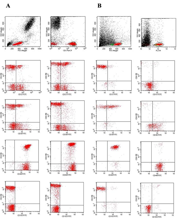
Journal of Thoracic Disease, Vol 13, No 4 April 2021                                                                                    2301

Introduction                                                                 Some cytokines, chemokines as well as immune cells
                                                                         derived mediators were proposed to be useful for the
Sarcoidosis is a systemic inflammatory disease caused by the
                                                                         diagnosis and follow-up of sarcoidosis patients; however,
combined effects of genetic susceptibility and environmental
                                                                         no single biomarker with proven unequivocal prognostic
exposures (1). Multiple potential infectious, non-infectious,
                                                                         value has yet been identified. Up-to-know only one serum
organic, and inorganic environmental etiologic agents,
                                                                         factor, angiotensin converting enzyme (ACE) is mentioned
unfortunately without any definitive demonstration of
                                                                         in International guidelines (15). Nevertheless its sensitivity
causality, have been proposed for sarcoidosis (2,3). It is
                                                                         and specificity are low (16,17), additionally, in the recent
likely that in predisposed individuals, sarcoidosis is the
                                                                         study (16) it was demonstrated that its clinical value as
result of the interplay between different etiologic agents
                                                                         biomarker can be further reduced for patients treated with
and the immune system (4).
                                                                         ACE inhibitors. Some other serum factors also have been
   Although the phenotypic expression of the disease
                                                                         proposed for sarcoidosis biomarkers, including activated
appears to vary in different populations, in general,
                                                                         macrophages and neutrophils secreted chitotriosidase,
patients with sarcoidosis tend to improve over time (5).
Many patients enter clinical remission within a few years                lysozyme, produced by monocyte-macrophage system and
of diagnosis; however, approximately one third experience                epithelioid cells and involved in granuloma formation,
chronic disease. Outcomes are generally less favourable for              mucin protein Krebs von den Lungen-6 (17,18).
parenchymal pulmonary sarcoidosis compared to lymph                          Bronchoalveolar lavage fluid examination is being used
node limited disease (6).                                                as diagnostic tool for forty years; best known indicators for
   Established experimental sarcoidosis models are still                 sarcoidosis are lymphocytosis and an increased CD4+ to
lacking, while studies on pathophysiologic mechanisms as                 CD8+ lymphocyte ratio. BAL cell patterns and lymphocyte
well as the search for diagnostic indicators are hampered                phenotyping often provide information useful for interstitial
by high variability in the course of the disease. One of the             lung diseases differentiation (18), and most of the studies
recent studies (7) using mass spectrometry based proteomics              shows that CD4 +/CD8 + ratio greater than 3.5 has high
identified 4,306 proteins in BAL cells, of which 272 proteins            diagnostic specificity for lung sarcoidosis. However, its
were differentially expressed in sarcoidosis compared to                 sensitivity is low, besides, specificity appears to be lower in
controls, and 121 were differentially expressed comparing                advanced radiographic stages of the disease (19).
progressive vs. non-progressive sarcoidosis subjects.                        Consequently, there is still great interest in cellular
   The prognosis of sarcoidosis is highly disparate                      biomarkers, which seem to be related to inflammation,
according to ethnic and genetic factors, the initial                     immune cell migration, and fibrosis (20-22), including
presentation, and other contributors (8). There is still                 cell surface molecules or their ligands. We had performed
no consensus on which tools are necessary to predict the                 literature search looking for a cellular markers that could
progression of the disease or how to monitor its activity (9).           help to predict the course of sarcoidosis and selected four
If present during an early phase, ongoing inflammation                   molecules, e.g., CD31, CD38, CD44, and CD103. Many
and interstitial fibrosis, specific to the chronic phase of the          inflammatory cells and tissue cells with various, sometimes
disease, may alert the presence of a more serious form and,              even opposite effects may be positive for CD31, CD38,
consequently, a tendicity to chronicity (10). Compared                   CD44, and CD103 (23-29), but while sarcoidosis is driven
to patients with persistently active non-fibrotic disease,               by T-cell mechanisms, particularly the accumulation of
the potential complications and long-term outcomes are                   activated CD4 T-cells in the lungs, allowing for T-cell
worse for those with fibrotic sarcoidosis (11). The extent               attachment and transmigration through the endothelium
of fibrosis on computed tomography is an independent                     (30-31), research was performed on T cell subpopulations
predictor of mortality (12). Since the mechanisms driving                CD4+ and CD8+.
the transition from inflammation to fibrosis in sarcoidosis                  CD31, also known as platelet endothelial cellular
are poorly understood (13) and the evolution and severity                adhesion molecule-1 (PECAM-1), is an integral membrane
of the disease vary substantially, it is difficult to predict the        protein expressed by endothelial cells, platelets, dendritic
disease course (14). It is not known if a fibrosis cascade is            cells, and blood cells, including T lymphocytes. CD31 is
activated at disease onset or if it is a response to poorly              known to be involved in leukocyte–leukocyte interactions,
controlled inflammation (11).                                            as well as in interactions between lymphocytes and the

© Journal of Thoracic Disease. All rights reserved.                 J Thorac Dis 2021;13(4):2300-2318 | http://dx.doi.org/10.21037/jtd-20-2396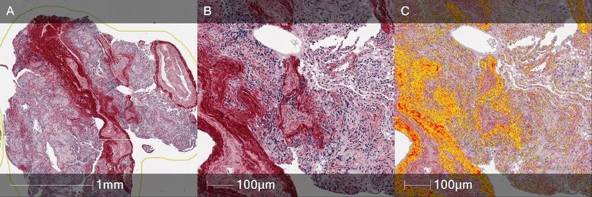
2302                                                                 Aleksonienė et al. CD31+, CD38+, CD44+, and CD103+ lymphocytes

vascular endothelium and migration to the inflamed tissues             Methods
through intercellular junctions (23,24). The ligand to the
                                                                       A total of 71 consecutive patients (25 were smokers and
CD31 molecule is a transmembrane glycoprotein CD38,
                                                                       46 were non-smokers) with newly diagnosed pulmonary
a multifunctional receptor and co-receptor involved
                                                                       sarcoidosis and 20 healthy controls (HCs) were enrolled in
in transmembrane signalling and cell adhesion, also
                                                                       the study from 2013 to 2015 at the Center of Pulmonology
contributing to modulation of antigen-mediated T-cell
                                                                       and Allergology (Vilnius University Hospital Santaros
responses (25,32). The human CD38 molecule is expressed
                                                                       Klinikos), and blood and BALF analyses were performed.
by immature hematopoietic cells, downregulated on
                                                                       Bronchoscopic lung biopsies were performed in 35 of these
mature cells, and re-expressed at high levels on activated
                                                                       patients with sarcoidosis. In addition, 5 samples were taken
lymphocytes, such as T cells, B cells, dendritic cells, and
                                                                       from biopsies that exhibited no histological changes, and
natural killer (NK) cells. CD38 ligation is followed by an
                                                                       these were used as a control for the tissue analyses. All study
increase in intracellular Ca2+, cell activation, proliferation,
                                                                       patients were Caucasian. None of the patients (including
differentiation, and migration, as well as by the production
and secretion of a panel of cytokines, such as interleukin             controls) had any relevant medical history or comorbidity
(IL)-6, IL-10, interferon- γ , granulocyte macrophage                  (e.g., tuberculosis) nor a history of exposure to organic or
colony stimulating factor (GM-CSF), and by proliferative               mineral dusts known to cause granulomatous lung disease,
effects (32). CD44 is a family of cell surface glycoproteins,          nor any immunosuppressive therapy—steroids, cytostatics,
also termed hyaladherin (HA), belonging to the group of                biotherapy.
cell adhesion molecules. HA is involved in cell-to-cell and               Forty-four patients had non-Löfgren sarcoidosis, and
cell-to-matrix interactions and participates in the regulation         the remaining 27 patients had Löfgren syndrome. Patients
of hyaluronic metabolism, activation, and migration of                 were grouped into stages according to chest radiography
lymphocytes, as well as the release of cytokines in areas of           and high-resolution computed tomography (HRCT).
inflammation (28-30). CD44 participates in a wide variety              Comparisons were made between the groups according to
of cellular functions, including lymphocyte activation,                smoking history (25 patients) and the presence of Löfgren
recirculation and homing. CD44 expression is known                     syndrome. All patients underwent diagnostic testing as
to be greater in the area of granuloma formation and                   part of the routine clinical investigation, including chest
fibrosis (33). One more biomarker, investigated in                     radiography, HRCT examination, pulmonary function
sarcoidosis patients is integrin αEβ7 (CD103), an adhesion             test, fibreoptic bronchoscopy with bronchoalveolar lavage
molecule expressed on 95% of intraepithelial CD4+                      (BAL), bronchoscopic lung biopsy, and BALF cell and
lymphocytes in the mucosa, but on less than 2% of                      blood examination. All study tests were performed over two
circulating peripheral blood lymphocytes (18). Differential            weeks, on average (in all cases, less than one month), from
expression of this marker on BAL CD4+ lymphocytes has                  the first visit to our center. The diagnosis was confirmed
been demonstrated, and CD103 has been proposed as a                    according to the American Thoracic Society/European
diagnostic marker of granulomatous lung disorders, such as             Respiratory Society/World Association for Sarcoidosis
sarcoidosis, albeit with controversial results.                        and other Granulomatous Disorders statement (15). The
   The aim of the present prospective study was to examine             study was conducted in accordance with the Declaration of
immune cell profiles that could be associated with the                 Helsinki (as revised in 2013). A signed informed consent
processes and pathways underlying the pathophysiology of               form was obtained from each participant. This study was
sarcoidosis. In this paper, we extended our previous study             approved by the Vilnius Regional Biomedical Research
(31-38) through the prospective and simultaneous analysis              Ethics Committee (No. 158200-12-5591160).
of lymphocyte subsets in the blood, bronchoalveolar lavage
fluid (BALF), and lung biopsy tissue of patients with newly
                                                                       Chest radiography and computed tomography
diagnosed pulmonary sarcoidosis at the time of initial
evaluation.                                                            Chest radiographic staging was performed according to
   We present the following study in accordance with the               the Scadding criteria, as follows: stage 0, normal chest
STROBE reporting checklist (available at http://dx.doi.                radiograph; stage I, mediastinal lymphadenopathy only;
org/10.21037/jtd-20-2396).                                             stage II, mediastinal lymphadenopathy with parenchymal

© Journal of Thoracic Disease. All rights reserved.               J Thorac Dis 2021;13(4):2300-2318 | http://dx.doi.org/10.21037/jtd-20-2396
Journal of Thoracic Disease, Vol 13, No 4 April 2021 2303
lesion; stage III, parenchymal disease only; and stage IV, follows: peridinin-chlorophyll-protein-complex (PerCP)
pulmonary fibrosis. HRCT scans were performed using conjugated anti-CD3; phycoerythrin (PE)-conjugated
64-slice CT (GE LightSpeed VCT, GE Healthcare, anti-CD4 and anti-CD8; and fluorescein-isothiocyanate
Milwaukee, Wisconsin). Scan parameters were as follows: (FITC)-conjugated anti-CD31, anti-CD38, anti-CD44,
collimation of 64×1.25 mm; tube voltage, 120 kV; tube and anti-CD103. For both blood and BALF lymphocyte
current, 660 mAs. subpopulation analysis, 100 µL of the cell suspensions
(1×10⁶ cells) were incubated with monoclonal antibodies
for 15 min: blood at room temperature, BALF at 4 ℃ in
Fibreoptic bronchoscopy, bronchoalveolar lavage, and
the dark. Afterwards, peripheral blood erythrocytes were
bronchoscopic lung biopsy
lysed using 2 ml of lysing solution (incubation for 10 min
Fibreoptic bronchoscopy, BAL, and bronchoscopic lung at room temperature in the dark), and then the suspension
biopsy were performed as described elsewhere (34,35). The was centrifuged at 300 g for 7 min at room temperature.
subjects were premedicated with atropine, and lidocaine The supernatant was discarded, and the remaining pellet
was delivered topically via an atomiser. The bronchoscope washed by adding 2 ml of phosphate buffered saline (PBS)
was inserted transnasally (in most cases) or orally and and centrifuging at 300 g for 7 min at room temperature
passed to segmental or subsegmental bronchus. BAL was (blood) or 4 ℃ (BALF). Afterwards, the supernatant was
performed in the right middle lobe, lingual, or in the area discarded, and the cells were re-suspended in 500 μL of 1%
of greatest radiologic abnormality. Sterile isotonic saline paraformaldehyde. Following cell fixation, flow cytometry
at room temperature was instilled in two 50-mL aliquots was run on an FACS Calibur (BD Biosciences, San Jose,
(as standard procedure in our centre). Each aliquot was CA, USA). After proper instrument settings, calibration,
retrieved with gentle manual aspiration (usually 30–40 mL and compensation, the results were analysed using
in total). The second aliquot was used to analyse BALF cells CellQuestPro software (BD Biosciences, San Jose, CA,
and lymphocyte subpopulations. USA). Fixed cells were analysed within 24 hours. Cells were
Bronchoscopic lung biopsy was performed under sequentially gated on lymphocytes [based on forward scatter
fluoroscopic guidance as follows: 6 to 10 pieces (3–4 mm2 (FSC) versus side scatter (SSC)] and on T lymphocytes
each) of lung tissue were taken from suspicious areas for (based on SSC versus CD3 PerCP histogram); after which,
histopathological examination at the National Center of markers of interest on CD4 and CD8 subpopulations were
Pathology, Affiliate of Vilnius University Hospital Santaros determined. The isotype control γ1/γ2a/CD3 was used for
Klinikos. the negative marker settings; these settings were applied for
subsequent lymphocyte analysis with a minimum of 10,000
events. Peripheral blood and BALF flow cytometry dot
Immunostaining and flow cytometry of peripheral blood
plots of representative patient are shown in Figure 1.
and bronchoalveolar lavage fluid samples
Peripheral blood and BALF samples were taken on the same
Tissue preparation and digital image analysis of histology
day, and both were analysed at the Laboratory of Clinical
specimens
Immunology and Blood Transfusion at the Center of
Laboratory medicine (Vilnius University Hospital Santaros Biopsy specimens were fixed in formalin and stained with
Klinikos). BALF for cell analysis was filtered through a 70 hematoxylin-eosin (HE) for histological investigation,
µm pore filter to remove mucus. The total cell numbers and Picro-Sirius Red was used for fibrosis assessment.
(Neubauer chamber) were determined, and the cellular Immunohistochemistry was performed using formalin-
material was sedimented by centrifugation (300 g for 7 min fixed paraffin-embedded sections, which were cut 3 μm
at 4 ℃). Trypan blue dye (0.4%) was used for the assessment thick and mounted on positively charged slides (Figure 2).
of BALF cell viability (the viability of the cells was Immunohistochemistry for CD8 +, CD38 +, CD44 +, and
95%±5%). Cell differentials were obtained by counting at CD103+ on the whole tissue sections was performed using
least 600 cells by light microscopy after staining with May- the Ultra View Universal DAB detection kit on a Ventana
Grünwald-Giemsa stain (Merck, Darmstadt, Germany). Benchmark Ultra staining system (Ventana Medical Systems,
The monoclonal antibodies (BD Biosciences, San Jose, Tucson, Arizona, USA). Epitope retrieval was performed
CA, USA) used for lymphocyte characterisation were as on the slides using Ventana Ultra CC1 buffer at 95 ℃ for
© Journal of Thoracic Disease. All rights reserved. J Thorac Dis 2021;13(4):2300-2318 | http://dx.doi.org/10.21037/jtd-20-23962304 Aleksonienė et al. CD31+, CD38+, CD44+, and CD103+ lymphocytes
A B
1000 1000 1000
1000
800 800 800 800
SSC-height
SSC-height
SSC-height
SSC-height
600 600 600 600
400 400 400 400
200 200 200 200
0 0 0 0
0 200 400 600 800 1000 100 101 102 103 104 0 200 400 600 800 1000 100 101 102 103 104
FSC-height CD3 PerCP FSC-height FL3-H
CD4 CD8 CD4 CD8
104 104 104 104
103 103 103 103
CD4 PE
CD8 PE
CD8 PE
CD4 PE
102 102 102 102
101 101 101 101
100 0 100 0 100100 100 0
10 101 102 103 104 10 101 102 103 104 101 102 103 104 10 101 102 103 104
CD31 FITC CD31 FITC CD31 FITC CD31 FITC
104
104 104 104
103
103 103 103
CD4 PE
CD4 PE
CD8 PE
CD8 PE
102
102 102 102
101 101
101 101
100 0 100 0 100 0
10 101 102 103 104 10 101 102 103 104 10 101 102 103 104 100 0
CD38 FITC 10 101 102 103 104
CD38 FITC CD38 FITC CD38 FITC
104 104 104 104
103 103 103 103
CD4 PE
CD4 PE
CD8 PE
CD8 PE
102 102 102 102
101 101 101 101
100 0 100 0 100 0 100 0
10 101 102 103 104 10 101 102 103 104 10 101 102 103 104 10 101 102 103 104
CD44 FITC CD44 FITC CD44 FITC CD44 FITC
104 104 104 104
103 103 103 103
CD4 PE
CD8 PE
CD4 PE
CD8 PE
102 102 102 102
101 101 101 101
100 0 100 0
10 101 102 103 104 10 101 102 103 104 100 0 100 0
10 101 102 103 104 10 10CD103
1
10FITC
2
103 104
CD103 FITC CD103 FITC CD103 FITC CD103 FITC
Figure 1 Typical histograms for CD31, CD38, CD44 and CD103 expression on CD4+ as well as CD8+ lymphocytes in blood and in
BALF. Flow cytometry dot plots demonstrating gating strategy in peripheral blood (dot plots A) and in BALF (dot plots B). Side scatter vs.
forward scatter gate for lymphocytes (R1), followed by a second gate on CD3+ T cell component (R2), CD3+ lymphocyte CD4+ and CD8+
subpopulations and finally CD31+, CD38+, CD44+ and CD103+ expression on CD4+ as well as CD8+ lymphocytes.
© Journal of Thoracic Disease. All rights reserved. J Thorac Dis 2021;13(4):2300-2318 | http://dx.doi.org/10.21037/jtd-20-2396Journal of Thoracic Disease, Vol 13, No 4 April 2021 2305
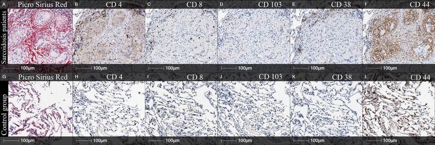
A Picro sirius red B CD4 C CD8 D CD103 E CD38 F CD44
Sarcoidosis patients
100 μm 100 μm 100 μm 100 μm 100 μm 100 μm
G Picro sirius red H CD4 I CD8 J CD103 K CD38 L CD44
Control group
100 μm 100 μm 100 μm 100 μm 100 μm 100 μm
Figure 2 Examples of lung tissue controls and sarcoid patients stained for CD4, CD8, CD103, CD38, CD44 immunohistochemistry.
Examples of lung tissue controls and sarcoid patients stained for Picro Sirius Red and CD4, CD8, CD103, CD38, CD44
immunohistochemistry. (A-F) Lung biopsy tissue containing non-necrotising granulomatous inflammation from sarcoid patients. Picro
Sirius Red staining reveals greater fibrosis in granulomatous inflammation areas compared to control tissues. CD4, CD8, CD103, CD38
immunohistochemistry shows slightly higher total numbers of lymphocyte populations in sarcoidosis patients, however only the percentage
of CD4+ and CD103+ cells was found to be significantly higher (Table 1).
64 min. The sections were then incubated with DAKO magnification (0.5 µm resolution).
anti-Human CD8+ mouse monoclonal antibody (C8/144B,
1:100 dilution), Cell Margue anti-CD38 rabbit monoclonal
Digital image analysis
antibody (SP149, 1:100 dilution), Abcamanti-CD44 rabbit
monoclonal: antibody (EPR1013Y, 1:100 dilution), and Digital image analysis (DIA) was performed using the
Abcamanti-Integrin alpha E rabbit monoclonal antibody HALO TM Classifier Module and Cyto Nuclear v1.5
(EPR4166, 1:500 dilution) at 37 ℃ for 32 min using algorithms (IndicaLabs, NM, USA) with a manually
the Ventana Ultraview DAB detection kit. Finally, the selected region of interest (ROI) enclosing the tissue
sections were developed in 3,3'-Diaminobenzidine as the section. The software enables automated recognition of
chromogen at 37 ℃ for 8 min, counter stained with Mayer’s selected tissue areas and cell segmentation in scanned
hematoxylin, and mounted. images. The nuclear/cytoplasmic analysis was calibrated to
Immunohistochemistry for CD4+ was performed using enumerate positive and negative inflammatory cell profiles
the EnVision FLEX + visualisation system on a DAKO in lung tissue. The positivity thresholds of DIA were
Autostainer Link 48+ system (Agilent DAKO, USA). Epitope monitored and individually calibrated by visual inspection
retrieval was performed on the slides using DAKO TRS in each immunohistochemical stain. The result of various
High pH buffer at 97 ℃ for 20 min using the PT-link module inflammatory cell densities was calculated by cell counts
(Agilent DAKO, USA). The sections were then incubated in lung tissue per square millimetre. Fibrosis areas were
with DAKO anti-Human CD4 + mouse monoclonal calculated using the HALO TM Area Quantification v1.0
antibody (4B12, 1:150 dilution) at 22 ℃ for 30 min algorithm (Figure 3).
using the EnVision FLEX + detection kit, developed in
DAB at 22 ℃ for 10 min, counter stained with Mayer’s
Statistical analysis
hematoxylin, and mounted. Digital images were captured
using the Aperio Scan-Scope XT Slide Scanner (Aperio Statistical analysis was performed using SPSS software,
Technologies, Vista, CA, USA) under 20× objective Version 20.0 (Statistical Package for Social Sciences, IBM,
© Journal of Thoracic Disease. All rights reserved. J Thorac Dis 2021;13(4):2300-2318 | http://dx.doi.org/10.21037/jtd-20-23962306 Aleksonienė et al. CD31+, CD38+, CD44+, and CD103+ lymphocytes
Table 1 Lung tissue lymphocyte subsets and collagen in sarcoidosis patients and controls.
Cells Sarcoidosis (n=35) Controls (n=5) P value
+
CD4 , total 7,374.5±8,391.0 1,183.4±844.5 0.198
CD8+, total 3,872.9±7,066.7 1,126.2±567.2 0.158
+
CD38 , total 2,803.4±5,166.9 558.4±465.5 0.090
+
CD44 , total 10,322.1±8,094.4 9,111.8±1,674.4 0.184
+
CD103 , total 1,532.3±1,588.7 460.8±270.5 0.069
+
CD4 , % 19.1±11.7 4.9±2.1 0.005**
+
CD8 , % 8.1±6.3 4.9±1.5 0.244
+
CD38 , % 6.0±6.2 2.4±1.4 0.098
+
CD44 , % 27.2±10.3 40.7±7.3 0.006**
CD103+, % 4.3±3.0 2.2±1.5 0.035*
+ 2
CD4 density, mm 704.8±518.5 181.5±73.3 0.005**
+ 2
CD8 density, mm 314.5±268.5 177.8±57.0 0.316
+ 2
CD38 density, mm 234.7±266.2 82.6±47.4 0.057
+ 2
CD44 density, mm 1002.3±501.6 1430.2±238.6 0.015*
+ 2
CD103 density, mm 158.2±118.0 73.8±45.7 0.026*
Collagen, % 20.2±7.4 18.2±8.4 0.379
Data are presented as the mean ± standard deviation. *PJournal of Thoracic Disease, Vol 13, No 4 April 2021 2307 Table 2 Characteristics of the patients with sarcoidosis and the controls Demographics Sarcoidosis patients (n=71) Controls (n=20) P value Sex (male/female) 38/33 12/8 – Age (years) 37 (21–68) 40 (24–57) – Löfgren syndrome (yes/no) 27/44 – – Smoker (yes/never) 25/46 5/15 – X-ray stage (I/II/III)^ 32/32/7 – – HRCT stage (I/II/III)^ 10/54/7 – – FVC, % pred 104±15 108±16 0.304 FEV1, % pred 97±13 103±17 0.140 FEV1/FVC, % 79±6 80±5 0.456 TLC, % pred 99±12 103±15 0.281 VC, % pred 106±14 107±16 0.927 RV, % pred 90±21 85±27 0.375 DLCO, % pred 76±11 87±7
2308 Aleksonienė et al. CD31+, CD38+, CD44+, and CD103+ lymphocytes
20
20 4040 **
**
***
***
**** **
**
%, CD3+CD4+CD38+
%,CD3+CD4+CD31+
+
15
15 3030
+
CD3 CD4 CD31
%, CD3 CD4 CD38
****
+
+
10
10 2020
+
+
12.2 13.1
24.2 23.6 24.7
55 9.8
8.4
1010 17.5
23.7 19.7
17.8
7.8
%,
4.9 5.9
3.1 7.0
00 00
Blood
Blood BALF
BALF Blood
Blood BALF
BALF
Controls (n=20)
Controls (n=20) II st.
st. (n=10)
(n=10) II st. (n=54)
II st. (n=54) IIIst.st.(n=7)
III (n=7) Controls (n=20)
Controls (n=20) II st.
st.(n=10)
(n=10) II st. (n=54)
II st. (n=54) IIIst.
III st.(n=7)
(n=7)
***
***
*** ***
40
40
100
100 ***
*** **
**
**
**
%, CD3+CD4+CD103++
%, CD3 CD4 CD103
30
30
CD3+CD4+CD44+
80
80
CD4+ CD44+
60
60
+
**
**
20
20
+
40
40 79.3 75.0
+
76.8
%, CD3
44.5 52.8 45.3 48.2 10
10 **
** 19.7
%,
20
20 37.1 1.4 12.1 12.1
0.4 0.5 8.1
5.1
00 00
Blood
Blood BALF
BALF Blood
Blood BALF
BALF
Controls (n=20)
Controls (n=20) I (n=10)
(n=10) II (n=54)
II (n=54) III (n=7)
III (n=7) Controls (n=20)
Controls (n=20) II (n=10)
(n=10) II (n=54)
II (n=54) III (n=7)
III (n=7)
Figure 4 Blood vs. BALF CD4 T lymphocyte subsets in different radiographic stages of sarcoidosis and controls groups. Data shown are
mean ± standard deviation. **PJournal of Thoracic Disease, Vol 13, No 4 April 2021 2309
3535 3030
3030 *
*
2525
CD3+CD8+CD38+
CD3+CD8+CD31+
CD3+ CD4+ CD31+
2525
** 2020
CD8+ CD38+
2020
1515
1515
21.3
1010
+
%,CD3
22.2 18.6 17.3
1010 17.9 19.3
17.1
%,%,
19.0
%,
13.2
10.7 55
55 7.7 9.0 6.6 6.3
4.9 4.3
00 00
Blood
Blood BALF
BALF Blood
Blood BALF
BALF
Controls(n=20)
Controls (n=20) II (n=10)
(n=10) IIII(n=54)
(n=54) III (n=7)
III (n=7) Controls(n=20)
Controls (n=20) II (n=10)
(n=10) IIII(n=54)
(n=54) III(n=7)
III (n=7)
7070 **
** ** 40
40
6060 **
**
***
***
**
%,%,CD3+CD8+CD44+
**
%,CD3+CD8+CD103+
**
CD3+ CD8+ CD44+
5050 30
30
CD3+ CD8+ CD103+
4040
20
20
3030
2020 43.0 40.9 39.2
32.7 33.9 10
10 20.7
14.0
%,
1010 17.3 21.6 20.7 2.1 10.7 11.5
3.2 5.0
3.6
00 00
Blood
Blood BALF
BALF Blood
Blood BALF
BALF
Controls(n=20)
Controls (n=20) II (n=10)
(n=10) IIII(n=54)
(n=54) III(n=7)
III (n=7) Controls(n=20)
Controls (n=20) II (n=10)
(n=10) IIII(n=54)
(n=54) III(n=7)
III (n=7)
Figure 5 Blood vs. BALF CD8 T lymphocyte subsets in different radiographic stages of sarcoidosis and controls groups. Data shown are
mean ± standard deviation. *P2310 Aleksonienė et al. CD31+, CD38+, CD44+, and CD103+ lymphocytes
%,on
onCD3
CD3+
+
% cells
cells
80
80 Blood, non-smokers
Blood, non-smokers
**
**
**
**
60
60
**
**
40
40 **
**
**
** **
20
20 **
00
CD31+
CD31+ CD38+
CD38+ CD44+
CD44+ CD103+ CD31+
CD103+ CD31+
CD38+
CD38+ CD44+
CD44+ CD103+
CD103+
CD4+ CD8+
+ +
CD4 CD8
Controls(N=15)
Controls (N = 15) Non-Löfgrensarcoidosis
Non-Löfgren sarcoidosis(N=29)
(N = 29) Löfgren'ssyndrome
Löfgren’s syndrome(N=18)
(N = 18)
+
%%,
onon
CD3 cells
CD3+cells BALF non-smokers
100
100
***
*** BALF non-smokers
**
***
***
8080
6060 ***
***
***
***
** **
**
**
***
***
4040 **
**
**
** **
**
** ***
***
** **
**
**
** **
2020
**
** **
00
CD31+
CD31+
CD38+
CD38+ CD44+
CD44+ CD103+ CD31+
CD103+ CD31+
CD38+
CD38+ CD44+
CD44+ CD103+
CD103+
CD4+ CD8+
+
CD4 CD8+
Controls(N=15)
Controls (N = 15) Non-Löfgrensarcoidosis
Non-Löfgren sarcoidosis(N=29)
(N = 29) Löfgren's syndrome(N=18)
Löfgren’s syndrome (N = 18)
Figure 6 BALF and blood lymphocyte subsets in non-Löfgren sarcoidosis and Löfgren syndrome non-smokers patients. Data shown are
mean ± standard deviation. *PJournal of Thoracic Disease, Vol 13, No 4 April 2021 2311
%,ononCD3
CD3+cellscells
+
% Blood, smokers
80
80
Blood, smokers
60
60
**
**
40
40
*
20
20 **
00
CD31+
CD31+
CD38+
CD38+ CD44+
CD44+ CD103+
CD103+ CD31+
CD31+
CD38+
CD38+ CD44+
CD44+ CD103+
CD103+
+
CD4+
CD4 CD8+
CD8 +
Controls (N = 5)
Controls (N=5) Non-Löfgren
Non-Löfgren sarcoidosis
sarcoidosis (N=15)(N = 15) Löfgren's syndrome
Löfgren’s syndrome (N=9)(N = 9)
%%,
onon CD3+
CD3+
cellscells
BALF, smokers
100
100 **
** BALF, smokers
**
80
80
60
60
**
** **
40
40 **
**
**
20
20
00
CD31
CD31 CD38
CD38 CD44
CD44 CD103
CD103 CD31
CD31 CD38
CD38 CD44
CD44 CD103
CD103
CD4
CD4 CD8
CD8
Controls(N=5)
Controls (N = 5) Non-Löfgren
Non-Löfgren sarcoidosis
sarcoidosis (N=15)(N = 15) Löfgren's
Löfgren’s syndrome
syndrome (N=9)(N = 9)
Figure 7 BALF and blood lymphocyte subsets in non-Löfgren sarcoidosis and Löfgren syndrome smoking patients. Data shown are mean ±
standard deviation. *P2312 Aleksonienė et al. CD31+, CD38+, CD44+, and CD103+ lymphocytes
Blood vs. BALF lymphocyte subsets in the non-Löfgren Correlations between blood, BALF, and lung tissue
and the Löfgren groups compared with non-smokers and lymphocyte subsets
smokers
Several significant correlations between blood, BALF,
We compared patients from the non-Löfgren group and and lung tissue lymphocyte subsets and other indices
the Löfgren group, the impact of smoking status on the were found (presented in Tables S1,S2,S3). Only the most
percentage markers studied was also clearly seen both in important ones are presented here. Positive correlations
blood and the BALF. In the blood of non-smoking groups, between the percentage of blood CD3+CD4+CD38+ and
most activity markers on CD4+ T cells differed significantly BALF CD3+CD4+CD38+ (r=0.510, P=0.0001) and between
in the Löfgren syndrome group vs. the non-Löfgren the percentage of blood CD3 +CD4 +CD44 + and BALF
syndrome patients (P=0.01 for CD4+CD31+, P=0.002 for CD3 +CD4 +CD44 + (r=0.362, P=0.002) were found. The
CD4+CD38+, P=0.002 for CD4+CD44+). In the BALF some percentage of blood CD3 +CD8 +CD103 + was positively
indices were higher in non-smoking Löfgren group patients, correlated with the tissue total CD44+ cell count (r=0.632,
specifically CD4+CD38+ (30.6%±15.2% vs. 17.9%±12.6%, P=0.0001).
P=0.002) and CD4+CD44+ (83.5%±8.3% vs. 74.2%±13.7%, Surprisingly, while comparing the corresponding
P=0.011). Nevertheless, in most cases percentages in the BALF and lung tissue lymphocyte subsets, only BALF
Löfgren group was significantly lower than in the non- CD3+CD4+CD103+ correlated with the tissue total CD103+
Löfgren syndrome patients: CD4+CD103 + (4.8%±5.4% cell count (r=0.473, P=0.020), CD103+ (%) cells (r=0.514,
vs. 10.5%±8.8%, P=0.002), CD8+CD31+ (5.7%±3.6% vs. P=0.010), and CD103+ density (r=0.408, P=0.048).
11.6%±8.4%, P=0.007), CD8 +CD44 + (13.8%±7.1% vs. Furthermore, we found weak but significant
22.3%±11.9%, P=0.006), and CD8+CD103+ (13.3%±8.9% negative correlations between the percentage of blood
vs. 3.9%±2.4%, P=0.004). CD3+CD4+CD103+ and the DLCO (r=−0.259, P=0.259)
Smoking had masked the differences mentioned value and between the BALF CD3+CD4+CD31+ and total
above, but in spite of this, for smokers CD4+CD38+ and lung capacity (TLC) (r=−0.261, P=0.029) and vital capacity
CD4+CD44+ were also higher in Löfgren group compared (VC) (r=−0.242, P=0.043). Moreover, we found significant
with non-Löfgren patients, while other markers (CD31 and negative correlations between the tissue CD44 + and
CD103) were lower. A significant difference was found only CD103+ cells with several pulmonary function test indices,
for CD4+CD103+ T cells (3.0%±4.4% vs. 12.7%±9.1%, as follows: CD44+ total with forced vital capacity (FVC)
P=0.01). (r=−0.406, P=0.044), TLC (r=−0.503, P=0.011) and VC
(r=−0.406, P=0.044); CD44+ (%) with DLCO (r=−0.414,
P=0.040); CD103 + total with FVC (r=−0.459, P=0.021),
Lung tissue lymphocyte subsets
TLC (r=−0.415, P=0.039) and VC (r=−0.481, P=0.015).
The lymphocyte subsets and percentage of collagen in the
lung tissue of sarcoidosis patients and controls are shown in
Discussion
Table 1. Surprisingly, few lymphocyte subsets were found to
be significantly different in the sarcoidosis group compared The most characteristic signs of sarcoidosis are granulomas,
with controls. Specifically, in the lung tissue, the percentage and the essential component of the immune response for
of CD4+ and CD103+ cells was found to be significantly granuloma formation is T cell activation and preferential
higher and the percentage of CD44+ cells was lower (the homing of activated T cells to the tissue. In spite of long-
patients with Löfgren syndrome were excluded from this lasting efforts of the scientific community, research in the
analysis). determination of the causative agents of sarcoidosis is still
We have not found a clear relationship between lacking. Studies of the key cellular players are essential for
lymphocyte subsets and the sarcoidosis stage (data not the understanding the etiopathogenesis of this disease. The
shown). Moreover, the analysis of the impact of smoking on mechanisms driving the transition from inflammation to
lung tissue surprisingly revealed that only the percentage fibrosis are also poorly understood; although, two phases are
of collagen was decreased in smoking sarcoid patients apparent. The first is chronic inflammation and the second
compared with non-smokers (17.9±8.5 vs. 22.6±5.3, is fibrotic transformation. It is likely that chronic sarcoidosis
P=0.015). may not simply be the persistence of acute sarcoidosis but
© Journal of Thoracic Disease. All rights reserved. J Thorac Dis 2021;13(4):2300-2318 | http://dx.doi.org/10.21037/jtd-20-2396Journal of Thoracic Disease, Vol 13, No 4 April 2021 2313 a fundamentally distinct form of remitting disease from the reporting that PECAM-1 concentrations were similar in very beginning (12). sarcoidosis patients and controls. In our study, we observed We investigated 71 patients with newly diagnosed a very similar phenomenon, e.g., CD31 expression on the sarcoidosis. For the analysis, study data were grouped peripheral blood T-cell surface in sarcoid patients and by radiologic disease stages, clinical manifestation, and controls was comparable. smoking status. While analysing CD31 expression on the immune In general, the majority of the investigated T lymphocyte cell surface we discovered that a much higher proportion subsets and activation marker expression profiles differed of blood CD4 + and CD8 + T cells express this receptor significantly between the sarcoidosis patients and the compared to BALF. This difference is possible due to a controls, especially in BALF. A marked difference in the higher number of BALF memory T lymphocytes, lacking percentage of lymphocyte subsets (in blood and in BALF) CD31 molecule on their surface. Moreover, expression of was found when comparing patients with and without CD31 on CD4+ T cells in BALF was found to increase with Löfgren syndrome. Additionally, the percentage of the sarcoidosis stage (p=0.002 for stage II vs. the control), lymphocyte subsets differed significantly in patients with while on CD8+ T lymphocytes, on the contrary, in stage different radiological stages of sarcoidosis (both in blood I sarcoid patients significantly less CD31 positive cells and in BALF). Smoking status also had considerable impact (P=0.035) were found. on the lymphocyte subset profiles of sarcoidosis patients. Ziora et al. demonstrated involvement of CD38 The most interesting and promising results were molecule in the early phases of lymphocyte binding to the obtained for the cell markers, represented by CD31 +, endothelium through direct interaction with CD31 (26). CD38+, CD44+, and CD103+ expression on T lymphocytes CD38–CD31 interactions upregulate integrin expression and especially on CD4 + T cells. These results are: (I) and promote the ensuing steps in the adhesion cascade. The increase of percentage of CD3+CD4+CD38+ both in BALF finding of selectin-like behaviour of the CD38 molecule and blood, and increase of percentage of CD3+CD4+CD44+ resulted in identification of CD31 as an endothelial cell in BALF in Löfgren syndrome patients comparing with surface ligand (26). It was shown that CD38 regulates patients without Löfgren syndrome (probable reflection inflammation by modulating leukocyte responses and of acute immune response), (II) increase of percentage migration to sites of inflammation. It has been proposed of CD3 + CD4 + 103 + T cells in BALF and in blood in as an early immune marker that reflects T-cell activation patients without Löfgren syndrome (compared with in allergies and some infectious diseases (25-27). CD38 Löfgren syndrome patients) and increase of percentage of is defined as both a cell surface enzyme (i.e., ectoenzyme) CD3+CD4+103+ T cells both in BALF and in blood with and as a receptor. The possible effect of CD38 receptor increasing (i.e., more advanced) sarcoidosis stage (probable on fibrosis formation was indicated by El-Chemaly at al., reflection of ongoing immune response), (III) percentage of showing that the blood CD38+ memory B cell count was BALF CD3+CD4+CD31+ is increased in sarcoidosis patients significantly higher in patients with pulmonary fibrosis when compared with controls independently of presence of compared to unaffected controls (20). Furthermore, Lee Löfgren syndrome, smoking status or stage of the disease et al. revealed a significantly increased proportion of (possible additional diagnostic marker). activated CD38+ cells within the naïve B-cell compartment CD31, is expressed in naïve recent thymic emigrants, but of severe chronic sarcoidosis patients in comparison with is downregulated after T-cell activation events and is absent healthy controls (39). Nevertheless, until now, no data on T from memory cells. In CD8+ T cells, CD31 expression is cell CD38 expression in sarcoidosis has been available. dynamically regulated, e.g., strongly downregulated during The impact of this molecule on mucosal immunity acute infection but re-expressed to intermediate levels in was studied by Deaglio et al. on intestinal lamina propria memory cells (26). CD31 is implicated in the development colonising T lymphocytes. This research showed that of atherosclerosis and its clinical complications (37). virtually all CD31+ cells co-expressed CD38, whereas only Nevertheless, papers describing CD31+ cells in sarcoidosis ~50% of CD38+ cells were CD31+ (40). When comparing or expression on BALF cells in other respiratory diseases the values of CD31 and CD38 marker expression observed are still lacking. To our knowledge, only Ziora et al. (38) in our study, it can be assumed that the same is valid for has analysed the serum concentrations of the soluble BALF. Unfortunately, as well as we applied three-color PECAM-1 molecule in the blood of sarcoidosis patients, cytometry, e.g., FITC conjugated monoclonal antibodies © Journal of Thoracic Disease. All rights reserved. J Thorac Dis 2021;13(4):2300-2318 | http://dx.doi.org/10.21037/jtd-20-2396
2314                                                              Aleksonienė et al. CD31+, CD38+, CD44+, and CD103+ lymphocytes

were used for both CD31 and CD38 molecules, it could not            the adhesion molecule CD44 was found to be significantly
be directly proven. However, we noted many similarities             reduced in Löfgren syndrome CD4+ T cells compared to
in CD31 and CD38 expression both on the peripheral                  the background. Conversely, non-Löfgren syndrome cells
blood and BALF T cell surface. On peripheral blood CD4+             showed a shift in CD44 expression of similar magnitude but
T cells, CD38 was also more highly expressed in stage               opposite direction. The reasons for the discrepancy between
II sarcoid patients compared with controls (P=0.004).               the work by Kaiser et al. and our findings are not clear. The
Unlike CD31, the effect of smoking was not seen on the              association between HLA and genetic background was not
blood or BALF cells. For both smoking and non-smoking               analysed in our study. Besides, in our study we had used
sarcoidosis patients, and especially the Löfgren syndrome           different BALF preparation and cell enumeration method.
group, patients’ CD3+CD4+ T cells had significantly higher          Lately, knowledge on the genetic architecture of sarcoidosis
CD38 expression compared with controls. Therefore,                  has been evolving, showing genetic differences between
the increase in CD38 expression on CD4+ T lymphocytes               Löfgren syndrome and non-Löfgren syndrome patients,
may be associated with the acute inflammatory response in           but the epigenetics events that determine the course of the
sarcoidosis.                                                        disease are not sufficiently known (43,44). Our findings
   Culty et al. had shown that CD44 expression is greater           on CD4 +CD38 + and CD4 +CD44 + T cells suggest that
in the area of granuloma formation and fibrosis (33). We            further investigations are required in order to assess these
did not find significant differences when comparing blood           markers as indicators of inflammatory activity in sarcoidosis
CD44 expression on CD4 + T cells between the control                patients. Analysis of these markers during active episodes of
group and the sarcoidosis group, only a tendency to increase        sarcoidosis seems to be most promising.
of expression (P=0.056) between stage I sarcoid patients and           Integrin CD103 was the only cell marker we chose that
controls. Meanwhile, on CD8 + T cells CD44 expression               had possible diagnostic value in terms of its expression as
was found to be significantly lower (P=0.035) when                  an additional biomarker of sarcoidosis. It is known that this
comparing stage III and control groups. When analysing              molecule can promote T-cell migration into the epithelium
blood lymphocyte subsets, grouped by sarcoidosis activity,          and is involved in the retention of lymphocytes in the
in non-smoking Löfgren syndrome patients significant                mucosa. Constant CD103 expression can reflect antigen(-s)
activation of CD4+CD44+ expression was revealed (P=0.007)           persistence in the lung tissue (22,45-48).
compared with controls. However, no difference was                     CD103 expression on control group peripheral blood
found in smokers or in the non-Löfgren group. Kasuga                T cells was very low compared to the other markers we
et al. evaluated soluble CD44 in the serum (sCD44) of 13            studied. CD103 was the only marker, which expression
sarcoidosis patients and 56 controls, as well as in the BALF        in BALF was significantly higher compared to peripheral
of 11 sarcoidosis patients and 10 controls (41). In patients        blood (P
Journal of Thoracic Disease, Vol 13, No 4 April 2021                                                                                    2315

that CD103+ expression on BALF CD4+ T cells displayed                 primary aim of investigating several biomarkers in newly
subgroup dependency: proportions significantly lower                  diagnosed sarcoidosis patients. Secondly, we investigated
than normal were noted in chest radiographic stage I, but             lymphocyte subsets simultaneously in blood, BALF, and
increased proportions in stages II and III were found. The            lung tissue. Thirdly, for enumeration of lung tissue cells
lowest expression was found in Löfgren syndrome patients.             we applied DIA. Moreover, the study population was fairly
Other authors (50-55) have also revealed that BALF CD4+               large, and the study was performed in a specialised tertiary
T lymphocytes from sarcoidosis patients compared with                 healthcare facility.
other interstitial diseases have reduced CD103 expression.               The limitations of the study are related to the
    Several explanations were proposed [discussed in (50)].           unavailability of lung biopsy data in the patients with
In brief, version is raised that in sarcoidosis CD4+ BALF             Löfgren syndrome (due to the study design). It was not
lymphocytes originate not from the mucosal inductive                  possible to analyse lymphocyte subsets in lung tissue for
sites, but from the peripheral blood. In other lung diseases          this group of patients. Also, lung tissue cells were analysed
a mucosal origin for BALF lymphocytes is suspected, with              by single immunohistochemistry, without the possibility to
CD103+ lymphocytes relocating from mucosal areas into                 obtain multiplex data on the lymphocyte subpopulations
the lung and proliferating in response to local stimuli (50).         and other lung tissue cells which should be removed from
    Braun et al. suggested that in patients with inflammatory         final analysis. In contrast, in blood and BALF, we used triple
lung diseases CD103-expressing CD4+ T cells in the lung are           staining with monoclonal antibodies. Another limitation is
continuously activated, long-living cells (53). However, Braun        that this was a single-centre study (this reflects the patient
et al. found no differences between the CD103+ and CD103–             population of our country since our centre is the primary
populations with respect to pro-inflammatory parameters.              centre for interstitial lung disease). Therefore, these results
Based on their data, Heron et al. proposed that a higher              may reflect the sarcoidosis manifestation of our patient
proportion of CD4+CD103+ T cells might be associated                  population.
with fibrosis formation in pulmonary sarcoidosis (54).
Bretagne et al. found a significant negative correlation
                                                                      Conclusions
between the BALF CD103+CD4+/CD4+ ratio and FVC,
TLC, and DLCO at last study visit. They suggested that                We f o u n d t h a t l y m p h o c y t e s u b t y p e s i n b l o o d ,
the BALF CD103+CD4+/CD4+ ratio may be of interest as a                BALF, and lung tissue were substantially different in
prognostic marker in sarcoidosis (55).                                sarcoidosis patients at the time of diagnosis compared
    Taking the data from this study and the reports of other          with healthy persons. Furthermore, the lymphocyte
authors together (53,54), it is reasonable to consider that           subtype profiles were significantly associated with the
CD4+CD103+ T cells in BALF and blood might be potential               clinical manifestation, radiological sarcoidosis stage, and
prognostic markers in sarcoidosis. At present, we speculate           smoking status. The lymphocyte profiles in BALF and
two possible scenarios: a relative deficit of CD4+CD103+              lung tissue did not correlate, which may indicate the
T cells in BALF reflects acute inflammation (immune                   need for assessment of both profiles in further studies.
response), or relative augmentation of CD4 +CD103 + T                 Increase of CD3 + CD4 + CD31 + in BALF may serve as
cells in BALF reflects persistent inflammation (immune                supporting evidence for a diagnosis of sarcoidosis.
response) in the lungs. It seems reasonable to address this           Increase of CD3 +CD4 +CD38 + in BALF and blood and
issue in future studies.                                              CD3+CD4+CD44+ in BALF—are markers of acute immune
    Like our previous preliminary findings (35,56) and                response in sarcoidosis. CD3+CD4+103+ T cells in BALF
the results of other authors (49), clinical manifestation,            and in blood are markers of a persistent immune response
radiological sarcoidosis stage, and smoking status should             in sarcoidosis patients and are a probable prognostic factor
always be taken into account when analysing lymphocyte                of the chronic course of the disease.
subsets and activation marker expression. Which lymphocyte
subpopulations are most important as prognostic factors
                                                                      Acknowledgments
and, maybe, additional diagnostic markers for sarcoidosis
still need to be resolved in future studies.                          Funding: The study was funded by the EU structural
    Our study has strengths and limitations. The strength             support project No VP1-3.1-ŠMM-01-V-03-002, (2011–
of our study is that it was carried out prospectively with the        2014): Improvement of Training of High Qualification

© Journal of Thoracic Disease. All rights reserved.              J Thorac Dis 2021;13(4):2300-2318 | http://dx.doi.org/10.21037/jtd-20-2396
2316 Aleksonienė et al. CD31+, CD38+, CD44+, and CD103+ lymphocytes
Specialists Conformed to the State and Society Needs in infectious antigens in sarcoidosis pathogenesis. Clin Chest
Biomedical field (BIOMEDOKT). Med 2015;36:561-8.
4. Beijer E, Veltkamp M, Meek B, et al. Etiology and
immunopathogenesis of sarcoidosis: Novel insights. Semin
Footnote
Respir Crit Care Med 2017;38:404-16.
Reporting Checklist: The authors have completed the 5. Judson MA, Boan AD, Lackland DT. The clinical course
STROBE reporting checklist. Available at http://dx.doi. of sarcoidosis: Presentation, diagnosis, and treatment
org/10.21037/jtd-20-2396 in a large white and black cohort in the United States.
Sarcoidosis Vasc Diffuse Lung Dis 2012;29:119-27.
Data Sharing Statement: Available at http://dx.doi. 6. Patterson KC, Chen ES. The pathogenesis of pulmonary
org/10.21037/jtd-20-2396 sarcoidosis and implications for treatment. Chest
2018;153:1432-42.
Peer Review File: Available at http://dx.doi.org/10.21037/jtd- 7. Bhargava M, Viken KJ, Barkes B, et al. Novel protein
20-2396 pathways in development and progression of pulmonary
sarcoidosis. Sci Rep 2020;10:13282.
Conflicts of Interest: All authors have completed the ICMJE 8. Nardi A, Brillet P-Y, Letoumelin P, et al. Stage
uniform disclosure form (available at http://dx.doi. IV sarcoidosis: Comparison of survival with the
org/10.21037/jtd-20-2396). The authors have no conflicts general population and causes of death. Eur Respir J
of interest to declare. 2011;38:1368-73.
9. Llabres M, Brito-Zerón P, Ramos-Casals M, et al.
Ethical Statement: The authors are accountable for all Synthetic pharmacotherapy for pulmonary sarcoidosis.
aspects of the work in ensuring that questions related Expert Opin Pharmacother 2019;20:1397-404.
to the accuracy or integrity of any part of the work are 10. Silva AL, Melo N, Caetano Mota P, et al. Pulmonary
appropriately investigated and resolved. The study was sarcoidosis: Prognostic factors at diagnosis in patients from
conducted in accordance with the Declaration of Helsinki North of Portugal. Reumatol Clin 2020;16:468-72.
(as revised in 2013). This study was approved by the Vilnius 11. Patterson KC, Strek ME. Pulmonary fibrosis in
Regional Biomedical Research Ethics Committee (No. sarcoidosis. Clinical features and outcomes. Ann Am
158200-12-5591160), and informed consent was taken from Thorac Soc 2013;10:362-70.
all individual participants. 12. Kirkil G, Lower EE, Baughman RP. Predictors
of mortality in pulmonary sarcoidosis. Chest
Open Access Statement: This is an Open Access article 2018;153:105-13.
distributed in accordance with the Creative Commons 13. Bonham CA, Strek ME, Patterson KC. From granuloma
Attribution-NonCommercial-NoDerivs 4.0 International to fibrosis: Sarcoidosis associated pulmonary fibrosis. Curr
License (CC BY-NC-ND 4.0), which permits the non- Opin Pulm Med 2016;22:484-91.
commercial replication and distribution of the article with 14. Valeyre D, Prasse A, Nunes H, et al. Sarcoidosis. Lancet
the strict proviso that no changes or edits are made and the 2014;383:1155-67.
original work is properly cited (including links to both the 15. Costabel U, Hunninghake GW. ATS/ERS/WASOG
formal publication through the relevant DOI and the license). statement on sarcoidosis. Sarcoidosis Statement
See: https://creativecommons.org/licenses/by-nc-nd/4.0/. Committee. American Thoracic Society. European
Respiratory Society. World Association for Sarcoidosis
and Other Granulomatous Disorders. Eur Respir J
References
1999;14:735-7.
1. Chen ES. Innate immunity in sarcoidosis pathobiology. 16. d'Alessandro M, Bergantini L, Perrone A, et al. Serial
Curr Opin Pulm Med 2016;22:469-75. investigation of angiotensin-converting enzyme in
2. Saidha S, Sotirchos ES, Eckstein C. Etiology of sarcoidosis patients treated with angiotensin-converting
sarcoidosis: Does infection play a role? Yale J Biol Med enzyme inhibitor. Eur J Intern Med 2020;78:58-62.
2012;85:133-41. 17. Bergantini L, Bianchi F, Cameli P, et al. Prognostic
3. Celada LJ, Hawkins C, Drake WP. The etiologic role of biomarkers of sarcoidosis: a comparative study of serum
© Journal of Thoracic Disease. All rights reserved. J Thorac Dis 2021;13(4):2300-2318 | http://dx.doi.org/10.21037/jtd-20-2396Journal of Thoracic Disease, Vol 13, No 4 April 2021 2317
chitotriosidase, ACE, lysozyme, and KL-6. Dis Markers between prognostic marker and therapeutic target. Trends
2019;2019:8565423. Mol Med 2008;14:210-8.
18. d'Alessandro M, Carleo A, Cameli P, et al. BAL 33. Culty M, O’Mara TE, Underhill CB, et al. Hyaluronan
biomarkers' panel for differential diagnosis of interstitial receptor (CD44) expression and function in human
lung diseases. Clin Exp Med 2020;20:207-16. peripheral blood monocytes and alveolar macrophages. J
19. Spagnolo P, Rossi G, Trisolini R, et al. Pulmonary Leukoc Biol 1994;56:605-11.
sarcoidosis. Lancet Respir Med 2018;6:389-402. 34. Danila E, Žurauskas E, Loskutovienė G, et al. Significance
20. El-Chemaly S, Cheung F, Kotliarov Y, et al. The of bronchoscopic lung biopsy in clinical practice. Adv Med
immunome in two inherited forms of pulmonary fibrosis. Sci 2008;53:11-16.
Front Immunol 2018;9:76. 35. Danila E, Norkūnienė J, Jurgauskienė L, et al. Diagnostic
21. Johnson P, Arif AA, Lee-Sayer SSM, et al. Hyaluronan role of BAL fluid CD4/CD8 ratio in different radiographic
and its interactions with immune cells in the healthy and and clinical forms of pulmonary sarcoidosis. Clin Respir J
inflamed lung. Front Immunol 2018;9:2787. 2009;3:214-21.
22. McMaster SR, Wein AN, Dunbar PR, et al. Pulmonary 36. Newman DK, Fu G, McOlash L, et al. PECAM-1 (CD31)
antigen encounter regulates the establishment of tissue- expression in naïve and memory, but not acutely activated,
resident CD8 memory T cells in the lung airways and CD8+ T cells. J Leukoc Biol 2018;104:883-93.
parenchyma. Mucosal Immunol 2018;11:1071-8. 37. Marelli-Berg FM, Clement M, Mauro C, et al. An
23. Schenkel AR, Chew TW, Chlipala E, et al. Different immunologist’s guide to CD31 function in T-cells. J Cell
susceptibilities of PECAM-deficient mouse strains to Sci 2013;126:2343-52.
spontaneous idiopathic pneumonitis. Exp Mol Pathol 38. Ziora D, Jastrzębski D, Adamek M, et al. Circulating
2006;81:23-30. concentration of markers of angiogenic activity in patients
24. Lishnevsky M, Young LC, Woods SJ, et al. with sarcoidosis and idiopathic pulmonary fibrosis. BMC
Microhemorrhage is an early event in the pulmonary Pulm Med 2015;15:113.
fibrotic disease of PECAM-1 deficient FVB/n mice. Exp 39. Lee NS, Barber L, Akula SM, et al. Disturbed homeostasis
Mol Pathol 2014;97:128-36. and multiple signalling defects in the peripheral blood
25. Gally F, Hartney JM, Janssen WJ, et al. CD38 plays a dual B-cell compartment of patients with severe chronic
role in allergen-induced airway hyperresponsiveness. Am J sarcoidosis. Clin Vaccine Immunol 2011;18:1306-16.
Respir Cell Mol Biol 2009;40:433-42. 40. Deaglio S, Mallone R, Baj G, et al. Human CD38 and its
26. Guedes AG, Jude JA, Paulin J, et al. Airway responsiveness ligand CD31 define a unique lamina propria T lymphocyte
in CD38-deficient mice in allergic airway disease: studies signaling pathway. FASEB J 2001;15:580-2.
with bone marrow chimeras. Am J Physiol Lung Cell Mol 41. Kasuga I, Minemura K, Nasu H, et al. Elevated serum
Physiol 2015;308:L485-93. soluble CD44 level in sarcoidosis. Int J Mol Med
27. Adekambi T, Ibegbu CC, Cagle S, et al. Biomarkers on 2000;6:679-82.
patient T cells diagnose active tuberculosis and monitor 42. Kaiser Y, Lakshmikanth T, Chen Y, et al. Mass cytometry
treatment response. J Clin Invest 2015;125:1827-38. identifies distinct lung CD4+ T cell patterns in Löfgren’s
28. Shimizu Y, Van Seventer GA, Siraganian R, et al. Dual role syndrome and non-Löfgren’s syndrome sarcoidosis. Front
of the CD44 molecule in T cell adhesion and activation. J Immunol 2017;8:1130.
Immunol 1989;143:2457-63. 43. Rivera NV, Ronninger M, Shchetynsky K, et al. High-
29. Goodison S, Urquidi V, Tarin D. CD44 cell adhesion density genetic mapping identifies new susceptibility
molecules. Mol Pathol 1999;52:189-96. variants in sarcoidosis phenotypes and shows genomic-
30. Rivera NV, Hagemann-Jensen M, Ferreira MAR, et al. driven phenotypic differences. Am J Respir Crit Care Med
Common variants of T-cells contribute differently to 2016;193:1008-22.
phenotypic variation in sarcoidosis. Sci Rep 2017;7:5623. 44. Kaiser Y, Eklund A, Grunewald J. Moving target: Shifting
31. Aleksonienė R, Zeleckienė I, Matačiūnas M, et al. the focus to pulmonary sarcoidosis as an autoimmune
Relationship between radiologic patterns, pulmonary spectrum disorder. Eur Respir J 2019;54:1802153.
function values and bronchoalveolar lavage fluid cells in 45. Sung SS, Fu SM, Rose CE Jr, et al. A major lung CD103
newly diagnosed sarcoidosis. J Thorac Dis 2017;9:88-95. (alphaE)-beta7 integrin-positive epithelial dendritic
32. Deaglio S, Aydin S, Vaisitti T, et al. CD38 at the junction cell population expressing Langerin and tight junction
© Journal of Thoracic Disease. All rights reserved. J Thorac Dis 2021;13(4):2300-2318 | http://dx.doi.org/10.21037/jtd-20-2396You can also read