Modulation of Cell Death and Promotion of Chondrogenic Differentiation by Fas/FasL in Human Dental Pulp Stem Cells (hDPSCs) - Frontiers
←
→
Page content transcription
If your browser does not render page correctly, please read the page content below
ORIGINAL RESEARCH
                                                                                                                                              published: 15 May 2020
                                                                                                                                        doi: 10.3389/fcell.2020.00279

                                            Modulation of Cell Death and
                                            Promotion of Chondrogenic
                                            Differentiation by Fas/FasL in Human
                                            Dental Pulp Stem Cells (hDPSCs)
                                            Alessandra Pisciotta 1† , Giulia Bertani 1† , Laura Bertoni 1† , Rosanna Di Tinco 1 ,
                                            Sara De Biasi 2 , Antonio Vallarola 3 , Elisa Pignatti 1 , Rossella Tupler 3 , Carlo Salvarani 4 ,
                                            Anto de Pol 1 and Gianluca Carnevale 1*
                                            1
                                              Department of Surgery, Medicine Dentistry and Morphological Sciences with Interest in Transplant, University of Modena
                                            and Reggio Emilia, Modena, Italy, 2 Department of Medical and Surgical Sciences for Children and Adults, University
                                            of Modena and Reggio Emilia, Modena, Italy, 3 Department of Biomedical, Metabolic and Neural Sciences, Center
                                            for Neuroscience and Neurotechnology, University of Modena and Reggio Emilia, Modena, Italy, 4 Rheumatology Unit,
                                            Azienda Unitá Sanitaria Locale-IRCCS di Reggio Emilia, Reggio Emilia, Italy

                         Edited by:
                    Giovanna Orsini,        Human dental pulp stem cells (hDPSCs) are characterized by high proliferation rate,
   Marche Polytechnic University, Italy     the multi-differentiation ability and, notably, low immunogenicity and immunomodulatory
                       Reviewed by:         properties exerted through different mechanisms including Fas/FasL pathway. Despite
                          Jangho Kim,
         Chonnam National University,
                                            their multipotency, hDPSCs require particular conditions to achieve chondrogenic
                          South Korea       differentiation. This might be due to the perivascular localization and the expression
             Gianandrea Pasquinelli,        of angiogenic marker under standard culture conditions. FasL stimulation was able to
           University of Bologna, Italy
                                            promote the early induction of chondrogenic commitment and to lead the differentiation
                  *Correspondence:
                   Gianluca Carnevale       at later times. Interestingly, the expression of angiogenic marker was reduced by
        gianluca.carnevale@unimore.it       FasL stimulation without activating the extrinsic apoptotic pathway in standard culture
     † These   authors have contributed     conditions. In conclusion, these findings highlight the peculiar embryological origin of
                    equally to this work
                                            hDPSCs and provide further insights on their biological properties. Therefore, Fas/FasL
                    Specialty section:      pathway not only is involved in determining the immunomodulatory properties, but also
         This article was submitted to      is implicated in supporting the chondrogenic commitment of hDPSCs.
                  Stem Cell Research,
                a section of the journal    Keywords: hDPSCs, Fas/FasL pathway, chondrogenic differentiation, 3D pellet culture, pericytes
  Frontiers in Cell and Developmental
                                Biology
         Received: 05 February 2020         INTRODUCTION
          Accepted: 31 March 2020
            Published: 15 May 2020          Human dental pulp stem cells (hDPSCs) are located in the perivascular area of the loose connective
                               Citation:    tissue enclosed within the pulp chamber. Human DPSCs are characterized by a low-invasive
      Pisciotta A, Bertani G, Bertoni L,    procedures required for isolation, high proliferation rate, low immunogenicity and the ability to
    Di Tinco R, De Biasi S, Vallarola A,    differentiate into different cell lineages (Gronthos et al., 2002). The multi/pluripotency of hDPSCs
      Pignatti E, Tupler R, Salvarani C,    can be attributed to their particular embryological derivation from the neural crest. Particularly,
     de Pol A and Carnevale G (2020)        neural crest cells originate during the formation of neural tube, at the third week of embryo
               Modulation of Cell Death
                                            development, then undergo an epithelial-mesenchymal transition (EMT) and migrate to different
      and Promotion of Chondrogenic
 Differentiation by Fas/FasL in Human
                                            body compartments under the control of several regulatory factors. Following migration, neural
    Dental Pulp Stem Cells (hDPSCs).        crest cells generate the majority of craniofacial tissues, including tooth, fat, muscle, bone and
            Front. Cell Dev. Biol. 8:279.   cartilage tissues, as well as cranial peripheral ganglia and nerves, among other cell types, such
        doi: 10.3389/fcell.2020.00279       as melanocytes (Pisciotta et al., 2020). It has been widely demonstrated that hDPSCs are able to

Frontiers in Cell and Developmental Biology | www.frontiersin.org                 1                                                  May 2020 | Volume 8 | Article 279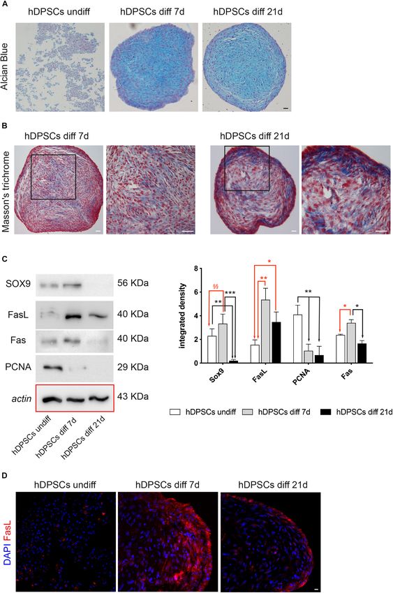
Pisciotta et al.                                                                                  Fas/FasL Promotes hDPSCs Chondrogenic Differentiation

differentiate toward lineages belonging to all the three germ               in immune system homeostasis maintenance and prevention
layers, as a matter of fact these stem cells can commit to glial            of autoimmunity, increasing evidence showed the involvement
cells and participate in peripheral nerve regeneration (Carnevale           of Fas/FasL signaling in further cellular responses, such as
et al., 2018), contribute in restoring urethral sphincter contractile       inflammation, proliferation and regeneration (Chang et al.,
function (Zordani et al., 2019), reduce fibrosis and ameliorate             2002). These data were further confirmed by previous findings
muscle trophism in Duchenne Muscular Dystrophy mouse                        demonstrating that hDPSCs do express Fas receptor under
model (Pisciotta et al., 2015b), besides promoting bone tissue              standard culture conditions (Pisciotta et al., 2018).
regeneration in critical size calvarial defects (Pisciotta et al.,             To this regard, the aim of our study was to investigate
2012). Further evidence of the regenerative potential of hDPSCs             whether and how Fas/FasL pathway can affect the stemness
is documented by their capability to promote vascularization in             features of hDPSCs and, particularly, the modulation of their
regenerating tissues in vivo and supported by their expression of           chondrogenic potential.
VEGF (Laino et al., 2005).
    On the other hand, the ability of hDPSCs to commit into
chondrogenic lineage is debated and controversial. Indeed,
                                                                            MATERIALS AND METHODS
findings from literature demonstrated that hDPSCs show high
variability when induced toward chondrogenic differentiation:
this is likely due to the heterogeneity of dental pulp cells
                                                                            Isolation of STRO-1+ /c-Kit+ Human
(Iohara et al., 2006; Zhang et al., 2006) and to high oxygen                Dental Pulp Stem Cells and
levels when differentiating hDPSCs in vitro, that do not reflect            Immunophenotype Characterization
the physiological in vivo oxygen tension required for articular             The study was conducted in accordance with the
chondrocyte differentiation (Chen et al., 2015). The only cartilage         recommendations of Comitato Etico Provinciale-Azienda
portion present in the craniofacial district is represented by              Ospedaliero-Universitaria di Modena (Modena, Italy), which
Meckel’s cartilage, hyaline cartilage formed in the mandibular              provided the approval of the protocol (ref. number 3299/CE;
process of the first branchial arch of vertebrate embryos. From             5 September 2017). Human DPSCs were isolated from third
an embryological point of view, chondrocytes are differentiated             molars of adult subjects (n = 3; 18–25 years) undergoing routine
from mesodermal cells in general, whereas cells forming Meckel’s            dental extraction. All subjects gave written informed consent in
cartilage, are differentiated from ectodermal mesenchymal cells             compliance with the Declaration of Helsinki.
of neural crest origin (Amano et al., 2010). Moreover, during                  Cells were isolated from human dental pulp as previously
intramembranous ossification cartilage is not present and MSCs              described (Bianchi et al., 2017). Briefly, dental pulp was harvested
from neural crest differentiate directly into osteoblasts.                  from the teeth and enzymatic digestion was carried out through
    Beside their differentiation potential, it has been widely              a digestive solution (3 mg/ml type I collagenase plus 4 mg/ml
demonstrated by several studies conducted in vitro and in vivo              dispase in α-MEM). Pulp was then filtered onto 100 µm Falcon
that hDPSCs can modulate the immune response through                        Cell Strainers, in order to obtain a cell suspension. Then, cell
different mechanisms (Wada et al., 2009; Zhao et al., 2012). To             suspension was plated in 25 cm2 culture flasks and expanded
this regard, it is renown that FasL is expressed in different cell          in standard culture medium (α-MEM supplemented with 10%
types residing in “immune-privileged” sites, such as the testis, the        heat inactivated foetal bovine serum (FBS), 2 mM L-glutamine,
eye and the nervous system (Brunlid et al., 2007). Therefore, it            100 U/ml penicillin, 100 µg/ml streptomycin; all from Sigma
would be helpful to understand how FasL expression modulates                Aldrich, St. Louis, MO, United States) at 37◦ C and 5% CO2 .
hDPSCs properties.                                                          Following cell expansion, hDPSCs underwent immune-selection
    Taken together, these properties represent the necessary                by using MACS separation kit, according to manufacturer’
                                                                                              R

criteria to define hDPSCs as a suitable stem cells source for               instructions. Two sequential immune-selections were performed
regenerative medicine.                                                      by using mouse IgM anti-STRO-1 and rabbit IgG anti-c-Kit
    Several findings in literature have highlighted the prominent           primary antibodies (Santa Cruz Biotechnology, Dallas, TX,
role of Fas/FasL pathway in immunomodulation. Particularly, the             United States). The following magnetically labeled secondary
activation of Fas/FasL pathway occurs following the exposure                antibodies were used: anti-mouse IgM and anti-rabbit IgG
to inflammatory microenvironment which induces apoptosis                    (Miltenyi Biotec, Bergisch Gladbach, Germany). The selection
in T cells (Pierdomenico et al., 2005; Zhao et al., 2012;                   of a homogeneous hDPSCs population expressing STRO-1 and
Riccio et al., 2014). Mechanistically, upon binding of FasL                 c-Kit was then evaluated by immunofluorescence analysis, as
to its receptor CD95, or Fas receptor, the extrinsic apoptotic              described below.
pathway is activated with Pro-Caspase 8 and Fas associated                     With the aim to provide a visual representation as to
death domain (FADD) being recruited to form the death                       the location and degree of overlap between two wavelengths
inducing signaling complex (DISC), in which Pro-Caspase                     corresponding of STRO-1 and c-Kit, co-localization map
8 undergoes activation. Then, Caspase 8 leaves the DISC,                    was performed by NIS software as previously described
activates caspase 3/7 and induces apoptosis (Chang et al.,                  (Gibellini et al., 2012). Briefly, the two-dimensional scatter plot
2002; Carnevale et al., 2017). Alternatively, c-FLIP, a protease-           diagram of the image was analyzed to evaluate the spatial
deficient caspase homolog can interact with FADD and act as                 colocalization of the signals and the Pearson’s correlation
an apoptosis inhibitor (Irmler et al., 1997). Besides its key role          was calculated. Moreover, areas with the strongest colocalized

Frontiers in Cell and Developmental Biology | www.frontiersin.org       2                                               May 2020 | Volume 8 | Article 279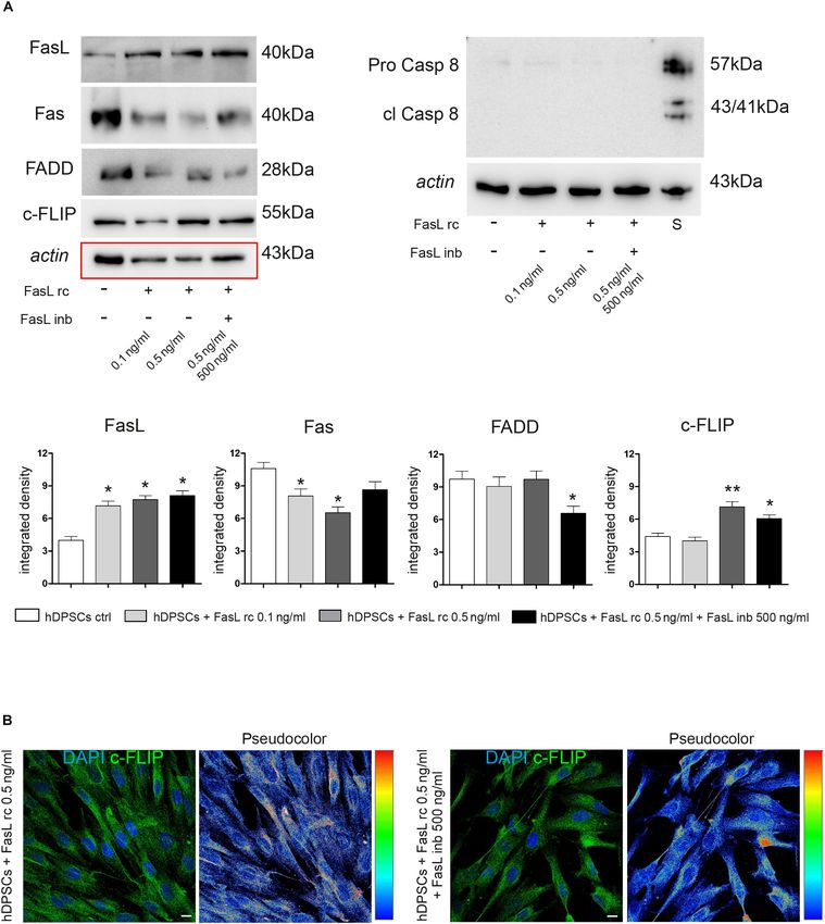
Pisciotta et al.                                                                             Fas/FasL Promotes hDPSCs Chondrogenic Differentiation

signals, corresponding to pixels for both detectors, were                 penicillin and 100 µg/ml streptomycin. Upon hDPSCs adhesion,
selected to generate colocalization binary maps. In order                 after 24 h, pre-activated PBMCs were seeded on hDPSCs at a
to evaluate the expression of the typical mesenchymal stem                5:1 ratio and kept in culture for 48 h. At the end of the direct
cells (MSCs) markers, immune-selected hDPSCs at passage 1                 co-culture, the floating PBMCs were removed together with the
underwent FACS analysis against CD73, CD90, CD105, CD34,                  supernatant and discarded, whereas the attached hDPSCs were
CD45, HLA-DR, as formerly described by Conserva et al.                    processed according to the subsequent experimental procedures.
(2019). Following trypsin dissociation, cells were resuspended               hDPSCs and pre-activated PBMCs cultured alone were
in culture medium and were stained with the following                     used as controls.
fluorochrome-conjugated antibodies (Abs): anti-human-CD73-
PE-CY7, -CD90-FITC, -CD105-APC, -CD45-PE, and -HLA-                       hDPSCs-PBMCs Co-culture: Evaluation
DR-PE-CY7 (all from BD Biosciences, Franklin Lakes, NJ,                   of Fas/FasL Pathway
United States); and -CD34-ECD (Beckman Coulter, Fullerton,                In order to evaluate Fas/FasL pathway in hDPSCs cultured alone
CA, United States). A minimum of 10,000 cells per sample was              and after co-culture with PBMCs, the expression of Fas, FasL,
acquired and analyzed by using the Attune Acoustic Focusing               Caspase 8 and c-FLIP, was investigated by Western blot analysis.
Flow Cytometer (Attune NxT, Thermo Fisher, Waltham, MA,                   Particularly, whole cell lysates were obtained as formerly reported
United States). Data were analyzed by FlowJo 9.5.7 (Treestar, Inc.,       (Pisciotta et al., 2018). Briefly, 30 µg of protein extract per
Ashland, OR, United States) under MacOS 10.                               specimen were quantified by a Bradford Protein Assay (Bio-Rad),
   STRO-1+ /c-Kit+ hDPSCs were also analyzed for the                      then SDS-polyacrylamide gel electrophoresis and subsequent
expression of HLA-ABC and HLA-DP-DQ-DR by confocal                        protein transfer to nitrocellulose membranes were performed.
immunofluorescence analysis. Cells were fixed with 4%                     The following antibodies were used: rabbit anti-FasL, mouse anti-
paraformaldehyde (PFA) in pH 7.4 phosphate buffer saline                  Fas, mouse anti-Caspase 8 (Cell Signaling Technology, Trask
(PBS) for 20 min and washed in PBS. After rinsing with PBS,               Lane Danvers, MA, United States), rabbit anti-c-FLIP (R&D
samples were blocked with 3% BSA in PBS for 30 min at room                systems, McKinley Place NE, Minneapolis, MN, United States),
temperature and then incubated with FITC-conjugated mouse                 diluted 1:1,000 in Tris-buffered saline (TBS) Tween 20 0.1%,
anti-HLA ABC and mouse anti-HLA-DP-DQ-DR primary                          plus 2% BSA and 3% non-fat milk and incubated overnight
antibodies (BD Biosciences, Franklin Lakes, NJ, United States),           at 4◦ C. Membranes were then incubated for 1 h at room
diluted 1:50 in PBS containing 3% BSA, for 1 h at room                    temperature with HRP-conjugated anti-mouse and anti-rabbit
temperature. Then, cells were rinsed thrice with PBS and finally,         secondary antibodies, diluted 1:2,000 in TBS Tween 20 0.1% plus
nuclei were stained with 1 µg/mL 4,6-diamidino-2-phenylindole             2% BSA and 3% non-fat milk.
(DAPI) in PBS for 5 min (Bianchi et al., 2017); then, samples                Membranes were then visualized by using Clarity Western
were mounted with anti-fading medium (FluoroMount, Sigma                  ECL Substrate (Bio-Rad, Alfred Nobel Drive Hercules, CA,
Aldrich, St. Louis, MO, United States) and were observed by a             United States), according to the manufacturer’s instructions.
Nikon A1 confocal laser scanning microscope. The confocal serial          Anti-actin antibody was used as control of protein loading.
sections were processed with ImageJ software to obtain three-                Densitometry of FasL, Pro-Caspase 8 and c-FLIP was carried
dimensional projections, and image rendering was performed                out with Fiji ImageJ software. An equal area was selected inside
using Adobe Photoshop Software (Carnevale et al., 2018).                  each band, and the mean of gray levels (in a 0–256 scale) was
                                                                          calculated. Data were then normalized to values of background
PBMCs Isolation and Co-culture With                                       and of control actin band (Pisciotta et al., 2018). Moreover, the
                                                                          expression of FasL in hDPSCs after co-culture with PBMCs was
hDPSCs                                                                    further evaluated by immunofluorescence analysis, as described
Human peripheral blood was collected from healthy donors                  above. Fas expression was also investigated in hDPSCs cultured
who gave written informed consent, according to the guidelines            alone, through FACS and immunofluorescence analyses.
of the ethics committee. Peripheral blood mononuclear cells
(PBMCs) were isolated by using Histopaque (Sigma Aldrich,  R

St. Louis, MO, United States), according to the manufacturer’
                                                                          Stimulation of hDPSCs With Human FasL
instructions, and re-suspended at a density of 106 cells/ml in            Recombinant Protein and Human FasL
RPMI 1640 medium (GIBCO Life Technologies Italy, Monza)
                                      R
                                                                          Inhibitor
supplemented with 10% FBS, 2 mM glutamine, 100 units/ml                   Prior to perform stimulation of hDPSCs with human FasL
penicillin, and 100 µg/ml streptomycin (all from Sigma-Aldrich,           recombinant protein (FasL rc) and human FasL inhibitor (FasL
St. Louis, MO, United States). PBMCs were pre-activated by                inb) for experimental purposes, the effects on hDPSCs viability
adding anti-CD3 and the costimulatory anti-CD28 monoclonal                and the potential toxicity of different concentrations of FasL
Abs (1 µg/106 PBMCs; BD Biosciences, Franklin Lakes, NJ,                  rc and FasL inb were evaluated, in order to determine the
United States) to culture medium and then used for co-culture             experimental doses to mimic the exposure of hDPSCs to pre-
experiments (Carnevale et al., 2017). Briefly, for co-culture             activated PBMCs.
experiments hDPSCs were seeded in 6-well plates at a cell                    hDPSCs were seeded at 7,000 cells/cm2 in a 96-well plate
density of 7,000 cells/cm2 and cultured in RPMI 1640 medium               in α-MEM plus 10% FBS, 2 mM glutamine, 100 units/ml
supplemented with 10% FBS, 2 mM glutamine, 100 units/ml                   penicillin, and 100 µg/ml streptomycin and cultured upon

Frontiers in Cell and Developmental Biology | www.frontiersin.org     3                                            May 2020 | Volume 8 | Article 279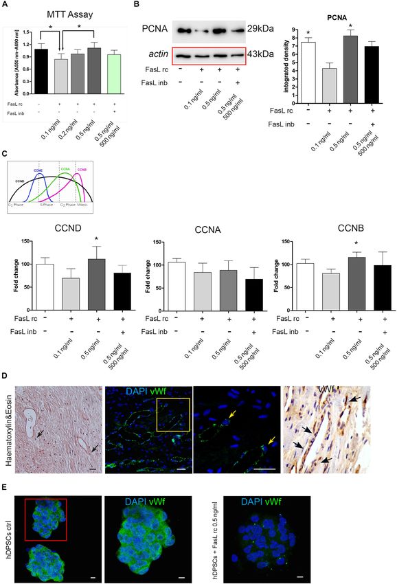
Pisciotta et al.                                                                             Fas/FasL Promotes hDPSCs Chondrogenic Differentiation

sub-confluence. Then, culture medium was supplemented with                streptomycin in order to obtain 3D floating spheres, as previously
different concentrations of FasL rc (0.1, 0.2, 0.5 ng/ml) and with        described by Pisciotta et al. (2018). When hDPSCs spheres
0.5 ng/ml FasL rc + 500 ng/ml FasL inb. Cells were kept under             aggregated, 0.5 ng/ml FasL rc and 0.5 ng/ml FasL rc + 500 ng/ml
stimulation for 24 h. hDPSCs cultured without stimulation were            FasL inb, respectively, were added to culture medium for 24 h.
used as controls.                                                         Then, culture medium was removed and cells were recovered
   Subsequently, the viability of hDPSCs after FasL rc                    and attached to slides by using Cytospin (CytospinTM 4, Thermo
stimulation was determined by using the standard MTT [3-                  Fisher Scientific, Third Avenue Waltham, MA, United States).
(4,5-dimethylthiazol-2-yl)-2,5-diphenyltetrazolium        bromide]        Briefly, hDPSCs spheres were collected and resuspended in
assay. Cells were incubated for 3 h with MTT reagent at                   300 µl of culture medium, then centrifuged at 1,500 rpm
37◦ C. After incubation, the purple formazan crystals were                for 10 min in CytospinTM 4 to let them adhere to slides.
dissolved in DMSO, at room temperature, then absorbance was               Subsequently, cells were fixed with 4% PFA in PBS for 20 min
measured at OD = 590 nm by using a multiwell plate reader                 at room temperature, then were permeabilized with Triton 0.1%
(Thermo Scientific Appliskan, Thermo Fisher Scientific). Besides          in PBS for 7 min and immunofluorescence analysis of von
this, cell proliferation in hDPSCs exposed to different FasL              Willebrand factor (vWf) was carried out by using a rabbit anti-
rc concentrations was evaluated by Western Blot analysis of               vWf primary antibody (1:100, Merck Millipore, Burlington, MA,
PCNA and densitometry analysis was carried out as described               United States), as described earlier.
above. To evaluate the expression levels of CCND, CCNA                        Human DPSCs cultured as floating spheres without stimuli
and CCNB, hDPSCs from each experimental group were                        were used as controls.
investigated by real-Time PCR. In particular, cells were                      At the same time, samples of human dental pulp (n = 3)
homogenized, and total RNA was extracted and purified                     were obtained from healthy donors, after obtaining written
using the PureLink RNA columns (Thermo Fisher Scientific).                informed consent, during routine tooth extraction procedures.
cDNA synthesis was performed by using Maxima First Strand                 After extraction, pulps were washed in PBS and fixed in PFA 4%
cDNA Synthesis Kit with DNase I treatment (Thermo Fisher                  in PBS for 1 h at room temperature. Then, PFA was removed, with
Scientific). Quantitative real-time PCRs were performed                   the samples being washed in PBS and then processed for paraffin
using SYBR Green Master mix (Bio-Rad) on CFX Connect                      embedding as described by Carnevale et al. (2018). Paraffin-
Real-time PCR instrument (Bio-Rad), with the following                    embedded dental pulps were sectioned by a Microm (Microm
oligonucleotides: hRPLP0 (F: TACACCTTCCCACTTGCTGA,                        HM 315) microtome and 6 sections per sample (5 µm thickness)
R: CCATATCCTCGTCCGACTCC) hCCND (F: CATCTACACC                             per experimental condition were cut. Half of the sections
GACAACTCCATC, R: TCTGGCATTTTGGAGAGGAAG), hC                               underwent routine hematoxylin and eosin (H&E) staining,
CNA (F: AATGGAACACTTGCTTCTGAAAG, R: CTTCAAGT                              whereas the other sections underwent immunofluorescence and
AGACTCAGCTCTGC), hCCNB (F: CCTCCCTTTTCAGTCC                               immunohistochemistry analyses against vWf in order to show the
GC, R: CTCCTGTGTCAATATTCTCCAAATC). Relative                               localization of vWf + cells within human dental pulp.
quantification was calculated from the ratio between the
cycle number (Ct) at which the signal crossed a threshold set
within the logarithmic phase of the given gene and that of the            Evaluation of FasL rc Stimulation on the
reference hRPLP0. Mean values of the duplicate results of three           Activation of Fas/FasL Pathway
independent experiments for each sample were used as individual           In order to investigate the effects triggered by human FasL rc
data for 2−11Ct statistical analysis (Zordani et al., 2019).              on hDPSCs at different concentrations, hDPSCs were processed
                                                                          for Western Blot analysis, as detailed above. The expression of
                                                                          FasL, Fas, Caspase 8 (Cell Signaling Technology, Trask Lane
Expression of von Willebrand Factor                                       Danvers, MA, United States), c-FLIP (R&D systems, McKinley
in hDPSCs and in Human Dental Pulp                                        Place NE, Minneapolis, MN, United States), FADD (mouse anti-
As well established in literature, hDPSCs reside within dental            FADD ab; Santa Cruz Biotechnology, Dallas, TX, United States)
pulp in close proximity of vessels, thus allowing to define them as       was evaluated in hDPSCs after exposure to 0.1 ng/ml, 0.5 ng/ml
pericytes (Janebodin et al., 2013). It is well known that pericytes       FasL rc and to 0.5 ng/ml FasL rc + 500 ng/ml FasL inb for 24 h.
play a key role in maintaining and regulating endothelial cell            To this regard, hDPSCs exposed to 1 µM Staurosporine were
structure. Moreover, pericytes in a synergistic fashion with              used as positive control of apoptosis. Densitometry analysis was
endothelial cells may regulate vessel formation and maturation,           performed as described earlier.
during angiogenesis process (Bergers and Song, 2005). In light               Furthermore, the expression of c-FLIP was also evaluated
of this evidence and previous findings demonstrating the ability          in hDPSCs after exposure to FasL rc 0.5 ng/ml and to
of hDPSCs to support tissue vascularization in vivo (Pisciotta            FasL rc 0.5 ng/ml + FasL inb 500 ng/ml, respectively, by
et al., 2012; Pisciotta et al., 2015b), the expression of a typical       immunofluorescence analysis with rabbit anti-c-FLIP ab (Santa
angiogenic marker such as von Willebrand factor was evaluated             Cruz Biotechnology, Dallas, TX, United States). Immunolabeling
in hDPSCs when stimulated with FasL rc. To this purpose,                  intensity of c-FLIP expression in both experimental groups was
hDPSCs were seeded at 10,000 cells/cm2 in ultra-low attachment            evaluated by pseudocolor analysis: blue to white arrays the colors
6-well plates, and cultured in DMEM-F12 containing 2% FBS,                in a spectrum with blue assigned to a lower value than white
2 mM glutamine, 100 units/ml penicillin, and 100 µg/ml                    (Carnevale et al., 2017).

Frontiers in Cell and Developmental Biology | www.frontiersin.org     4                                            May 2020 | Volume 8 | Article 279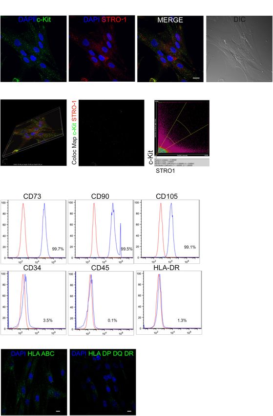
Pisciotta et al. Fas/FasL Promotes hDPSCs Chondrogenic Differentiation
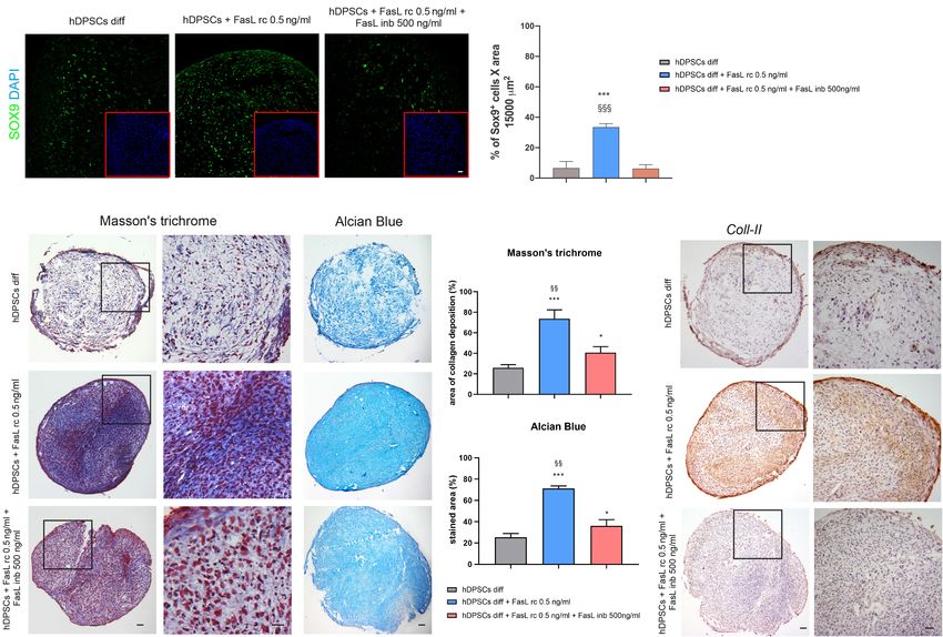
Chondrogenic Differentiation of hDPSCs (n = 6 slides per experimental group). Then, after 21 days
As mentioned before, hDPSCs hold a wide differentiation of chondrogenic induction, Masson’s trichrome and Alcian
potential, although findings in literature report that the ability of Blue stain were carried out in order to evaluate collagen
these stem cells to reach chondrogenic commitment is debated deposition and the production of sulphured acid mucins
(Iohara et al., 2006; Zhang et al., 2006). To this purpose, and GAG, respectively. Values calculated on six slides per
chondrogenic differentiation of STRO-1+ /c-Kit+ hDPSCs was experimental group were expressed as mean % area ± standard
performed under 3D pellet culture, attempting to mimic the deviation (SD). All these measurements were conducted by
physiological conditions required for articular chondrocyte using Fiji ImageJ software. Finally, immunohistochemistry with
differentiation (Chen et al., 2015). Briefly, hDPSCs were cultured DAB against Coll-II (mouse anti-Coll-II; Abcam, Cambridge,
for 7 and 21 days, respectively, in polypropylene tubes at United Kingdom) was performed at 21 days of differentiation to
a density of 5 × 10⁵ cells/tube in chondrogenic medium further analyze the achievement of chondrogenic commitment.
consisting in DMEM-HG supplemented with 5% FBS, 100 nM Immunohistochemistry was carried out as previously described
dexamethasone and 10 ng/ml TGFβ-3, 10 mM 2P-ascorbic acid (Zordani et al., 2019). All the antibodies used in the study are
(Sigma Aldrich, St. Louis, MO, United States), 1% v/v sodium indicated in Table 1.
pyruvate (Life Technologies, Carlsbad, CA, United States),
50 mg/ml ITS premix (BD Biosciences, Franklin Lakes, NJ, Statistical Analysis
United States). To evaluate chondrogenic differentiation, cell All the experiments were performed in triplicate. Data were
pellets from each experimental group were embedded in expressed as mean ± SD. Differences between two experimental
paraffin and 5 µm thick sections were obtained by microtome. conditions were analyzed by paired, Student’s t-test. Differences
Histological analysis by using Alcian Blue and Masson’s among three or more experimental samples were analyzed by
trichrome staining were performed (Pisciotta et al., 2015b; ANOVA followed by Newman–Keuls post hoc test (GraphPad
Conserva et al., 2019). Prism Software version 5 Inc., San Diego, CA, United States). In
Moreover, chondrogenic commitment was also confirmed any case, significance was set at P < 0.05.
by Western Blot analysis of SOX9 (rabbit anti-SOX9 ab; Cell
Signaling Technology, Trask Lane Danvers, MA, United States)
on hDPSCs after 7 and 21 days of induction. Besides, the RESULTS
expression of Fas, FasL and PCNA was investigated as well
to evaluate how Fas/FasL pathway is modulated during the Isolation of STRO-1+ /c-Kit+ Human
induction of chondrogenic differentiation. Densitometry analysis Dental Pulp Stem Cells and
of SOX9, FasL, Fas and PCNA were carried out as detailed above. Immunophenotype Characterization
The expression of FasL was further evaluated by
After isolation and expansion of hDPSCs in vitro, magnetic
immunofluorescence analysis with a rabbit anti-FasL ab
immunoselection allowed to obtain a STRO-1+ /c-Kit+ hDPSCs
(Santa Cruz Biotechnology, Dallas, TX, United States) after 7
population. The expression of these two surface markers
and 21 days of chondrogenic differentiation. Undifferentiated
in the selected stem cell population was confirmed by
hDPSCs were used as controls.
confocal immunofluorescence analysis (Figures 1A,B). Almost
all immune-sorted cells were positively labeled against c-Kit and
Chondrogenic Differentiation of hDPSCs STRO-1 surface antigens, which were distinctly expressed and
With the Addition of FasL rc detected, as shown by the lack of overlapping signals revealed
In order to further evaluate the role of FasL stimulation on by the colocalization map (Figure 1C). Moreover, immune-
hDPSCs capability to commit toward chondrogenic lineage, FasL phenotypical characterization through FACS analysis revealed
rc was added to cells differentiated under pellet culture method. that all the typical MSCs markers were expressed by STRO-
In particular, hDPSCs were seeded in polypropylene tubes at a 1+ /c-Kit+ hDPSCs, while being CD45/HLA-DR negative and,
density of 5 × 10⁵ cells/tube in chondrogenic medium additioned only to a lesser extent, CD34 positive (Figure 1D) which is in
with 0.5 ng/ml Fas rc and 0.5 ng/ml FasL rc + 500 ng/ml FasL accordance with previous findings (Pisciotta et al., 2015a). At
inb, respectively, for 24 h. Then, the stimuli were removed and the same time, confocal immunofluorescence analysis showed
chondrogenic differentiation was carried out for 7 and 21 days. the positive labeling against HLA-ABC antigens and the lack of
During the 21 days induction time, FasL stimulation was added expression of HLA DP-DQ-DR antigens in the selected hDPSCs
to chondrogenic medium once a week. population (Figure 1E).
The chondrogenic commitment was evaluated on hDPSCs
3D pellets sections (5 µm thickness), formerly processed for hDPSCs-PBMCs Co-culture: Evaluation
paraffin embedding and microtome cut. Immunofluorescence of Fas/FasL Pathway
analysis of SOX9 (Cell Signaling Technology, Trask Lane After co-culture of hDPSCs and PBMCs for 48 h (Figure 2A),
Danvers, MA, United States) was performed after 7 days the expression of FasL, Caspase 8 and c-FLIP was evaluated
of induction and quantification of SOX9+ cells was carried by Western Blot analysis in hDPSCs cultured alone and in
out by counting the number of positively labeled cells in hDPSCs after co-culture (Figure 2B). Densitometry analysis
6 randomly selected fields of 15,000 µm2 per pellet section showed a significant increase in FasL expression in hDPSCs after
Frontiers in Cell and Developmental Biology | www.frontiersin.org 5 May 2020 | Volume 8 | Article 279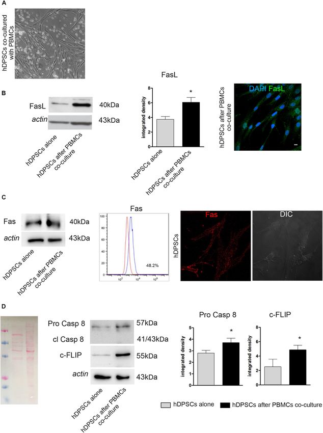
Pisciotta et al.                                                                                Fas/FasL Promotes hDPSCs Chondrogenic Differentiation

TABLE 1 | List of used antibodies.

Name                                                    Host                 Source                   Dilution                    Application

anti-human-CD73-PE-CY7                                 mouse            BD Biosciences                  1:50                          FACS
anti-human-CD90-FITC                                   mouse            BD Biosciences                  1:50                          FACS
anti-human-CD105-APC                                   mouse            BD Biosciences                  1:50                          FACS
anti-human-CD45-PE                                     mouse            BD Biosciences                  1:50                          FACS
anti-human-HLA-DR-PE-CY7                               mouse            BD Biosciences                  1:50                          FACS
anti-human-CD34-ECD                                    mouse           Beckman Coulter                  1:50                          FACS
anti-human-HLA ABC-FITC                                mouse            BD Biosciences                  1:50                            IF
anti-HLA-DP-DQ-DR-FITC                                 mouse            BD Biosciences                  1:50                            IF
anti−FasL                                              rabbit       Cell Signaling Technology          1:1000                          WB
anti−FasL                                              rabbit       Santa Cruz Biotechnology            1:50                            IF
anti-Fas                                               mouse        Cell Signaling Technology       1:50 1:1000                     FACS WB
anti-Caspase 8                                         mouse        Cell Signaling Technology          1:1000                          WB
anti-c-FLIP                                            rabbit            R&D systems                   1:1000                          WB
anti-c-FLIP                                            rabbit       Santa Cruz Biotechnology            1:50                            IF
anti-vWf                                               rabbit            Merck Millipore               1:100                            IF
anti-FADD                                              mouse        Santa Cruz Biotechnology           1:1000                          WB
anti-SOX9                                              rabbit       Cell Signaling Technology          1:1000                          WB
anti-Coll-II                                           mouse                 Abcam                      1:50                           IHC

co-culture with PBMCs, when compared to hDPSCs cultured                      almost reverted to control conditions (Figure 3A). Western Blot
alone (∗ P < 0.05 vs hDPSCs alone; Figure 2B). This data                     analysis of PCNA was also performed on hDPSCs after 24 h
was confirmed by immunofluorescence analysis (Figure 2B).                    of stimulation (Figure 3B). A statistically significant increase
Contemporarily, Fas expression was detected in hDPSCs after co-              in PCNA expression was detected in hDPSCs stimulated with
culture with PBMCs, as demonstrated by Western Blot analysis.                FasL rc 0.5 ng/ml when compared to hDPSCs stimulated with
Interestingly, no statistically significant difference was shown             FasL rc + FasL inb (∗ P < 0.05 vs hDPSCs with FasL rc + FasL
when compared to hDPSCs cultured alone, indeed FACS and                      inb; Figure 3B). The untreated control showed statistically
immunofluorescence analyses showed that hDPSCs express Fas                   significant higher levels of PCNA, with respect to hDPSCs
yet in standard culture conditions (almost 48% of the cells;                 stimulated with FasL rc 0.1 ng/ml (∗ P < 0.05 vs hDPSCs + FasL
Figure 2C). Western Blot analysis also allowed to evaluate                   rc 0.1 ng/ml; Figure 3B). These data confirmed the results
the expression of Pro-Caspase 8, which revealed an increased                 obtained through MTT assay. Likewise, real-Time PCR analysis
expression in hDPSCs after co-culture (∗ P < 0.05 vs hDPSCs                  of mRNA expression of CCND, CCNA and CCNB revealed a
alone; Figure 2D). On the other hand, the antibody did not show              statistically significant mRNA fold increase in CCND and CCNB
any labeling against cleaved Caspase 8, therefore demonstrating              in hDPSCs treated with FasL rc 0.5 ng/ml, thus confirming
that activation of the apoptotic cascade did not occur in any                that the stimulation with FasL rc at its highest concentration
of hDPSCs experimental groups. Western Blot analysis also                    triggered a greater proliferation in hDPSCs (∗ P < 0.05 vs hDPSCs
showed an increase in c-FLIP expression in hDPSCs after co-                  ctrl; Figure 3C).
culture, with respect to hDPSCs alone (∗ P < 0.05 vs hDPSCs
alone; Figure 2D).                                                           Expression of von Willebrand Factor
                                                                             in hDPSCs and in Human Dental Pulp
Stimulation of hDPSCs With Fas-L rc and                                      As highlighted in Figure 3D, pericytes can be recognized
Fas-L inb: Cell Viability and Proliferation                                  within human dental pulp close to vessels, by their cell
Following the stimulation with FasL rc and FasL inb, MTT                     morphology, as shown by H&E stain (black arrows; Figure 3D).
analysis showed an increase in cell viability at increasing                  Immunofluorescence and immunohistochemistry analyses
concentrations of FasL rc, indeed, when the highest concentration            confirmed the localization in vivo of vWf+ dental pulp
was used (0.5 ng/ml) cell viability was higher than in the                   cells adjacent to blood vessels (yellow square and arrows;
untreated control (Figure 3A). In particular, the untreated                  black arrows; Figure 3D). Immunofluorescence analysis
control and hDPSCs treated with the highest concentration                    performed on hDPSCs cultured as 3D floating spheres
of FasL rc showed a statistically significant higher viability,              confirmed that cells express the angiogenic marker von
compared to hDPSCs treated with the lowest concentration                     Willebrand factor, when cultured under standard expansion
of FasL rc (0.1 ng/ml) (∗ P < 0.05 vs hDPSCs + FasL rc                       conditions. On the contrary, hDPSCs lost the expression of
0.1 ng/ml; Figure 3A). When hDPSCs treated with FasL rc                      vWf after stimulation with FasL rc at its highest concentration
(0.5 ng/ml) were additioned with FasL inb, MTT values were                   (0.5 ng/ml; Figure 3E).

Frontiers in Cell and Developmental Biology | www.frontiersin.org        6                                            May 2020 | Volume 8 | Article 279
Pisciotta et al. Fas/FasL Promotes hDPSCs Chondrogenic Differentiation FIGURE 1 | Mesenchymal profile of immune-selected hDPSCs. (A) Immunofluorescence analysis on hDPSCs after immune-selection with MACS shows the expression of stemness markers c-Kit (green) and STRO-1 (red). DIC image shows the morphological phenotype of hDPSCs. Nuclei are counterstained with DAPI. Bar = 10 µm. (B) Three dimensional reconstruction of optical sections acquired for each fluorescent signal. (C) Co-localization binary map of STRO-1 and c-Kit signals is shown. On the right, representative dot plots of co-localization signals are reported. (D) Fluorescence-activated cell sorting (FACS) analysis performed on immune-selected hDPSCs. (E) Expression of major histocompatibility complex class I (HLA ABC) and class II (HLA DP DQ DR). Bar = 10 µm. Frontiers in Cell and Developmental Biology | www.frontiersin.org 7 May 2020 | Volume 8 | Article 279
Pisciotta et al.                                                                                          Fas/FasL Promotes hDPSCs Chondrogenic Differentiation

  FIGURE 2 | Evaluation of Fas/FasL pathway on both hDPSCs cultured alone and after co-culture with PBMCs. (A) Phase contrast image of hDPSCs cultured with
  PBMCs. (B) Western Blot analysis of FasL in both hDPSCs cultured alone and after co-culture with PBMCs. Histograms represent the mean ± SD (n = 3) of
  densitometry of FasL, *P < 0.05 hDPSCs after PBMCs co-culture vs hDPSCs cultured alone. The expression of FasL after co-culture with PBMCs was confirmed by
  confocal immunofluorescence analysis. Nuclei are counterstained with DAPI. Bar = 10 µm. (C) Western Blot analysis of Fas (CD95) in both hDPSCs cultured alone
  and after co-culture with PBMCs. FACS analysis confirms the positive expression of Fas in hDPSCs cultured alone. The expression of Fas on live hDPSCs cultured
  alone was also evaluated by Immunofluorescence analysis. DIC Image shows the morphological phenotype of hDPSCs. (D) Western blot analysis of pro-caspase 8,
  cleaved caspase 8 and c-FLIP performed on hDPSCs cultured alone and after co-culture with PBMCs. Actin bands were presented as control of protein loading;
  histograms represent mean ± SD, *P < 0.05 hDPSCs after PBMCs co-culture vs hDPSCs cultured alone.

Frontiers in Cell and Developmental Biology | www.frontiersin.org               8                                                May 2020 | Volume 8 | Article 279
Pisciotta et al.                                                                                             Fas/FasL Promotes hDPSCs Chondrogenic Differentiation

  FIGURE 3 | Stimulation of hDPSCs with human recombinant FasL. (A) MTT assay was performed on hDPSCs after stimulation with different concentrations of FasL
  rc. (B) Western blot analysis of PCNA in hDPSCs stimulated with FasL rc. Histograms represent the mean ± SD (n = 3) of densitometry of PCNA; *P < 0.05
  hDPSCs ctrl and hDPSCs treated with FasL rc 0.5 ng/ml vs hDPSCs treated with FasL rc 0.1 ng/ml. (C) Real–time PCR analysis showing fold increase of mRNA
  levels of CCND, CCNA and CCNB in hDPSCs following stimulation with FasL rc. Data represent mean ± SD of fold change obtained from five independent
  experiments; *P < 0.05 hDPSCs treated with FasL rc 0.5 ng/ml vs hDPSCs treated with FasL rc 0.1 ng/ml. FasL inhibitor was used for FasL rc blocking.
  (D) Representative image of histological section of human dental pulp stained with H&E, magnification 10×. Black arrows indicate the pericytes close to vessels.
  Immunofluorescence analysis of vWf was performed on paraffin-embedded section of dental pulp. Yellow square indicates the area of high magnification (right side).
  Yellow arrows highlight vWf positive pericytes. On the right, immunohistochemistry analysis of vWf with black arrows indicating the presence of vWf+ cells close to
  vessels, magnification 20×. Bar = 50 µm. (E) Immunofluorescence analysis of vWf was carried out on hDPSCs 3D spheres after treatment with FasL rc 0.5 ng/ml.
  hDPSCs 3D spheres untreated were used as control. Bar = 10 µm.

Frontiers in Cell and Developmental Biology | www.frontiersin.org                  9                                                 May 2020 | Volume 8 | Article 279
Pisciotta et al.                                                                                Fas/FasL Promotes hDPSCs Chondrogenic Differentiation

Stimulation of hDPSCs With FasL rc and                                      statistically significant decreasing after 21 days of induction,
FasL inb: Evaluation of Fas/FasL                                            when compared to undifferentiated control (§§ P < 0.01 hDPSCs
                                                                            diff 7-day vs hDPSCs undiff; ∗∗ P < 0.01 hDPSCs diff 21-
Pathway                                                                     day vs hDPSCs undiff). Besides, the expression of SOX9 after
Following stimulation with FasL rc and FasL inb, Western Blot               21 days of induction was statistically significant lower, when
analyses were carried out to investigate the expression of different        compared to the undifferentiated control and to hDPSCs after
proteins related to the Fas/FasL pathway, such as FasL, Fas,                7 days of induction, respectively (∗∗ P < 0.01 vs hDPSCs undiff;
FADD, c-FLIP, Pro-Caspase 8 and cleaved Caspase 8 (Figure 4A).              ∗∗∗ P < 0.001 vs hDPSCs diff 7-day). Western blot analysis also
As shown by densitometry analyses, there was a statistically                evaluated the expression of other proteins, such as FasL, Fas
significant increase in FasL expression in stimulated hDPSCs                and PCNA. The expression of FasL was statistically significant
compared to untreated control (∗ P < 0.05 vs hDPSCs ctrl).                  increased after 7 and 21 days of induction (∗∗ P < 0.01 hDPSCs
    As far as Fas receptor is concerned, a significant                      diff 7-day vs hDPSCs undiff, ∗ P < 0.05 hDPSCs diff 21-day
downregulation was observed in hDPSCs stimulated with                       vs hDPSCs undiff), while PCNA expression was statistically
0.5 ng/ml FasL rc, compared to the untreated control exhibiting             significant lower at 7 and 21 days of induction, compared
basal levels of Fas (∗ P < 0.05 vs hDPSCs ctrl). Similarly, a               to undifferentiated control (∗∗ P < 0.01 vs hDPSCs diff 7-day
statistically significant decrease in Fas expression was observed           and hDPSCs diff 21-day). Fas expression showed a statistically
in hDPSCs stimulated with 0.1 ng/ml FasL rc (∗ P < 0.05 vs                  significant increase in hDPSCs at 7 days of induction, when
hDPSCs ctrl). On the other hand, a slight – although statistically          compared to undifferentiated hDPSCs and to hDPSCs at 21 days
not significant – decrease in Fas expression was revealed in                of induction, respectively (∗ P < 0.05 vs hDPSCs undiff, ∗ P < 0.05
hDPSCs treated with FasL rc + FasL inb, showing values similar              vs hDPSCs diff 21-day; Figure 5C). FasL expression was also
to control cells. The expression of FADD was not affected by                confirmed by confocal immunofluorescence analysis performed
stimulation, indeed a slight decrease was noticed only when                 on hDPSCs 3D pellet sections at different times of chondrogenic
hDPSCs were treated with FasL rc + FasL inb (∗ P < 0.05 vs                  commitment. The data are in agreement with Western Blot
hDPSCs ctrl). Conversely, a statistically significant augmentation          analysis, in fact, the expression of FasL showed an increase after 7
of c-FLIP expression was demonstrated in hDPSCs following                   and 21 days of induction to differentiation (Figure 5D).
stimulation with 0.5 ng/ml FasL rc and the treatment with FasL
rc + FasL inb, respectively (∗∗ P < 0.01, ∗ P < 0.05 vs hDPSCs
ctrl). As shown by Western Blot analysis, there was no activation           Role of FasL Stimulation in
of the apoptotic pathway, as cleaved Caspase 8 was not expressed            Chondrogenic Induction of hDPSCs
(Figure 4A). Subsequently, confocal immunofluorescence                      The induction to chondrogenic differentiation of hDPSCs with
analysis also confirmed the increase in c-FLIP expression in                the addition of FasL rc and FasL rc + FasL inb was evaluated
hDPSCs treated with 0.5 ng/ml FasL rc, when compared to                     by confocal immunofluorescence, Masson’s trichrome, Alcian
unstimulated control (Figure 4B). The pseudocolor analysis                  Blue stain and immunohistochemistry analyses. As reported
highlighted a higher intensity in c-FLIP immunolabeling in                  in Figure 6A, SOX9 showed a differential expression among
hDPSCs stimulated with FasL rc compared to hDPSCs treated                   the three experimental groups after 7 days of chondrogenic
with FasL rc + FasL inb (Figure 4B).                                        induction. In particular, as shown in Figure 6B, hDPSCs
                                                                            3D pellets stimulated with FasL rc 0.5 ng/ml after 7 days
Evaluation of Chondrogenic                                                  of differentiation, revealed a statistically significant higher
Differentiation of hDPSCs in vitro                                          number of SOX9+ cells when compared to hDPSCs 3D pellet
After in vitro expansion, the ability of STRO-1+ /c-Kit+ hDPSCs             differentiated without FasL rc stimulation and to hDPSCs 3D
to differentiate toward the chondrogenic lineage was evaluated.             pellet differentiated plus FasL rc 0.5 ng/ml and FasL inb 500
After 7 and 21 days of induction to chondrogenic differentiation,           ng/ml, respectively (∗∗∗ P < 0.001 vs hDPSCs diff; §§§ P < 0.001
the commitment of hDPSCs, cultured as 3D cell pellets, was                  vs hDPSCs + FasL rc 0.5 ng/ml + FasL inb 500 ng/ml). These
investigated through histochemical staining with Alcian Blue                data suggest that the stimulation of hDPSCs 3D pellets with FasL
and Masson’s trichrome stain. In particular, Alcian blue staining           rc + FasL inb triggered a reduction in SOX9 expressing cells
demonstrated the presence of sulphured acid mucins and                      to levels resembling hDPSCs diff. As far as later differentiation
glycosaminoglycans (GAG) containing carboxylic groups, at 7                 time is concerned, Masson’s trichrome and Alcian Blue stain
and 21 days of induction (Figure 5A). Masson’s trichrome                    highlighted that collagen deposition and sulphured acid mucins
staining highlighted the presence of collagen (blue) after 7 and            and GAG production were greater in hDPSCs differentiated
21 days of chondrogenic differentiation of hDPSCs. In particular,           plus the addition of FasL rc stimulation (Figure 6C). As
an increased amount of collagen deposition was revealed after               shown in Figure 6D, Masson’s trichrome and Alcian Blue stain
21 days of induction, with respect to the earlier time of induction         quantifications revealed that hDPSCs differentiated plus FasL
(Figure 5B). After 7 and 21 days of culture in chondrogenic                 rc stimulation reached the chondrogenic differentiation to a
medium, the commitment was also evaluated by Western Blot                   greater extent, when compared to hDPSCs differentiated without
analysis (Figure 5C). In particular, the expression of SOX9,                FasL rc stimulation (∗∗∗ P < 0.001 vs hDPSCs diff). When
a typical marker of early chondrogenic differentiation, was                 both FasL rc and FasL inb were added, hDPSCs still showed
statistically significant increased after 7 days of induction, while        a greater induction with respect to hDPSCs diff (∗ P < 0.05),

Frontiers in Cell and Developmental Biology | www.frontiersin.org      10                                             May 2020 | Volume 8 | Article 279
Pisciotta et al. Fas/FasL Promotes hDPSCs Chondrogenic Differentiation FIGURE 4 | Evaluation of Fas/FasL pathway in hDPSCs following stimulation with FasL rc. (A) Western Blot analysis of FasL, Fas, FADD, c-FLIP, pro-caspase 8 and cleaved caspase 8 in hDPSCs after stimulation with FasL rc at different concentrations. hDPSCs treated with 1 µM Staurosporine were used as positive control of cleaved caspase 8. At the bottom, histograms represent mean ± SD (n = 3) of densitometry of FasL, Fas, FADD and c-FLIP; *P < 0.05 and **P < 0.01 vs hDPSCs ctrl. (B) Confocal immunofluorescence analysis of c-FLIP was performed in hDPSCs stimulated with FasL rc 0.5 ng/ml and with FasL rc 0.5 ng/ml + FasL inb 500 ng/ml. On the right of each immunofluorescent image is reported pseudocolor analysis of c-FLIP staining intensity. Bar = 10 µm. although resulting in a statistically significant lower induction, Immunohistochemistry analysis of Coll-II further showed that when compared with hDPSCs differentiated plus FasL rc matrix deposition was notably higher in hDPSCs differentiated stimulation (§§ P < 0.01 vs hDPSCs diff + FasL rc 0.5 ng/ml). plus FasL rc stimulation, when compared to hDPSCs diff and Frontiers in Cell and Developmental Biology | www.frontiersin.org 11 May 2020 | Volume 8 | Article 279
Pisciotta et al.                                                                                           Fas/FasL Promotes hDPSCs Chondrogenic Differentiation

  FIGURE 5 | Chondrogenic differentiation of hDPSCs. (A) Evaluation of chondrogenic commitment of hDPSCs at 7 and 21 days of induction. Cell pellet sections
  were stained by Alcian Blue, magnification 10×. Bar = 50 µm. (B) Masson’s trichrome staining was carried out to analyze the collagen deposition (blue) in hDPSCs
  at different time points. Black squares highlight areas of higher magnification on the right side of each image. Bar = 50 µm. (C) Western Blot analysis showing
  expression of SOX9, FasL, Fas and PCNA through the chondrogenic induction of hDPSCs. Histograms represent mean ± SD (n = 3) of densitometry of SOX9, FasL,
  PCNA and Fas. With regard to SOX9: **P < 0.01 hDPSCs undiff vs hDPSCs diff 21-day, ***P < 0.001 hDPSCs 7-day vs hDPSCs diff 21-day; §§ P < 0.01 hDPSCs
  diff 7-day vs hDPSCs undiff; with regard to FasL: *P < 0.05 hDPSCs diff 21-day vs hDPSCs undiff; **P < 0.01 hDPSCs diff 7-day vs hDPSCs undiff; with regard to
  PCNA: **P < 0.01 hDPSCs undiff vs hDPSCs diff 7-day and **P < 0.01 hDPSCs undiff vs hDPSCs diff 21-day; with regard to Fas: *P < 0.05 hDPSCs diff 7-day vs
  hDPSCs undiff and *P < 0.05 hDPSCs diff 7-day vs hDPSCs diff 21-day. (D) Immunofluorescence images showing FasL expression at different times of
  commitment. Bar = 20 µm.

Frontiers in Cell and Developmental Biology | www.frontiersin.org               12                                                 May 2020 | Volume 8 | Article 279
Pisciotta et al.                                                                                             Fas/FasL Promotes hDPSCs Chondrogenic Differentiation

  FIGURE 6 | Effects of FasL stimulation on chondrogenic differentiation of hDPSCs. (A) Expression of early chondrogenic marker SOX9 was assayed in hDPSCs by
  confocal immunofluorescence analysis at 7 days of chondrogenic induction supplemented with FasL rc 0.5 ng/ml and with FasL rc 0.5 ng/ml + FasL inb 500 ng/ml,
  respectively. hDPSCs differentiated without adding FasL stimulation were used as controls (hDPSCs diff). Red squares show nuclei stained with DAPI. Bar = 20 µm.
  (B) Histograms representing the mean percentage of SOX9+ cells/15,000 µm2 area ± SD. ***P < 0.001 vs hDPSCs diff; §§§ P < 0.001 vs hDPSCs diff + FasL rc
  0.5 ng/ml + FasL inb 500 ng/ml. (C) Representative images of Masson’s trichrome and Alcian Blue stained sections from hDPSCs 3D pellet show collagen
  deposition and the presence of acid mucins and GAG among the three different experimental groups. Black squares indicate areas of higher magnification, reported
  on the right side. Bar = 50 µm. (D) Histograms showing percentage areas of collagen deposition and Alcian Blue stain of 3D pellet sections from each experimental
  group (n = 6). Data are expressed as mean ± SD. *P < 0.05, ***P < 0.001 vs hDPSCs diff; §§ P < 0.01 vs hDPSCs + FasL rc 0.5 ng/ml + FasL inb 500 ng/ml.
  (E) Immunohistochemistry images showing the expression of late chondrogenic marker Coll-II in differentiated hDPSCs exposed to FasL rc 0.5 ng/ml and FasL rc
  0.5 ng/ml + FasL inb 500 ng/ml. hDPSCs differentiated without adding FasL stimulation (hDPSCs diff), were used as controls. Black squares indicate areas of higher
  magnification, reported on the right side, magnification 10×. Bar = 50 µm.

hDPSCs diff + FasL rc + FasL inb, confirming the achievement                          when applied to different animal models of tissue injuries. In
of chondrogenic commitment (Figure 6E).                                               particular, when hDPSCs were studied for the regeneration
                                                                                      of bone, muscle and peripheral nerve tissues, not only
                                                                                      they committed toward the host tissue cells but also they
DISCUSSION                                                                            promoted the tissue healing by triggering angiogenesis
                                                                                      (D’Aquino et al., 2007; Pisciotta et al., 2015b; Zordani
Human dental pulp stem cells (hDPSCs) represent a                                     et al., 2019). To this regard, it is noteworthy that hDPSCs
heterogeneous cell population enclosed in the loose connective                        can be localized within dental pulp in the perivascular
tissue. The immune-selection against the stemness markers                             area, thus being defined as pericytes (Janebodin et al.,
c-Kit and STRO-1 allows to obtain a purer stem cell niche                             2013). Although it has been demonstrated that hDPSCs are
with a typical mesenchymal phenotype. It has been widely                              able to commit toward the mesenchyme related lineages,
demonstrated that c-Kit+ /STRO-1+ hDPSCs are able to                                  the chondrogenic differentiation ability of hDPSCs is
differentiate in vitro toward all the three germ layer derived                        still controversial.
lineages (Laino et al., 2005; Carnevale et al., 2013) due to                              It is well known that from an embryological point of
their peculiar embryological origin from the neural crest.                            view, neural crest derived cells contribute to the formation
The multipotency of hDPSCs has been further confirmed                                 of the craniofacial district tissues. During the embryological
by several findings reporting their regeneration potential                            development the poorly represented chondrogenic tissue is

Frontiers in Cell and Developmental Biology | www.frontiersin.org                13                                                 May 2020 | Volume 8 | Article 279
Pisciotta et al. Fas/FasL Promotes hDPSCs Chondrogenic Differentiation
immediately replaced by newly synthesized bone tissue supported able to decrease the expression of angiogenic marker and favor
by neo-angiogenesis. The only portion of cartilage tissue is more suitable microenvironment for chondrogenic induction.
represented by Meckel’s cartilage. This hyaline cartilage, formed In conclusion, we can affirm that Fas/FasL pathway (1) not
in the mandibular process of the first branchial arch of vertebrate only confers hDPSCs the ability to avoid apoptosis induction
embryos, is not involved in bone formation although supporting when exposed to immune cells but also is primary for the
the intramembranous ossification (Amano et al., 2010). Based on maintenance of proliferation properties of hDPSCs, and (2)
these considerations it can be argued that neural crest derived is inductive and conductive of chondrogenic commitment by
stem cells require particular conditions to differentiate toward inhibiting the expression of angiogenic marker of neural crest
chondrogenic tissue. derived pericytes.
In order to understand chondrogenic potential of hDPSCs,
3D culture pellet mimicking the physiological conditions
of chondrocytes differentiation was performed. Data from DATA AVAILABILITY STATEMENT
the early phase of chondrogenic induction revealed high
levels of FasL suggesting that it may exert a key role in All datasets generated for this study are included in the
promoting the early induction of chondrogenic commitment, manuscript files.
besides being expressed along the whole differentiation time.
Therefore, it may be assumed that Fas/FasL pathway is not
only involved in the immunomodulation (Zhao et al., 2012; ETHICS STATEMENT
Riccio et al., 2014; Carnevale et al., 2017). To investigate
The study was conducted in accordance with the
how Fas/FasL pathway affects hDPSCs biological properties,
recommendations of Comitato Etico Provinciale Azienda
stem cells expressing high levels of Fas were stimulated with
Ospedaliero-Universitaria di Modena (Modena, Italy), which
different concentrations of human recombinant FasL protein.
provided the approval of the protocol (ref. number 3299/CE;
This stimulation aimed to mimic the effects of the exposure
5 September 2017). All subjects gave written informed consent in
to inflammatory microenvironment and revealed that, not
compliance with the Declaration of Helsinki.
only undifferentiated hDPSCs avoided apoptosis, but were still
proliferating. These findings are in accordance with a previous
report showing that Fas, a classically pro-apoptotic molecule, is
also able to promote increased cell proliferation in different types
AUTHOR CONTRIBUTIONS
of cells including hepatocytes (Budd, 2002). Further studies also AP, GB, and LB designed and performed the experiments,
demonstrated that high levels expression of c-FLIP counteract evaluated the data, and wrote the manuscript. RD and
the signals of cell death toward a growth signal pathway EP performed the experiments and wrote the manuscript.
(Ashany et al., 1999). SD performed the hDPSCs characterization experiments and
Interestingly, at early times of differentiation, an increase in data interpretation. AV and RT performed molecular analysis
SOX9+ hDPSCs was evident, thus suggesting a pro-inductive and provided guidance on the data interpretation. AP and
effect of FasL toward chondrogenic commitment. At later times CS performed the experiments, contributed to the data
of differentiation, an augmentation in Coll-II deposition was interpretation and edited the manuscript. GC managed the
revealed, demonstrating how FasL is able to play a conductive overall project, contributed to the data interpretation, and
role by leading the chondrogenic commitment of hDPSCs. These edited the manuscript.
findings confirm previous evidence reporting that FasL expressed
by mature chondrocytes exerts a cytoprotective effect (Ryu et al.,
2012). At the same time, FasL plays an inhibitory role in the FUNDING
promotion of angiogenesis by inducing Fas mediated apoptosis
of vascular endothelial cells, and consequently might avoid the GB was supported by research fellowship funded by Department
cartilage replacement with bone tissue (Sun et al., 2013). Indeed, of Surgery, Medicine Dentistry and Morphological Sciences.
as reported the FasL stimulation, added to culture medium, was University of Modena and Reggio Emilia prot n 20862/2017.
REFERENCES Bianchi, M., Pisciotta, A., Bertoni, L., Berni, M., Gambardella, A., Visani, A., et al.
(2017). Corrigendum to “Osteogenic Differentiation of hDPSCs on Biogenic
Amano, O., Doi, T., Yamada, T., Sasaki, A., Sakiyama, K., Kanegae, H., et al. Bone Apatite Thin Films”. Stem Cells Int. 2017:6587384. doi: 10.1155/2017/
(2010). Meckel’s cartilage: discovery, embryology and evolution: —overview of 6587384
the specificity of meckel’s cartilage—. J. Oral Biosci. 52, 125–135. doi: 10.1016/ Brunlid, G., Pruszak, J., Holmes, B., Isacson, O., and Sonntag, K.-C. (2007).
S1349-0079(10)80041-6 Immature and neurally differentiated mouse embryonic stem cells do not
Ashany, D., Savir, A., Bhardwaj, N., and Elkon, K. B. (1999). Dendritic cells are express a functional Fas/Fas ligand system. Stem Cells 25, 2551–2558. doi:
resistant to apoptosis through the Fas (CD95/APO-1) pathway. J. Immunol. 163, 10.1634/stemcells.2006-0745
5303–5311. Budd, R. C. (2002). Death receptors couple to both cell proliferation
Bergers, G., and Song, S. (2005). The role of pericytes in blood-vessel formation and apoptosis. J. Clin. Invest. 109, 437–442. doi: 10.1172/JCI
and maintenance. Neuro Oncol. 7, 452–464. doi: 10.1215/s1152851705000232 15077
Frontiers in Cell and Developmental Biology | www.frontiersin.org 14 May 2020 | Volume 8 | Article 279You can also read