Rosmarinic acid inhibits cell proliferation, migration, and invasion and induces apoptosis in human glioma cells
←
→
Page content transcription
If your browser does not render page correctly, please read the page content below
INTERNATIONAL JOURNAL OF MOlecular medicine 47: 67, 2021

           Rosmarinic acid inhibits cell proliferation, migration, and
             invasion and induces apoptosis in human glioma cells
       YUNSHENG LIU1*, XIANGPING XU1*, HAN TANG1, YUCHEN PAN1, BING HU2 and GUODONG HUANG1

                  1
                      Department of Neurosurgery, Shenzhen Second People's Hospital, The First Affiliated Hospital of
                               Shenzhen University Health Science Center, Shenzhen, Guangdong 518035;
                         2
                           Department of Otolaryngology Head and Neck Surgery, The First Affiliated Hospital of
                                  Sun Yat‑sen University, Guangzhou, Guangdong 510080, P.R. China

                                        Received September 17, 2020; Accepted January 25, 2021

                                                       DOI: 10.3892/ijmm.2021.4900

Abstract. There is a growing evidence that Fyn kinase is                 U251 and U343 cells. Collectively, the present study suggested
upregulated in glioblastoma multiforme (GBM), where it                   RA as a drug candidate for the treatment of GBM.
plays a key role in tumor proliferation and invasion. In the
present study, the antitumor effects of rosmarinic acid (RA),            Introduction
a Fyn inhibitor, were explored in human‑derived U251 and
U343 glioma cell lines. These cells were treated with various            Glioblastoma multiforme (GBM) is the most malignant
concentrations of RA to determine its effects on proliferation,          primary brain tumor in humans and the most lethal cancer
migration, invasion, apoptosis, and gene and protein expres‑             of the central nervous system, with an annual incidence of
sion levels. The CCK‑8 assay revealed that RA significantly              3.19 cases per 100,000 persons (1‑3). Despite significant
suppressed cell viability of U251 and U343 cells. Furthermore,           advances in tumor resection, radiation therapy, and chemo‑
RA significantly reduced proliferation rates, inhibited migra‑           therapy, the prognosis of GBM remains poor. Treatment failure
tion and invasion, and decreased the expression levels of                and continued disease progression results in a median overall
invasion‑related factors, such as matrix metalloproteinase               survival of approximately 12‑15 months, with a 5‑year survival
(MMP)‑2 and MMP‑9. TUNEL staining revealed that RA                       rate of less than 10% (4). Studies have revealed that the rapid
resulted in a dose‑dependent increase of U251 and U343                   proliferation and high invasiveness of GBM cells lead to treat‑
cell apoptosis. In line with this finding, the expression of             ment failure and tumor recurrence (5,6). Therefore, it is urgent
apoptosis suppressor protein Bcl‑2 was downregulated and                 to develop an effective treatment for GBM.
that of the pro‑apoptotic proteins Bax and cleaved caspase‑3                 Src‑family kinases (SFKs) are non‑receptor tyrosine
was increased. In addition, it was revealed that the phospha‑            kinases. The family of proteins contains nine members
tidylinositol 3‑kinase (PI3K)/Akt/nuclear factor‑κ B (NF‑κ B)            (Fyn, c‑Src, Yes, Lyn, Lck, Blk, Hck, Fgr and Yrk), five
signaling pathway was involved in RA‑induced cytotoxicity in             of which (Fyn, c‑Src, Yes, Lyn and Lck) are expressed in
                                                                         human gliomas (7‑10). SFKs are frequently overexpressed
                                                                         and/or activated in numerous human cancers (11‑13), where
                                                                         they play a role in tumor invasion, proliferation, metastasis,
                                                                         survival, and angiogenesis (14). By knocking down indi‑
                                                                         vidual SFKs in cultured cells (LN229, SF767, and GBM8),
Correspondence to: Dr Bing Hu, Department of Otolaryngology              Lewis‑Tuffin et al (7) determined that reduced c‑Src, Fyn,
Head and Neck Surgery, The First Affiliated Hospital of Sun Yat‑sen      and Yes expression also reduced growth and migration and
University, 58 Zhong Shan Er Lu, Guangzhou, Guangdong 510080,
                                                                         altered motility‑related protein phosphorylation patterns,
P.R. China
                                                                         while reduced Lyn expression had little effect on growth
E‑mail: szshent@email.szu.edu.cn
                                                                         and migration. Other in vitro studies have revealed that Fyn
Professor Guodong Huang, Department of Neurosurgery, Shenzhen            knockdown is associated with decreased glioma cell prolifera‑
Second People's Hospital, The First Affiliated Hospital of Shenzhen      tion and migration (15,16). Fyn tyrosine kinase, a downstream
University Health Science Center, 3002 Sungang West Road, Shenzhen,
                                                                         target of the oncogenic receptor tyrosine kinase pathway, is
Guangdong 518035, P.R. China
                                                                         rarely mutated, yet significantly overexpressed, in human
E‑mail: huang_guodong_sz@126.com
                                                                         GBM (17,18). The mechanisms of Fyn tyrosine kinase over‑
*
    Contributed equally                                                  expression in human glioma cells are poorly known yet they
                                                                         are important since small‑molecule inhibitors of Fyn may be
Key words: apoptosis, Fyn,             glioma,   invasion,   PI3K/Akt,   therapeutic options for glioma.
proliferation, rosmarinic acid                                               Rosmarinic acid (RA) is a natural phenolic compound
                                                                         that acts as a Fyn inhibitor by homology modeling of the
                                                                         human Fyn structure (19). RA can be found in species of the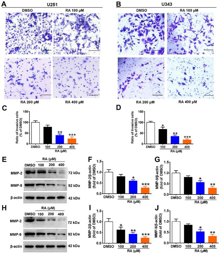
2                            LIU et al: ANTICANCER EFFECTS OF ROSMARINIC ACID IN GLIOMA CELLS

Boraginaceae and Lamiaceae families, mainly in the leaves of        Technologies, Inc.) as per the manufacturer's instruc‑
Rosmarinus officinalis, from which it can be easily isolated; it    tions. Briefly, cells were seeded in 96‑well microplates at
can also be found in peppermint, lemon balm, oregano, sage,         2x10³ cells/well and medium containing various concentrations
and thyme (20,21). The molecular structure of RA (chemical          of RA (0, 100, 200 and 400 µM) were added to the wells. After
formula: C18H16O8) contains two benzene rings located at            incubation for 24 or 48 h, the medium was replaced with 10 µl
the extremities of the molecule and a pair of ortho‑hydroxyl        CCK‑8 solution. After further incubation for 1 h at 37˚C, the
groups in each benzene ring (Fig. 1). It has been reported that     absorbance of each well was measured at 450 nm using a micro‑
RA exerts a variety of beneficial biological properties, mainly     plate reader (Multiskan Go 1510; Thermo Fisher Scientific,
antioxidant (22), anti‑inflammatory (23), pro‑apoptotic (24),       Inc.). In addition, cell viability assessed by CCK‑8 assay, was
and neuroprotective (25) effects. Furthermore, recent studies       also performed on cells that were treated with or without RA
have revealed that RA has antineoplastic activity in leukemia,      (200 µM) for 24 h after PI3K agonist (740 Y‑P, 25 µg/ml) or
hepatocellular carcinoma, gastric carcinoma, colorectal cancer,     inhibitor (LY294002, 30 µM) pretreatment for 2 h.
breast cancer, and small‑cell carcinoma of the lung (26‑29).
However, the effects of RA on tumor biological characteristics,     Immunofluorescence. U251 and U343 cells were seeded on a
such as proliferation, migration, and invasion of human glioma      cover slide in a 24‑well plate at a density of 5x10 4 cells/well
cells and their mechanisms, have not been clearly reported.         and maintained in a CO2 incubator for overnight growth. Then,
    The phosphatidylinositol 3‑kinase (PI3K)/Akt/nuclear            the cells were treated with RA (0, 100, 200 and 400 µM) or
factor‑κ B (NF‑κ B) signaling pathway is an important pathway       vehicle for 24 h. Subsequently, the cells were washed three
in the regulation of tumorigenesis, and is significantly acti‑      times with PBS and blocked for 1 h in PBS containing
vated in glioma (30). The activation of PI3K/Akt/NF‑κ B             0.2% Triton X‑100 and 10% skimmed milk powder at 37˚C.
signaling cascades is inhibited by Fyn knockdown in primary         Immunodetection was performed by incubation with a
astrocytes (31). Therefore, it was hypothesized that RA may         Fyn‑specific antibody at 37˚C for 1 h, followed by overnight
have an anti‑glioma effect by inhibiting the PI3K/Akt/NF‑κ B        incubation at 4˚C. After washing three times in PBS, the cover
signaling pathway.                                                  slides were incubated for 1 h at 37˚C with an anti‑rabbit Alexa
    In the present study, the effects of RA on glioma growth,       Fluor 488‑conjugated secondary antibody (1:200; Invitrogen;
migration, invasion, and apoptosis in vitro were explored, and      Thermo Fisher Scientific, Inc.). The cells were then washed
the findings suggest that RA may be a potential therapeutic         three times with PBS and mounted using ProLong™ Gold
agent for glioma.                                                   antifade reagent (P36930; Thermo Fisher Scientific, Inc.).
                                                                    Stained cells were analyzed at a magnification of x200 using a
Materials and methods                                               confocal microscope (LSM 800; Zeiss AG).

Reagents and antibodies. RA was purchased from Aladdin              Wound healing assay. Cell migration was evaluated using
(cat. no. R109804). LY294002 and 740 Y‑P were purchased             the wound healing assay as previously described (32).
from MedChemExpress (cat. nos. HY‑10108 and HY‑P0175,               Briefly, U251 and U343 cells were seeded in 24‑well plates at
respectively). The following primary antibodies were used:          5x10⁴ cells/well and allowed to adhere. Then, a wound/scratch
Fyn (product code ab125016; 1:2,000) was purchased from             was produced using a 200‑µl pipette tip. After washing off
Abcam , PI3K (product no. 4249S; 1:1,000), MMP‑2 (product           the separated cells with phosphate‑buffered solution (PBS),
no. 87809S; 1:1,000), MMP‑9 (product no. 13667S; 1:1,000),          serum‑free medium containing various concentrations of
Bcl‑2 (product no. 3498S; 1:1,000), Bax (product no. 2772S;         RA (0, 100, 200, and 400 µM) was added to the wells. The
1:1,000), cleaved caspase‑3 (product no. 9661S; 1:1,000), fibril‑   wound was observed at regular intervals between 0 and 48 h.
larin (product no. 2639S; 1:1,000), and caspase‑3 (product          Randomly selected areas were photographed at a magnifica‑
no. 9662S; 1:1,000), were all from Cell Signaling Technology,       tion of x100 using a phase‑contrast microscope (CKX41;
Inc., and phosphorylated Akt (p‑Akt) (cat. no. sc‑514032; 1:500),   Olympus Corporation) and the wound area was calculated by
Akt (cat. no. sc‑81434; 1:1,000), NF‑κ B p65 (cat. no. sc‑8008;     ImageJ software (version 1.8.0; National Institutes of Health).
1:1,000), β‑actin (cat. no. sc‑47778; 1:2,000), and glyceralde‑
hyde‑3‑phosphate dehydrogenase (GAPDH) (cat. no. sc‑47724;          Invasion assay. The invasion assay was conducted using
1:2,000) were purchased from Santa Cruz Biotechnology, Inc.         Corning® BioCoat™ Matrigel® Invasion Chambers (product
                                                                    no. 354480; Corning, Inc.) with 8‑µm pore chambers inserted
Cell culture. Human‑derived glioma cell lines (U251 and             into 24‑well plates. U251 and U343 cells (5x10⁴ cells) were
U343) and the normal human astrocyte (NHA) cell line were           cultured in 500 µl of serum‑free DMEM with RA in the inserts
purchased from the American Type Culture Collection (ATCC).         and 500 µl of DMEM containing 15% FBS in the bottom of the
All cells were maintained under 5% CO2 at 37˚C in Dulbecco's        wells. After 24 h of incubation, the inserts were washed three
modified Eagle's medium (DMEM) (cat. no. 11995040)                  times with PBS and the cells were fixed with 4% paraformal‑
containing 100 U/ml penicillin, 100 µg/ml streptomycin              dehyde at room temperature for 10 min. After washing with
(cat. no. 15070063), and 10% fetal bovine serum (FBS) (cat.         PBS, the cells were stained with 0.1% crystal violet at room
no. 10100147; all from Gibco; Thermo Fisher Scientific, Inc.)       temperature for 15 min. Following another wash with PBS,
in water‑jacketed humidity‑controlled incubators.                   the inner sides of the chamber were wiped with a cotton swab
                                                                    and images of the cells that invaded through the Matrigel®
Cell viability. Cell proliferation was calculated using             were obtained using a phase‑contrast microscope (magnifica‑
Cell Counting Kit‑8 (CCK‑8) assay (Dojindo Molecular                tion, x200). Finally, the number of invading cells was counted.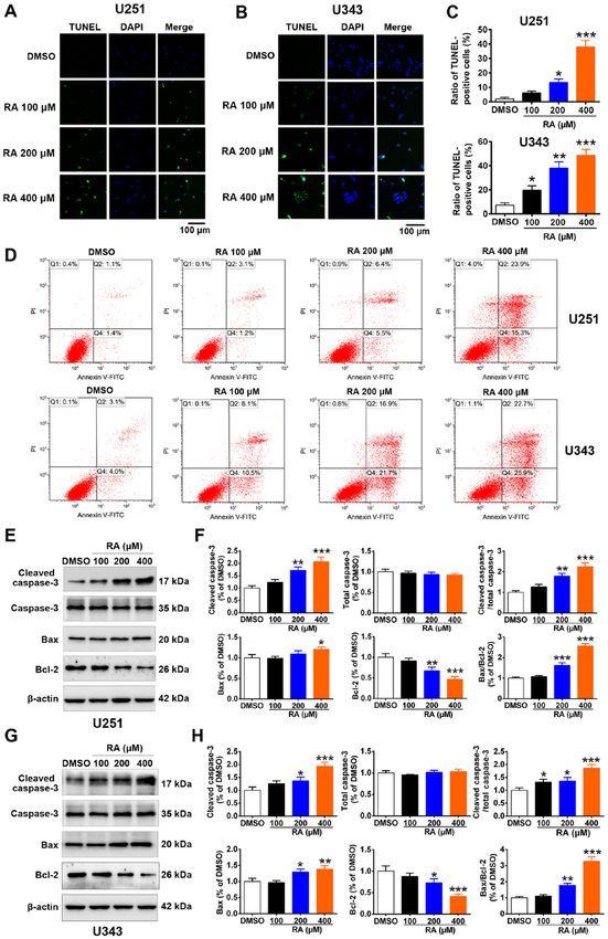
INTERNATIONAL JOURNAL OF MOlecular medicine 47: 67, 2021                                              3

                                                Figure 1. Chemical structure of rosmarinic acid.

TUNEL and DAPI staining. U251 and U343 cells were seeded                  1:4,000; Cell Signaling Technology, Inc.) were added for 1 h
on a cover slide in a 24‑well plate at a density of 3x10⁴ cells/well      at room temperature. The membranes were visualized with
and maintained in a CO2 incubator for overnight growth. Cells             Western Bright™ reagent (cat. no. K‑12043‑D10; Advansta,
were treated with various concentrations of RA (0, 100, 200,              Inc.). The band signals were captured by The ChemiDoc MP
and 400 µM) or vehicle for 24 h. Terminal deoxynucleotidyl                system (Bio‑Rad Laboratories, Inc.). ImageJ software (version
transferase (TdT) dUTP nick‑end labeling (TUNEL) staining                 1.8.0; National Institutes of Health) was used for densitometric
was performed according to the manufacturer's instructions                analysis. The experiments were repeated at least three times
(product no. C1086; Beyotime Institute of Biotechnology).                 independently.
Briefly, the cells were fixed with 4% paraformaldehyde at
room temperature for 30 min, incubated for 5 min with 0.3%                Statistical analyses. The data are expressed as the
Triton X‑100 in PBS, and then incubated with a reaction                   mean ± standard deviation (SD) of at least three independent
mixture containing terminal deoxynucleotidyl transferase and              experiments, and statistical differences of cell viability were
fluorescent labeling solution for 60 min at 37˚C according to             determined using two‑way analysis of variance (ANOVA)
the manufacturer's protocol. The cells were then stained with             followed by Dunnett's multiple comparison test; other
4',6‑diamidino‑2‑phenylindole (DAPI) for 5 min at 37˚C and                comparisons were performed using one‑way ANOVA followed
mounted using ProLong™ Gold antifade reagent. Stained                     by the Dunnett's multiple comparison test. All statistical
cells were analyzed using a confocal microscope (magnifica‑               analyses were computed by SPSS statistical analysis software
tion, x200).                                                              version 22.0 (IBM Corp.). Values of P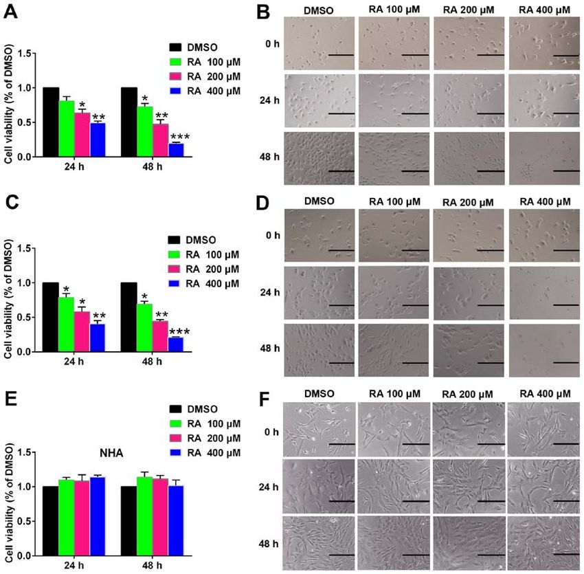
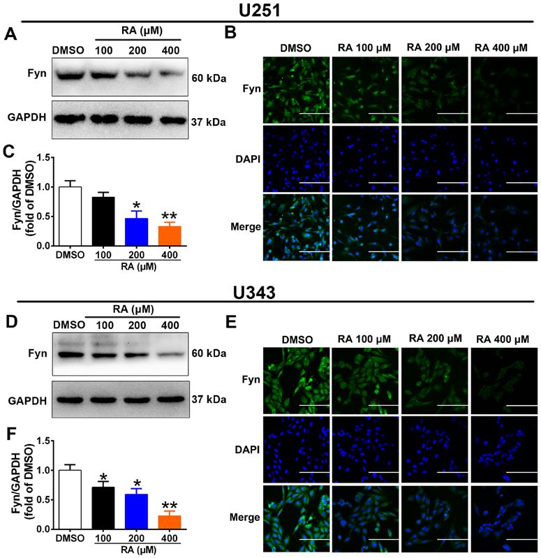
4                                LIU et al: ANTICANCER EFFECTS OF ROSMARINIC ACID IN GLIOMA CELLS

Figure 2. RA inhibits Fyn expression in glioma cells. (A and D) Western botting revealed that RA treatment suppressed the expression level of Fyn in a
dose‑dependent manner in U251 and U343 cells. (B and E) Representative areas of Fyn‑positive immunofluorescent staining in U251 and U343 cells. Scale
bar, 100 µm. (C and F) Densitometric quantification of the bands in A and D. GAPDH was used as a loading control. *P
INTERNATIONAL JOURNAL OF MOlecular medicine 47: 67, 2021                                                             5

Figure 3. RA inhibits glioma cell growth. (A and C) U251 and U343 cells were treated with the indicated doses of RA for 24 and 48 h, viability was determined
using the CCK‑8 assay, and data in control cells were normalized to 100%. Data are presented as the means ± SD of three experiments. (B and D) The
morphological changes of U251 and U343 cells. U251 and U343 cells were treated with various doses of RA for 24 and 48 h and observed using inverted
phase‑contrast microscopy. Scale bar, 100 µm. (E) The viability of NHA was determined after treatment with the indicated doses of RA for 24 and 48 h.
(F) Cell morphology of NHA after treatment with the indicated doses of RA for 24 and 48 h. Scale bar, 100 µm. *P
6                                 LIU et al: ANTICANCER EFFECTS OF ROSMARINIC ACID IN GLIOMA CELLS

Figure 4. RA reduces glioma cell migration. (A) Images of U251 cells immediately after, and at 24 and 48 h post‑scratch with the indicated RA concentrations.
Scale bar, 200 µm. (B and C) Quantification the wound healing of A. (D) Images of U343 cells immediately after, and at 24 and 48 h post‑scratch with the
indicated RA concentrations. Scale bar, 200 µm. (E and F) Quantification the wound healing of D. *P
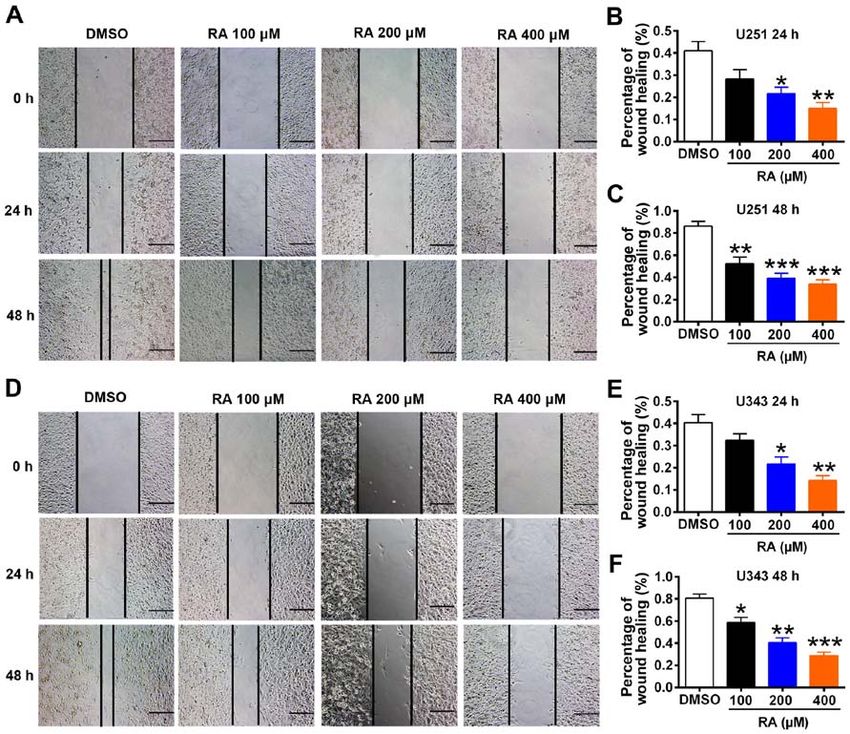
INTERNATIONAL JOURNAL OF MOlecular medicine 47: 67, 2021                                                               7

Figure 5. RA reduces glioma cell invasion and inhibits the expression of related factors. (A) Images of U251 cells after treatment with the indicated concentra‑
tions of RA for 24 h. Scale bar, 50 µm. (B) Images of U343 cells after treatment with indicated concentrations of RA for 24 h. Scale bar, 50 µm. (C) Percentage
of invasive U251 cells. (D) Percentage of invasive U343 cells. (E) The expression levels of MMP‑2 and MMP‑9 were detected by western blotting in U251
cells after treatment with the indicated concentrations of RA for 24 h. (F and G) Densitometric quantification of the bands in E. (H) The expression levels of
MMP‑2 and MMP‑9 were detected by western blotting in U343 cells after treatment with the indicated concentrations of RA for 24 h. (I and J) Densitometric
quantification of the bands in H. The histogram indicates the mean ± SD of three independent experiments. *P
8                                 LIU et al: ANTICANCER EFFECTS OF ROSMARINIC ACID IN GLIOMA CELLS

Figure 6. RA induces glioma cell apoptosis. (A and B) U251 and U343 cells were treated with the indicated doses of RA for 24 h, and then collected for TUNEL
staining. Scale bar, 100 µm. (C) Statistical analysis of apoptotic U251 and U343 cells. (D) U251 and U343 cells were exposed to the indicated concentrations
of RA for 24 h and then analyzed for apoptosis by flow cytometry using the Annexin V/PI dual‑staining assay. (E and G) U251 and U343 cells were treated
with RA for 24 h. Then, western blotting was conducted to detect the expression of cleaved caspase‑3, caspase‑3, Bax, and Bcl‑2. (F and H) Densitometric
quantification of the bands in E and G. The expression of GAPDH was used as an endogenous control. *P
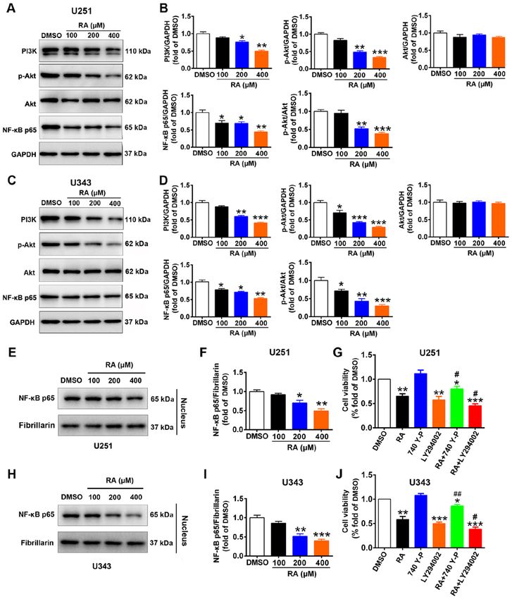
INTERNATIONAL JOURNAL OF MOlecular medicine 47: 67, 2021                                                            9

Figure 7. The PI3K/Akt/NF‑κ B signaling pathway is involved in RA‑induced cytotoxicity in glioma cells. (A) The protein expression of PI3K, p‑Akt, Akt,
and NF‑κ B p65 in U251 was assessed by western blotting. (B) Densitometric quantification of the bands in A. (C) The protein expression of PI3K, p‑Akt, Akt,
and NF‑κ B p65 in U343 was assessed by western blotting. (D) Densitometric quantification of the bands in C. (E and H) Expression levels of NF‑κ B p65 in
the nucleus were detected by western blotting. (F and I) Densitometric quantification of the bands in E and H. (G and J) Viability of U251 and U343 cells as
measured by CCK‑8 assay. The results are expressed as the mean ± SD. GAPDH was used as a loading control. *P
10                          LIU et al: ANTICANCER EFFECTS OF ROSMARINIC ACID IN GLIOMA CELLS

in a time‑ and dose‑dependent manner. Concurrently, it had        Ethics approval and consent to participate
little effect on NHA cell proliferation and morphology. This
suggested that RA may be a promising antitumor drug based         Not applicable.
on its weak toxicity. The present study also demonstrated
that RA inhibited glioma cell invasion. Since MMP‑2 and           Patient consent for publication
MMP‑9 are important regulators of invasion, the effect of RA
on their expression levels was also examined. Western blot‑       Not applicable.
ting revealed that treatment with RA decreased MMP‑2 and
MMP‑9 expression in glioma cells. The main feature of tumor       Competing interests
metastasis is the migration of cancer cells from the initial
tumor site to the circulatory system or lymphatic system (55).    The authors declare that they have no conflicts of interest.
Therefore, inhibiting tumor cell migration could reduce metas‑
tasis. In the present study, RA inhibited migration of U251 and   References
U343 cells. Considering that apoptosis is a crucial antitumor
mechanism (56), it was investigated whether RA could induce        1. Lapointe S, Perry A and Butowski NA: Primary brain tumours in
apoptosis in glioma cells. The percentage of apoptotic U251           adults. Lancet 392: 432‑446, 2018.
                                                                   2. Senders JT, Harary M, Stopa BM, Staples P, Broekman MLD,
and U343 cells was significantly increased after treatment            Smith TR, Gormley WB and Arnaout O: Information‑based
with RA. As Bcl‑family proteins are main regulators of                medicine in glioma patients: A clinical perspective. Comput
apoptosis (57), the effect of RA on the expression of Bax and         Math Methods Med 2018: 8572058, 2018.
                                                                   3. Batash R, Asna N, Schaffer P, Francis N and Schaffer M:
Bcl‑2 was also examined. RA increased Bax expression and              Glioblastoma multiforme, diagnosis and treatment; recent litera‑
decreased Bcl‑2 expression in glioma cells. Correspondingly,          ture review. Curr Med Chem 24: 3002‑3009, 2017.
the expression of cleaved (activated) caspase‑3, but not           4. Stupp R, Mason WP, van den Bent MJ, Weller M, Fisher B,
                                                                      Taphoorn MJ, Belanger K, Brandes AA, Marosi C, Bogdahn U,
that of total caspase‑3, was increased by RA. In addition,            et al: Radiotherapy plus concomitant and adjuvant temozolomide
the Bax/Bcl‑2 and cleaved caspase‑3/caspase‑3 ratios were             for glioblastoma. N Engl J Med 352: 987‑996, 2005.
increased by RA, thus providing a mechanistic basis for the        5. Zhang S, Liao K, Miao Z, Wang Q, Miao Y, Guo Z, Qiu Y,
                                                                      Chen B, Ren L, Wei Z, et al: CircFOXO3 promotes glioblastoma
induction of apoptosis by RA in glioma cells. These results are       progression by acting as a competing endogenous RNA for
similar to the reported role of RA in colorectal cancer (37).         NFAT5. Neuro Oncol 21: 1284‑1296, 2019.
Numerous studies have revealed that the PI3K/Akt/NF‑κ B            6. El‑Sehemy A, Selvadurai H, Ortin‑Martinez A, Pokrajac N,
                                                                      Mamatjan Y, Tachibana N, Rowland K, Lee L, Park N,
pathway is closely related to glioma progression (46,58,59).          Aldape K, et al: Norrin mediates tumor‑promoting and ‑suppres‑
In line with these studies, the present results revealed that         sive effects in glioblastoma via Notch and Wnt. J Clin Invest 130:
the PI3K/Akt/NF‑κ B signaling pathway was involved in the             3069‑3086, 2020.
                                                                   7. Lewis‑Tuffin LJ, Feathers R, Hari P, Durand N, Li Z,
antitumor effects of RA in glioma.                                    Rodriguez FJ, Bakken K, Carlson B, Schroeder M, Sarkaria JN
     Collectively, these findings indicated that the antitumor        and Anastasiadis PZ: Src family kinases differentially influence
effects of RA in glioma may be mediated by Fyn inhibition.            glioma growth and motility. Mol Oncol 9: 1783‑1798, 2015.
                                                                   8. Irby RB and Yeatman TJ: Role of Src expression and activation
The detailed mechanisms warrant further investigation in vitro        in human cancer. Oncogene 19: 5636‑5642, 2000.
and in vivo.                                                       9. Summy JM and Gallick GE: Src family kinases in tumor progres‑
                                                                      sion and metastasis. Cancer Metastasis Rev 22: 337‑358, 2003.
                                                                  10. Frame MC: Src in cancer: Deregulation and consequences for
Acknowledgements                                                      cell behaviour. Biochim Biophys Acta 1602: 114‑130, 2002.
                                                                  11. Tornillo G, Knowlson C, Kendrick H, Cooke J, Mirza H,
Not applicable.                                                       Aurrekoetxea‑Rodríguez I, Vivanco MDM, Buckley NE,
                                                                      Grigoriadis A and Smalley MJ: Dual mechanisms of LYN kinase
                                                                      dysregulation drive aggressive behavior in breast cancer cells.
Funding                                                               Cell Rep 25: 3674‑3692 e10, 2018.
                                                                  12. Emaduddin M, Bicknell DC, Bodmer WF and Feller SM: Cell
                                                                      growth, global phosphotyrosine elevation, and c‑Met phosphory‑
The present research was funded by China Postdoctoral                 lation through Src family kinases in colorectal cancer cells. Proc
Science Foundation (grant no. 2019M663104) and the Science            Natl Acad Sci USA 105: 2358‑2362, 2008.
and Technology Planning Project of Guangdong Province             13. Masaki T, Okada M, Tokuda M, Shiratori Y, Hatase O, Shirai M,
                                                                      Nishioka M and Omata M: Reduced C‑terminal Src kinase (Csk)
(grant no. 2017A020215089).                                           activities in hepatocellular carcinoma. Hepatology 29: 379‑384,
                                                                      1999.
Availability of data and materials                                14. Yadav V and Denning MF: Fyn is induced by Ras/PI3K/Akt
                                                                      signaling and is required for enhanced invasion/migration. Mol
                                                                      Carcinog 50: 346‑352, 2011.
The datasets used and/or analyzed during the current study        15. Lu KV, Zhu S, Cvrljevic A, Huang TT, Sarkaria S, Ahkavan D,
are available from the corresponding author on reasonable             Dang J, Dinca EB, Plaisier SB, Oderberg I, et al: Fyn and SRC are
                                                                      effectors of oncogenic epidermal growth factor receptor signaling
request.                                                              in glioblastoma patients. Cancer Res 69: 6889‑6898, 2009.
                                                                  16. Han X, Zhang W, Yang X, Wheeler CG, Langford CP, Wu L,
Authors' contributions                                                Filippova N, Friedman GK, Ding Q, Fathallah‑Shaykh HM, et al:
                                                                      The role of Src family kinases in growth and migration of glioma
                                                                      stem cells. Int J Oncol 45: 302‑310, 2014.
BH and GH conceived and designed the study. YL, XX, HT            17. Comba A, Dunn PJ, Argento AE, Kadiyala P, Ventosa M, Patel P,
and YP performed the experiments. YL, XX and HT analyzed              Zamler DB, Núñez FJ, Zhao L, Castro MG and Lowenstein PR:
                                                                      Fyn tyrosine kinase, a downstream target of receptor tyrosine
the data. YL, BH and GH wrote the manuscript. All authors             kinases, modulates antiglioma immune responses. Neuro
have read and approved the final manuscript.                          Oncol 22: 806‑818, 2020.
INTERNATIONAL JOURNAL OF MOlecular medicine 47: 67, 2021 11
18. Bowman RL, Wang Q, Carro A, Verhaak RG and Squatrito M: 39. Wang Y, Tang H, Zhang Y, Li J, Li B, Gao Z, Wang X, Cheng G
GlioVis data portal for visualization and analysis of brain tumor and Fei Z: Saponin B, a novel cytostatic compound purified from
expression datasets. Neuro Oncol 19: 139‑141, 2017. Anemone taipaiensis, induces apoptosis in a human glioblastoma
19. Jelić D, Mildner B, Kostrun S, Nujić K, Verbanac D, Culić O, cell line. Int J Mol Med 32: 1077‑1084, 2013.
Antolović R and Brandt W: Homology modeling of human Fyn 40. Su CC, Wang MJ and Chiu TL: The anti‑cancer efficacy of
kinase structure: Discovery of rosmarinic acid as a new Fyn curcumin scrutinized through core signaling pathways in glio‑
kinase inhibitor and in silico study of its possible binding modes. blastoma. Int J Mol Med 26: 217‑224, 2010.
J Med Chem 50: 1090‑1100, 2007. 41. Zhang WF, Yang Y, Li X, Xu DY, Yan YL, Gao Q, Jia AL and
20. Şengelen A and Önay‑Uçar E: Rosmarinic acid and siRNA Duan MH: Angelica polysaccharides inhibit the growth and
combined therapy represses Hsp27 (HSPB1) expression promote the apoptosis of U251 glioma cells in vitro and in vivo.
and induces apoptosis in human glioma cells. Cell Stress Phytomedicine 33: 21‑27, 2017.
Chaperones 23: 885‑896, 2018. 42. Yang SX, Polley E and Lipkowitz S: New insights on PI3K/AKT
21. Juskowiak B, Bogacz A, Wolek M, Kamiński A, Uzar I, pathway alterations and clinical outcomes in breast cancer.
Seremak‑Mrozikiewicz A and Czerny B: Expression profiling Cancer Treat Rev 45: 87‑96, 2016.
of genes modulated by rosmarinic acid (RA) in MCF‑7 breast 43. Arlt A, Gehrz A, Müerköster S, Vorndamm J, Kruse ML,
cancer cells. Ginekol Pol 89: 541‑545, 2018. Fölsch UR and Schäfer H: Role of NF‑kappaB and Akt/PI3K
22. Yang SY, Hong CO, Lee GP, Kim CT and Lee KW: The hepato‑ in the resistance of pancreatic carcinoma cell lines against
protection of caffeic acid and rosmarinic acid, major compounds gemcitabine‑induced cell death. Oncogene 22: 3243‑3251, 2003.
of Perilla frutescens, against t‑BHP‑induced oxidative liver 44. Tomar VS, Patil V and Somasundaram K: Temozolomide
damage. Food Chem Toxicol 55: 92‑99, 2013. induces activation of Wnt/β ‑catenin signaling in glioma cells
23. Chu X, Ci X, He J, Jiang L, Wei M, Cao Q, Guan M, Xie X, via PI3K/Akt pathway: Implications in glioma therapy. Cell Biol
Deng X and He J: Effects of a natural prolyl oligopeptidase Toxicol 36: 273‑278, 2020.
inhibitor, rosmarinic acid, on lipopolysaccharide‑induced acute 45. Song L, Chen X, Mi L, Liu C, Zhu S, Yang T, Luo X, Zhang Q,
lung injury in mice. Molecules 17: 3586‑3598, 2012. Lu H and Liang X: Icariin‑induced inhibition of SIRT6/NF‑κ B
24. Lin CS, Kuo CL, Wang JP, Cheng JS, Huang ZW and triggers redox mediated apoptosis and enhances anti‑tumor
Chen CF: Growth inhibitory and apoptosis inducing effect of immunity in triple‑negative breast cancer. Cancer Sci 111:
Perilla frutescens extract on human hepatoma HepG2 cells. 4242‑4256, 2020.
J Ethnopharmaco 112: 557‑567, 2007. 46. Fahey JM, Korytowski W and Girotti AW: Upstream signaling
25. Kelsey NA, Wilkins HM and Linseman DA: Nutraceutical events leading to elevated production of pro‑survival nitric oxide
antioxidants as novel neuroprotective agents. Molecules 15: in photodynamically‑challenged glioblastoma cells. Free Radic
7792‑7814, 2010. Biol Med 137: 37‑45, 2019.
26. Wu CF, Hong C, Klauck SM, Lin YL and Efferth T: Molecular 47. Furnari FB, Fenton T, Bachoo RM, Mukasa A, Stommel JM,
mechanisms of rosmarinic acid from Salvia miltiorrhiza in acute Stegh A, Hahn WC, Ligon KL, Louis DN, Brennan C, et al:
lymphoblastic leukemia cells. J Ethnopharmaco 176: 55‑68, 2015. Malignant astrocytic glioma: Genetics, biology, and paths to
27. Xavier CP, Lima CF, Fernandes‑Ferreira M and Pereira‑Wilson C: treatment. Genes Dev 21: 2683‑2710, 2007.
Salvia fruticosa, Salvia officinalis, and rosmarinic acid induce 48. Karachi A, Dastmalchi F, Mitchell DA and Rahman M:
apoptosis and inhibit proliferation of human colorectal cell lines: Temozolomide for immunomodulation in the treatment of glio‑
The role in MAPK/ERK pathway. Nutr Cancer 61: 564‑571, blastoma. Neuro Oncol 20: 1566‑1572, 2018.
2009. 49. Patil SA, Hosni‑Ahmed A, Jones TS, Patil R, Pfeffer LM and
28. Yu C, Chen DQ, Liu HX, Li WB, Lu JW and Feng JF: Rosmarinic Miller DD: Novel approaches to glioma drug design and drug
acid reduces the resistance of gastric carcinoma cells to 5‑fluo‑ screening. Expert Opin Drug Discov 8: 1135‑1151, 2013.
rouracil by downregulating FOXO4‑targeting miR‑6785‑5p. 50. Xie JH, Lai ZQ, Zheng XH, Xian YF, Li Q, Ip SP, Xie YL,
Biomed Pharmacother 109: 2327‑2334, 2019. Chen JN, Su ZR, Lin ZX and Yang XB: Apoptosis induced by
29. Yesil‑Celiktas O, Sevimli C, Bedir E and Vardar‑Sukan F: bruceine D in human non‑small‑cell lung cancer cells involves
Inhibitory effects of rosemary extracts, carnosic acid and rosma‑ mitochondrial ROS‑mediated death signaling. Int J Mol Med 44:
rinic acid on the growth of various human cancer cell lines. Plant 2015‑2026, 2019.
51. Ong SKL, Shanmugam MK, Fan L, Fraser SE, Arfuso F, Ahn KS,
Foods Hum Nutr 65: 158‑163, 2010. Sethi G and Bishayee A: Focus on formononetin: Anticancer
30. Li W, Du Q, Li X, Zheng X, Lv F, Xi X, Huang G, Yang J and potential and molecular targets. Cancers 11: 611, 2019.
Liu S: Eriodictyol Inhibits Proliferation, Metastasis and induces 52. Petersen M and Simmonds MS: Rosmarinic acid. Phytochemistry 62:
apoptosis of glioma cells via PI3K/Akt/NF‑κ B signaling pathway. 121‑125, 2003.
Front Pharmacol 11: 114, 2020. 53. Elansary HO and Mahmoud EA: Egyptian herbal tea infusions'
31. Ko HM, Lee SH, Kim KC, Joo SH, Choi WS and Shin CY: The antioxidants and their antiproliferative and cytotoxic activities
role of TLR4 and fyn interaction on lipopolysaccharide‑stimulated against cancer cells. Nat Prod Res 29: 474‑479, 2015.
PAI‑1 expression in astrocytes. Mol Neurobiol 52: 8‑25, 2015. 54. Alcaraz M, Alcaraz‑Saura M, Achel DG, Olivares A, López-
32. Zhang T, Ji D, Wang P, Liang D, Jin L, Shi H, Liu X, Meng Q, Morata JA and Castillo J: Radiosensitizing effect of rosmarinic
Yu R and Gao S: The atypical protein kinase RIOK3 contributes acid in metastatic melanoma B16F10 cells. Anticancer Res 34:
to glioma cell proliferation/survival, migration/invasion and the 1913‑1921, 2014.
AKT/mTOR signaling pathway. Cancer Lett 415: 151‑163, 2018. 55. McCall B, McPartland CK, Moore R, Frank‑Kamenetskii A
33. Yachida S, Jones S, Bozic I, Antal T, Leary R, Fu B, Kamiyama M, and Booth BW: Effects of astaxanthin on the proliferation and
Hruban RH, Eshleman JR, Nowak MA, et al: Distant metastasis migration of breast cancer cells in vitro. Antioxidants (Basel) 7:
occurs late during the genetic evolution of pancreatic cancer. 135, 2018.
Nature 467: 1114‑1117, 2010. 56. Shin J, Song MH, Oh JW, Keum YS and Saini RK: Pro‑oxidant
34. Marín‑Ramos NI, Thein TZ, Cho HY, Swenson SD, Wang W, actions of carotenoids in triggering apoptosis of cancer cells: A
Schönthal AH, Chen TC and Hofman FM: NEO212 inhibits review of emerging evidence. Antioxidants (Basel) 9: 532, 2020.
migration and invasion of glioma stem cells. Mol Cancer Ther 17: 57. Chowdhury I, Thompson WE, Welch C, Thomas K and
625‑637, 2018. Matthews R: Prohibitin (PHB) inhibits apoptosis in rat granulosa
35. Pan H, Xue W, Zhao W and Schachner M: Expression and func‑ cells (GCs) through the extracellular signal‑regulated kinase 1/2
tion of chondroitin 4‑sulfate and chondroitin 6‑sulfate in human (ERK1/2) and the Bcl family of proteins. Apoptosis 18:
glioma. FASEB J 34: 2853‑2868, 2020. 1513‑1525, 2013.
36. Szabo E, Schneider H, Seystahl K, Rushing EJ, Herting F, 58. Yan J, Xu C, Li Y, Tang B, Xie S, Hong T and Zeng E: Long
Weidner KM and Weller M: Autocrine VEGFR1 and VEGFR2 non‑coding RNA LINC00526 represses glioma progression via
signaling promotes survival in human glioblastoma models in forming a double negative feedback loop with AXL. J Cell Mol
vitro and in vivo. Neuro Oncol 18: 1242‑1252, 2016. Med 23: 5518‑5531, 2019.
37. Han YH, Kee JY and Hong SH: Rosmarinic Acid Activates AMPK 59. Nakabayashi H and Shimizu K: Involvement of Akt/NF‑ κ B
to inhibit metastasis of colorectal cancer. Front Pharmacol 9: 68, pathway in antitumor effects of parthenolide on glioblastoma
2018. cells in vitro and in vivo. BMC Cancer 12: 453, 2012.
38. Niknejad H, Yazdanpanah G and Ahmadiani A: Induction of
apoptosis, stimulation of cell‑cycle arrest and inhibition of angio‑ This work is licensed under a Creative Commons
genesis make human amnion‑derived cells promising sources for Attribution-NonCommercial-NoDerivatives 4.0
cell therapy of cancer. Cell Tissue Res 363: 599‑608, 2016. International (CC BY-NC-ND 4.0) License.You can also read