Laser-stimulated uorescence reveals unseen details in fossils from the Solnhofen Limestone (Upper Jurassic, Bavaria, Germany)
←
→
Page content transcription
If your browser does not render page correctly, please read the page content below
Laser-stimulated uorescence reveals unseen
details in fossils from the Solnhofen Limestone
(Upper Jurassic, Bavaria, Germany)
Luke Barlow ( up812045@myport.ac.uk )
University of Portsmouth https://orcid.org/0000-0002-0867-649X
Michael Pittman
University of Hong Kong https://orcid.org/0000-0002-6149-3078
David Martill
University of Portsmouth
Thomas Kaye
Foundation for Scienti c Advancement https://orcid.org/0000-0001-7996-618X
Anthony Butcher
Article
Keywords: Laser-Stimulated Fluorescence, ultraviolet, techniques, uorescence, Jurassic, Solnhofen
Limestone, Altmühtal Formation, Painten Formation, Mörnsheim Formation
DOI: https://doi.org/10.21203/rs.3.rs-59716/v1
License: This work is licensed under a Creative Commons Attribution 4.0 International License.
Read Full License
Page 1/32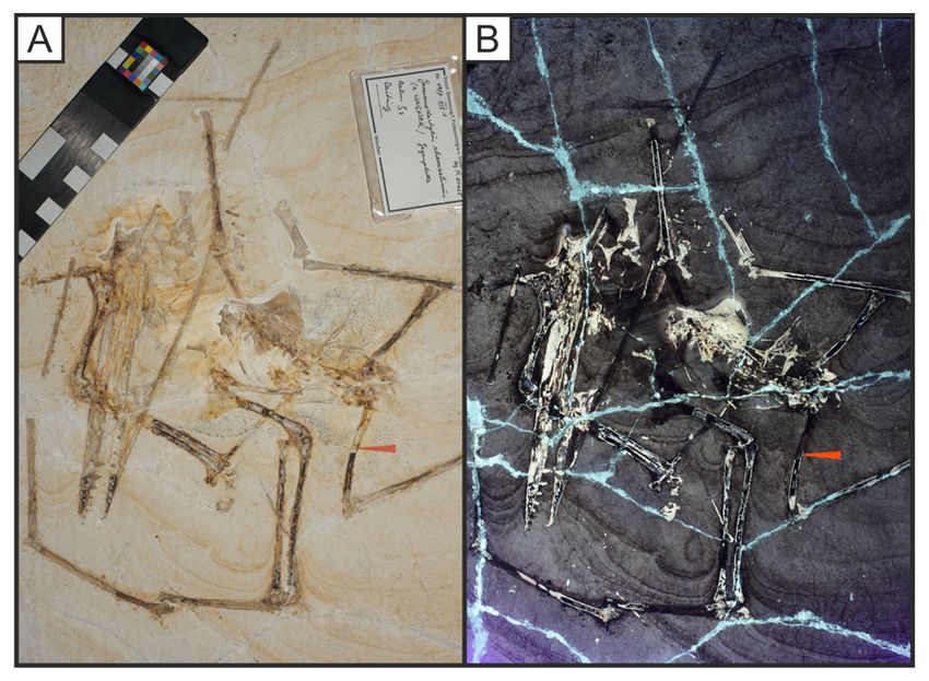
Abstract
Laser-Stimulated Fluorescence (LSF) has seen increased use in palaeontological investigations in recent
years. The method uses the high ux of laser light to reveal details sometimes missed by ultraviolet (UV)
and optical wavelengths. In this study, we compare the results of LSF with UV on a range of fossils from
the Upper Jurassic Solnhofen Limestone Konservat-Lagerstätte of Bavaria, Germany. The methodology
follows previous protocols with modi cations made to enhance laser beam intensity. Our experiments
show the value of LSF in revealing shallow subsurface detail of specimens, previously not widely applied
to Solnhofen fossils. In particular, fossil decapods from the Solnhofen Limestone reveal full body
outlines, even under the matrix, along with details of segmentation within the appendages such as limbs
and antennae. The results indicate that LSF can be used on both vertebrate and invertebrate fossils and
may surpass the information provided by traditional UV methods in some specimens.

Introduction
Since the introduction of ultraviolet (UV) uorescence for the analysis of fossils from the Upper Jurassic
Solnhofen Limestone lithographic limestones of Germany in the early 20th century (Miethe & Born 1928),
the technique has been increasingly used in the analysis of exceptionally preserved fossils from this
famous fossil Lagerstätte. Fossils from a wide range of phyla have been studied using UV including
decapods (Schwiegert 2011), ammonites (Keupp 2007), sh (Tischlinger & Arratia 2013), pterosaurs
(Frey et al. 2003) and dinosaurs (Göhlich & Chiappe 2006; Rauhut et al. 2012) among others. More
recently, laser light techniques have been applied to fossils to induce uorescence (Kaye et al. 2010),
especially on ‘iconic’ fossil vertebrates from sites of exceptional preservation like Anchiornis,
Confuciusornis, Jianianhualong, Psittacosaurus and Sapeornis (Vinther et al. 2016; Mayr et al. 2016;
Xu et al. 2017; Kaye et al. 2019a,b; Pittman & Xu 2020; Serrano et al. 2020), including the rst discovered
fossil feather which is from the Solnhofen Limestone (Kaye et al. 2019a).. Fossils from the lithographic
limestone horizons of the Solnhofen Limestone have been widely studied using UV and thus provides an
ideal opportunity to compare the two uorescent techniques on a range of fossils including cephalopods,
decapods, and small vertebrates (Hess 1999; Zhou & Wang 2010) (Fig. 1). The Solnhofen Limestone is
famous for its well-bedded, ultra ne-grained lithographic limestones (often called plattenkalk) and
referred to as such herein) that formed in the calm basins of the Solnhofen lagoons on the northern
margin of the Tethys Ocean (Viohl 1998; Munnecke et al. 2008). The palaeoenvironment represented by
these limestones is a closed lagoonal system with high evaporation rates leading to a strati ed water
column with anoxic bottom waters largely devoid of macroorganisms (Viohl 1998). Occasional mixing
through storms brought the toxic water to the aerated surface zone leading to the mass mortality of
nektonic organisms (Pan et al. 2019). These organisms often became exceptionally well preserved due to
a lack of scavenging, bacterial sealing, and rapid burial (Wellnhofer 2009). Here we compare images of
Solnhofen fossils under LSF and UV and evaluate the use of LSF on fossils in this important Konservat-
Lagerstätte.

                                                 Page 2/32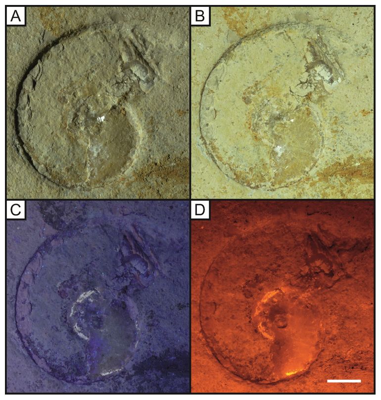
Methods
The specimens used in this study labelled LB 1–13, abbreviated from the initials of the primary author,
were collected during a series of eld visits to the Solnhofen region over twenty years and are
accessioned in the collection of the School of the Environment, Geography and Geosciences (SEGG),
University of Portsmouth. Additional specimens from the Staatliche naturwissenschaftliche Sammlungen
Bayerns, Bayerische Staatssammlung für Paläontologie und Geologie (SNSB-BSPG) were studied by MP
and TGK during a visit to the Museum für Naturkunde (Kaye et al. 2019a) (Figs 13–16). The specimens
used are based on availability and represent some of the main groups found in the Solnhofen
Limestones.

Photography
Photographs were taken in a blacked-out room to avoid natural light contamination. An LED lamp
illuminated the specimen obliquely (c. 45 degrees) or directly for the white light photographs.

The method of Laser-Stimulated Fluorescence modi ed from Kaye et al. (2015) used an MGL-III–532–
1~300mW green diode-pumped solid-state (DPSS) laser with a PSU-III-LCD power supply with a set
output of 85mW. Alterations to the method included mounting the laser onto a camera track where only a
trucking motion was permitted, so the 532 nm green laser moved through the x-axis to scan the specimen
whilst maintaining the same perpendicular relationship (Fig. 2). Through substituting trucking for the
panning motion in previous publications, the laser module maintains a constant distance from the
specimen and therefore a constant beam intensity across the entire fossil. For Figs 13–16 the same
methodology was used in Kaye et al. 2019a with an abbreviated method stated here. A 1W 405nm blue
laser was used to induce uorescence and a Nikon DSLR was used to take the photographs with a
425nm blocking lter. Post-processing (equalisation, saturation, and colour balance) was then performed
in Photoshop CS6.

The method of ultraviolet uorescence consisted of a 365 nm lamp as used by Tischlinger & Frey (2002)
with the specimen illuminated as close as possible.

The long-exposures for each image under all three lighting regimes were taken using a Nikon D5300
DSLR camera mounted on a tripod with a 2-second self-timer setting that prevents camera movement
from affecting the image. Aperture priority mode controlled the length of exposure following the method
described by Eklund et al. (2018). They suggested a 10 second exposure for UV and the ISO was adjusted
accordingly. Using a low ISO prevents grainy photos and with a small aperture, other light sources are
prevented from contaminating the image. 30 second exposures were used for imaging under LSF with the
ISO left at 100. The position and distance of the specimen from the camera remained constant for each
method so that LSF, white light, and UV images could be collected e ciently. This lighting sequence
allowed the O56 blocking lter that prevents camera over-saturation with LSF to be applied effectively.
This lter was removed for white light and UV photography, as although the lter can pick up UV
                                                 Page 3/32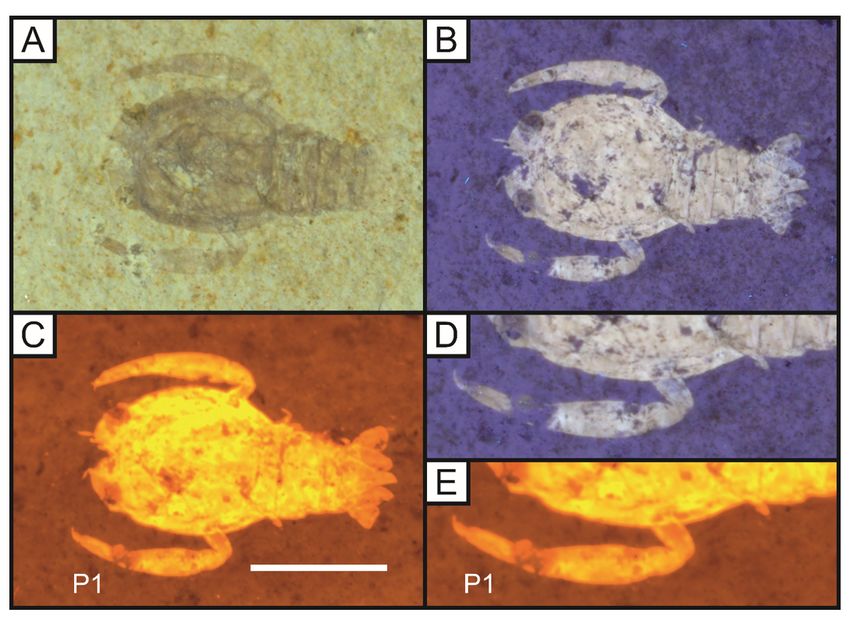
uorescence, the uorescence is clearer without a blocking lter. Due to the attened nature of the fossils
within Solnhofen laminites, repetitive photography and photo stacking techniques were unnecessary.

Health and Safety
It is important that when using LSF or UV techniques, appropriate safety precautions are observed. The
green laser and UV lamp require laser and UV blocking goggles during operation and these methods were
conducted in a locked room with a suitable exterior notice to prevent people from being harmed. Humans
are most sensitive to green laser light (Galang et al. 2010) and by following these guidelines, the
operators and others can be safeguarded. When using UV, 10-minute breaks were taken every 10–30
minutes to prevent eye damage and headaches (Tischlinger & Arratia 2013). Since pale colours uoresce
under UV light, dark clothing was worn while conducting scans.

Results
Cephalopoda
Cephalopods in the form of ammonites, belemnites and teuthoids occur frequently in the Solnhofen
Limestone and are sometimes exceptionally well preserved (Fuchs et al. 2015). Many have been reported
with aspects of their soft tissues preserved, including impressions of tentacles with hooklets in
belemnites and teuthoids (Klug et al. 2016), the musculature of the mantle in teuthoids (Klug et al. 2015)
and the siphuncle and pellicula in ammonites (Keupp 2007). Although ammonites make up a large
portion of the fossils from the Solnhofen plattenkalks, they are often poorly preserved due to the
aragonitic composition of the shell which is readily dissolved during diagenesis (Seilacher et al. 1976).
This dissolution leaves behind an external or composite mould in the matrix, occasionally with the
original outline and the calcitic aptychi in the body chamber (Keupp 2007).

Although rarely preserved, the original shell can be observed once replaced by calcite in the phragmacone
(Fig. 4) and the body chamber (Arratia et al. 2015). The body chamber is present within the halo of the
dissolved shell and can be enhanced under UV and LSF (Fig. 6). The siphuncle, pellicula and other non-
mineralised elements are often phosphatised in Solnhofen ammonites and these may uoresce more
intensely than the remaining shell (Keupp 2007). UV uorescence displays colour differences on these
ammonites (Fig. 6C), but the contrast between non-mineralised parts and the surrounding shell is lacking,
when compared with the LSF image of specimen LB 4 (Fig. 6D). It appears that the raised darker areas on
isolated aptychi (LB 1) (Fig. 3) are the thick spongy layer on the inside of the aptychus underlying the
thinner crenulated outer layer, rather than soft tissues following Lehmann (1976).

Lumbricaria Goldfuss, 1831, a coprolite attributed to ammonites (Janicke 1970) lies on the same slab as
an ammonite (Fig. 6) and appears to contain aptychi of a smaller ammonite. Plesioteuthis prisca
(Rüppell, 1829), a squid from the Solnhofen Limestone with a central rachis that is revealed in its entirety

                                                  Page 4/32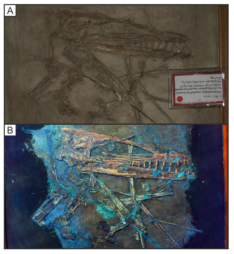
and uoresces at two distinct levels under LSF (Fig. 7D). This rachis runs along the centre of the visible
body outline, although no soft tissues of the mantle are present (Fig. 7).

Decapoda
Decapod crustacea occur frequently in the Solnhofen plattenkalks and are often well preserved as fully
articulated individuals, even in the larval stage (Haug et al. 2008; Winkler 2014). The fossils are valuable
for understanding arthropod ontogeny, with specimens that display growth cycles only known from a few
other Lagerstätten (J. T. Haug et al. 2009). Numerous studies have been carried out using UV
  uorescence, revealing unseen details to allow distinctions between taxa or increasing the specimen-
matrix contrast (Haug et al. 2011; Winkler 2012; Audo et al. 2014). The decapods lend themselves to
techniques using uorescence as they hold auto uorescent compounds within their exoskeleton (Haug et
al. 2011; Glenn et al. 2013).

The preservation of the fossils studied (LB 6–9), vary from isolated appendages to complete articulated
examples. A single walking leg attributed to Aeger tipularius von Schlotheim, 1822 (Fig. 8) is near
complete with only a few setae missing from the exposed surface. UV uorescence enhances the
specimen-matrix contrast but fails to uoresce below the matrix where the complete setae are revealed
through LSF (Fig. 8).

Anatomical details of complete specimens of Cycleryon propinquus von Schlotheim, 1822 (Fig. 9)
and Antrimpos speciosus Münster, 1839 (Figs 10, 11) are enhanced through uorescent techniques as
previous studies demonstrate (Schweigert & Garassino 2004). However, it must be reiterated that previous
studies on Solnhofen material have only used ultraviolet as a means of uorescence (Keupp 2007;
Schweigert 2011; Tischlinger & Arratia 2013). A distal portion of the left rst pereiopod on LB 7 is unseen
under UV light but is revealed, albeit faintly, under LSF (Fig. 8). In Antrimpos speciosus (LB 8) (Figs 10–
11), more detail of the body outline is exposed by LSF under a green 532 nm laser compared with UV and
this is emphasised in the magni ed portions (Figs 9, 10 B-C, E-F).

As the gures of Solnhofen decapods herein show, the detail revealed under Laser-Stimulated
Fluorescence, often surpasses that revealed by ultraviolet uorescence, with the outline of the entire
animal outline being revealed, along with small elements such as antennae and swimmerets (Figs 10,
11). The veneer of the matrix on LB 8 in Figs 10–11 may have obstructed the UV uorescence and could
be prepared further to assist the use of this method. Ultraviolet uorescence on a specimen
of Alcmonacaris winkleri Polz, 2008 (LB 9) reveals a faint outline of the animal while recording colour
patterning (Fig. 12). Under LSF, green laser light, this colour information is lost but the animal is revealed
in its entirety (Fig. 12D). Techniques to rectify this loss of information have been developed using
multiple wavelengths (Kaye et al. 2015). As Figs 9–12 show, the preserved exoskeleton and the body
outline uoresce at different levels because of the auto uorescent compounds within the arthropod
exoskeleton.

                                                   Page 5/32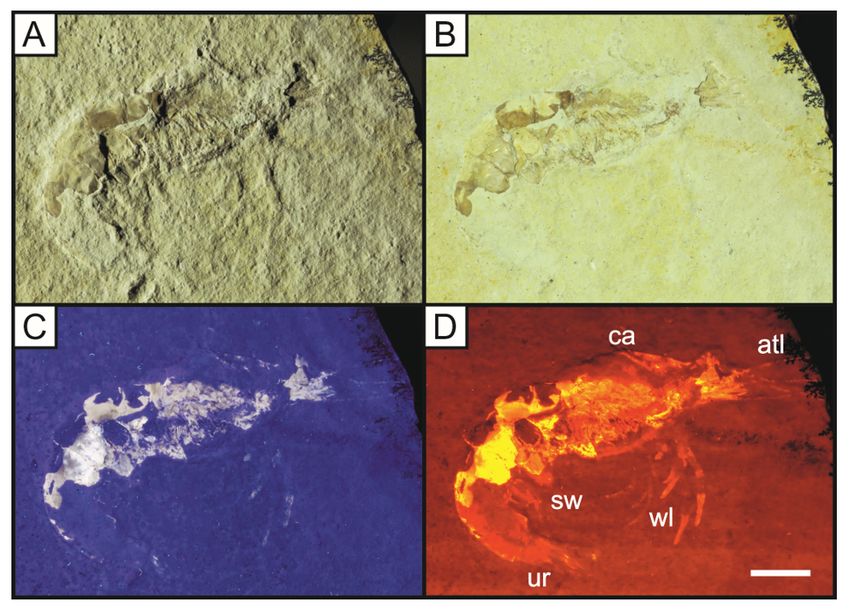
Vertebrata
The Solnhofen Limestone has achieved much of its fame as a fossil Konservat-Lagerstätten because of
the exceptional preservation of its vertebrate fossils, especially those of volant animals such as the
earliest unequivocal fossil bird Archaeopteryx and a diverse assemblage of pterosaurs (Beardmore et al.
2017; Schwarz et al. 2019; Longrich et al. 2020) where parts of the ight surfaces are preserved, including
wing membranes and feathers (Frey et al. 2003; Jäger et al. 2018; Benton et al. 2019; Kaye et al. 2019a;
Foth et al. 2020; Wilkin 2020). Some of this exceptional preservation can be seen in the examples below
(Figs 13–16). The hollow bones of pterosaurs are rarely preserved and through LSF the contrast between
preserved bone and the imprints of the skeleton in the matrix is exempli ed. The dwarf crocodilyform
Alligatorellus (Fig. 16) under the blue laser displays soft tissues around the entire body as well as a
brighter section at the base of the tail that may represent partial preservation of the caudofemoralis
muscle.

However, the exceptional preservation is not restricted to the Tetrapoda, with many shes also
exceptionally well preserved with full articulation and soft part preservation (Konwert 2016; Arratia et al.
2019; Ebert & Lane 2019) as well as reptiles (Tennant & Mannion, 2014) (see Fig. 17). Specimens LB 10–
13 (Figs 17–20) vary in completeness from articulated individuals to isolated sources of phosphate,
resulting in the high level of uorescence under UV and LSF (Fig. 12).

The specimens studied vary between right and left lateral views, seen in specimens LB 10 and 11 (Figs 17
and 18 respectively). The uorescence of these specimens (LB 10–13), results from the vertebrate
endoskeleton containing high levels of phosphorus (Tischlinger & Arratia 2013). The fossils in white light
contrast little with the surrounding sediment as in many other Solnhofen fossils. Indeed, it may be
di cult to see some fossils, especially when weathered, without using uorescence.

Many Solnhofen fossil sh possess a fully ossi ed skull, ns, neural and haemal spines made apparent
through uorescence along with thin autocentra (Arratia & Schultze 2013). The autocentra surrounding
the dark chordocentra are thin and almost translucent and uoresce under both UV and LSF. Species of
the teleost family Orthogonikleithridae are the most common vertebrates in the Solnhofen Limestones
(Konwert 2016). The ossi cation patterns observed on a specimen of Orthogonikleithrus hoelli Arratia,
1997 (LB 12) (Fig. 19), correspond with previous studies using UV for uorescence (Tischlinger & Arratia
2013).

Fish uoresce well under both UV and LSF and although the difference is often minimal, clearly in Fig. 18
LSF surpasses the UV photograph with an increased uorescence of both the skull and vertebral column
completing the structures seen and this results from the higher ux and subsurface illumination.

Discussion
Laser-Stimulated Fluorescence has various applications as illustrated by the numerous examples gured
by Kaye et al. (2015) including an automated microvertebrate sorting machine developed to reduce time
                                                  Page 6/32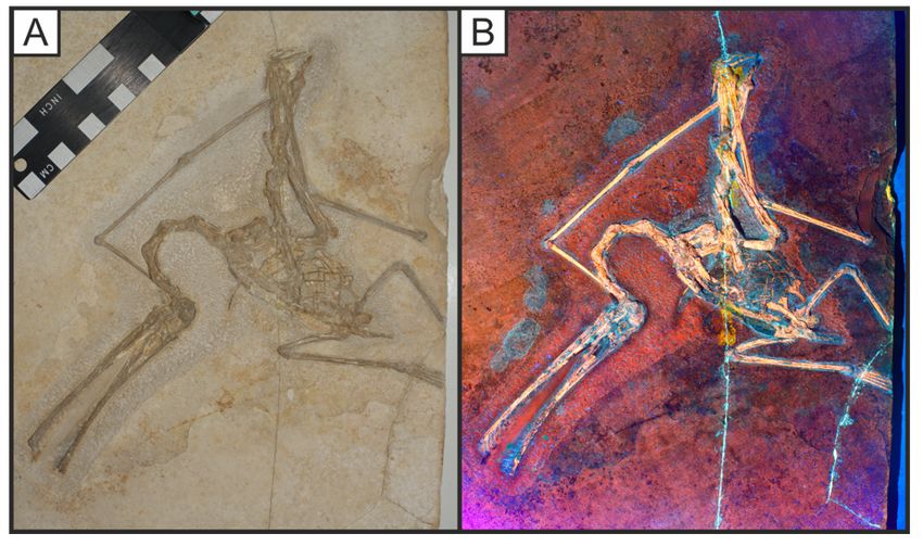
spent on manual sorting along with demonstrating the potential of LSF to reveal subsurface fossils (Kaye
et al. 2015, Figs 10 and 8 respectively). Recently, it has even been applied as part of an automated drone
system to seek out fossils on the ground at night (Kaye & Pittman 2020). The technique has revealed
geochemical halos around the lost calamus of 'Archaeopteryx’ (Kaye et al. 2019b),, akin to the effect
observed in Figs 10–12 where the uorescence of decapods extends beyond the preserved exoskeleton.
The methodology is simple and provides a rapid means for producing high-quality images of uorescing
fossils, allowing for a camera image to be available within 30 seconds of exposure. This exposure time is
longer than UV or white light photography as the uorescence occurs through a ne laser beam scanning
an entire specimen rather than the instant illumination under an LED or UV lamp. The same wavelengths
of 532nm have been employed previously (Kaye et al. 2015), although this green light is often substituted
for blue or violet wavelengths (T. Kaye, pers. comm. 2019). Blue/violet lasers increase the contrast further
and reveal uorescence colour differences akin to those under UV (Schwarz et al. 2019). The 532nm laser
in this study results in varying levels of orange uorescence through the O56 blocking lter that can
cause di culties in distinguishing between different parts of a specimen. As reported by several studies
(Mayr et al. 2016; Vinther et al. 2016; Wang et al. 2017; Saitta et al. 2018, Falk et al. 2019; Yang et al.
2019; Pittman & Xu 2020; Serrano et al. 2020), LSF continues to reveal new and exciting details on well-
preserved fossil assemblages (Konservat-Lagerstätten) like the Jehol Lagerstätte of northeastern China
and the Las Hoyas Lagerstätte of Spain. The high uorescence we observed in decapods (Figs 8–12)
likely results from the auto uorescence present in crustacean exoskeletons (Charbonnier et al. 2017). The
Solnhofen specimens analysed here uoresce brightly due to high levels of phosphate in both original
and diagenetic minerals (Wilby et al. 1996). The principal ndings in the Solnhofen specimens are that
LSF reveals morphology not visible under other UV techniques and more often to a greater degree of
clarity and depth.

Costs
With reductions to the cost of laser systems, the method could be replicated using a 532nm laser, an LCD
power supply and a Zecti 31.5in”/80cm camera slider. This study using an 85mW laser shows the
technique can be used with less powerful laser equipment than the 300–500mW laser used by Falk et
al. (2016) and Wang et al. (2017). With two wavelengths in tandem, different structures uoresce,
allowing for a more complete picture (Wang et al. 2017), providing an option for further research. The use
of a less powerful laser also allows for LSF to be more accessible on the grounds of cost, but with a
necessary trade-off in uorescence signal.

Conclusions
The list of non-destructive techniques available to palaeontologists is increasing. X-rays were rst
implemented in 1896 on fossils from another German Lagerstätte, the Devonian Hunsrück Slate
(Hohenstein 2004), and was widely implemented from the 1930s by Lehmann on this site along with
other fossil-rich areas like Messel pit and the Solnhofen region. The use of UV has become standard in
                                                 Page 7/32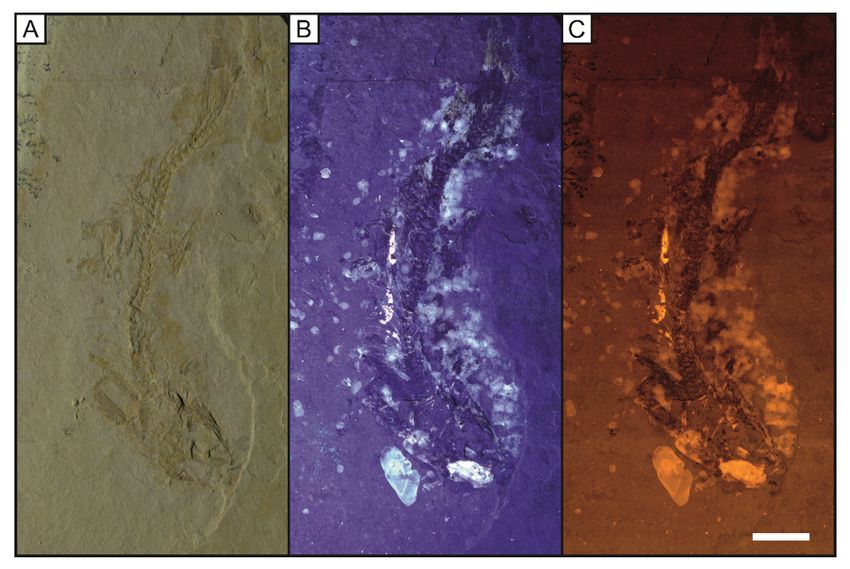
many palaeontological studies especially those investigating soft tissue preservation (Hone et al. 2010;
Kellner et al. 2010; Cuesta et al. 2015; Schwarz et al. 2019; Hoffman et al. 2020) since its rst use in 1926
on fossil vertebrates within the Solnhofen plattenkalks (Simpson 1926). Composite imaging and 3D
computer modelling have also been used on Solnhofen fossils and provide a valuable alternative,
especially on small and delicate specimens (Haug et al. 2008). Synchrotron Rapid Scanning X-ray
Fluorescence (SRS-XRF) combined with chemical images allowed for the mapping of plumage patterns
in the iconic Solnhofen bird Archaeopteryx (Manning et al. 2013).

LSF was added to the literature as recently as 2015. The use of lower-powered equipment in this study
illustrates that the technique can still be employed to good effect and could provide a valuable teaching
resource. The method can be performed in as little as 30 seconds, allowing for almost instant data
collection. Previous publications have focused entirely on vertebrate material. This study shows the
effect of LSF on invertebrates for the rst time. In addition, it adds the Solnhofen plattenkalks to the list
of Konservat-Lagerstätten where the effects under LSF are now known. This study underscores LSF as an
alternative tool to UV for non-destructive palaeontological investigation using extensive comparative
 gures that show unseen details to an equal and often greater extent than UV.

Declarations
Acknowledgements
MP and TGK’s participation in this study was supported by the RAE Improvement Fund of the Faculty of
Science, The University of Hong Kong (HKU) and funds from the HKU MOOC Dinosaur Ecosystems. MP
was also supported by Research Grant Council General Research Fund 17103315. TGK was also
supported by the Foundation for Scienti c Advancement. We would like to thank Oliver Rauhut for
granting us access to specimens in his care at the Bavarian State Collections of Palaeontology and
Geology.

References
  1. ARRATIA, G. 1997. Basal teleosts and teleostean phylogeny. Palaeo Ichthyologica, 7, 1-168.
  2. ― and SCHULTZE, H. P. and TISCHLINGER, H. 2019. On a remarkable new species of Tharsis, a Late
     Jurassic teleostean sh from southern Germany: its morphology and phylogenetic
     relationships. Fossil Record, 22(1), 23 pp.
  3. ― and SCHULTZE, H., TISCHLINGER, H. and VIOHL, G. 2015. Solnhofen - ein fenster in die Jurazeit
    1+2 - gesamtausgabe. 1st edn. Verlag Friedrich Pfeil, Munchen, 607 pp.
  4. AUDO, D. and CHARBONNIER, S. 2012. Late Cretaceous crest-bearing shrimps from the Sahel Alma
     Lagerstätte of Lebanon. Acta Palaeontologica Polonica, 58(2), 335-349.
  5. ― and SCHWEIGERT, G., HAUG, J. T., HAUG, C., SAINT MARTIN, J. P. and CHARBONNIER, S. 2014.
     Diversity and palaeoecology of the enigmatic genus Knebelia (Eucrustacea, Decapoda, Eryonidae)

                                                 Page 8/32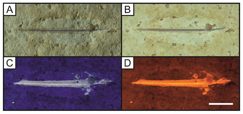
from Upper Jurassic plattenkalks in southern Germany. Palaeontology, 57(2), 397-416.
 6. BARTELS, C. 2009. The fossils of the Hunsruck slate: Marine life in the Devonian. 2nd edn.
    Cambridge University Press, Cambridge, UK, 86–88.
 7. BEARDMORE, S. R., LAWLOR, E. and HONE, D. W. E. 2017. Using taphonomy to infer differences in
    soft tissues between taxa: an example using basal and derived forms of Solnhofen pterosaurs. The
    Science of Nature, 104(7-8), 65 pp.
 8. BENTON, M. J., DHOUAILLY, D., JIANG, B. and MCNAMARA, M. 2019. The early origin of
   feathers. Trends in ecology & evolution, 34, 856-869.
 9. BLAINVILLE, H.D. 1818. Poissons fossils. Nouveau Dictionnaire d’Histoire Naturelle, Nouvelle édition
    Chez Deterville, 27, 319–359.
10. CHARBONNIER, S., AUDO, D., GARASSINO, A. and HYŽNÝ, M. 2017. Fossil crustacea of Lebanon.
    Publications Scienti ques du Muséum, Paris, 252 pp.
11. CONROY, G. C. and VANNIER, M. W. 1984. Noninvasive three-dimensional computer imaging of
    matrix- lled fossil skulls by high-resolution computed tomography. Science, 226(4673), 456-458.
12. CUESTA, E., DIAZ-MARTINEZ, I., ORTEGA, F. and SANZ, J. L. 2015. Did all theropods have chicken-like
   feet? First evidence of a non-avian dinosaur podotheca. Cretaceous Research, 56, 53-59.
13. EBERT, M. 2019. Zandtfuro and Schernfeldfuro, New Genera of Halecomorphi (Actinopterygii) from
    the Upper Jurassic Solnhofen Archipelago. Journal of Vertebrate Paleontology, 39(2). doi:
    10.1080/02724634.2019.1592759
14. ― and KÖLBL-EBERT, M. and LANE, J. A. 2015. Fauna and predator-prey relationships of Ettling, an
    actinopterygian sh-dominated Konservat-Lagerstätte from the Late Jurassic of southern
   Germany. PLoS One, 10(1). doi: 10.1371/journal.pone.0116140
15. EKLUND, M. J., AASE, A. K. and BELL, C.J. 2018. Progressive Photonics: methods and applications of
    sequential imaging using visible and non-visible spectra to enhance data-yield and facilitate forensic
    interpretation of fossils. Journal of Paleontological Techniques, 20, 1-36.
16. FALK, A. R., KAYE, T. G., ZHOU, Z. and BURNHAM, D. A. 2016. Laser uorescence illuminates the soft
    tissue and life habits of the Early Cretaceous bird Confuciusornis. PLoS One, 11(12). doi:
   10.1371/journal.pone.0167284
17. ― and O’CONNOR, J., WANG, M. and ZHOU, Z. 2019. On the preservation of the beak in
    Confuciusornis (Aves: Pygostylia). Diversity, 11(11), 212 pp. doi: 10.3390/d11110212
18. FOTH, C., HAUG, C., HAUG, J. T., TISCHLINGER, H. and RAUHUT, O. W. 2020. Two of a feather: a
    comparison of the preserved integument in the juvenile theropod dinosaurs Sciurumimus and
    Juravenator from the Kimmeridgian Torleite Formation of southern Germany. In The Evolution of
    Feathers. Springer, Cham, 79-101.
19. FREY, E., TISCHLINGER, H., BUCHY, M. C. and MARTILL, D. M. 2003. New specimens of Pterosauria
   (Reptilia) with soft parts with implications for pterosaurian anatomy and locomotion. Geological
   Society, London, Special Publications, 217(1), 233-266.

                                                Page 9/32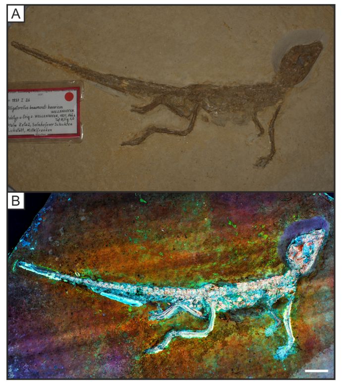
20. GALANG, J., RESTELLI, A., HAGLEY, E. W. and CLARK, C. W. 2010. A red light for green laser
    pointers. Optics and Photonics News, 21(10), 11-13.
21. GLENN, D., PAKES, M. J. and CALDWELL, R. L. 2013. Fluorescence in Arthropoda informs ecological
    studies in anchialine crustaceans, Remipedia, and Atyidae. Journal of Crustacean Biology, 33(5),
    620-626.
22. GÖHLICH, U. B. and CHIAPPE, L. M. 2006. A new carnivorous dinosaur from the Late Jurassic
   Solnhofen archipelago. Nature, 440, 329-332.
23. GOLDFUSS, A. 1831. Petrefacta Germaniae - Abbildungen und Beschreibungen der Petrefacten
    Deutschlands und der angränzenden Länder. Erster Theil. Arnz and Company, Dusseldorf, 1-252.
24. HAUG, C., HAUG, J. T., WALOSZEK, D., MAAS, A., FRATTIGIANI, R. and LIEBAU, S. 2009. New methods
    to document fossils from lithographic limestones of southern Germany and
    Lebanon. Palaeontologia Electronica, 12(3), 12 pp.
25. HAUG, J. T., HAUG, C. and EHRLICH, M. 2008. First fossil stomatopod larva (Arthropoda: Crustacea)
   and a new way of documenting Solnhofen fossils (Upper Jurassic, Southern
   Germany). Palaeodiversity, 1, 103-109.
26. ― and HAUG, C., WALOSZEK, D., MAAS, A., WULF, M. and SCHWEIGERT, G. 2009. Development in
    Mesozoic scyllarids and implications for the evolution of Achelata (Reptantia, Decapoda,
    Crustacea). Palaeodiversity, 2, 97-110.
27. ― and HAUG, C., KUTSCHERA, V., MAYER, G., MAAS, A., LIEBAU, S., CASTELLANI, C., WOLFRAM, U.,
   CLARKSON, E. N. K. and WALOSZEK, D. 2011. Auto uorescence imaging, an excellent tool for
   comparative morphology. Journal of Microscopy, 244(3), 259-272.
28. HILL, C. R. 1990. Scanning electron microscopy in palaeobotany. Scanning Electron Microscopy in
    Taxonomy and Functional Morphology. Systematics Association Special Volume Series, 41, 193-234.
29. HOFFMANN, R., BESTWICK, J., BERNDT, G., BERNDT, R., FUCHS, D. and KLUG, C. 2020. Pterosaurs ate
    soft-bodied cephalopods (Coleoidea). Scienti c Reports, 10(1), 1-7.
30. HOHENSTEIN, P. 2004. X-ray imaging for palaeontology. The British Journal of Radiology, 77(917),
   420-425.
31. HONE, D. W., TISCHLINGER, H., XU, X. and ZHANG, F. 2010. The extent of the preserved feathers on
    the four-winged dinosaur Microraptor gui under ultraviolet light. PloS one, 5(2).
    doi: 10.1371/journal.pone.0009223
32. JÄGER, K. R., TISCHLINGER, H., OLESCHINSKI, G. and SANDER, P. M. 2018. Goldfuss was right: Soft
    part preservation in the Late Jurassic pterosaur Scaphognathus crassirostris revealed by re ectance
    transformation imaging and ultraviolet light and the auspicious beginnings of paleo-
   art. Palaeontologia Electronica, 21(3). doi 10.26879/713
33. JANICKE, V. 1970. Lumbricaria, ein cephalopoden–koprolith. Neues Jahrbuch für Geologie und
    Paläontologie, Monatshefte, 1, 50-60.
34. KAYE, T. G. and PITTMAN, M. 2020. Fluorescence‐based detection of eld targets using an
    autonomous unmanned aerial vehicle system. Methods in Ecology and Evolution. doi:
                                               Page 10/32
10.1111/2041-210X.13402
35. ― and MARTIN, L., BURNHAM, D. and GONG, E. 2010. Multispectral imaging and analysis of a
Liaoning 'mystery specimen'. In 70th Annual Meeting of the Society of Vertebrate Paleontology.
36. ― and FALK, A. R., PITTMAN, M., SERENO, P. C., MARTIN, L. D., BURNHAM, D. A., GONG, E., XU, X. and
WANG, Y. 2015. Laser-stimulated uorescence in paleontology. PloS one, 10(5). doi:
10.1371/journal.pone.0125923
37. ― and PITTMAN, M., MAYR, G., SCHWARZ, D. and XU, X. 2019a. Detection of lost calamus
challenges identity of isolated Archaeopteryx feather. Scienti c reports, 9(1), 1-6.
38. ― and PITTMAN, M., MARUGÁN-LOBÓN, J., MARTÍN-ABAD, H., SANZ, J. L. and BUSCALIONI, A. D.
2019b. Fully edged enantiornithine hatchling revealed by laser-stimulated uorescence supports
precocial nesting behaviour. Scienti c reports, 9(1), 1-4.
39. KELLNER, A. W., WANG, X., TISCHLINGER, H., DE ALMEIDA CAMPOS, D., HONE, D. W. and MENG, X.
2010. The soft tissue of Jeholopterus (Pterosauria, Anurognathidae, Batrachognathinae) and the
structure of the pterosaur wing membrane. Proceedings of the Royal Society B: Biological
Sciences, 277(1679), 321-329.
40. KEUPP, H. 2007. Complete ammonoid jaw apparatuses from the Solnhofen plattenkalks:
implications for aptychi function and microphagous feeding of ammonoids. Neues Jahrbuch für
Geologie und Paläontologie-Abhandlungen, 245(1), 93-101.
41. ― and RIEDEL, F. 2010. Remarks on the possible function of the apophyses of the Middle Jurassic
microconch ammonite Ebrayiceras sulcatum (Zieten, 1830), with a discussion on the palaeobiology
of Aptychophora in general. Neues Jahrbuch für Geologie und Paläontologie-Abhandlungen, 255(3),
301-314.
42. KLUG, C., FUCHS, D., SCHWEIGERT, G., RÖPER, M. and TISCHLINGER, H. 2015. New anatomical
information on arms and ns from exceptionally preserved Plesioteuthis (Coleoidea) from the Late
Jurassic of Germany. Swiss Journal of Palaeontology, 134(2), 245-255.
43. ― and SCHWEIGERT, G., FUCHS, D., KRUTA, I. and TISCHLINGER, H. 2016. Adaptations to squid-style
high-speed swimming in Jurassic belemnitids. Biology letters, 12(1). doi 10.1098/rsbl.2015.0877
44. KONWERT, M. 2016. Orthogonikleithrus francogalliensis, sp. nov. (Teleostei, Orthogonikleithridae)
from the Late Jurassic Plattenkalks of Cerin (France). Journal of Vertebrate Paleontology, 36(3), 1-
10.
45. KOWALSKI, J., BODZIOCH, A., JANECKI, P. A., RUCIŃSKI, M. R. and ANTCZAK, M. 2019. Preliminary
report on the microvertebrate faunal remains from the Late Triassic locality at Krasiejów, SW Poland.
Annales Societatis Geologorum Poloniae, 89(3), 291-305.
46. LEHMANN, U. 1976. Ammonites: their lives and their environment. Enke Verlag, Stuttgart, 171 pp.
47. LONGRICH, N. R., TISCHLINGER, H. and FOTH, C. 2020. The Feathers of the Jurassic Urvogel
Archaeopteryx. In The Evolution of Feathers. Springer, Cham, 119-146.
48. MANNING, P. L., EDWARDS, N. P., WOGELIUS, R. A., BERGMANN, U., BARDEN, H. E., LARSON, P. L.,
SCHWARZ-WINGS, D., EGERTON, V. M., SOKARAS, D., MORI, R. A. and SELLERS, W. I. 2013.
Page 11/32Synchrotron-based chemical imaging reveals plumage patterns in a 150 million year old early
bird. Journal of Analytical Atomic Spectrometry, 28(7), 1024-1030.
49. MAYR, G., PITTMAN, M., SAITTA, E., KAYE, T. G. and VINTHER, J. 2016. Structure and homology of
Psittacosaurus tail bristles. Palaeontology, 59(6), 793-802.
50. MIETHE, A. and BORN, A. 1928. Die uorographie von fossilien. Paläontologische Zeitschrift, 9(4),
343-356.
51. MUNNECKE, A., WESTPHAL, H. and KÖLBL-EBERT, M. 2008. Diagenesis of plattenkalk: examples
from the Solnhofen area (Upper Jurassic, southern Germany). Sedimentology, 55(6), 1931-1946.
52. MÜNSTER, G. V. 1839. Decapoda Macroura. Abbildung und Beschreibung der fossilen
langschwiinzigen Krebse in den Kalkschiefern von Bayern. Beitraege zur Petrefactenkunde, 2, 1-88.
53. OPPEL A. 1863. Über jurassische Cephalopoden, Palaeontologische Mittheilungen aus dem Museum
des königlich Bayerischen Staates, 3, 163- 266.
54. PAN, Y., FÜRSICH, F. T., CHELLOUCHE, P. and HU, L. 2019. Taphonomy of sh concentrations from the
Upper Jurassic Solnhofen Plattenkalk of Southern Germany. Neues Jahrbuch für Geologie und
Paläontologie-Abhandlungen, 292(1), 73-92.
55. PARENT, H. and WESTERMANN, G. E. 2016. Jurassic ammonite aptychi: functions and evolutionary
implications. Swiss Journal of Palaeontology, 135(1), 101-108.
56. PITTMAN, M. and XU, X. (editors). 2020. Pennaraptoran theropod dinosaurs: past progress & new
frontiers. Bulletin of the American Museum of Natural History, 320 pp, in press.
57. POLZ H. 2008. Alcmonacaris winkleri g. nov. sp. nov. (Crustacea: Decapoda: Pleocyemata: Caridea)
aus den Solnhofener Plattenkalken von Eichstätt. Archaeopteryx, 26, 1–9.
58. RACICOT, R. 2016. Fossil secrets revealed: X-ray CT scanning and applications in paleontology. The
Paleontological Society Papers, 22, 21-38.
59. RAUHUT, O. W., FOTH, C., TISCHLINGER, H. and NORELL, M. A. 2012. Exceptionally preserved juvenile
megalosauroid theropod dinosaur with lamentous integument from the Late Jurassic of
Germany. Proceedings of the National Academy of Sciences, 109(29), 11746-11751.
60. RÜPPELL, E. 1829. Abbildung und Beschreibung einiger neuer oder wenig bekannten Versteinerungen
aus der Kalkschieferformation von Solnhofen. Brönner Verlag, Frankfurt, 12 pp.
61. SAITTA, E. T., FLETCHER, I., MARTIN, P., PITTMAN, M., KAYE, T. G., TRUE, L. D., NORELL, M.A.,
ABBOTT, G. D., SUMMONS, R. E., PENKMAN, K. and VINTHER, J. 2018. Preservation of feather bers
from the Late Cretaceous dinosaur Shuvuuia deserti raises concern about immunohistochemical
analyses on fossils. Organic Geochemistry, 125, 142-151.
62. SCHLOTHEIM, E.F. VON. 1822. Nachtrage zur Petrefacktenkunde. Becker, Gotha, 114 pp.
63. SCHWARZ, D., KUNDRÁT, M., TISCHLINGER, H., DYKE, G. and CARNEY, R. M. 2019. Ultraviolet light
illuminates the avian nature of the Berlin Archaeopteryx skeleton. Scienti c reports, 9(1), 1-11.
64. SCHWEIGERT, G. 2011. The decapod crustaceans of the Upper Jurassic Solnhofen Limestones: a
historical review and some recent discoveries. Neues Jahrbuch für Geologie und Paläontologie-
Page 12/32Abhandlungen, 260(2), 131-140.
65. ― and GARASSINO, A. 2004. New genera and species of shrimps (Crustacea: Decapoda:
Dendrobranchiata, Caridea) from the Upper Jurassic lithographic limestones of S. Germany.
Stuttgarter Beiträge zur Naturkunde, Staatliches Museum für Naturkunde, Rosenstein 1, D-70191
Stuttgart, 350, 33 pp.
66. SEILACHER, A., ANDALIB, F., DIETL, G. and GOCHT, H. 1976. Preservational history of compressed
Jurassic ammonites from southern Germany. Neues Jahrbuch für Geologie und Paläontologie,
Abhandlungen, 152, 307-356.
67. SERRANO, F. J., PITTMAN, M., KAYE, T.G., WANG, X.L., ZHENG, X.T. and CHIAPPE, L.M. 2020. Laser-
Stimulated Fluorescence re nes ight modelling of the Early Cretaceous bird Sapeornis.
68. STÜRMER, W., SCHAARSCHMIDT, F. and MITTMEYER, H. 1980. Versteinertes Leben Im Röntgenlicht.
W. Kramer, Frankfurt am Main, 80 pp.
69. SYLVESTER-BRADLEY, P. C. 1969. Aluminum Coating in Scanning Electron Microscopy.
Micropaleontology, 15(3), 366 pp.
70. TISCHLINGER, H. and ARRATIA, G. 2013. Ultraviolet light as a tool for investigating Mesozoic shes,
with a focus on the ichthyofauna of the Solnhofen archipelago. Mesozoic shes, 5, 549-560.
71. ― and FREY, E. 2002. Ein Rhamphorhynchus (Pterosauria, Reptilia) mit ungewöhnlicher
Flughauterhaltung aus dem Solnhofener Plattenkalk. Archaeopteryx, 20, 1-20.
72. VINTHER, J., NICHOLLS, R., LAUTENSCHLAGER, S., PITTMAN, M., KAYE, T. G., RAYFIELD, E., MAYR, G.
and CUTHILL, I. C. 2016. 3D camou age in an ornithischian dinosaur. Current Biology, 26(18), 2456-
2462.
73. VIOHL, G. 1998. The Solnhofen lithographic limestone - genesis and habitats. Archaeopteryx, 16, 37-
68.
74. WANG, X., PITTMAN, M., ZHENG, X., KAYE, T. G., FALK, A. R., HARTMAN, S. A. and XU, X. 2017. Basal
paravian functional anatomy illuminated by high-detail body outline. Nature communications, 8, 1-6.
75. WELLNHOFER, P. 2009. Archaeopteryx: the icon of evolution. Verlag Friedrich Pfeil, Munchen, 208 pp.
76. WILBY, P. R., BRIGGS, D. E., BERNIER, P. and GAILLARD, C. 1996. Role of microbial mats in the
fossilization of soft tissues. Geology, 24(9), 787-790.
77. WILKIN, J. 2020. The south German Plattenkalks. Geology Today, 36(1), 27-32.
78. WINKLER, N. 2012. Libanocaris annettae nov. sp. (Crustacea: Dendrobranchiata: Penaeidae) from
the Upper Jurassic Solnhofen Lithographic Limestones of Eichstätt. Zitteliana, 59-65.
79. WINKLER, N. 2014. A new caridean shrimp (Crustacea: Decapoda: Dendrobranchiata) from the Upper
Jurassic Solnhofen Lithographic Limestones of Schernfeld (South Germany). Zitteliana, 83-90.
80. XU, X., CURRIE, P., PITTMAN, M., XING, L., MENG, Q., LÜ, J., HU, D. and YU, C. 2017. Mosaic evolution
in an asymmetrically feathered troodontid dinosaur with transitional features. Nature
Communications, 8(1), 1-12.
Page 13/32Figures
Figure 1
A facies map of the Solnhofen Limestones in Bavaria, southern Germany with the location X indicates the
source of the fossils used in this study. Modi ed from Ebert et al. (2015).
Page 14/32Figure 2
A simpli ed diagram of the camera and specimen table setup used to perform LSF imaging. 1, The image
area of the DSLR camera; 2, Carriage of the camera track holding the laser module; 3, The imaged
specimen; 4, Laser illumination using a line generator; 5, 85mW 532nm laser module; 6, DSLR 5300; 7,
Tripod for long exposure photography. Not to scale.
Page 15/32Figure 3
Isolated specimen of Lamellaptychus (LB 1), a genus for ammonite mouthparts not associated with a
speci c taxon, under white light (A-B), UV (C) and LSF (D). A. Direct white light image differentiating
between the brown thick spongy layer and the thin crenulated layer beneath; B, oblique white light casting
a shadow on the specimen revealing the full outline; C, ultraviolet light of 365 nm wavelength used to
display colour differences between the two layers; D, Increased contrast under LSF using a 532 nm green
laser allows the full outline of the original shape to be observed. Scale bar = 10mm.
Page 16/32Figure 4
The oppeliid ammonite Neochetoceras bous Oppel, 1863 with a calci ed phragmacone, full body
chamber outline and preserved aptychi (LB 2). A, oblique white light displaying remnants of the body
chamber at the left inside edge; B, UV image displaying the siphuncle, phragmacone and jaw apparatus
with colour differences along with possible stomach contents; C, LSF image of Neochetoceras bous
Page 17/32under a 532 nm green laser with phosphatised siphuncle along with a clear boundary between the
phragmacone and body chamber unseen in other methods. Scale bar = 10 mm.
Figure 5
The phragmacone of the oppeliid ammonite Fontannesiella prolithographica (Fontannes) (LB 3) with
phosphatised siphuncle under white light (A-B), UV (C), and LSF under 532 nm green laser light (D). Note
the colour variation seen under 365 nm UV is not consistent with Fig. 6 and may be because of
differential preservation. Scale bar = 10 mm.
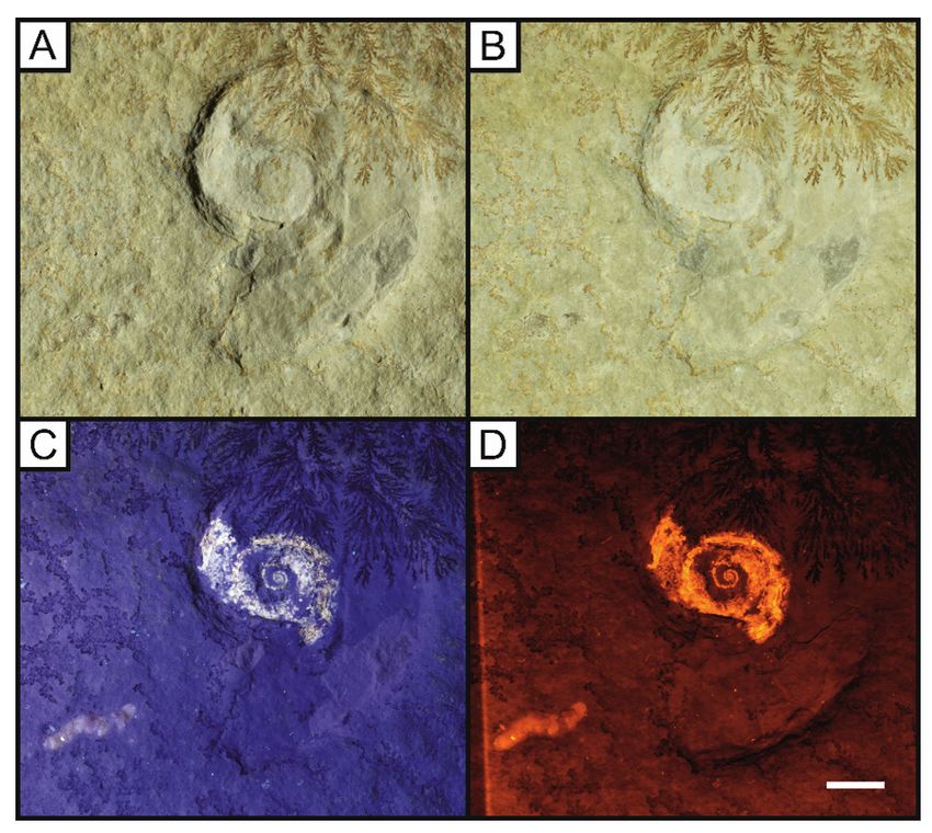
Page 18/32Figure 6
The oppeliid ammonite Neochetoceras sp. with some phragmacone uorescence and the ammonite
coprolite Lumbricaria attributed to LB 4. A, direct white light recording a slight colour difference around
the phragmacone and very little evidence of the coprolite; B, oblique white light; C, UV light uorescing the
coprolite, siphuncle and phragmacone; D, LSF displaying full uorescence of the phragmacone,
increasing the observed contrast. Note the brighter siphuncle under 532 nm green laser light in D, allowing
for a greater distinction than under UV light seen in C. Scale bar = 10mm.
Page 19/32Figure 7
The plesioteuthid squid Plesioteuthis prisca with a uorescent gladius (LB 5). This squid can be seen
under oblique (A) and direct white light (B). 365nm UV light uoresces the rachis in this specimen,
showing that the central rachis is raised (C). LSF using the 532 nm laser uoresces this gladius at
different levels with the central vane picked out through its higher uorescence. Scale bar = 10mm.
Figure 8
An isolated appendage of the fossil prawn Aeger tipularius under different lighting conditions (A-D) (LB
6). A, oblique white light image, casting shadows on the slight relief present; B, direct white light; C, Same
specimen under 365 nm ultraviolet uorescence, highlighting the specimen from the background and
recovering some unseen setae that appear broken; D, Laser-stimulated uorescence image of the
Page 20/32specimen under 532 nm green laser displaying an improvement over the UV image seen in C with the
complete leg revealed along with none of the gaps present under UV. Scale bar = 10mm.
Figure 9
The eryonid crustacean Cycleryon propinquus von Schlotheim, 1822 under white light (A), 365 nm UV (B)
and LSF (C) revealing the full outline of the animal under a 532 nm green laser (LB 7). D and E represent
comparisons of the uorescent techniques on the rst pereopod (P1). Note the missing section under UV
light is revealed through the subsurface illumination of LSF. Scale represents 1cm. D and E magni ed
x1.5.
Page 21/32Figure 10
Complete fossil of the penaeid shrimp Antrimpos speciosus Münster, 1839 (LB 8) under different lighting
conditions; A, oblique white light showing the split rock revealing the original exoskeleton; B, direct white
light; C, UV uorescence increases the contrast with the background by illuminating the specimen and
leaving the matrix dark; D, LSF reveals the entire body outline of the animal and this not seen in the white
light or UV photographs. Note the antennulae (atl), swimmerets (sw), walking legs (wl) and urostyle (ur)
Page 22/32which were not fully visible in white light. Abbreviations following C. Haug et al. (2009). The green laser
wavelength used on this specimen was 532 nm. Scale bar = 10 mm.
Figure 11
Isolated UV (A-C) and LSF (D-F) images from Fig. 10 highlighting the differences in revealed detail. B and
E are UV and LSF images of the revealed antennae with clear segmentation visible under LSF. C and F
highlight the swimmerets that are fully revealed under LSF with full segmentation. Abbreviations as
Page 23/32above with the addition of the antennular peduncle (ped) from Audo and Charbonnier (2012). Scale bar =
10mm. B and E magni ed x2 and C and F magni ed x3.6.
Figure 12
Specimen LB 9 of the caridean shrimp Alcmonacaris winkleri under oblique white light (A), direct white
light (B), UV uorescence (C) and LSF (D). Notice the faint outline under UV is enhanced through LSF to
reveal the full animal, allowing the fossil to be labelled. Scale = 10mm. The features revealed use the
same abbreviations as Fig. 9 with the addition of the carapace (ca). Abbreviations following C. Haug et
al. (2009). The LSF wavelength used was 532 nm.
Page 24/32Figure 13
Specimen SNSB-BSPG 1935 I 24 of the pterodactyloid pterosaur Ctenochasma gracile under white light
(A) and LSF (B). This individual is fully articulated with a blue uorescent soft tissue body outline and
blue cartilage between the light orange bones. A 405 nm blue laser was used to uoresce this specimen.
Page 25/32Figure 14
Specimen SNSB-BSPG AS I 745a of the pterodactyloid pterosaur Germanodactylus rhamphastinus under
white light (A) and LSF (B). The skull and upper torso have an increased contrast under LSF through a
405 nm blue laser, separating the specimen from the surrounding matrix. Specimen badge measures
5cm.
Page 26/32Figure 15
Specimen SNSB-BSPG 1977 XIX 1 of the pterodactyloid pterosaur Germanodactylus rhamphastinus on a
counterplate slab under white light (A) and LSF (B). The counterplate slab has some missing bone
material where it remains dark under LSF. A 405 nm blue laser was used to carry out LSF and uoresces
any preserved bone material.
Page 27/32Figure 16
Specimen SNSB-BSPG 1937 I 27 of the atoposaurid crocodyliform Alligatorellus beaumonti bavaricus
under white light (A) and LSF (B). The colour patterning makes clear distinction possibly between the
skeleton and osteoderms and the surrounding soft tissues. Note the brightly uorescing soft tissues at
the base of the tail, possibly phosphatised remnants of muscle tissue. LSF was carried out using a 405
nm blue laser. Scale represents 2cm.
Page 28/32Figure 17
A specimen of the teleost sh Leptolepides sprattiformes Blainville, 1818 (LB 10), under white light (A),
UV uorescence (B) and LSF (C). Not the completed soft tissue outline that is brighter than the ossi ed
material. Gut contents uoresce brightly at the centre of the body cavity along with a coprolite illuminated
at the posterior. The laser used on this specimen was a 532 nm green laser. Scale = 10 mm.
Page 29/32Figure 18
A near complete specimen of the salmoniform sh Orthogonikleithrus hoelli, lacking the dorsal section of
caudal n (LB 11). A, oblique white light photograph showing a complete vertebral column and faint skull
and caudal n elements; B, direct white light image; C, UV uorescence highlighting the bones of the skull
along with the pectoral n; C, enhanced uorescence from B showing the caudal n is incomplete along
with uorescing the entire skull. LSF was carried out with a 532 nm green laser. Scale bar = 20 mm.
Page 30/32Figure 19
Two small specimens of the salmoniform sh Orthogonikleithrus hoelli on the same slab (LB 12) under
different light conditions. A, oblique white light photograph with a dark vertebral column and lighter skull
and ns present; B, UV photograph with complete skull and tail revealed. Dorsal and pelvic ns are also
revealed through uorescence with illumination from the left; C, LSF image revealing the entire dorsal and
pelvic ns on the right specimen that are faint under UV. Note the erect position of the dorsal and pelvic
ns on the left and relaxed position on the right. A 532 nm green laser was used to carry out LSF. Scale
bar = 10mm.
Page 31/32Figure 20
Specimen LB 13 of the extinct bulldog sh Allothrissops salmoneus (Blainville, 1818) under white light
(A), UV (B) and LSF (C) conditions. The uorescence around the articulated skeleton reveals loose scales
along with gular plates and gut contents. A 532 nm green laser was used to carry out LSF. Scale bar = 10
mm.
Page 32/32You can also read