Preventive Vaccination with Telomerase Controls Tumor Growth in Genetically Engineered and Carcinogen-Induced Mouse Models of Cancer
←
→
Page content transcription
If your browser does not render page correctly, please read the page content below
Research Article

Preventive Vaccination with Telomerase Controls Tumor Growth
in Genetically Engineered and Carcinogen-Induced
Mouse Models of Cancer
                               1                       2,3                         1                               1                            5
Carmela Mennuni, Stefano Ugel, Federica Mori, Barbara Cipriani, Manuela Iezzi,
                 5                 1                   1                1
Tania Pannellini, Domenico Lazzaro, Gennaro Ciliberto, Nicola La Monica,
                2                4                   1
Paola Zanovello, Vincenzo Bronte, and Elisa Scarselli
1
  Istituto di Ricerca di Biologia Molecolare, Pomezia, Italy; 2Department of Oncology and Surgical Science; 3Venetian Institute for Molecular
Medicine; 4Istituto Oncologico Veneto, Padova, Italy; and 5Aging Research Center (CeSI), ‘‘G. d’Annunzio’’ University Foundation,
Chieti Scalo, Italy

Abstract                                                                                      recognized by T lymphocytes specific against telomerase (2, 3).
The telomerase reverse transcriptase, TERT, is an attractive                                  These findings have justified cancer vaccination clinical trials
target for human cancer vaccination because its expression is                                 based either on autologous dendritic cells transfected with human
reactivated in a conspicuous fraction of human tumors.                                        TERT–derived peptides (4, 5).
Genetic vaccination with murine telomerase (mTERT) could                                         To exploit the immunogenicity versus tolerance issue, we vac-
break immune tolerance in different mouse strains and                                         cinated mice with mouse TERT (mTERT)–based DNA vaccines.
resulted in the induction of both CD4+ and CD8+ telomerase-                                   Gene-based vaccination strategies are probably the most promis-
specific T cells. The mTERT-derived immunodominant epi-                                       ing approaches to induce effective cell-mediated immunity (CMI)
topes recognized by CD8+ T cells were further defined in these                                against cancer (6). Delivery of antigens by DNA injection allows
mouse strains and used to track immune responses. Anti-                                       access to multiple antigen-presenting pathways. Moreover, in-
tumor efficacy of telomerase-based vaccination was investi-                                   creased expression of the antigen and enhanced immunogenicity
gated in two cancer models closely resembling human                                           can be achieved by physical methods, including in vivo electro-
diseases: the TRAMP transgenic mice for prostate cancer                                       poration (7, 8), and molecular modifications, such as the use of
                                                                                              codon-optimized sequences and the fusion of the target antigen
and a carcinogen-induced model for colon cancer. TERT
overexpression in tumor lesions was shown in both models                                      to microbial-derived proteins (9, 10). DNA vaccines are safe and
by immunohistochemistry, thus reinforcing the similarity of                                   easy to produce; moreover, they can be used repeatedly for long-
these tumors to their human counterparts. Repeated immu-                                      term maintenance of antitumor CMI.
nizations with mTERT-encoding DNA resulted in a significant                                      There are differences in telomerase activity between humans and
delay of tumor formation and progression in both the prostate                                 rodents: in particular, telomerase has higher basal levels of activity
cancer and the colon cancer models. Moreover, evaluation                                      in mouse tissues compared with humans. Nevertheless, reactiva-
of the intratumoral infiltrate revealed the presence of telo-                                 tion of telomerase is commonly occurring in mouse tumors as well
merase-specific T cells in vaccinated mice. The safety of                                     and abrogation of telomerase activity in mouse cancerous cells
vaccination was confirmed by the absence of histomorpho-                                      impairs their metastatic potential (11, 12). Many of the data about
                                                                                              the efficacy of genetic vaccination with mTERT were obtained with
logic changes on postnecropsy analysis of several organs
and lack of adverse effects on blood cell counts. These results                               transplantable tumor models but the correlation of responses to
indicate that TERT vaccination can elicit antigen-specific                                    immunization with overexpression of telomerase was not investi-
immunosurveillance and imply this antigen as a potential                                      gated (13). Transplantable tumor cell lines are useful for immu-
candidate for preventive cancer vaccines. [Cancer Res 2008;                                   nologic studies but genetically engineered and chemically induced
68(23):9865–74]                                                                               tumors in rodents are considered better models for evaluating
                                                                                              results transferable to the clinical setting (14). The TRAMP mouse
                                                                                              model is genetically engineered to expresses SV40 large T antigen
Introduction                                                                                  preferentially in the prostate epithelium to develop prostate tumors
   TERT is considered a very attractive antigen for cancer therapy                            (15). The carcinogen 1,2-dimethylhydrazine (DMH) is widely used
(1). Telomerase is a ribonucleoprotein in which the protein                                   for the induction of colon cancers in mouse (16, 17). Because our
component, TERT, uses its RNA as template for adding telomeric                                immunohistochemistry data showed mTERT overexpression in
repeat sequences to the ends of chromosomes. TERT is shut down                                both prostate and colon tumors, we used these experimental
in most human somatic tissues but reactivated in 85% of tumors,                               models to monitor the effect of mTERT-based vaccination. Here, we
conferring unlimited replicative potential to neoplastic cells. When                          show that TERT-based genetic vaccination induces telomerase-
telomerase is reactivated in tumor cells, TERT is processed and                               specific CD8+ T cells able to infiltrate tumor lesions and affects
presented together with class I MHC molecules and tumors are                                  various stages of tumor progression in both tumor models without
                                                                                              causing any detectable adverse effect.
   Note: C. Mennuni and S. Ugel contributed equally to this work.
   Requests for reprints: Vincenzo Bronte, Istituto Oncologico Veneto, Via                    Materials and Methods
Gattamelata 64, 35128 Padova, Italy. Phone: 39-049-8215897; Fax: 39-049-8072854;
E-mail: enzo.bronte@unipd.it.
                                                                                                 Mice and cell lines. TRAMP mice and TRAMP-C2 cell line were a gift
   I2008 American Association for Cancer Research.                                            from N. M. Greenberg (Fred Hutchinson Cancer Research Center, Seattle,
   doi:10.1158/0008-5472.CAN-08-1603                                                          WA). The presence of PB-Tag gene in TRAMP mice was tested by PCR as

www.aacrjournals.org                                                                   9865                              Cancer Res 2008; 68: (23). December 1, 2008

            Downloaded from cancerres.aacrjournals.org on December 23, 2020. © 2008 American Association for Cancer
                                                           Research.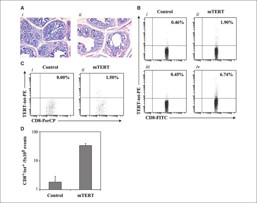
Cancer Research

described (15) and heterozygous TRAMP mice were used in the experi-            The Lasso tool (Adobe Photoshop software) was used to perform quan-
ments. C57Bl/6 (H-2b) and BALB/c (H-2d) mice were from Charles River.          titative image analysis.
Mice were treated in accordance with national legislation and European            Immunohistochemistry. For the detection of telomerase, cryostat
guidelines by means of an internal review process. MBL-2 (H-2b) is a C57Bl/    sections (4–10 Am) of colon and prostate were incubated, respectively,
6 leukemia line, B16 (H-2b) is a C57Bl/6 melanoma line, and CT26 (H-2d) is a   with the anti-mouse telomerase ab177 and with ab23699 (Abcam run out of
BALB/c colon carcinoma.                                                        the ab177 during our study; Abcam) for overnight at 4jC. Briefly, sections
   DNA vectors and immunization procedures. The synthetic codon                were rinsed in PBS-0.1% Triton and incubated with the secondary anti-
optimized sequence coding for TPA-mTERT-LTB (mTERT-LTB) was                    rabbit antibody. To reveal the reaction, we used an ABC Complex (Vector)
obtained by GENEART and cloned into vector pV1JnsA (18). cDNA                  for 30 min at room temperature. Only in colon tumors, staining was
encoding mTERT wild-type sequence was obtained by Geron Corp. and              enhanced by nickel-3,3¶-diaminobenzidine.
the relevant gene sequence was excited with NotI and HindIII and cloned           For the detection of tumor-infiltrating lymphocytes (TIL), prostate tissue
in VR1055 (Vical). As control, empty pV1JnsA vector and VR1055 expressing      slides were incubated with rat anti-mouse CD8 (AbD Serotec) for 30 min
h-galactosidase (h-gal) were used. DNA immunization was performed              followed by 30-min incubation with goat anti-rat IgG (Jackson ImmunoR-
according to commonly used protocols available at the DNA vaccine Web          esearch). To reveal the reaction, slides were incubated for 30 min with
site (8).6                                                                     alkaline phosphatase–conjugated streptavidin and stained with fuxin
   Leukocyte cultures. Spleens were removed from the animals and cell          chromogen substrate for 15 min followed by a hematoxylin counterstain.
suspension was prepared. Mouse peripheral blood mononuclear cells                 Cytofluorimetric evaluation of TERT-specific TILs. TILs were isolated
(PBMC) were obtained by retroocular bleeding. RBCs were removed by             by means of a mechanical separation and by enzymatic digestion with
osmotic lyses. Mixed leukocyte cultures (MLC) were set up with TRAMP           300 units/mL DNase, 0.1% hyaluronidase, and 1% collagenase buffer. After
splenocytes (3  106) coincubated with g-irradiated allogeneic BALB/c          30-min incubation at 37jC, the cells were filtered through a 70-Am sieve and
splenocytes (3  106). Mixed leukocyte peptide cultures (MLPC) were set up     resuspended in fluorescence-activated cell sorting (FACS) buffer with anti-
with either naive or vaccinated TRAMP splenocytes (5  106) restimulated       mouse Fc-gR for 10 min at room temperature. After washing, cells were
in vitro with 1 Amol/L of mTERT198-205 peptide (VGRNFTNL) or h-gal96-103       labeled with mTERT198-205 (H-2b) tetramer-PE (5 Ag/mL; Beckman Coulter)
peptide (DAPIYTNV). Culture was grown for 5 d in DMEM-10% fetal bovine         for 20 min at room temperature. Samples were then stained at 4jC with rat
serum at 37jC in 5% CO2. Peptides were purchased from JPT.                     anti-CD8-PerCP(Ly-2) or FITC. Cells were analyzed with FACS Canto II and
   Intracellular staining for IFN-;. Intracellular staining (ICS) was          data were elaborated using DIVA software (BD Biosciences).
performed as described (19). Cells were then fixed with formaldehyde 1%           Induction of colon tumors by DMH and evaluation of tumor lesions.
in PBS and analyzed on a FACS Canto II using DIVA software (Becton             Treatment of BALB/c mice with DMH [20 mg/kg in Tris-HCl (pH 6.5)] was
Dickinson).                                                                    started at week 6 of age and was performed once a week for 6 wk. Animals
   Enzyme-linked immunosorbent spot assays. Ninety-six–well MAIP               were sacrificed to evaluate tumor lesions and colons were dissected, split
plates (Millipore) were coated overnight with a 2.5 Ag/mL solution of rat      open on filter paper, and fixed in 4% formalin-1 PBS. Lesions were stained
anti-mouse IFN-g (BD Pharmingen). Cells were plated at 1  106 per well in     in 1% methylene blue observed microscopically and scored by two
triplicate and incubated for 20 h at 37jC with 2 Ag/mL peptides. After         independent operators. Colons were discolored by 70% ethanol and paraffin
incubation, plates were washed with PBS and 0.05% Tween 20 and                 embedded, and 5-mm-thick sections were H&E stained for tumor grading.
incubated overnight at 4jC with anti-mouse biotin-conjugated anti-IFN-g        Tumor grading has been assessed and G1-G3 stages were assigned based on
antibody. The day after, streptavidin-alkaline phosphatase conjugate was       tumor differentiation grade and mitotic event number following AJCC
added for 2 h. Plates were developed by adding nitroblue tetrazolium/          Cancer Staging Manual, Sixth Edition.
5-bromo-4-chloro-3-indolyl phosphate (Pierce) and spots were then                 Safety assessment. Twenty BALB/c female mice, 6 wk old, were
counted using an automated enzyme-linked immunosorbent spot (ELI-              immunized as described in the previous section. Immunized mice and age-
SPOT) reader.                                                                  matched control mice were sacrificed at 38 wk of age. Hematologic counts
   ELISA. Splenocytes (105 cells) from MLPCs were restimulated for 24 h in     were performed (ADVIA 120) and several organs were collected (liver, lungs,
triplicates with an equal amount of target cells, the supernatants were        spleen, kidneys, hearth, injection sites in femoral quadriceps, lymph nodes,
harvested, and released IFN-g was measured by ELISA (Endogen).                 bone marrow, and Peyers patches). Tissues were immediately fixed in 10%
   Cytotoxicity assay. Cytotoxicity was measured by 51Cr release assay.        buffered formalin. Histopathologic evaluation was performed by the Safety
MLPCs were incubated either with MBL-2 cells pulsed with the peptides or       Assessment and Pathology (MSD-Chibret, Paris, France).
with B16 target cells pretreated when indicated for 24 h with IFN-g (25 ng/       Statistical analysis. The Wilcoxon-Mann-Whitney U test was used to
mL). Target cells were labeled with 51Cr (100 ACi; Amersham) and then          examine the null hypothesis of rank identity between two sets of
mixed with effector cells. Assays were performed in triplicate and             nonparametric data. Student’s t test was used to compare parametric
supernatants were harvested to measure 51Cr released. The percent of           groups. Kaplan-Maier plots and the Mantel-Haenszel test were used to
specific lysis was calculated as previously described (20).                    compare survival of mice belonging to different treatment groups. All
   Histologic evaluation of prostate lesions in TRAMP. The urogenital          P values presented are two sided.
tract was removed at necroscopy and prepared for pathologic evaluation:
tissues were fixed in paraformaldehyde/lysine/periodate for 3 h at 4jC,
infiltrated with 30% sucrose in PBS overnight at 4jC, and frozen in OCT.       Results
Histologic sections were analyzed with a standard H&E stain. The sec-            DNA vaccination with mTERT breaks tolerance in healthy
tions were evaluated by a pathologist blinded to the treatment groups          mice. The mTERT gene sequence was modified by fusing Escherichia
according to Kaplan-Lefko guidelines (21). The presence of normal tissue,      coli LTB sequence to its COOH terminal part (22). LTB is a secreted
prostate intraepithelial neoplasia (PIN; epithelial stratification with        protein; therefore, to fully incorporate its adjuvant function in the
occasional mitotic figures or cribriform pattern), adenocarcinoma (ADC;        vaccine, we introduced a secretion signal before the telomerase
abnormal glands or multiple papillae with vessel-rich stromal axes lined by
                                                                               sequence. Moreover, for safety purposes, the mTERT sequence was
epithelial cells with numerous mitoses), poorly differentiated carcinoma
(PDC; sheets of small cells with a low nucleus-cytoplasm ratio with no         modified in two amino acid positions to be catalytically inactive (23).
glandular formation, numerous mitotic, and apoptotic figures) was scored.      This fusion construct was used to exploit the induction of anti-TERT
                                                                               CMI in healthy mice of both C57Bl/6 and BALB/c strains. Mice
                                                                               were therefore subjected to five weekly injections before measuring
                                                                               CMI. The immune response was evaluated among splenocytes by
  6
      http://dnavaccine.com                                                    ICS for IFN-g–producing cells. TERT peptides were divided into

Cancer Res 2008; 68: (23). December 1, 2008                                9866                                                   www.aacrjournals.org

            Downloaded from cancerres.aacrjournals.org on December 23, 2020. © 2008 American Association for Cancer
                                                           Research.
Telomerase-Based Genetic Vaccination
three pools (mTERT pool 1 containing the sequence from amino with that obtained with the LTB fusion construct, but a weaker CD8+
acids 1–388, pool 2 containing the sequence from amino acids 377– T-cell response (Fig. 1C). CD4+ T-cell reactivity was also detected
811, and pool 3 containing the sequence from amino acids 801– toward LTB sequence but only when present in the construct (Fig.
1122). This approach allows detection of both CD4+ and CD8+ 1C). Overall, these results indicate that DNA vaccination with
antigen-activated T cells (24). ICS indicated that the CD8+ T-cell mTERT is immunogenic in a ‘‘self ’’ context.
epitope was contained within the mTERT pool 1, whereas CD4+ Immune response induced by vaccination against telomerase
T-cell reactivity was stimulated by pools 2 and 3 in BALB/c mice is long lived and safe. Twenty BALB/c mice were immunized
(Fig. 1A). C57Bl/6 mice showed CD8+ T-cell reactivity against both by five weekly injections with mTERT-LTB. Immune response was
mTERT pools 1 and 2, whereas CD4+ T-cell response in this strain analyzed from each individual mouse by ICS on PBMC. PBMCs
was detected only toward the NH2-terminal region of the mTERT were collected before starting the immunization (week 6 of age),
protein (mTERT-1 peptide pool; Fig. 1B). Intervals of 1 or 2 weeks shortly after the end of the immunization protocol (week 13),
between immunizations were comparable in terms of CMI induction and finally at 38 weeks of age. Blood samples were evaluated for
and two DNA injections were sufficient to induce an immune their reactivity against mTERT peptide pool 1, previously shown
response against mTERT (data not shown). Finally, to contain the class I MHC-restricted epitope for BALB/c mice.
we compared our optimized mTERT fusion construct to mTERT The anti-TERT CD8+ T-cell response was not changed even a long
plain construct in BALB/c mice. The mTERT plain construct was time after the immunization (Fig. 1D). In spite of the presence of
also immunogenic, inducing a CD4+ T-cell response comparable circulating antitelomerase CD8+ T cells, no alteration of blood cell
Figure 1. Evaluation of immune reactivity in mice immunized with mTERT. A, BALB/c mice immunized with mTERT-LTB. B, C57Bl/6 mice immunized with
mTERT-LTB. For all four panels, cutoff values were set as the average reactivity of six mice immunized with mock plasmid plus 2 SD (0.05% for CD4+ and 0.09%
for CD8+ T cells, respectively). In A and B, splenocytes were exposed to the three TERT peptides pools. Columns, average frequencies from 10 individual mice;
bars, SD. *, responses significantly different from mice immunized with mock plasmid (P < 0.05, Student’s t test). C, comparison of CMI evaluated by ICS in BALB/c
mice immunized either with mTERT construct (black columns ) or with mTERT-LTB fusion (gray columns ). Columns, average of eight mice per group; bars, SD. *,
responses significantly different in the two groups (P < 0.05, Student’s t test). D, longevity of CD8+ T cells induced by vaccination in BALB/c mice immunized with
mTERT-LTB. PBMCs were exposed to mTERT-1 peptide pool and analyzed by ICS for IFN-g production before (week 6) and twice after the immunization (weeks 13
and 38). Columns, average values of 10 mice; bars, SD.
www.aacrjournals.org 9867 Cancer Res 2008; 68: (23). December 1, 2008
Downloaded from cancerres.aacrjournals.org on December 23, 2020. © 2008 American Association for Cancer
Research.Cancer Research
counts was observed between vaccinated (n = 10) and age-matched 9-mer epitopes were synthesized and tested individually. Only
control (n = 4; average WBC of 7.1 103/mm3 and RBC of 11.1 one epitope corresponding to the mTERT167-175, AYQVCGSPL, was
106/mm3 in vaccinated mice versus WBC of 7.6 103/mm3 and RBC recognized in all BALB/c mice (Fig. 2B). Two different epitopes
of 11.5 106/mm3 in untreated mice). Moreover, no significant recognized by CD8+ T cells were instead identified in C57Bl/6 mice:
histomorphologic changes were noted in mice from vaccinated mTERT198-205 VGRNFTNL and mTERT505-512 SLGKYGKL. However,
group compared with untreated mice. Two mice among those although the peptide mTERT198-205 was recognized by freshly
receiving the vaccine exhibited focal skeletal muscle fiber atrophy isolated splenocytes of immunized mice (Fig. 2C) and could also
with focal or multifocal mineralization, which suggested marginal expand mTERT-specific CD8+ T cells following in vitro stimula-
postinjury scar, possibly related to the immunization procedure. tion (shown in next figures), mTERT505-512 failed to expand primed
Identification of mTERT immunogenic epitopes in BALB/c T cells (data not shown). We thus decided to use mTERT198-205
and C57Bl/6 mice strains. To characterize better mTERT epitopes VGRNFTNL epitope for subsequent monitoring of the immune
recognized by CD8+ T cells in different mouse strains, midi-pools response in C57Bl/6 mice.
consisting of twelve 15-mers were prepared so that each individual DNA vaccination with mTERT induces CD8+ T-cell response
peptide was present into two different midi-pools. Each pair of in TRAMP mice. The TRAMP model of prostate cancer was
peptide pools, including the same 15-mer, was plated on 96-well genetically engineered to target SV40 T antigen expression almost
plates using a matrix scheme. Reactivity was found against the exclusively in the prostate tissue, leading to progressive prostate
midi-pools D, Q, and R as shown in Fig. 2A for BALB/c mice. Once cancer development in all the mice (15). Despite tumor growth,
the reactive overlapping 15-mers were identified, the putative TRAMP mice remained immunocompetent at different stages,
Figure 2. Identification of the epitopes recognized by CD8+ T cells in BALB/c and C57Bl/6 mice. A, ELISPOT analysis for the identification of reactive 15-mer
peptide contained in mTERT pool 1 was performed by determining the reactivity against a series of midi-pools indicated with letters from A to W. B, ELISPOT analysis of
the reactivity of five single BALB/c mice (1–5 ) toward the H-2d–restricted CD8+ T-cell epitope mTERT167-175 (AYQVCGSPL). C, ELISPOT analysis of the reactivity of
five individual C57Bl/6 mice (1–5 ) toward the H-2b–resticted epitope mTERT198-205 (VGRNFTNL). In B and C , IFN-g–producing cells are enumerated as SFC/106
splenocytes from mice immunized with mTERT-LTB plasmid. Cutoff values were set as the average reactivity of six mice immunized with mock plasmid plus 2 SD
(12 SFC/106 splenocytes).
Cancer Res 2008; 68: (23). December 1, 2008 9868 www.aacrjournals.org
Downloaded from cancerres.aacrjournals.org on December 23, 2020. © 2008 American Association for Cancer
Research.Telomerase-Based Genetic Vaccination
including the late stages of tumor development (week 48). In fact, prostatic tissue was more represented (30.64% versus 7.37%;
splenocytes of TRAMP mice of 8, 24, 36, and 48 weeks of age P < 0.05) in mTERT-vaccinated mice (Fig. 4B, i–iii). A second
recognized in vitro allogeneic target cells (Fig. 3A). Surprisingly, in group of TRAMP mice was monitored until either spontaneous
48-week-old TRAMP mice, there was a weak but reproducible death or the appearance of either painful or life-threatening
spontaneous immune response against the immunodominant complications. A clear effect of vaccination on overall survival was
mTERT198-205 epitope, which was absent in younger TRAMP mice observed with both plasmid constructs expressing TERT (P = 0.004,
(P = 0.021; Fig. 3A). However, it was clear that this spontaneous cumulative data are shown in Fig. 4C).
response was unable to control tumor progression, similarly to Telomerase-specific T cells infiltrate prostate tumors.
what was observed in humans (25). TRAMP mice immunized at the 6th week of age were euthanized
Starting from week 6, TRAMP mice were vaccinated twice, at at week 24 and tumor sections were evaluated by immunohisto-
2-week intervals, with either h-gal–encoding or mTERT-encoding chemistry to detect tumor-infiltrating CD8+ T cells. CD8+ T cells
plasmids. Two weeks after the second vaccine dose, IFN-g ELISPOT were present in both mock- and mTERT-vaccinated mice, indi-
showed that mTERT immunization was able to prime and expand cating that this tumor is permissive for T-cell infiltration (Fig. 5A,
antigen-specific CD8+ T cells recognizing only the mTERT198-205 i and ii). However, many CD8+ T cells were in direct contact with
epitope and not the control h-gal96-103 peptide (P = 0.035; Fig. 3B). tumor epithelial cells in mTERT-vaccinated mice (Fig. 5A, ii). To
The numbers of spot-forming colonies (SFC)/106 cell obtained in identify mTERT-specific CD8+ T cells, we used mTERT198-205-Kb
this assay were comparable with those reported in Fig. 2C for wild- fluorescent tetramers (tet). Firstly, we showed the specificity of this
type C57Bl/6 mice. Mice vaccinated with h-gal–coding plasmid reagent by staining, both ex vivo and after in vitro stimulation with
recognized only h-gall96-103 peptide and not mTERT198-205 peptide mTERT198-205 peptide, telomerase-specific splenocytes induced by
(P = 0.0045; Fig. 3B). The numbers of IFN-g–producing splenocytes vaccination (Fig. 5B). Once this reagent was validated, 24-week-old
in TRAMP mice vaccinated with ether mTERT or mTERT-LTB TRAMP mice were sacrificed and single-cell suspension derived
plasmid DNA were similar, suggesting the induction of comparable from prostate tissues was stained with mTERT198-205-Kb fluorescent
immune responses in C57Bl/6 mice (Fig. 3B). tetramers and anti-CD8 monoclonal antibodies (mAb) and
The antigen-specific T-cell population was expanded in vitro by analyzed by FACS (Fig. 5C, i and ii). CD8+/tet+ cells were detected
stimulating effector lymphocytes with mTERT198-205 peptide in among tumor-infiltrating CD8+ T cells in three of three mTERT-
MLPC. As expected, these peptide-stimulated cultures were able vaccinated mice, with an average of 33.4 F 6 CD8+/tet+/5 105
to release IFN-g when coincubated with mTERT198-205-pulsed cell total events analyzed by FACS (Fig. 5D). CD8+ infiltrating T cells
targets (Fig. 3C). Moreover, the TERT-specific T cells also were also detected in three of three mice vaccinated with the
recognized to the same extent unpulsed, syngeneic B16 melanoma control plasmid but the tet+ cells were not present in the infiltrate
and TRAMP-C2 prostate cancer cells. This recognition of naturally (average, 1.8 F 1 CD8+/tet+/5 105 total events; Fig. 5D, mTERT
processed TERT peptides was increased on pretreatment of B16 versus mock vaccinated, P = 0.02). Confirming histologic data, the
cells with IFN-g, known to up-regulate surface class I MHC percentage of tumor-infiltrating CD8+ T cells was comparable in
expression (Fig. 3C). Finally, we also evaluated the cytotoxicity of the two groups (3,220 F 1,200 CD8+/5 105 total events in the
TERT-specific MLPC. Lytic activity was shown both against target control and 2,868 F 712 CD8+/5 105 total events in mTERT-
cells pulsed with mTERT198-205 epitope and, to a lower extent, vaccinated mice).
against B16 and TRAMP-C2 tumor cells naturally processing the Vaccination of DMH-treated BALB/c controls tumor growth.
endogenous TERT antigen (Fig. 3D). A second cancer model was used to evaluate efficacy of DNA
Vaccination of TRAMP mice reduces the prostate areas vaccination approach. Mouse exposure to DMH carcinogen led to
affected by the tumor and increases long-term survival. As progressive development of multiple tumors in the colon in all
described in human prostatic carcinoma, overexpression of exposed mice (16). Tumor progression resembles human colorectal
telomerase was appreciable from the early stages of prostate cancer, both macroscopically and microscopically, being charac-
epithelial transformation in TRAMP mice (26). A large proportion terized by the formation of aberrant crypt proceeding to adenoma
of the nuclei of the hyperplastic epithelium, in fact, showed a and finally carcinoma (17). We firstly showed mTERT over-
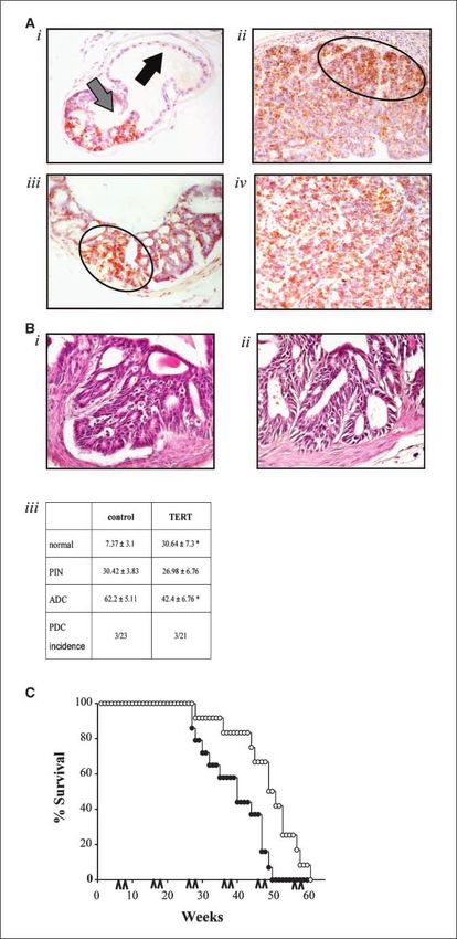
moderate to strong staining for mTERT (Fig. 4A, i–iv). The expression in samples obtained from DMH-treated animals. In
positivity was conserved along the neoplastic progression to PIN control animals (vehicle treated), mTERT staining was mostly
and ADC. Noteworthy, the staining was stronger at the edges of restricted to the lower segment of the colon crypts, where proli-
ADC, correlating with the proliferative activity of the tumor. PDC ferative cells are residing (stem cells and committed stem cells;
tumors also expressed mTERT in the majority of cells (Fig. 4A, iv). Fig. 6A, i). On the contrary, in DMH-induced animals, the immu-
To evaluate the in vivo vaccination efficacy, TRAMP mice were nostaining showed an upper shift of mTERT pattern of expression
immunized with both TERT plasmids from the 6th week of age. and positive cells were detected in all three segments of the crypt
DNA was administered at biweekly intervals and the immunization (bottom, mid, and upper segments; Fig. 6A, ii) already 6 weeks after
schedule was repeated every 10 weeks. A first group of vaccinated DMH induction of tumors. This observation agrees with the
TRAMP mice was euthanized at week 24 for histologic examina- proliferative shift induced by the carcinogen, which has been
tion. Pathologic evaluation indicated that prostates of control extensively described in this model of colon rectal cancer (27).
plasmid–treated animals had extensive areas of PIN and ADC with It is noteworthy that mTERT not only is expressed in the mid-third
unpolarized proliferating cells (Fig. 4B, i), whereas prostates of of the crypt, where transit cells migrate and are still differentiat-
mTERT-vaccinated TRAMP mice showed areas of PIN constituted ing and partly proliferating, but also in the upper third of the crypt,
by more differentiated and polarized cells (Fig. 4B, ii). Quantitative where terminally differentiated cells and apoptotic cells are resid-
image analysis of prostates confirmed that only 42.4% of their area ing in normal situation. At very late stages after DMH induction
was occupied by ADC in the mouse group vaccinated with mTERT (35 weeks), several ADCs were detectable, and mTERT signal was
compared with 62.2% in the controls (P < 0.05). Conversely, normal
www.aacrjournals.org 9869 Cancer Res 2008; 68: (23). December 1, 2008
Downloaded from cancerres.aacrjournals.org on December 23, 2020. © 2008 American Association for Cancer
Research.Cancer Research
Figure 3. Evaluation of TERT-specific immune response in vaccinated TRAMP mice. A, TRAMP mice of different ages (8, 24, 36, and 48 wk) were euthanized
and their splenocytes were used to set up allogeneic and peptide-stimulated cultures (MLC and MLPC, respectively). MLCs (black columns) and MLPCs (gray columns)
were tested in IFN-g ELISA. MLPC values of IFN-g released in the presence of cells pulsed with control h-gal96-103 peptide were subtracted from the values
obtained with the mTERT198-205 peptide-pulsed cells. For allogeneic MLCs, IFN-g release is reported as the difference between the values obtained in the presence
of the allogeneic targets (CT26 cells) and syngeneic targets (MBL-2 cells). Columns, mean; bars, SD. Student’s t test: mTERT198-205 responsiveness of 48-wk
TRAMP mice versus 8, 24, and 36 wk, P = 0.02. Where no SD is present, data are greater than the assay limit. B, splenocytes of TRAMP mice, vaccinated at
6th week of age, with either TERT-LTB fusion (TERT-LTB ), full-length TERT (TERT ), h-gal plasmid (b-gal ), or control empty plasmid (control ) were tested ex vivo
in an IFN-g ELISPOT assay with either mTERT198-205 (right ) or h-gal96-103 (left) peptides. Columns, mean of triplicate wells of one representative experiment;
bars, SD. Student’s t test statistics: mTERT198-205 mice vaccinated with mTERT plasmid DNA versus mice vaccinated with h-gal–encoding plasmid DNA, P = 0.005;
mTERT198-205 mice vaccinated with mTERT-LTB fusion plasmid DNA versus mice vaccinated with h-gal–encoding plasmid DNA, P = 0.014; mTERT198-205 mice
vaccinated with mTERT plasmid DNA versus mice vaccinated with mTERT-LTB fusion plasmid DNA, P = 0.6 (not significant). C, MLPCs from mice immunized
with either mTERT (black columns ) or h-gal (gray columns ) were tested in IFN-g ELISA against mTERT198-205 peptide-pulsed or h-gal96-103 peptide-pulsed
MBL-2 cells, TRAMP-C2 cells, and B16 melanoma cell line either untreated (B16) or pretreated with IFN-g (B16 IFN-c ) for 24 h. As additional control, B16 melanoma
cells pulsed with mTERT198-205 peptide was included (B16 mTERT ). Columns, mean of triplicate wells; bars, SD. Where no SD is present, data are greater than
the assay limit. D, MLPCs were tested in a cytotoxicity test by 51Cr release assay against mTERT198-205 peptide-pulsed or h-gall96-103 peptide-pulsed MBL-2,
TRAMP-C2 prostate cancer cells, and B16 melanoma cells, either treated or untreated with IFN-g.
clearly appreciated in the mucosa adjacent to large tumors and in development was investigated by starting the immunization
several areas of the tumoral mass (Fig. 6A, iii and iv). schedule 15 weeks after DMH induction. Mice received five weekly
We performed two different vaccination experiments in this DNA injections of the TERT-LTB fusion construct. DMH treatment
model. In the ‘‘prophylactic setting,’’ we immunized mice 4 weeks did not compromise the immune responsiveness in mice (28), and
after DMH induction, when macroscopic tumor lesions were in agreement with previous findings, we found that the immune
not yet detectable in the large bowel. In the ‘‘early therapeutic responsiveness against mTERT198-205 was indistinguishable from
setting,’’ the effect of the mTERT vaccination on later tumor stage that measured in control BALB/c mice (average, 155 F 94 SFC/106
Cancer Res 2008; 68: (23). December 1, 2008 9870 www.aacrjournals.org
Downloaded from cancerres.aacrjournals.org on December 23, 2020. © 2008 American Association for Cancer
Research.Telomerase-Based Genetic Vaccination
Figure 4. Efficacy of TERT vaccination controlling
prostate cancer progression in TRAMP mice.
A, immunohistochemistry of TERT expression during
prostate carcinogenesis. i, mTERT, scarcely
expressed in the normal portion of the prostatic duct
(black arrow ), is hyperexpressed in numerous cells of
the early hyperplastic lesions (gray arrow ). ii, in ducts
affected by PIN (irregularly shaped glands with
cribriform architecture), TERT remains widely
hyperexpressed, particularly in zones with evident
architectural disorder (circled ). iii, TERT positivity
seems diffuse in the ADC cells and particularly marked
along the invasion front (circled ). iv, in PDC tumors,
TERT is irregularly expressed in most cells. B,
histology of prostate tissues in vaccinated TRAMP
mice sacrificed at the 24th week of age. i, prostates of
control plasmid–treated animals showed extensive
areas of PIN and ADC with unpolarized proliferating
cells. ii, prostates of TERT-vaccinated TRAMP mice
showed areas of PIN constituted by more differentiated
and polarized cells. iii, table summarizing the
percentage of prostate areas occupied by normal
tissue, PIN, and ADC in control and mTERT-
vaccinated mice. Values are expressed as mean F
SD. *, statistically significant difference from the control
(P < 0.05, n = 23 mice in each experimental group). C,
survival curves. The cycle of vaccination,
composed of two DNA injections at biweekly interval,
was repeated every 10 wk, starting from week 6
(bottom arrowheads ). Survival curves are from
TRAMP mice vaccinated with either mTERT
(empty circle ; n = 15) or control plasmids (black circle ;
n = 15). Mantel-Haenszel test: mTERT plasmid
DNA versus empty plasmid DNA, P = 0.004.
www.aacrjournals.org 9871 Cancer Res 2008; 68: (23). December 1, 2008
Downloaded from cancerres.aacrjournals.org on December 23, 2020. © 2008 American Association for Cancer
Research.Cancer Research
in vehicle treated versus 118 F 83 SFC/106 in DMH treated; n = 6 effect of vaccination on prevention of early benign lesions (aberrant
mice/group). crypt foci and early adenomas), whereas the effect on more advanced
Vaccination efficacy was evaluated by euthanizing mice 12 weeks stages is mainly on progression of existing lesions toward larger size
after DMH induction for the prophylactic setting. At this time, and less differentiated tumors.
only benign tumors are detectable; the most frequent lesion is
multiple aberrant foci crypts and few early adenomas are also
present. A significant reduction in the number of multiple aberrant Discussion
foci crypts and of early adenomas was observed (P = 1 10 8, To the best of our knowledge, this article shows, for the first
Fig. 6B, and P = 9 10 9, data not shown, respectively). In the time, that mTERT-based vaccination can induce mTERT epitope-
second experiment (vaccination in early therapeutic setting), specific CD8+ T cells and alter the complex chain of cancerogenesis
mice were sacrificed at week 28 from DMH induction. Analysis of events in experimental models other than transplantable tumors,
tumor was performed by counting the number of late adenoma leading to a significant prolongation of survival. TERT antigen thus
lesions and measuring their size. Results showed that the joins, to all intents and purposes, HER2/NEU oncogene and
vaccination significantly reduced the number of late adenomas, prostate stem cell antigen (PSCA) in the list of potential candidates
as well as the adenoma size, compared with untreated mice to design new vaccines for tumor prevention (29–31). Moreover,
(P = 0.026, data not shown; P = 0.0001, Fig. 6C). In addition, given the broader expression of telomerase in cancer, compared
histologic evaluation was performed on 12 late adenomas isolated with HER2/NEU oncogene and PSCA, TERT-based immunotherapy
from mice in each experimental group. Notably, 70% of the widens preventive vaccination application to a larger fraction of
adenomas isolated from mice vaccinated with mTERT-LTB were human cancers.
in the G1-G2 differentiation state, whereas 85% of those isolated Although telomerase overexpression was described in both
from control mice were already in a more advanced G3 histology models of colon and prostate cancers, once neoplastic lesions were
grade (Fig. 6D, P = 0.01). Overall, these results indicate a powerful established, the initial phase of cancer progression seemed to be
Figure 5. Analysis of TILs in prostates from TRAMP mice. A, immunohistochemistry
evaluation of CD8+ T cells. Sections from 24-wk-old TRAMP mouse vaccinated
either with the control plasmid (i) or with mTERT plasmid (ii ). Arrows, lymphocytes
in close contact with epithelial cells. B, FACS staining of splenocytes from TRAMP
mice with anti-CD8 mAb and TERT-tet. Ex vivo analysis from a pool of five mice
immunized with either the control plasmid (i) or the mTERT plasmid (ii ).
Cytofluorimetric analysis of splenocytes week after in vitro stimulation with
mTERT198-205 peptide: iii, mice immunized with control plasmid; iv, mice immunized
with mTERT plasmid. C, FACS analysis of TILs in mouse prostates of 24-wk-old
TRAMP mice. The frequencies of double-positive CD8+/tet+ population are shown
in a representative control (i) and mTERT-vaccinated mouse (ii ). D, average
numbers of double-positive CD8+/tet+ cells on the total events acquired by
FACS (5 105) in the two groups (P = 0.02, Student’s t test; n = 3 mice/group).
Cancer Res 2008; 68: (23). December 1, 2008 9872 www.aacrjournals.org
Downloaded from cancerres.aacrjournals.org on December 23, 2020. © 2008 American Association for Cancer
Research.Telomerase-Based Genetic Vaccination
Figure 6. Efficacy of TERT vaccination in a carcinogen-induced colon cancer model. A, immunohistochemistry for detection of TERT in DMH-treated mice. i, vehicle
(VEH )-treated animals 5 wk after the beginning of treatment: the staining is mostly restricted to the lower third of the colon crypts (arrows ). ii, DMH-treated
animals 5 wk after the beginning of treatment: staining is distributed in all the segments of the colon crypts (arrows ). Bar, 70 Am. iii, 35 wk after the beginning of
DMH treatment: the mucosa adjacent to a large tumor mass is showing TERT-specific staining in the different segments of the crypts (arrows ) and also in hyperplastic
crypts (arrowhead). iv, portion of large ADC: positive (arrows ) and negative areas (asterisks ) are depicted. Bar, 80 Am. B, average number of aberrant crypt foci/
mouse in mice vaccinated with TERT-LTB fusion in a prophylactic setting compared with mice treated with the control plasmid (20 mice/group; P = 1 10 8,
Student’s t test). C, average size of adenomas in mice vaccinated with TERT-LTB in an early therapeutic setting compared with mice treated with control plasmid
(20 mice/group; P = 0.0001, Wilcoxon Rank sum test). D, frequencies of G1 (well differentiated), G2 (moderately differentiated), and G3 (poorly differentiated)
tumors in groups treated in an early therapeutic setting (12 late adenomas/group; P = 0.01, Wilcoxon-Mann-Whitney U test).
more affected from active vaccination. This was also true when est avidity for mTERT198-205 peptide-Kb complexes.7 TERT-based
comparing the higher significance of the mTERT vaccination on immunotherapy might thus benefit from the adoptive transfer
either ‘‘prophylactic’’ or ‘‘early therapeutic’’ settings of the DMH- of TERT-specific CD8+ T cells and our initial studies indicate that
induced colon cancer model (Fig. 6). This effect might depend this approach might affect more advanced forms of prostate cancer
either on progressive weakening of the vaccination efficacy or in TRAMP mice.
establishment of evasive maneuvers from neoplastic cells. Indeed, There are no data supporting TERT antigen loss in vaccinated
the data about the functional activity of mTERT-specific CD8+ versus untreated mice at late disease stages (data not shown), and
T cells suggest that the overall avidity of the effectors generated by thus, progressive weakening of the immune response due to active
vaccination is low (Fig. 3C and D). These CD8+ T cells, in fact, escape from tumor seems a more likely possibility. In TRAMP mice,
recognize very efficiently mTERT198-205 peptide-pulsed target we found that the combination of arginase (ARG) and nitric oxide
cells but much less wild-type tumors (Fig. 3C and D). This low synthase (NOS) inhibitors was essential to restore full tumor
functional avidity is the consequence of the low frequency of recognition by TILs infiltrating advanced prostate tumors, and
mTERT-specific CD8+ T cells and this population can be enriched
by in vitro repeated stimulations with the antigen followed by
cloning of CD8+ T cells possessing a T-cell receptor with the high- 7
S. Ugel et al., in preparation.
www.aacrjournals.org 9873 Cancer Res 2008; 68: (23). December 1, 2008
Downloaded from cancerres.aacrjournals.org on December 23, 2020. © 2008 American Association for Cancer
Research.Cancer Research
similar data were obtained with human prostate organ cultures immune response against telomerase is observed in TRAMP mice
(32). These results allowed us to identify a dominant mechanism (Fig. 3A) and in some patients with cancer (33). In line with our
based on L-arginine metabolism by which mouse and human observations, a naturally arising response against the ubiquitous
prostate cancers restrain tumor-specific T lymphocytes and offered histone H4 protein was recently described among CD8+ T cells
novel perspectives for the immunotherapy of cancer. We are, in infiltrating prostate cancer in TRAMP mice, indicating that ubiqui-
fact, developing ARG and NOS dual inhibitors to be administered tous proteins may become tumor antigens only in the context
to tumor-bearing mice in combination with either vaccination or of the tumor (34). Telomerase and histone H4 thus belong to a cate-
the adoptive transfer of mTERT-specific CD8+ T cells. gory of antigens that are normally ignored by the immune system,
The results of our studies have also other important implications for which a T-cell repertoire with antitumor activity exists and can
for the development of clinical cancer vaccines. The TERT-LTB be rescued by active immunization without apparent side effects.
fusion construct seems to prime a better CD8+ T-cell response in
some mouse strains (Fig. 1C) and this might even be more relevant
Disclosure of Potential Conflicts of Interest
when the genetic heterogeneity of human beings is considered.
Moreover, we showed that repeated immunization with mTERT No potential conflicts of interest were disclosed.
resulted in a sustained immune response for prolonged periods
without the appearance of overt autoimmune manifestations and Acknowledgments
hematopoiesis impairment. These findings suggest that normal Received 4/30/2008; revised 9/23/2008; accepted 9/25/2008.
tissues and hematopoietic stem cells do not became target of Grant support: Italian Ministry of Health, Italian Association for Cancer Research,
Fondazione CRT, Progetto Prostata, and Istituto Superiore Sanità-Alleanza Contro il
antitelomerase T cells. Telomerase, under normal circumstances, Cancro (project no. ACC8).
is likely segregated in molecular complexes and not efficiently The costs of publication of this article were defrayed in part by the payment of page
processed and presented in the context of MHC class I molecules charges. This article must therefore be hereby marked advertisement in accordance
with 18 U.S.C. Section 1734 solely to indicate this fact.
and therefore ignored by the immune system. This hypothesis is We thank Francesco Calvaruso and Raffaele Cerino for their excellent technical
further strengthened by the observation that a spontaneous assistance and Arthur Fridman for statistical analysis.
References 12. Bagheri S, Nosrati M, Li S, et al. Genes and pathways oligomerization and telomerase activity. J Biol Chem
downstream of telomerase in melanoma metastasis. 2002;277:8538–44.
1. Harley CB. Telomerase and cancer therapeutics. Nat Proc Natl Acad Sci U S A 2006;103:11306–11. 24. Draenert R, Altfeld M, Brander C, et al. Comparison
Rev Cancer 2008;8:167–79. 13. Nair SK, Heiser A, Boczkowski D, et al. Induction of of overlapping peptide sets for detection of antiviral
2. Vonderheide RH. Universal tumor antigens for cancer cytotoxic T cell responses and tumor immunity against CD8 and CD4 T cell responses. J Immunol Methods
vaccination: targeting telomerase for immunopreven- unrelated tumors using telomerase reverse transcriptase 2003;275:19–29.
tion. Discov Med 2007;7:103–8. RNA transfected dendritic cells. Nat Med 2000;6:1011–7. 25. Nagorsen D, Scheibenbogen C, Letsch A, et al. T cell
3. Chen DY, Vance BA, Thompson LB, Domchek SM, 14. Gutmann DH, Hunter-Schaedle K, Shannon KM. responses against tumor associated antigens and
Vonderheide RH. Differential lysis of tumors by Harnessing preclinical mouse models to inform human prognosis in colorectal cancer patients. J Transl Med
polyclonal T cell lines and T cell clones specific for clinical cancer trials. J Clin Invest 2006;116:847–52. 2005;3:3.
hTERT. Cancer Biol Ther 2007;6:1991–6. 15. Greenberg NM, DeMayo F, Finegold MJ, et al. 26. Puebla-Mora AG, Heras A, Cano-Valdez AM, Domi-
4. Su Z, Dannull J, Yang BK, et al. Telomerase Prostate cancer in a transgenic mouse. Proc Natl Acad nguez-Malagon H. Human telomerase and a-methyl-
mRNA-transfected dendritic cells stimulate antigen- Sci U S A 1995;92:3439–43. acyl-coenzyme A racemase in prostatic carcinoma. A
specific CD8+ and CD4+ T cell responses in patients 16. Chang WW. The mode of formation and progression comparative immunohistochemical study. Ann Diagn
with metastatic prostate cancer. J Immunol 2005;174: of chemically induced colonic carcinoma. Prog Clin Biol Pathol 2006;10:205–8.
3798–807. Res 1985;186:217–35. 27. Mori F, Piro FR, Della Rocca C, et al. Survivin
5. Domchek SM, Recio A, Mick R, et al. Telomerase- 17. Gilbert JM. Experimental colorectal cancer as a model and cyclooxygenase-2 are co-expressed in human
specific T-cell immunity in breast cancer: effect of of human disease. Ann R Coll Surg Engl 1987;69:48–53. and mouse colon carcinoma and in terminally
vaccination on tumor immunosurveillance. Cancer Res 18. Montgomery DL, Shiver JW, Leander KR, et al. differentiated colonocytes. Histol Histopathol 2007;
2007;67:10546–55. Heterologous and homologous protection against influ- 22:61–77.
6. Rice J, Ottensmeier CH, Stevenson FK. DNA vaccines: enza A by DNA vaccination: optimization of DNA 28. Coca S, Enrech S, Moreno Garcia V, et al. Evaluation
precision tools for activating effective immunity against vectors. DNA Cell Biol 1993;12:777–83. of the antitumor activity of interleukin-12 in an
cancer. Nat Rev Cancer 2008;8:108–20. 19. Cipriani B, Fridman A, Bendtsen C, et al. Therapeutic experimental murine model of colorectal cancer in-
7. Widera G, Austin M, Rabussay D, et al. Increased DNA vaccination halts disease progression in BALB-neuT mice: duced by 1,2 dimethyl-hydrazine (DMH). Rev Esp
vaccine delivery and immunogenicity by electroporation the amplitude of elicited immune response is predictive Enferm Dig 2005;97:619–28.
in vivo . J Immunol 2000;164:4635–40. of vaccine efficacy. Hum Gene Ther 2008;19:670–80. 29. Lollini PL, Cavallo F, Nanni P, Forni G. Vaccines for
8. Cappelletti M, Zampaglione I, Rizzuto G, Ciliberto G, 20. De Palma R, Marigo I, Del Galdo F, et al. Therapeutic tumour prevention. Nat Rev Cancer 2006;6:204–16.
La Monica N, Fattori E. Gene electro-transfer improves effectiveness of recombinant cancer vaccines is associated 30. Garcia-Hernandez Mde L, Gray A, Hubby B, Klinger
transduction by modifying the fate of intramuscular with a prevalent T-cell receptor a usage by melanoma- OJ, Kast WM. Prostate stem cell antigen vaccination
DNA. J Gene Med 2003;5:324–32. specific CD8+ T lymphocytes. Cancer Res 2004;64:8068–76. induces a long-term protective immune response
9. Mennuni C, Calvaruso F, Facciabene A, et al. Efficient 21. Bono AV, Montironi R, Pannellini T, et al. Effects of against prostate cancer in the absence of autoimmunity.
induction of T-cell responses to carcinoembryonic castration on the development of prostate adenocarci- Cancer Res 2008;68:861–9.
antigen by a heterologous prime-boost regimen using noma from its precursor HGPIN and on the occurrence 31. Finn OJ. Cancer immunology. N Engl J Med 2008;358:
DNA and adenovirus vectors carrying a codon usage of androgen-independent, poorly differentiated carcino- 2704–15.
optimized cDNA. Int J Cancer 2005;117:444–55. ma in TRAMP mice. Prostate Cancer Prostatic Dis, 32. Bronte V, Kasic T, Gri G, et al. Boosting antitumor
10. Rice J, Dossett ML, Ohlen C, et al. DNA fusion Eur J Immunol 2008;38:2118–30. responses of T lymphocytes infiltrating human prostate
gene vaccination mobilizes effective anti-leukemic cyto- 22. Facciabene A, Aurisicchio L, Elia L, et al. Vectors cancers. J Exp Med 2005;201:1257–68.
toxic T lymphocytes from a tolerized repertoire. Eur J encoding carcinoembryonic antigen fused to the B subunit 33. Filaci G, Fravega M, Setti M, et al. Frequency of
Immunol 2008;38:2118–30. of heat-labile enterotoxin elicit antigen-specific immune telomerase-specific CD8+ T lymphocytes in patients
11. Blasco MA, Rizen M, Greider CW, Hanahan D. responses and antitumor effects. Vaccine 2007;26:47–58. with cancer. Blood 2006;107:1505–12.
Differential regulation of telomerase activity and 23. Arai K, Masutomi K, Khurts S, Kaneko S, Kobayashi 34. Savage PA, Vosseller K, Kang C, et al. Recognition of a
telomerase RNA during multi-stage tumorigenesis. Nat K, Murakami S. Two independent regions of human ubiquitous self antigen by prostate cancer-infiltrating
Genet 1996;12:200–4. telomerase reverse transcriptase are important for its CD8+ T lymphocytes. Science 2008;319:215–20.
Cancer Res 2008; 68: (23). December 1, 2008 9874 www.aacrjournals.org
Downloaded from cancerres.aacrjournals.org on December 23, 2020. © 2008 American Association for Cancer
Research.Preventive Vaccination with Telomerase Controls Tumor
Growth in Genetically Engineered and Carcinogen-Induced
Mouse Models of Cancer
Carmela Mennuni, Stefano Ugel, Federica Mori, et al.
Cancer Res 2008;68:9865-9874.
Updated version Access the most recent version of this article at:
http://cancerres.aacrjournals.org/content/68/23/9865
Cited articles This article cites 34 articles, 11 of which you can access for free at:
http://cancerres.aacrjournals.org/content/68/23/9865.full#ref-list-1
Citing articles This article has been cited by 9 HighWire-hosted articles. Access the articles at:
http://cancerres.aacrjournals.org/content/68/23/9865.full#related-urls
E-mail alerts Sign up to receive free email-alerts related to this article or journal.
Reprints and To order reprints of this article or to subscribe to the journal, contact the AACR Publications
Subscriptions Department at pubs@aacr.org.
Permissions To request permission to re-use all or part of this article, use this link
http://cancerres.aacrjournals.org/content/68/23/9865.
Click on "Request Permissions" which will take you to the Copyright Clearance Center's
(CCC)
Rightslink site.
Downloaded from cancerres.aacrjournals.org on December 23, 2020. © 2008 American Association for Cancer
Research.You can also read


