Human RTEL1 associates with Poldip3 to facilitate responses to replication stress and R-loop resolution - Genes Dev
←
→
Page content transcription
If your browser does not render page correctly, please read the page content below
Downloaded from genesdev.cshlp.org on October 23, 2020 - Published by Cold Spring Harbor Laboratory Press

Human RTEL1 associates with Poldip3
to facilitate responses to replication stress
and R-loop resolution
Andrea Björkman,1 Søren L. Johansen,2 Lin Lin,3,4 Mike Schertzer,5 Dimitris C. Kanellis,1
Anna-Maria Katsori,1 Søren T. Christensen,2 Yonglun Luo,3,4 Jens S. Andersen,6 Simon J. Elsässer,1
Arturo Londono-Vallejo,5 Jiri Bartek,1,7 and Kenneth B. Schou1
1
 Division of Genome Biology, Department of Medical Biochemistry and Biophysics, Science for Life Laboratory, Karolinska
Institute, Solna 171 77, Sweden; 2Department of Cell Biology and Physiology, University of Copenhagen, DK-2100 Copenhagen,
Denmark; 3Department of Biomedicine, Aarhus University, Aarhus 8200, Denmark; 4Steno Diabetes Center Aarhus, Aarhus
University Hospital, Aarhus 8200, Denmark; 53UMR 3244 (Telomere and Cancer Laboratory), Centre National de la Recherche
Scientifique, Institut Curie, PSL Research University, Sorbonne Universités, Paris 75005, France; 6Department of Biochemistry and
Molecular Biology, University of Southern Denmark, DK-5230 Odense M, Denmark; 7Danish Cancer Society Research Centre,
DK-2100 Copenhagen, Denmark

RTEL1 helicase is a component of DNA repair and telomere maintenance machineries. While RTEL1’s role in DNA
replication is emerging, how RTEL1 preserves genomic stability during replication remains elusive. Here we used a
range of proteomic, biochemical, cell, and molecular biology and gene editing approaches to provide further insights
into potential role(s) of RTEL1 in DNA replication and genome integrity maintenance. Our results from comple-
mentary human cell culture models established that RTEL1 and the Polδ subunit Poldip3 form a complex and are/
function mutually dependent in chromatin binding after replication stress. Loss of RTEL1 and Poldip3 leads to
marked R-loop accumulation that is confined to sites of active replication, enhances endogenous replication stress,
and fuels ensuing genomic instability. The impact of depleting RTEL1 and Poldip3 is epistatic, consistent with our
proposed concept of these two proteins operating in a shared pathway involved in DNA replication control under
stress conditions. Overall, our data highlight a previously unsuspected role of RTEL1 and Poldip3 in R-loop sup-
pression at genomic regions where transcription and replication intersect, with implications for human diseases
including cancer.
[Keywords: RTEL1; helicase; R-loop; DNA:RNA hybrid; Poldip3; POLδ; polymerase δ; DNA repair; DNA damage
response; telomere maintenance; dyskeratosis congenita; Hoyeral-Hreiderson syndrome]
Supplemental material is available for this article.
Received June 25, 2019; revised version accepted May 18, 2020.

RTEL1 (regulator of telomere length 1) was identified as a                  mediates and quadruplex structures during DNA repair
cancer susceptibility gene (Shete et al. 2009; Wrensch                      and meiotic crossover (Barber et al. 2008; Youds et al.
et al. 2009; Vannier et al. 2014) and implicated in a num-                  2010; Vannier et al. 2012). More recently, RTEL1 was
ber of telomere dysfunction syndromes (Ballew et al.                        shown to play a broader role in replication through associ-
2013a,b; Deng et al. 2013; Le Guen et al. 2013; Walne                       ation with the proliferating cell nuclear antigen (PCNA) of
et al. 2013). Studies in both mice and human cells have                     the replisome, pointing to a critical role of RTEL1 at the
shown that RTEL1 plays dual roles in preserving both                        replication fork for genetic stability (Vannier et al. 2013).
telomere integrity and prevention of chromosomal abnor-                     However, whether PCNA represents the key physiologi-
malities on a genome-wide scale (Ding et al. 2004; Vannier                  cal partner for the function(s) of RTEL1 in DNA replica-
et al. 2013; Speckmann et al. 2017; Porreca et al. 2018). Ac-               tion remains to be addressed. Likewise, the localization
cordingly, RTEL1 was previously demonstrated to act as                      and function of RTEL1 at the replisome remains unclear.
an antirecombinase by evading excessive sister chromatid                       In this study, we examined the emerging role of RTEL1
exchange and resolving DNA D-loop recombination inter-                      in DNA replication. We identified a functional interplay
                                                                            between RTEL1 and the Poldip3 protein. Poldip3 has
Corresponding authors: jb@cancer.dk; kenneth.schou@ki.se
Article published online ahead of print. Article and publication date are
online at http://www.genesdev.org/cgi/doi/10.1101/gad.330050.119. Free-     © 2020 Björkman et al. This article, published in Genes & Development,
ly available online through the Genes & Development Open Access             is available under a Creative Commons License (Attribution 4.0 Interna-
option.                                                                     tional), as described at http://creativecommons.org/licenses/by/4.0/.

                  GENES & DEVELOPMENT 34:1–10 Published by Cold Spring Harbor Laboratory Press; ISSN 0890-9369/20; www.genesdev.org              1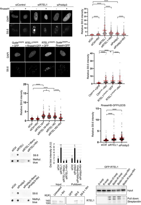
Downloaded from genesdev.cshlp.org on October 23, 2020 - Published by Cold Spring Harbor Laboratory Press

Björkman et al.

putative functions in two different complexes in human                  A                                B
cells, namely the DNA polymerase δ (POLδ) and the
TREX complex, respectively, implicating Poldip3 in
diverse biological processes including DNA synthesis
and mRNA trafficking. We found that RTEL1 and Poldip3
physically interact and recruit to chromatin in a mutually
dependent manner in response to replication stress. Con-
sistently, RTEL1 and Poldip3 depletion leads to elevated
RNA–DNA hybrid (R-loop) accumulation and these R-
loops are confined to sites of active replication. Further-
more, we show here that the R-loop accumulation after
RTEL1 and Poldip3 depletion occurs at genomic common
fragile sites, rDNA and telomeres. Our results reveal a
new role of RTEL1 (and Poldip3) in protection against
genome instability by preventing excessive R-loop accu-
mulation after replication stress, a condition emerging
as a hallmark of cancer and implicated in aging (Halazone-              C
tis et al. 2008; Merchut-Maya et al. 2019), thus providing
mechanistic insights into the role of RTEL1 in safe-                                                     D
guarding proper genome-wide replication and genomic
integrity.

Results

Poldip3 is a novel RTEL1-associated protein
To explore the function of RTEL1 in the regulation of cell
proliferation and DNA repair, we searched for novel                    Figure 1. RTEL1 associates with Poldip3. (A) Schematic flow
RTEL1-interacting proteins using the Flag tag affinity pu-             chart of the Flag affinity purification protocol of Flag-RTEL1.
rification method. To enrich crude lysates for nuclear pro-            Flag-CHFR or Flag-RTEL1 or expressing HeLa cells were harvest-
                                                                       ed and whole cell lysates were treated with a cocktail of DNases
teins including those proteins enriched on chromatin, we
                                                                       and RNases. Flag-RTEL1 fusion proteins were Flag immunopuri-
digested whole-cell extracts with a panel of DNA and
                                                                       fied, eluted with Flag peptides, and immunocomplexes resolved
RNA nucleases prior to Flag affinity purification analysis             by PAGE. (B) Silver staining of resolved proteins from the Flag-
(Fig. 1A). The Flag-purified protein complexes were re-                RTEL1 immunoprecipitation. Control immunoprecipitation
solved by gel electrophoresis and proteins were visualized             was performed with a Flag-CHFR fusion protein. CHFR was
by silver staining (Fig. 1B). Flag-RTEL1 protein complexes             used as control since it, like RTEL1, is a RING finger-containing
revealed several specifically coeluted proteins, one                   protein. Flag-CHFR, Flag-RTEL1 and Poldip3 are indicated with
of which was identified by mass spectrometry as the                    arrows. (C) GFP-RTEL1 binds HA-Poldip3. HEK293T cells were
PDIP46/Poldip3/SKAR (subsequently referred to as Pol-                  cotransfected with HA-Poldip3 and either GFP or GFP-RTEL1
dip3) (Supplemental Fig. S1A), a poorly understood protein             and incubated for 24 h prior to harvest. Extracts were subjected
                                                                       to GFP immunoprecipitation followed by immunoblotting with
with dual roles in the nuclear mRNA trafficking TREX/
                                                                       HA and GFP antibodies. (D) Endogenous RTEL1 binds endoge-
THO complex and DNA polymerase δ (Polδ) complex                        nous Poldip3. U2OS cells were harvested in EBC buffer and cell
(Heath et al. 2016; Lee et al. 2017). We confirmed this in-            lysates were treated as in A. Precleared lysates were subjected
teraction by coimmunoprecipitation where GFP-tagged                    to immunoprecipitation with either IgG or RTEL1 antibody.
RTEL1 specifically pulled down HA-tagged Poldip3                       RTEL1 immunocomplexes were released from the RTEL1-conju-
when expressed in human HEK293T cells (Fig. 1C). This                  gated resin by treatment with glycine-HCl buffer.
interaction was not mediated by unspecific DNA inter-
mediates since DNA nuclease treatment of whole-cell ly-
sates did not affect the interaction (Supplemental Fig.                holocomplex. Indeed, besides binding to Poldip3, coim-
S1B). In addition, we could demonstrate that endogenous                munoprecipitation assays also identified specific physical
RTEL1 and Poldip3 proteins interacted in U2OS cells (Fig.              interactions between RTEL1 and POLD1 and POLD3 (Fig.
1D), suggesting a biologically relevant function of this               2A). Supporting this finding, upon fractionation of whole-
interplay.                                                             cell extracts by size exclusion chromatography, RTEL1
   Given that Poldip3 was recently demonstrated to be a                cofractionated with Poldip3 as well as POLD1 and
stoichiometric subunit of the DNA polymerase δ (POLδ)                  POLD3 subunits (Fig. 2B) thus establishing that RTEL1
holocomplex (Lee et al. 2017) and additional POLδ com-                 and the POLδ holoenzyme reside in a genuine protein
plex subunits have previously been identified in RTEL1                 complex. Since the evidence suggested that RTEL1 and
immunocomplexes (i.e., POLD1 and POLD3) (Vannier                       Poldip3 might exist in a stable complex, we assessed
et al. 2013; Schertzer et al. 2015), we asked whether                  whether their respective protein stabilities might be af-
RTEL1 might bind additional components of the POLδ                     fected by each other, with immunoblot analyses of

2      GENES & DEVELOPMENT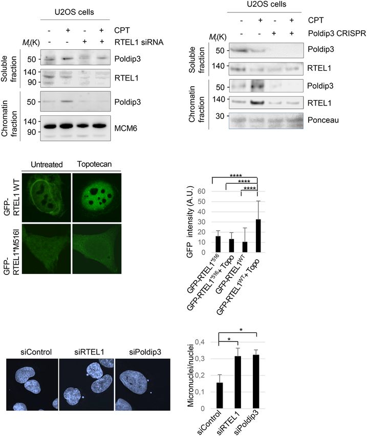
Downloaded from genesdev.cshlp.org on October 23, 2020 - Published by Cold Spring Harbor Laboratory Press

                                                                                                        RTEL1 and R-loop resolution

A                              C                        D                        Figure 2. RTEL1 binds other Poldδ subunits
                                                                                 and directly to Poldip3 through its helicase
                                                                                 domain. (A) RTEL1 binds POLD3 and POLD1
                                                                                 subunits of the Polδ complex. Cell extracts
                                                                                 from HEK293T cells transfected with Flag-
                                                                                 RTEL1 or mock transfected were cleared and
                                                                                 subjected to Flag immunoprecipitation with
                                                                                 anti-Flag beads followed by PAGE. Proteins
                                                                                 were probed by immunoblotting using the in-
                                                                                 dicted antibodies. (B) Gel filtration analysis
B                                                                                of HEK293T cell extracts. Proteins were
                                                                                 probed by immunoblotting using the indicted
                                                                                 antibodies. (C) Codepletion of RTEL1 by Pol-
                                                                                 dip3 silencing. U2OS cells transfected with ei-
                                                                                 ther control or Poldip3 siRNAs were incubated
                                                                                 4 d and subsequently harvested and proteins
                                                                                 immunoblotted using the indicated antibod-
                                                                                 ies. (D) Purified RTEL1 and Poldip3 interact
                                                                                 in vitro. Bacterially purified GST-RTEL1 was
E                                     F                                          incubated with high-salt-purified HA (control)
                                                                                 or HA-Poldip3 on HA-conjugated beads. Elud-
                                                                                 ed protein complexes were subjected to immu-
                                                                                 noblot analysis and proteins were probed using
                                                                                 either HA or RTEL1 antibodies. (E) Schematic
                                                                                 representation of Flag-RTEL1 deletion con-
                                                                                 structs. (F ) RTEL1 binds Poldip3 primarily
                                                                                 via its 4FeS helicase domain. HEK293T cells
                                                                                 were transfected with Flag-RTEL1 full-length
G                                                                                or deletion constructs spanning the indicated
                                                                                 regions of RTEL1 to determine the Poldip3-
                                                                                 binding site. The cell lysates were subjected
                                                                                 to Flag immunoprecipitation followed by
                                                                                 3xFlag peptide elution and, subsequently, pro-
                                                                                 teins were subjected to immunoblotting with
                                                                                 Flag or Poldip3 antibodies. (G) GFP pull-
                                                                                 down analysis of mutated GFP-RTEL1.
                                                                                 HEK293T cells were cotransfected with HA-
                                                                                 Poldip3 and either GFP-RTEL1 WT or GFP-
RTEL1 containing the Hoyeraal-Hreidarsson syndrome mutations at M516I, L734R, G763V, or R1264H. Cell extracts were subjected
to GFP immunoprecipitation followed by immunoblotting with GFP or HA antibodies.

RTEL1 and Poldip3 in U2OS cells that had been depleted             previously associated with dyskeratosis congenita/Hoyer-
of Poldip3 using RNA interference. RTEL1 protein levels            aal-Hreidarsson syndrome are scattered throughout the
were dramatically decreased in U2OS cells depleted of              tertiary structure of RTEL1, including some in the heli-
Poldip3 (Fig. 2C), further supporting the notion that              case domain, we asked whether any of these mutations
RTEL1 and Poldip3 associate in a stable complex. As                might disrupt the integrity of the emerging RTEL1–Pol-
RTEL1 also interacts with PCNA (Vannier et al. 2013),              dip3 complex (Supplemental Fig. S2A). Interestingly,
similarly to the replisome component POLδ, we exam-                among the mutated versions of RTEL1, RTEL1 containing
ined whether RTEL1 interacts directly with Poldip3.                a methionine-to-isoleucine substitution at position 516
High-salt-purified HA-Poldip3 coupled to an anti-HA res-           (M516I) demonstrated a dramatically reduced capacity
in was examined for its capacity to bind to bacterially pu-        to bind Poldip3 (Fig. 2G; Supplemental Fig. S2B). Methio-
rified GST-tagged RTEL1 or GST alone. A HA pull-down               nine 516 is a highly conserved residue in the helicase
assay revealed that HA-Poldip3 bound specifically to               domain of RTEL1 (Supplemental Fig. S2B), suggesting
GST-RTEL1, indicating that RTEL1 and Poldip3 interact              that the structural integrity of the helicase domain is crit-
directly (Fig. 2D). To gain further insight into the struc-        ical for RTEL1 binding to Poldip3.
tural requirements for the RTEL1–Poldip3 interaction,
we performed a deletion analysis to delineate the minimal
                                                                   RTEL1 and Poldip3 recruit to chromatin after
region of RTEL1 required for its interaction with Poldip3
                                                                   topoisomerase I inhibition
(Fig. 2E). Truncation of RTEL1 indicated that Poldip3
bound prominently to the RTEL1 N terminus harboring                Because RTEL1 function has been implicated in DNA rep-
the helicase domain (Fig. 2F), further supporting the no-          lication (Vannier et al. 2013), analogous to POLδ, we inves-
tion that RTEL1 and Poldip3 reside in a physical complex.          tigated the possible regulatory role of RTEL1 toward
Considering that disease-associated mutations in RTEL1             Poldip3 functions in the DNA damage response upon

                                                                                                  GENES & DEVELOPMENT            3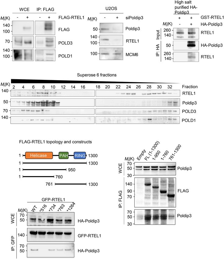
Downloaded from genesdev.cshlp.org on October 23, 2020 - Published by Cold Spring Harbor Laboratory Press

Björkman et al.

replication stress. Exposure to replication inhibitors                 dip3 act together on chromatin in response to TOPOI inhi-
hydroxyurea (HU) or camptothecin (CPT) failed to aug-                  bition-induced replication stress. Supporting the notion
ment the association of ectopically expressed RTEL1 and                that RTEL1 function relies on its interaction with Poldip3,
Poldip3 (Supplemental Fig. S2C), suggesting that the                   we found that GFP-tagged RTEL1 wild type but not M516I
RTEL1–Poldip3 complex stability is unaffected by replica-              mutated RTEL1 translocated from the cytoplasm to the
tion stress. RTEL1- and Poldip3-deficient cells were previ-            nucleus after TOPOI inhibition (Fig. 3C,D). A key pheno-
ously shown to be hypersensitive to topoisomerase I                    type of RTEL1 deficiency is hyperrecombination, reflected
(TOPOI) poisons (Barber et al. 2008; Tumini et al. 2016).              in an increase of RAD51 nuclear foci due to the accumula-
This prompted us to test whether RTEL1 is involved in                  tion of recombination intermediates that persist and fail to
the chromatin loading of Poldip3 in TOPOI-inhibited cells              be appropriately repaired (Barber et al. 2008). Likewise, we
in S phase. Interestingly, while TOPOI inhibition by CPT               found that Poldip3 loss augmented nuclear RAD51 stain-
treatment increased chromatin retention of Poldip3 in                  ing (Supplemental Fig. S2D,E), indicating that Poldip3 defi-
RTEL1-proficient cells, RTEL1 ablation markedly im-                    ciency phenocopies the hyperrecombination phenotype of
paired this outcome (Fig. 3A), indicating that RTEL1 is in-            RTEL1. Consistent with the occurrence of accumulated
volved in chromatin accumulation of Poldip3 after TOPOI                recombination intermediates, the silencing of either
inhibitor-induced replication fork stalling. Because                   RTEL1 or Poldip3 resulted in increased chromosomal in-
RTEL1 and Poldip3 reside in a stable complex, we assessed              stability as evident from the marked increase of micronu-
whether Poldip3 depletion might reciprocally com-                      clei (Fig. 3E,F). The increased genomic instability was also
promise RTEL1 accumulation on chromatin after TOPOI                    apparent from the increase in the nuclear staining of
inhibition. Indeed, whereas Poldip3 silencing by clustered             γH2A.X, a marker of DNA damage, upon RTEL1 or Pol-
regularly interspaced short palindromic repeats (CRISPR/               dip3 loss (Supplemental Fig. S3A,B). These results indicate
Cas9) recapitulated the RTEL1 protein reduction observed               that the loss of either protein leads to persistent DNA dam-
with Poldip3 siRNA treatment (Fig. 2C), residual RTEL1                 age. Hence, these findings further support the notion that
protein showed reduced recruitment to chromatin after                  RTEL1 and Poldip3 exert their functions on chromatin in a
CPT treatment (Fig. 3B), indicating that RTEL1 and Pol-                mutually dependent manner.

A                                      B                                               Figure 3. RTEL1 and Poldip3 bind chromatin
                                                                                       in a mutually dependent manner after CPT
                                                                                       treatment. (A) RTEL1 is required for chromatin
                                                                                       recruitment of Poldip3 after CPT treatment.
                                                                                       Whole cell extracts of WT or RTEL1 silenced
                                                                                       U2OS cells were treated with CPT for 24 h, har-
                                                                                       vested, and split into soluble and chromatin frac-
                                                                                       tions and chromatin bound proteins were
                                                                                       released by acid extraction. Proteins were de-
                                                                                       tected by immunoblotting with the indicated
                                                                                       antibodies. (B) Poldip3 is required for chromatin
                                                                                       recruitment of RTEL1 after CPT treatment.
                                                                                       Whole-cell extracts of WT or Poldip3 CRISPR si-
    C                                                                                  lenced U2OS cells were treated with CPT for
                                        D
                                                                                       24 h, harvested, and split into soluble and chro-
                                                                                       matin fractions and chromatin-bound proteins
                                                                                       were released by acid extraction. Proteins were
                                                                                       detected by immunoblotting with the indicated
                                                                                       antibodies. (C ) GFP-RTEL1 WT or M516I mutat-
                                                                                       ed GFP-RTEL1 nuclear localization. U2OS cells
                                                                                       stably expressing GFP-RTEL1 WT or M516 mu-
                                                                                       tated GFP-RTEL1 were left untreated or treated
                                                                                       with topotecan for 24 h. (D) Quantification of
                                                                                       nuclear localization of GFP-RTEL1in U2OS
                                                                                       cells stably expressing GFP-RTEL1 WT or GFP-
                                                                                       RTEL1∗ M516I. Nuclear minus cytoplasmic
                                                                                       staining is shown. (∗∗∗∗ ) P < 0.0001, two-tailed
                                            F                                          Mann-Whitney test. (E) Increased micronuclei
E                                                                                      formation in cells depleted of RTEL1 or Poldip3.
                                                                                       Representative images showing DAPI staining
                                                                                       in U2OS cells treated with control, RTEL1, or
                                                                                       Poldip3 siRNA. (F) Quantification of micronu-
                                                                                       clei in U2OS cells treated with control, RTEL1,
                                                                                       or Poldip3 siRNAs. Mean and SD of three inde-
                                                                                       pendent experiments are shown. (∗ ) P < 0.05, Stu-
                                                                                       dent’s t-test.

4       GENES & DEVELOPMENT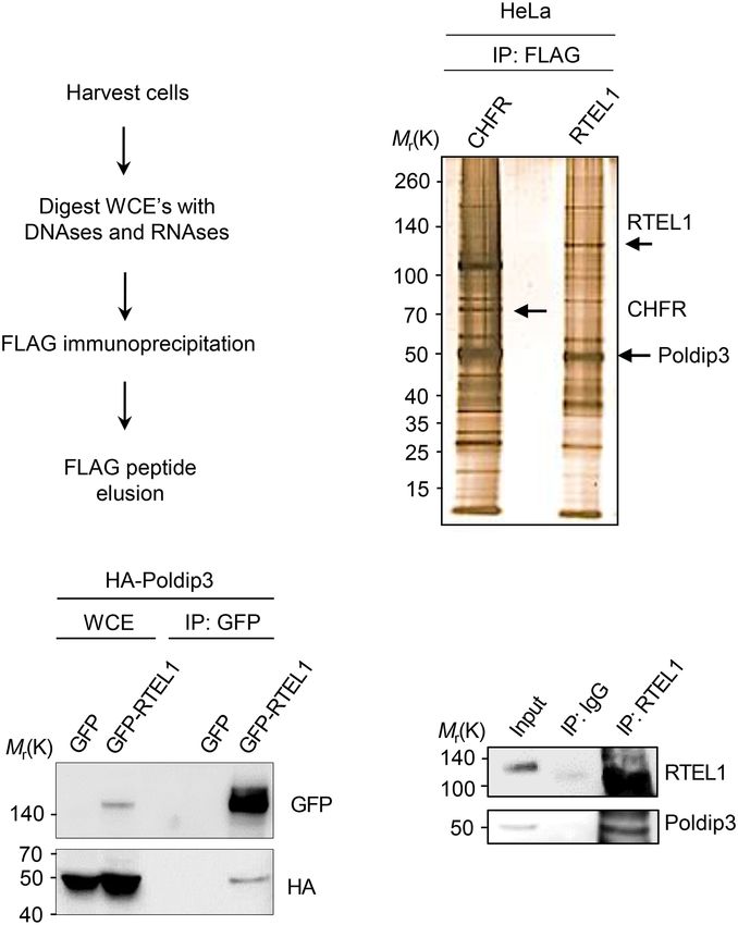
Downloaded from genesdev.cshlp.org on October 23, 2020 - Published by Cold Spring Harbor Laboratory Press

                                                                                                        RTEL1 and R-loop resolution

Poldip3 and RTEL1 suppress R-loop accumulation                     judged by the increased nuclear GFP intensity in RTEL1-
                                                                   depleted cells (Fig. 4F), suggesting that the aberrantly en-
Besides its presence in the POLδ complex, Poldip3 is also a        hanced accumulation of R-loops after RTEL1 loss is genu-
putative component of THO/TREX, a complex implicated               ine. As our evidence suggests that RTEL1 and Poldip3
in several steps of nuclear mRNP biogenesis, including             reside in a physical complex with mutually dependent
transcription, 3′ end processing, and export (Lee et al.           functions on chromatin, we assessed whether these pro-
2017). Notably, THO/TREX disruption is associated                  teins function epistatically to protect cells against
with the accumulation of R-loops; i.e., discrete RNA:              R-loop accumulation. Indeed, U2OS cells codepleted of
DNA hybrids that are formidable barriers to the replica-           RTEL1 and Poldip3 showed no additional increase in
tion fork progression (Heath et al. 2016). Furthermore,            R-loops compared with cells depleted for either protein
DinG, RTEL1’s ancient ortholog in bacteria, has been re-           alone (Supplemental Fig. S3G), indicating that RTEL1
ported to resolve R loop structures (Boubakri et al.               and Poldip3 function in the same pathway. To further val-
2010). To investigate whether Poldip3 or RTEL1 defi-               idate the notion that loss of RTEL1 leads to increased
ciency leads to excess R-loop accumulation, we examined            R-loop accumulation, we isolated genomic DNA from
Poldip3 and RTEL1 silenced cells for basal levels of R-loop        U2OS cells with and without RTEL1 and performed a
formation by immunofluorescence microscopy with the                dot blot analysis using the S9.6 antibody to probe for
widely used S9.6 antibody recognizing DNA:RNA hy-                  R-loops. Again, we found an increase in the amount of
brids. To exclude the possibility that the S9.6 antibody de-       S9.6 signal in the genomic DNA of U2OS cells depleted
tects RNA species different from R-loops, we assessed R-           of RTEL1 as compared with siRNA-treated control cells
loops after RTEL1 and Poldip3 depletion in cells generated         or RTEL1-depleted cells subsequently incubated with
to conditionally overexpress the R-loop-specific degrading         RNase H (Fig. 4G,I). Similarly, Poldip3 loss also leads to
nuclease RNase H. U2OS cells deficient for Poldip3 exhib-          a marked increase of genomic R-loops as detected by the
ited elevated R-loops compared with control cells (Fig. 4A,        dot-blot analysis compared with siRNA control cells or
B; Supplemental Fig. S3C). As with Poldip3-deficient cells,        in DNA from Poldip3-ablated cells subsequently incubat-
cells depleted for RTEL1 by siRNA treatment displayed              ed with RNase H (Fig. 4H,I), further implicating these pro-
increased R-loop accumulation as compared with control             teins in preventing excessive accumulation of potentially
siRNA-treated cells (Fig. 4A,B; Supplemental Fig. S3D).            harmful R-loops.
The S9.6 signal was particularly strong in the nucleolus,             Next, we asked whether RTEL1 might physically inter-
a region prone to R-loop accumulation due to highly ac-            act with RNA:DNA hybrids. Using the S9.6 antibody af-
tive transcription of repetitive rDNA (Hage et al. 2010;           finity purification approach in the presence of excess
Stuckey et al. 2015). Interestingly, doxycycline-induced           RNase A (to reduce unspecific RNA-mediated interac-
expression of RNase H effectively eliminated the R-loop            tions and avoid S9.6 recognition of double-stranded
accumulation after RTEL1 and Poldip3 depletion (Fig.               RNA [dsRNA]), we were able to pull down endogenous
4A,B), indicating that the R-loop accumulation after               RTEL1, whereas pretreatment of nuclear extracts with
depletion of either protein is specific. The increase in           the DNase prevented RTEL1 pull-down, indicating specif-
S9.6 staining in control cells treated with RNase H could          ic binding of RTEL1 to the RNA:DNA hybrid (Fig. 4J). In-
be due to the stress induced by RNase H overexpression, a          terestingly, RTEL1 was pulled down stronger after CPT
phenomenon that has been reported previously (Paulsen              treatment (Fig. 4J), indicating that CPT-induced R-loop
et al. 2009; Salas-Armenteros et al. 2017). The increase           accumulation augmented RTEL1 binding to RNA:DNA
in R-loops was effectively recapitulated in cells treated          hybrids. We then assessed whether RTEL1 could bind
with different siRNAs against the RTEL1 transcripts, an            pure RNA:DNA hybrids in vitro. Using immobilized bio-
accumulation that was also inhibited by the induction              tin-labeled RNA and DNA oligonucleotides annealed in
of RNase H (Fig. 4E), indicating that the increase in R-           vitro, we found that stably expressed wild-type GFP-
loops upon RTEL1 siRNA treatment is genuine. Likewise,             RTEL1 was efficiently retrieved in biotin pull-downs
CRISPR/Cas9-based targeted knockout of RTEL1 lead to a             (Fig. 4K). As a control, an annealed biotin-labeled D-loop
marked increase in R-loop accumulation, an effect that             was tested for its ability to pull down GFP-RTEL1. We
was also reversed in cells overexpressing RNase H (Fig.            found that RTEL1 was retrieved by both biotin R-loops
4C,D; Supplemental Fig. S3E), Notably, elevated R-loops            or D-loops, indicating that RTEL1 binds R-loops as effi-
were also detected in RTEL1-depleted normal human dip-             ciently as D-loops. Treatment of annealed biotin R-loops
loid cells RPE-1 (Supplemental Fig. S3F), thereby exclud-          with pure RNase H prior to pull-down also reduced the
ing the possibility that the R-loop-antagonizing function          coelution of RTEL1 (Fig. 4K), indicating that the binding
of RTEL1 is restricted to cancer cells.                            of RTEL1 to R-loops is genuine.
   To further rule out the possibility of nonspecific bind-
ing of the S9.6 antibody, we tested the enrichment of R-
loops after RTEL1 loss using U2OS cells stably expressing          Genomic localization of R-loops after RTEL1
HB-GFP, a fusion of green fluorescent protein (GFP) with           and Poldip3 loss
the DNA–RNA hybrid-binding (HB) domain of RNase H                  Given that RTEL1 and Poldip3 knockdown resulted in in-
that can be used to detect R-loops in cells (Tanikawa              creased R loop levels, we set out to explore in more detail
et al. 2016). Indeed, RTEL1 ablation resulted in R-loop ac-        the genomic regions showing abundant R-loops in such
cumulation compared with control cells in this setting, as         cells. Common R-loop-prone loci are typically very long

                                                                                                  GENES & DEVELOPMENT            5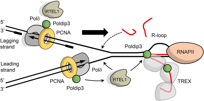
Downloaded from genesdev.cshlp.org on October 23, 2020 - Published by Cold Spring Harbor Laboratory Press

Björkman et al.

                                                  B                                        Figure 4. Increased accumulation of R loops
A
                                                                                           in RTEL1- or Poldip3-depleted cells. (A) In-
                                                                                           creased accumulation of R loops in cells de-
                                                                                           pleted of RTEL1 or Poldip3. Representative
                                                                                           images of DAPI and S9.6 fluorescence stain-
                                                                                           ing in U2OS cells treated with control,
                                                                                           RTEL1, or Poldip3 siRNAs. (B) Quantifica-
                                                                                           tion of nuclear S9.6 fluorescence intensity
                                                                                           in U2OS cells treated with control, RTEL1,
                                                                                           or Poldip3 siRNA. Cells conditionally over-
  C                                                                                        expressing GFP-RNase H (RH) with doxycline
                                                                                           were used. (C ) Increased amount of R-loops
                                                 D                                         in RTEL1 knockout U2OS cells. U2OS cells
                                                                                           transiently transfected with CRISPR–CAS9
                                                                                           guides against RTEL1 or control guide and
                                                                                           RNase H or GFP. (D) Quantification of nucle-
                                                                                           ar S9.6 fluorescence intensity in U2OS cells
                                                                                           transiently transfected with CRISPR-CAS9
                                                                                           guides against RTEL1 or control guide and
                                                                                           RNase H or GFP. (E) Increased accumulation
    E
                                                                                           of R-loops in U2OS cells depleted of RTEL1
                                                                                           using two different RTEL1 siRNAs. Quantifi-
                                                                                           cation of nuclear S9.6 fluorescence intensity
                                                                                           in U2OS cells conditionally overexpressing
                                                   F                                       GFP-RNnase H (RH) treated with control or
                                                                                           RTEL1 siRNAs. (F ) Increased GFP-RNase
                                                                                           H/R-loop binding domain recruitment to nu-
                                                                                           cleus in cells depleted of RTEL1 or Poldip3.
                                                                                           Quantification of GFP fluorescence intensity
                                                                                           in U2OS-RNase HB-GFP cells treated
                                                                                           with control, RTEL1, or Poldip3 siRNA.
                                                                                           (G) R-loop dot blot analysis. U2OS cells
    G                          I                                                           were treated with either control, RTEL1, or
                                                                                           Poldip3 siRNA followed by a 3-d incubation.
                                                                                           Genomic DNA was isolated from cell lysates,
                                                                                           bulk RNA digested with RNase A, and DNA
                                                                                           was subsequently hand spotted on activated
                                                        K                                  nylon membranes as indicated. DNA digest-
                                                                                           ed with the nuclease benzonase (Benz.) or
                                                                                           RNase H (RH) was used as a control. (H) R-
    H                        J                                                             loop dot blot analysis. U2OS cells were treat-
                                                                                           ed with either control or Poldip3 siRNA fol-
                                                                                           lowed by a 3-d incubation. Genomic DNA
                                                                                           was isolated from cell lysates, bulk RNA di-
                                                                                           gested with RNase A, and DNA was subse-
                                                                                           quently hand-spotted on activated nylon
                                                                                           membranes as indicated. DNA digested
                                                                                           with the nuclease benzonase (Benz.) or RN-
ase H (RH) was used as a control. (I) Quantification of G and H. Mean and SD are shown. (∗ ) P < 0.05; (∗∗ ) P < 0.01, Student’s t-test. (J)
RTEL1 binds to R-loops. U2OS cells left untreated or treated with CPT for 24 h were harvested and R-loops were immunoprecipitated
from cleared nuclear extracts with the S9.6 antibody. Nuclear extracts from cells treated with RNase H (RH) was used as a control. (K)
RTEL1 interacts with R-loops and D-loops in vitro. Extracts of U2OS cells stably expressing GFP-RTEL1 WT were incubated with bio-
tin-coupled R-loop or D-loop hybrids immobilized on streptavidin beads, and GFP-RTEL1 was immunoblotted with GFP antibody.
Cell extracts incubated with purified RNase H (RH) was used as a control. Mean and SD are plotted. (B,D,E,F) (∗ ) P < 0.05; (∗∗∗∗ ) P <
0.0001, two-tailed Mann-Whitney test. Data from three or more independent experiments are combined and immunofluorescence inten-
sities are normalized to siCtrl.

genes vulnerable to replication–transcription complexes                  ing G1 cell cycle phase (Lukas et al. 2011). Indeed, we
collisions (Helmrich et al. 2011). Such affected loci, also              found that RTEL1 and Poldip3 depletion lead to an in-
known as common fragile sites (CFS), are readily revealed                creased number of nuclear 53BP1 bodies in G1 of U2OS
as chromosome breaks after replication inhibition by                     cells (Fig. 5A,B), suggesting that RTEL1/Poldip3 defi-
aphidicolin (Aph) treatment and, if unrepaired before mi-                ciency and R-loops accumulation are associated with
tosis, such persistent replication stress-associated lesions             enhanced replication stress and CFS expression. To fur-
including CFS are marked as 53BP1 bodies in the follow-                  ther understand whether CFSs and other genomic loci

6      GENES & DEVELOPMENT
Downloaded from genesdev.cshlp.org on October 23, 2020 - Published by Cold Spring Harbor Laboratory Press
RTEL1 and R-loop resolution
A B
C D
E F G
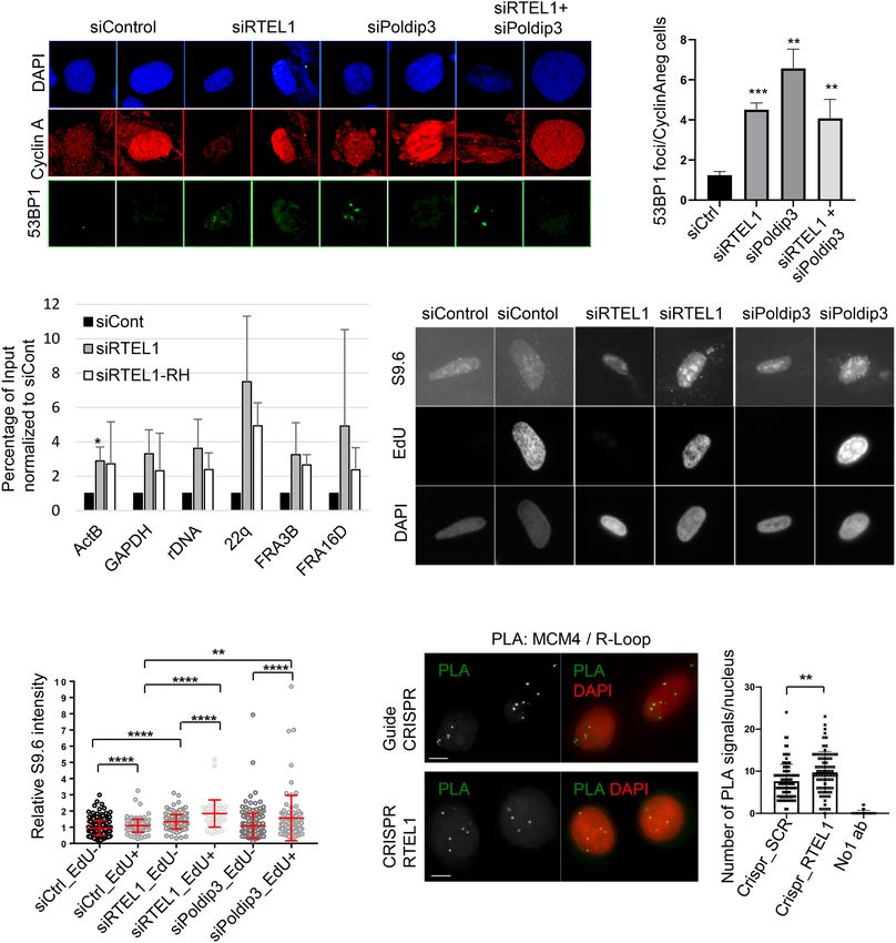
Figure 5. R loops accumulate at specific genomic loci and associate with replication after RTEL1 depletion. (A) Increased 53BP1 bodies in
Cyclin A-negative U2OS cells depleted of RTEL1, Poldip3, or RTEL1 and Poldip3. Representative images showing DAPI and 53BP1 stain-
ing in U2OS cells treated with control, RTEL1, Poldip3, or RTEL1 and Poldip3 siRNA. (B) Quantification of A. The average of three in-
dependent experiments are shown. (∗∗ ) P < 0.01; (∗∗∗ ) P < 0.001, Student’s t-test. (C ) DNA:RNA hybrids accumulate at commonly
expressed genes (GAPDH and ActB), common fragile sites (FRA3B and FRA16D), telomeres (22q) and rDNA in U2OS cells depleted of
RTEL1. Results are obtained with qPCR from DNA samples captured by DNA–RNA immunoprecipitation (DRIP) from siCont or siR-
TEL1 U2OS samples untreated or treated with RNase H (RH). Percentage of input normalized to siCont from three experiments are
shown. (∗ ) P < 0.01, Student’s t-test. (D) Increased amount of R loops in EdU-positive RTEL1- or Poldip3-depleted cells. Representative im-
ages of S9.6, EdU, and DAPI staining in RTEL1- and Poldip3-depleted cells. (E) Quantification of S9.6 nuclear fluorescence intensity in
control, RTEL1 or Poldip3 siRNA-treated EdU-positive or EdU-negative U2OS cells. Values normalized to siCtrl. Mean and SD are plotted
(∗∗ ) P < 0.01; (∗∗∗∗ ) P < 0.0001, two-tailed Mann-Whitney test. (F ) Proximity ligation assay (PLA) of S9.6 and MCM4 after RTEL1 knockout.
HeLa cells sorted (GFP) 48 h after transfection with either CRISPR–Cas9 against RTEL1 or CRISPR control and put on slides. PLA reveal-
ing MCM4 and R-loops. (G) Quantification of F. (SCR) Scrambled control guide. (∗∗ ) P < 0.01, Mann-Whitney test.
intersect with R-loop hotspots after RTEL1 and Poldip3 S9.6 antibody (Fig. 5C; Supplemental Fig. S4A,B). Further-
deficiency, we assessed three genomic regions in more de- more, overexpression of RNase H reduced the DRIP assay
tail using the DNA–RNA immunoprecipitation (DRIP) as- values of these loci indicating that the R-loop accumula-
say. We focused on CFSs (represented by FRA3B and tion at these genomic sites is genuine. Finally, considering
FRA16D) and other likely R-loop-prone loci in the ge- that R-loop accumulation has detrimental consequences,
nome; namely, telomeric repeats (represented by 22q), re- particularly during transcription and replication, we test-
petitive ribosomal DNA (rDNA) regions, and highly ed whether RTEL1 knockdown has any effect on these
transcribed genes (GAPDH and ActB). Indeed, we found processes. RTEL1 siRNA-treated cells showed normal
that both RTEL1 and Poldip3 depletion increased the R- staining with the modified nucleotide ethynyl uridine
loop accumulation at all the above-mentioned genomic (EU) (a marker for RNA synthesis) (Supplemental Fig.
regions, but not at a control region (Rag1), as judged by S4C,D), suggesting that global transcription was unaltered
the increased pull-down of these DNA regions with the in RTEL1-depleted cells. In contrast, incorporation of 5-
GENES & DEVELOPMENT 7Downloaded from genesdev.cshlp.org on October 23, 2020 - Published by Cold Spring Harbor Laboratory Press
Björkman et al.
ethynyl-2′ -deoxyuridine (Edu; a marker for DNA synthe-
sis) (Supplemental Fig. S4E,F) was reduced in siRTEL1
cells, which is consistent with previous publications
(Uringa et al. 2012; Vannier et al. 2013) supporting a role
for RTEL1 in replication. We therefore asked whether
the R-loop accumulation after RTEL1 or Poldip3 deple-
tion is confined to actively replicating cells in S phase
by comparing R-loops in cells with the neighboring cells
outside S phase. Here we found an enrichment of R-loops
in S-phase cells (Fig. 5E), indicating that R-loop accumula-
tion after RTEL1/Poldip3 loss occurs mainly during DNA
replication. An increase in R-loops was also detected in
non-S-phase cells, although to a lesser degree compared Figure 6. Cartoon showing our model of the role of RTEL1 and
Poldip3 in R-loop resolution. RTEL1 associates with the repli-
with S phase, suggesting that RTEL1 and Poldip3 are need-
some, here represented by Polδ (gray) and PCNA (yellow) via Pol-
ed for R-loop prevention/resolution throughout the cell
dip3 and/or PCNA, as it replicates DNA. As the replication fork
cycle. We further examined whether R-loop accumulation encounters the transcription machinery, RTEL1 might resolve
after RTEL1 loss might occur in close proximity to repli- R-loops that block the replication fork and/or the R-loops pro-
cation forks. Indeed, using the proximity ligation assay voked by stalling of replication (shown by arrows from RTEL1).
(PLA), we found that R-loops are enriched in proximity (RNAPII) RNA polymerase II. The black lines represent single-
to the replisome helicase component MCM4 (Fig. 5F,G), stranded DNA and the red lines depict RNA. The big arrow indi-
suggesting that RTEL1 ablation leads to R-loop accumula- cates the direction of the replication fork.
tion preferentially at replication forks.
Collectively, these data suggest that RTEL1 and Poldip3
prevent potential R-loop accumulation, a so-far unsus- (Fig. 5D–G) and RTEL1 depletion has no overt effect on
pected role that helps protect genome integrity at vulner- global transcription (Supplemental Fig. S4C,D) points
able regions such as CFS or rDNA and is particularly mainly to the first possibility. We hypothesize that Pol-
apparent during DNA replication. dip3 as a subunit of Polδ together with other components
of the replisome (e.g., PCNA) are critical for the timely re-
cruitment of RTEL1 to the stalled replication fork where
Discussion it resolves replication fork stalling at sites of transcrip-
tion. One likely obstacle to replication forks is the RNA
Overall, our present study identifies a previously unrecog- polymerase as well as other components of the transcrip-
nized protein complex of RTEL1 and Poldip3 that operates tion machinery. This is also supported by the increased R-
at the crossroad of the fundamental cellular processes of loop accumulation at CFSs in RTEL1- or Poldip3-depleted
DNA replication and transcription, as schematically de- cells (Fig. 5C; Supplemental Fig. S4A,B), which are located
picted in our model presented in Figure 6. Excessive accu- in very long genes that often replicate late in S phase and
mulation of R-loops represents a formidable barrier to the are thus more sensitive to replication stress-inducing rep-
replication fork progression and needs to be reversed to lication–transcription conflicts. We propose that RTEL1
prevent replication failure and genomic instability (Heath might strip off such transcription proteins from DNA be-
et al. 2016). The surprising R-loop accumulation when fore they collide with the replisome. As such, the function
RTEL1 is lost might have two likely causes that in both of RTEL1 would resemble that of its ancient ortholog in
cases might be explained by a defective physical interac- bacteria, DinG, which functions to prevent collisions be-
tion between RTEL1 and Poldip3. Poldip3 functions as a tween replication forks and the RNA polymerase (Bou-
subunit in both the DNA polymerase δ (Polδ) complex bakri et al. 2010). This notion is consistent with the
during replication and in the RNA sequestering TREX previous reports demonstrating that RTEL1-deficient
complex during transcription. cells are hypersensitive specifically to topoisomerase I
Our observation that RTEL1 and Poldip3 reside in a (TOPOI) poisons (Barber et al. 2008), agents that induce in-
complex thus indicates that RTEL1 functions in either crease of R-loops specifically at highly transcribed regions
replication, as previously proposed (Vannier et al. 2013), (Marinello et al. 2013). Likewise, POLδ deficiency also
and/or transcription. RTEL1 loss could lead to defective renders cells hypersensitive to TOPOI inhibitors (Tumini
replication fork progression and replication fork collapse et al. 2016), raising the possibility that both RTEL1 and
that would result in naked DNA strands accessible to POLδ play a critical role in overcoming TOPOI-induced
RNA species consequently producing excessive DNA: DNA damage during replication. In further support of
RNA hybrids. Alternatively, defective TREX complex this concept, two reports published at the time of submis-
function could result in untimely RNA sequestration of sion of our revised manuscript suggest that RTEL1 is in-
newly transcribed RNA and an increased pool of RNA volved in suppressing replication–transcription conflicts
moieties competent to invade adjoining DNA template (Takedachi et al. 2020; Wu et al. 2020) and R-loop accumu-
strands. Our results that RTEL1 and Poldip3 loss lead to lation (Wu et al. 2020).
R-loop accumulation most prominently in S phase during Last but not least, this previously unsuspected role of
DNA replication and in a close proximity to the replisome the human RTEL1–Poldip3 complex, identified in our
8 GENES & DEVELOPMENTDownloaded from genesdev.cshlp.org on October 23, 2020 - Published by Cold Spring Harbor Laboratory Press
RTEL1 and R-loop resolution
present study, contributes to genome integrity mainte- Danish National Research Foundation (project CARD, no.
nance under replication stress, an important homeostatic DNRF125), and the Lundbeck Foundation (R266-2017-4289).
function with implications for proper organismal develop- Work in the Londono-Vallejo laboratory was supported by grants
ment and avoidance of a range of grave pathologies includ- from Agence Nationale de la Recherche NR (ANR-14-CE10-
0006) and Fondation ARC pour la Recherche contre le Cancer.
ing cancer.
Author contributions: K.B.S., J.B., and A.B. designed the re-
search. K.B.S., A.B., S.L.J., L.L., M.S., D.C.K., and A.-M.K. per-
Materials and methods
formed the experiments. All authors analyzed and discussed the
data. K.B.S., A.B., A.-M.K., and J.B. wrote the paper. All co-authors
Mass spectrometry analysis
reviewed and edited the paper. K.B.S. and J.B. obtained funding for
the study.
SDS-PAGE-resolved, 4%–12% gradient gels were silver-stained
with SilverQuest (Life Technologies), and appropriate bands
were cut and subjected to in-gel digestion followed by liquid chro-
matography-MS with the Q Exactive HF mass spectrometer References
(Thermo Fisher Scientific). Resulting peptides were identified Ballew BJ, Joseph V, De S, Sarek G, Vannier JB, Stracker T,
by protein-sequence database searches using MaxQuant software Schrader KA, Small TN, O’Reilly R, Manschreck C, et al.
(Tyanova et al. 2016). 2013a. A recessive founder mutation in regulator of telomere
elongation helicase 1, RTEL1, underlies severe immunodefi-
Plasmids and gene silencing ciency and features of Hoyeraal Hreidarsson syndrome. PLoS
Genet 9: e1003695. doi:10.1371/journal.pgen.1003695
A cDNA for human RTEL1, KIAA1088, was obtained from Ballew BJ, Yeager M, Jacobs K, Giri N, Boland J, Burdett L, Alter
HUGE protein database (http://www.kazusa.or.jp/techcgi/view_ BP, Savage SA. 2013b. Germline mutations of regulator of
direct.cgi?id=hk02589s1) and was inserted by PCR into pFlag- telomere elongation helicase 1, RTEL1, in dyskeratosis conge-
CMV2 (Sigma) or into the pEGFP-C1 vector (Clontech). To gener- nita. Hum Genet 132: 473–480. doi:10.1007/s00439-013-
ate GFP-RTEL1 mutant versions, the causative dyskeratosis con- 1265-8
genita mutations were introduced by standard PCR using mutant Barber LJ, Youds JL, Ward JD, McIlwraith MJ, O’Neil NJ, Petal-
primers and the same flanking primers as used to generate GFP- corin MI, Martin JS, Collis SJ, Cantor SB, Auclair M, et al.
RTEL1 WT (Schmid et al. 2018). 2008. RTEL1 maintains genomic stability by suppressing ho-
All plasmid transfections were performed using FuGene6 mologous recombination. Cell 135: 261–271. doi:10.1016/j
(Roche). siRNA oligonucleotides (Dharmacon) were synthesized .cell.2008.08.016
to the following human sequences: (sense strand) siRTEL1#1
Boubakri H, de Septenville AL, Viguera E, Michel B. 2010. The
(5′ -UGAAGAAACAAAGAGUAAUU-3′ ) and (antisense strand)
helicases DinG, Rep and UvrD cooperate to promote replica-
siRTEL1#1 (3′ -UUACUUCUUUGUUCUCUCAUU-5′ ), (sense
tion across transcription units in vivo. EMBO J 29: 145–157.
strand) siRTEL1#2 (5′ -GCCUGUGUGUGGAGUAUGA-3′ ) and
doi:10.1038/emboj.2009.308
(antisense strand) siRTEL1#2 (5′ -UCAUACUCCACACACAG
Deng Z, Glousker G, Molczan A, Fox AJ, Lamm N, Dheekollu J,
GC-3′ ), and (sense strand) siPOLDIP3 (5′ -GGGAAAGUGCAG
Weizman OE, Schertzer M, Wang Z, Vladimirova O, et al.
GAUGCCA-3′ ) and (antisense strand) (5′ -UGGCAUCCUGCA
2013. Inherited mutations in the helicase RTEL1 cause telo-
CUUUCCC-3′ ). For efficient knockdown of RTEL1 and Poldip3
mere dysfunction and Hoyeraal-Hreidarsson syndrome. Proc
in all experiments, cells were transfected twice with the siRNAs
Natl Acad Sci 110: E3408-16.
as follows. On day 1, cells were incubated for 6 h with transfec-
Ding H, Schertzer M, Wu X, Gertsenstein M, Selig S, Kammori M,
tion reagents containing control or target siRNA. Then the trans-
Pourvali R, Poon S, Vulto I, Chavez E, et al. 2004. Regulation
fection medium was replaced with fresh growth medium and
of murine telomere length by Rtel: an essential gene encoding
cells were incubated for an additional 18 h. On day 2, cells were
a helicase-like protein. Cell 117: 873–886. doi:10.1016/j.cell
treated as in day 1 and then incubated for an additional 42
.2004.05.026
h. siCONTROL (Dharmacon) was used as a control siRNA. All
siRNA transfections were performed with 100 nM siRNA duplex- El Hage A, French SL, Beyer AL, Tollervey D. 2010. Loss of topo-
es using Lipofectamine RNAi MAX (Invitrogen). Cell culture hu- isomerase I leads to R-loop-mediated transcriptional blocks
man U2OS and HEK293T were cultured in DMEM containing during ribosomal RNA synthesis. Genes Dev 24: 1546–1558.
10% fetal bovine serum. U2OS-derived cell lines capable of ex- doi:10.1101/gad.573310
pressing ectopic RTEL1 alleles from pEGFP-C1-RTEL1 con- Halazonetis TD, Gorgoulis VG, Bartek J. 2008. An oncogene-in-
structs were generated and maintained as described. duced DNA damage model for cancer development. Science
Additional information about the Materials and Methods is 319: 1352–1355. doi:10.1126/science.1140735
available in the Supplemental Material. Heath CG, Viphakone N, Wilson SA. 2016. The role of TREX in
gene expression and disease. Biochem J 473: 2911–2935.
doi:10.1042/BCJ20160010
Acknowledgments Helmrich A, Ballarino M, Tora L. 2011. Collisions between repli-
cation and transcription complexes cause common fragile site
We thank Professor Karlene Cimprich for kindly providing U2OS- instability at the longest human genes. Mol Cell 44: 966–977.
RNase HB-GFP cells. The work was funded by grants from the doi:10.1016/j.molcel.2011.10.013
Karolinska Institutet, the Alfred Benzon’s Foundation, the Swed- Lee MYWT, Wang X, Zhang S, Zhang Z, Lee EYC. 2017. Regula-
ish Cancer Society (170176), the Swedish Research Council (VR- tion and modulation of human DNA polymerase δ activity
MH 2014-46602-117891-30), the Strategic Research Program in and function. Genes 8: 190.
Cancer at Karolinska Institutet (2201), the Novo Nordisk Founda- Le Guen T1, Jullien L, Touzot F, Schertzer M, Gaillard L, Perder-
tion (16854), the Danish Cancer Society (R1123-A7785-15-S2), the iset M, Carpentier W, Nitschke P, Picard C, Couillault G, et al.
Danish Council for Independent Research (DFF-7016-00313), the 2013. Human RTEL1 deficiency causes Hoyeraal-Hreidarsson
GENES & DEVELOPMENT 9Downloaded from genesdev.cshlp.org on October 23, 2020 - Published by Cold Spring Harbor Laboratory Press
Björkman et al.
syndrome with short telomeres and genome instability. Hum priming in a eukaryotic system. Proc Natl Acad Sci 112: 5779–
Mol Genet 22: 3239–3249. doi:10.1093/hmg/ddt178 5784. doi:10.1073/pnas.1501769112
Lukas C, Savic V, Bekker-Jensen S, Doil C, Neumann B, Pedersen Takedachi A, Despras E, Scaglione A, Guérois R, Guervilly JH,
RS, Grøfte M, Chan KL, Hickson ID, Bartek J, et al. 2011. Blin M, Audebert S, Camoin L, Hasanova Z, Schertzer M,
53BP1 nuclear bodies form around DNA lesions generated et al. 2020. SLX4 interacts with RTEL1 to prevent transcrip-
by mitotic transmission of chromosomes under replication tion-mediated DNA replication perturbations. Nat Struct
stress. Nat Cell Biol 13: 243–253. doi:10.1038/ncb2201 Mol Biol 27: 438–449. doi:10.1038/s41594-020-0419-3
Marinello J, Chillemi G, Bueno S, Manzo SG, Capranico G. 2013. Tanikawa M, Sanjiv K, Helleday T, Herr P, Mortusewicz O. 2016.
Antisense transcripts enhanced by camptothecin at divergent The spliceosome U2 snRNP factors promote genome stability
CpG-island promoters associated with bursts of topoisomer- through distinct mechanisms; transcription of repair factors
ase I–DNA cleavage complex and R-loop formation. Nucleic
and R-loop processing. Oncogenesis 5: e280. doi:10.1038/onc
Acids Res 41: 10110–10123. doi:10.1093/nar/gkt778
sis.2016.70
Merchut-Maya JM, Bartek J, Maya-Mendoza A. 2019. Regulation
Tumini E, Barroso S, -Calero CP, Aguilera A. 2016. Roles of hu-
of replication fork speed: mechanisms and impact on genomic
man POLD1 and POLD3 in genome stability. Sci Rep 6;
stability. DNA Repair 81: 102654. doi:10.1016/j.dnarep.2019
38873. doi:10.1038/srep38873
.102654
Tyanova S, Temu T, Cox J. 2016. The MaxQuant computational
Paulsen RD, Soni DV, Wollman R, Hahn AT, Yee MC, Guan A,
Hesley JA, Miller SC, Cromwell EF, Solow-Cordero DE, platform for mass spectrometry-based shotgun proteomics.
et al. 2009. A genome-wide siRNA screen reveals diverse cel- Nat Protoc 11: 2301–2319. doi:10.1038/nprot.2016.136
lular processes and pathways that mediate genome stability. Uringa EJ, Lisaingo K, Pickett HA, Brind’Amour J, Rohde JH,
Mol Cell 35: 228–239. doi:10.1016/j.molcel.2009.06.021 Zelensky A, Essers J, Lansdorp PM. 2012. RTEL1 contributes
Porreca RM, Glousker G, Awad A, Matilla Fernandez MI, Gibaud to DNA replication and repair and telomere maintenance.
A, Naucke C, Cohen SB, Bryan TM, Tzfati Y, Draskovic I, et al. Mol Biol Cell 23: 2782–2792. doi:10.1091/mbc.e12-03-0179
2018. Human RTEL1 stabilizes long G-overhangs allowing Vannier JB, Pavicic-Kaltenbrunner V, Petalcorin MI, Ding H,
telomerase-dependent over-extension. Nucleic Acids Res 46: Boulton SJ. 2012. RTEL1 dismantles T loops and counteracts
4533–4545. doi:10.1093/nar/gky173 telomeric G4-DNA to maintain telomere integrity. Cell 149:
Salas-Armenteros I, Pérez-Calero C, Bayona-Feliu A, Tumini E, 795–806. doi:10.1016/j.cell.2012.03.030
Luna R, Aguilera A. 2017. Human THO-Sin3A interaction re- Vannier JB, Sandhu S, Petalcorin MI, Wu X, Nabi Z, Ding H, Boul-
veals new mechanisms to prevent R-loops that cause genome ton SJ. 2013. RTEL1 is a replisome-associated helicase that
instability. EMBO J 36: 3532–3547. doi:10.15252/embj promotes telomere and genome-wide replication. Science
.201797208 342: 239–242. doi:10.1126/science.1241779
Schertzer M1, Jouravleva K, Perderiset M, Dingli F, Loew D, Le Vannier JB, Sarek G, Boulton SJ. 2014. RTEL1: functions of a dis-
Guen T, Bardoni B, de Villartay JP, Revy P, Londoño-Vallejo ease-associated helicase. Trends Cell Biol 24: 416–425. doi:10
A. 2015. Human regulator of telomere elongation helicase 1 .1016/j.tcb.2014.01.004
(RTEL1) is required for the nuclear and cytoplasmic traffick- Walne AJ, Vulliamy T, Kirwan M, Plagnol V, Dokal I. 2013. Con-
ing of pre-U2 RNA. Nucleic Acids Res 43: 1834–1847. doi:10 stitutional mutations in RTEL1 cause severe dyskeratosis
.1093/nar/gku1402
congenita. Am J Hum Genet 92: 448–453. doi:10.1016/j.ajhg
Schmid FM, Schou KB, Vilhelm MJ, Holm MS, Breslin L, Farinelli
.2013.02.001
P, Larsen LA, Andersen JS, Pedersen LB, Christensen ST. 2018.
Wrensch M, Jenkins RB, Chang JS, Yeh RF, Xiao Y, Decker PA,
IFT20 modulates ciliary PDGFRα signaling by regulating the
Ballman KV, Berger M, Buckner JC, Chang S, et al. 2009. Var-
stability of Cbl E3 ubiquitin ligases. J Cell Biol 217: 151–
iants in the CDKN2B and RTEL1 regions are associated with
161. doi:10.1083/jcb.201611050
high-grade glioma susceptibility. Nat Genet 41: 905–908.
Shete S, Hosking FJ, Robertson LB, Dobbins SE, Sanson M,
Malmer B, Simon M, Marie Y, Boisselier B, Delattre JY, doi:10.1038/ng.408
et al. 2009. Genome-wide association study identifies five sus- Wu W, Bhowmick R, Vogel I, Özer Ö, Ghisays F, Thakur RS, San-
ceptibility loci for glioma. Nat Genet 41: 899–904. doi:10 chez de Leon E, Richter PH, Ren L, Petrini JH, et al. 2020.
.1038/ng.407 RTEL1 suppresses G-quadruplex-associated R-loops at diffi-
Speckmann C, Sahoo SS, Rizzi M, Hirabayashi S, Karow A, Ser- cult-to-replicate loci in the human genome. Nat Struct Mol
was NK, Hoemberg M, Damatova N, Schindler D, Vannier Biol 27: 424–437. doi:10.1038/s41594-020-0408-6
JB, et al. 2017. Clinical and molecular heterogeneity of Youds JL, Mets DG, McIlwraith MJ, Martin JS, Ward JD, ONeil
RTEL1 deficiency. Front Immunol 8: 449. NJ, Rose AM, West SC, Meyer BJ, Boulton SJ. 2010. RTEL-1
Stuckey R, García-Rodríguez N, Aguilera A, Wellinger RE. 2015. enforces meiotic crossover interference and homeostasis. Sci-
Role for RNA:DNA hybrids in origin-independent replication ence 327: 1254–1258. doi:10.1126/science.1183112
10 GENES & DEVELOPMENTDownloaded from genesdev.cshlp.org on October 23, 2020 - Published by Cold Spring Harbor Laboratory Press
Human RTEL1 associates with Poldip3 to facilitate responses to
replication stress and R-loop resolution
Andrea Björkman, Søren L. Johansen, Lin Lin, et al.
Genes Dev. published online June 19, 2020
Access the most recent version at doi:10.1101/gad.330050.119
Supplemental http://genesdev.cshlp.org/content/suppl/2020/06/18/gad.330050.119.DC1
Material
Published online June 19, 2020 in advance of the full issue.
Creative This article, published in Genes & Development, is available under a Creative Commons
Commons License (Attribution 4.0 International), as described at
License http://creativecommons.org/licenses/by/4.0/.
Email Alerting Receive free email alerts when new articles cite this article - sign up in the box at the top
Service right corner of the article or click here.
Published by © 2020 Björkman et al.; Published by Cold Spring Harbor Laboratory PressYou can also read


