Kinetic and subcellular analysis of PS-ASO/protein interactions with P54nrb and RNase H1
←
→
Page content transcription
If your browser does not render page correctly, please read the page content below
Nucleic Acids Research, 2019 1
                                                                                                                            doi: 10.1093/nar/gkz771

Kinetic and subcellular analysis of PS-ASO/protein
interactions with P54nrb and RNase H1
Timothy A. Vickers* , Meghdad Rahdar, Thazha P. Prakash and Stanley T. Crooke

                                                                                                                                                                  Downloaded from https://academic.oup.com/nar/advance-article-abstract/doi/10.1093/nar/gkz771/5565286 by guest on 17 September 2019
Department of Core Antisense Research, Ionis Pharmaceuticals, Inc., Carlsbad, CA, USA

Received February 13, 2019; Revised August 21, 2019; Editorial Decision August 26, 2019; Accepted August 28, 2019

ABSTRACT                                                                          binding more cellular proteins with higher affinity than
                                                                                  non-toxic PS-ASOs (1). These sequence and chemistry de-
The rapid RNase H1-dependent mislocalization of                                   pendent ASO–protein interactions were shown to corre-
heterodimer proteins P54nrb and PSF to nucleoli is                                late with toxic potentials of cEt-modified PS-ASOs in vitro
an early event in the pathway that explains the effects                           and in vivo altering the stability, function, or distribution
of most toxic phosphorothioate ASOs (PS-ASOs).                                    of many cellular proteins and resulting in significant tox-
Using a recently developed NanoLuciferace (NLuc)-                                 icity. Toxic, but not safe cEt PS-ASOs, caused rapid mis-
based structural complementation reporter system                                  localization of the paraspeckle proteins P54nrb and PSF
which allows us to observe ASO/protein interactions                               to nucleoli, resulting in nucleolar stress and fragmentation,
in real time in live cells, we have determined that                               upregulation of P21 mRNA and activation of caspase activ-
safe and toxic PS-ASOs associate with these pro-                                  ity, and ultimately, apoptotic cell death. In animals, because
teins with kinetics and impact on subcellular local-                              PS-ASOs accumulate in high concentrations in the liver, the
                                                                                  most common manifestation of the toxicity is the death of
ization that differ. Toxic PS-ASOs interact in a com-
                                                                                  hepatocytes which results in liver failure most easily demon-
plex that includes RNase H1, P54nrb and PSF; but                                  strated by increases in liver enzymes, aminoalanine trans-
RNase H1/P54nrb complexes were observed in only                                   ferase (ALT) and aspartate aminotransferase (AST) and
the cells treated with toxic, but not safe PS-ASOs.                               histological changes. Furthermore, in the same manuscript,
In addition, experiments performed in vitro suggest                               the substitution of a 2 -methoxy nucleotide at position 2 of
that RNA is also a required component of the com-                                 the DNA ‘gap’ portion of the PS-ASOs was demonstrated
plex. The protein–protein interaction between P54nrb                              to ablate or greatly ameliorate the toxicity without mean-
and RNase H1 requires the spacer region of RNAse                                  ingful loss of potency. Given the breadth of use of antisense
H1, while the P54nrb core domains are required for                                technology and drugs and the potential impact of these in-
association with RNase H1. In addition, we have de-                               sights on the therapeutic index of PS-ASO drugs, it is im-
termined that PS-ASOs bind P54nrb via RRM1 and                                    portant to better understand how these proteins interact
                                                                                  with PS-ASOs and identifying the nucleic acid sequences
RRM2, while they bind RNase H1 primarily via the
                                                                                  (and possibly structures) required to assemble these com-
hybrid binding domain, however catalytic domain in-                               plexes is vital to understanding the role these complexes
teractions also contribute to overall affinity. These                             play in inducing cellular toxicity.
ASO–protein interactions are highly influenced by                                    Proteins interact with DNA and RNA through electro-
the chemistry of the PS-ASO binding environment,                                  static interactions, hydrogen bonding, hydrophobic interac-
however little correlation between affinity for specific                          tions and base stacking (2–5). These forces contribute in
proteins and PS-ASO toxicity was observed.                                        varying degrees to proteins binding in a structure and se-
                                                                                  quence specific or non-sequence specific manner (6). We re-
                                                                                  cently reported the development of a highly sensitive and
INTRODUCTION                                                                      high throughput BRET affinity assay which relies on the
The broad and increasing use of oligonucleotides of var-                          transfer of light energy from a Nanoluc luciferase (Nluc)
ious types as research tools and platforms for drug dis-                          tagged binding protein acting as the BRET donor, to a flu-
covery and development demand a more thorough under-                              orescently tagged ASO acting as the BRET acceptor (7). In
standing of how these pharmacological agents interact with                        the current work we use this assay to define to domains of
various proteins and how chemical modifications, sequence                         P54nrb with which PS-ASOs interact. We further charac-
and structure influence interactions with proteins. We re-                        terize the contribution of PS-ASO chemistry to these inter-
cently reported that protein-binding contributes profoundly                       actions as well as that of the binding environment.
to the toxic potentials of PS-ASOs, with toxic PS-ASOs

* To   whom correspondence should be addressed. Tel: +1 760 931 9200; Email: tvickers@ionisph.com

C The Author(s) 2019. Published by Oxford University Press on behalf of Nucleic Acids Research.
This is an Open Access article distributed under the terms of the Creative Commons Attribution Non-Commercial License
(http://creativecommons.org/licenses/by-nc/4.0/), which permits non-commercial re-use, distribution, and reproduction in any medium, provided the original work
is properly cited. For commercial re-use, please contact journals.permissions@oup.com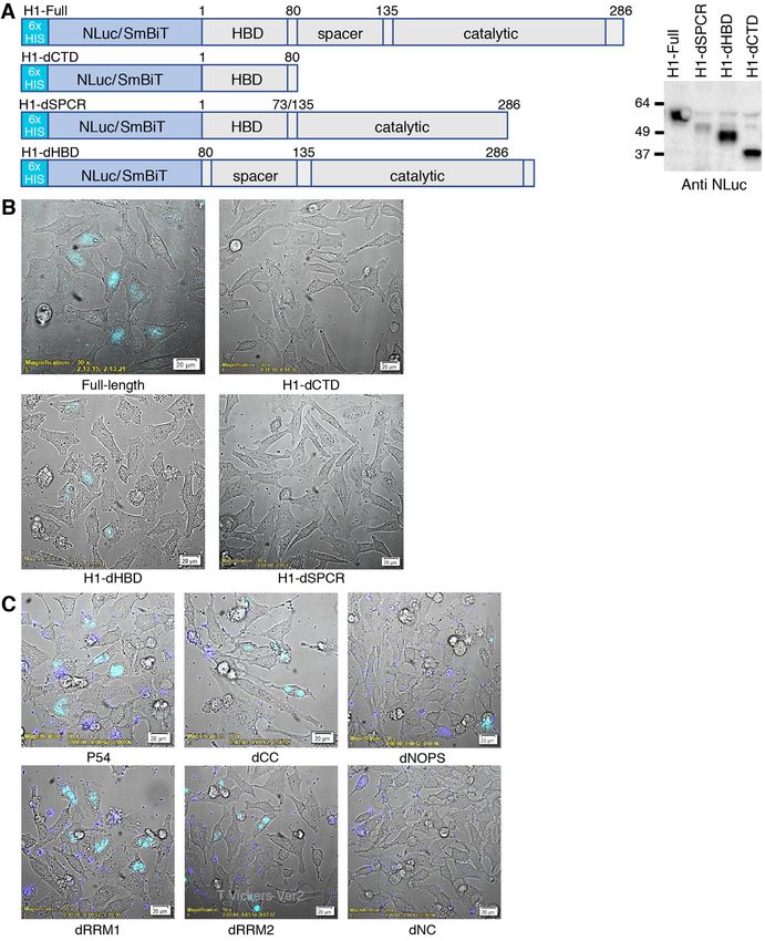
2 Nucleic Acids Research, 2019
We have also developed a NLuc-based structural com- luted with water (3 ml) and desalted by HPLC on reverse
plementation reporter system which enables observation of phase column to yield the3 - BCN carbamate ASO conju-
ASO–protein interactions, in real time in live cells (1). Using gate in 87–90% yield. A solution of 1.5 umol SmBiT pep-
this assay, we elucidate the kinetics and subcellular localiza- tide (Val-Thr-Gly-Tyr-Arg-Leu-Phe-Glu-Glu-Ile-Leu-Gly-
tion of ASO–protein interactions for toxic and safe cEt PS- Gly-Ser-Gly-Gly-Lys(N3)-NH2) containing an azide group
ASOs. In addition, using a variation of the same assay, we at the C-terminus (CPC Scientific, 1245 Reamwood Ave,
follow the association of various proteins with the P54nrb– Sunnyvale, CA, USA) in DMSO (0.5 ml) was added and
PS-ASO complex. We show that this complex is made up of the reaction mixture was stirred at room temperature for 5
Downloaded from https://academic.oup.com/nar/advance-article-abstract/doi/10.1093/nar/gkz771/5565286 by guest on 17 September 2019
not only P54nrb and PSF, but significantly includes RNase h. The reaction mixture was diluted with water (13 ml) and
H1 in the presence of toxic, but not safe PS-ASO. We fi- purified by HPLC on a strong anion exchange column (GE
nally characterize the interaction of PS-ASOs with RNase Healthcare Bioscience, Source 30Q, 30 m, 2.54 × 8 cm, A
H1as well as the protein–protein interaction of P54nrb and = 100 mM ammonium acetate in 30% aqueous CH3CN, B
RNase H1. = 1.5 M NaBr in A, 0–60% of B in 60 min, flow 14 ml min–1 ).
The fraction containing full length conjugates (assessed by
LC–MS analysis) were pooled together and concentrated.
MATERIALS AND METHODS The residue dissolved in water and desalted by HPLC on re-
Preparation of antisense oligonucleotides verse phase column to yield the peptide conjugated ASOs in
an isolated yield of 75–80%. The PS-ASO conjugates were
Synthesis and purification of phosphorothioate oligonu- characterized by ion-pair-HPLC–MS analysis with Agilent
cleotides was performed as described previously (7). All 1100 MSD system.
ASOs were ‘gapmers’ 16–20 nucleotides in length with 2 -
Fluoro (2 -F), 2 -O-methoxyethyl (MOE) or constrained
ethyl (cEt) (8) substitutions at the positions indicated in
in Table 1. Standard phosphodiester and phosphorothioate Construction, expression and purification of fusion proteins
deoxy and ribo oligonucleotides were obtained from Inte- Amino-terminal NLuc fusions were created using the vector
grated DNA technologies (Coralville, IA, USA). pFN31K Nluc CMV-neo (Promega). Briefly, P54nrb and
The synthesis of 3 -amino PS-ASOs was performed on an RNase H1 were amplified from plasmids RC206688 and
ÄKTAoligopilot synthesizer (GE Healthcare Bioscience) in RC200595 (Origene) respectively using PCR primers com-
40-mol scale. The oligonucleotide was synthesized on 3 - plementary to the full length cDNAs. The forward PCR
amino-modifier C7 CPG (Glen Research Inc.) solid sup- primer was comprised of sequence complimentary to the
port. A solution of phosphoramidites in acetonitrile (0.1 sequence following the AUG start codon preceded by an
M), and standard sulfurization reagent, coupling reagents XhoI site for cloning in frame with NLuc, whereas the re-
and capping reagents were used. For each of the modi- verse primer was complementary to the sequence preced-
fied analogs 4-fold excess of nucleoside 3 -phosphoramidite ing the stop codon followed by an EcoRI site. The PCR
were delivered with a 12-min coupling time. The 5 -end amplified product was digested with XhoI and EcoRI then
dimethoxytrityl group was left on to facilitate purifica- ligated into the pFN31K Nluc CMV-neo vector prepared
tion. Post-synthetically, oligonucleotide bearing solid sup- with the same enzymes using standard techniques. A 6X
port was treated with 50% piperidine in DMF for 1 h to HIS-tag (CAT CAT CAT CAC CAC CAC) was inserted up-
remove Fmoc from 3 -amino group, and treated with 1:1 stream of the NLuc cassette by site directed mutagenesis us-
triethylamine: acetonitrile to remove cyanoethyl protecting ing a Q5 Site-Directed Mutagenesis Kit (New England Bio-
groups from the phosphorothioate linkages. Subsequently, Labs) according to the manufacturers protocol. Sequences
solid support was treated with aqueous NH4 OH (28–30 of PCR primers can be found in Supplementary Table S1.
wt%) at room temperature for 4 h. 10% (v/v) of 40% methy- The LgBiT fusions were created by replacing the existing
lamine in water was added then incubated at room tem- NLuc with LgBiT in the fusion plasmid. The NLuc cassette
perature for additional 14 h to cleave ASO from support was excised by digestion with NheI and XhoI. LgBiT was
and remove all protecting groups. Oligonucleotide was pu- amplified from pBiT1.1-C[TK/LgBiT] vector (Promega)
rified by ion-exchange chromatography using a NaBr gra- with a forward primer consisting of NheI site Kozak se-
dient across a column packed with Source 30Q resin. Pure quence and sequence complimentary to the sequence fol-
fractions were desalted using HPLC on a reverse phase col- lowing the AUG start codon (gcat tcga GCTAGCGCT
umn. Purity and mass of oligonucleotides were determined CACC ATG GTC TTC ACA CTC GAA GAT TTC) and
using ion-pair LCMS analysis. a reverse primer complementary to the sequence preceding
SmBiT peptides were conjugated to 3 -amino PS-ASOs the stop codon followed by an XhoI site (GTT CCG AGT
as described previously (9) (Supplementary Figure S1). AAC CAT CAA CAG T ctcgag gcat tcga).
3 Amino ASO (1 umol) in 0.1 M sodium tetraborate Deletion mutants, inserts and point mutations were also
buffer, pH 8.5 (0.15 ml) was combined with a solution of generated by site directed mutagenesis of the parent NLuc
BCN N-hydroxysuccinimide ester 2 (2 umol, (1R,8S,9S)- fusion plasmids using a Q5 Site-Directed Mutagenesis Kit
bicyclo[6.1.0]non-4-yn-9-ylmethyl N-succinimidyl carbon- (New England BioLabs). Primers were designed to delete
ate) in DMSO (0.04 ml) and the reaction mixture was stirred the indicated amino acids using the NEBaseChanger tool
at room temperature for 18 h. The reaction mixture was di- (Supplementary Table S2).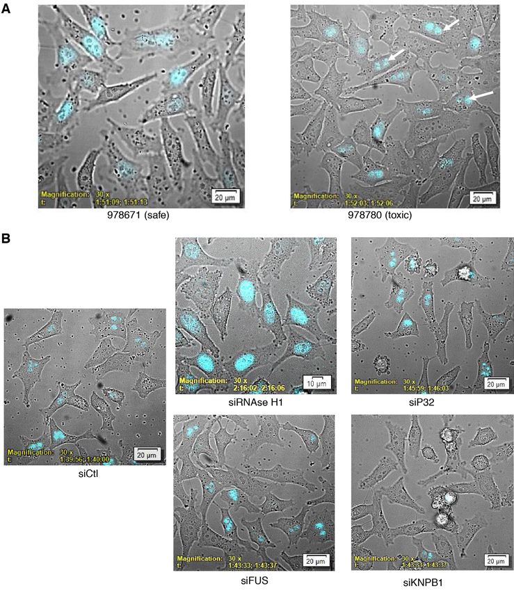
Nucleic Acids Research, 2019 3
Table 1. Sequence of ASOs used in study
ASO Length Sequence Chemistry Conjugate
N/A 20 CTGCTAGCCTCTGGATTTGA PO DNA/PS DNA none
N/A 20 CUGCUAGCCUCUGGAUUUGA PO RNA/PS RNA none
766638 20 CTGCTAGCCTCTGGATTTGA PS 2 -F gap-mer 3 Alexa Fluor 594
766634 20 CTGCTAGCCTCTGGATTTGA PS MOE gap-mer 3 Alexa Fluor 594
766636 20 CTGCTAGCCTCTGGATTTGA PS cEt gap-mer 3 Alexa Fluor 594
936532 16 GTCTGTGCATCTCTCC PS cEt gap-mer 3 Alexa Fluor 594
Downloaded from https://academic.oup.com/nar/advance-article-abstract/doi/10.1093/nar/gkz771/5565286 by guest on 17 September 2019
936533 16 GGCTACTACGCCGTCA PS cEt gap-mer 3 Alexa Fluor 594
978780 16 GTCTGTGCATCTCTCC PS cEt gap-mer 3 SmBiT peptide
978671 16 GGCTACTACGCCGTCA PS cEt gap-mer 3 SmBiT peptide
464917 16 GTCTGTGCATCTCTCC PS cEt gap-mer none
549148 16 GGCTACTACGCCGTCA PS cEt gap-mer none
982034 16 GTCTgTGCATCTCTCC PS cEt gap-mer none
558807 16 GCATGTTCTCACATTA PS cEt gap-mer none
936053 16 GCATgTTCTCACATTA PS cEt gap-mer none
482050 16 ATCATGGCTGCAGCTT PS cEt gap-mer none
982037 16 ATCAtGGCTGCAGCTT PS cEt gap-mer none
508031 16 TGAGGTCCTGCACTGG PS cEt gap-mer none
895155 16 TGAGgTCCTGCACTGG PS cEt gap-mer none
N/A 20 CTGCTAGCCTCTGGATTTGA PO DNA 3 Alexa Fluor 594
N/A 20 UCAAAUCCAGAGGCUAGCAG PO RNA compliment none
Sequence of ASOs used in study. Substitution with chemically modified nucleotides in gap-mer wings is indicated by bold type. 2 -O-Methyl modified bases
are indicated by lower case. Insertion of modified nucleotides is indicated by bold type.
Gene editing for the generation of the LgBiT-P54nrb 15 min, substrate addition and BRET readings were carried
HeLa cell line has been described previously (1). out as detailed above. P54nrb 1× binding buffer: 100 mM
NaCl, 20 mM Tris–HCl pH 7.5, 1 mM EDTA, 0.1% NP-
NanoBRET/NanoBiT assays for binding affinity 40. RNase H1 1 X binding buffer: 50 mM NaCl, 20 mM
Tris–HCl pH 7.5, 1 mM McCl2 , 1 mM TCEP.
Fusion proteins were expressed by transfecting the plasmids
into 6 × 10⁵ HEK 293 cells using Effectene transfection
reagent according to the manufacturer’s protocol (Qiagen). Western blot analysis
Following a 24-h incubation, cells were removed from the Western blots were carried out as detailed previously (7).
plate by trypsinization, washed with PBS, then resuspended Cells in 6 cm plates were transfected with NLuc-fusion plas-
in 250 l Pierce IP Lysis Buffer (Thermo Scientific). Lysates mids (500 ng) using Effectene transfection reagent (Qiagen).
were incubated 30 min at 4◦ C while rotating, then debris pel- Following a 24-h incubation, cells were removed from the
leted by centrifugation at 15,000 rpm for 5 min. The fusion plate by trypsinization, pelleted, washed 1× with PBS, then
protein was purified by adding 20 l HisPur Ni-NTA Mag- resuspended in IP Lysis buffer (Pierce) supplemented with
netic Beads (Thermo Scientific) and 10 mM imidazol then 1× Protease and Phosphatase Inhibitor Cocktail (Thermo
incubating at 4◦ C for 2 h. Beads were then washed 4 times Fisher Scientific), quantitated using a BCA protein assay
with PBS + 10 mM imidazol and 0.01% Tween-20. Fusion (Pierce), and separated on a 4–12% gradient SDS-PAGE
protein was eluted from the beads in 100 l PBS + 200 mM gel. Proteins were transferred to a nitrocellulose membrane
imidazol, followed by dilution with of 200 l IP buffer. using iBlot Gel Transfer Device (Thermo Fisher Scientific).
BRET assays were performed in white 96-well plates as The membranes were blocked at room temperature for 30
previously described (7). Alexa-linked ASOs at the indi- min with blocking buffer containing 5% (w/v) nonfat dry
cated concentrations were incubated at room temperature milk in 1× PBS and incubated with primary antibodies
for 15 min in 1× binding buffer with 106 RLU/well of Ni- in blocking buffer at room temperature for 2 h or 4◦ C
NTA purified NLuc fusion protein. For the PO DNA/RNA overnight. After washing three times with washing buffer
heteroduplex, 200 uM of each strand was first annealed (0.1% Tween-20 in 1× PBS) for 5 min each wash, mem-
in 1× PBS by slow cooling from 95◦ C. Following the in- branes were incubated with secondary antibodies in block-
cubation, NanoGlo substrate (Promega) was added at 0.1 ing buffer at room temperature for 1 h. After washing three
l/well. Readings were performed for 0.3 s using a Glo- times with washing buffer for 5 min each wash, proteins
max Discover system using 450 nm/8 nm band pass for were detected based on enhanced chemiluminescence (Ab-
the donor filter, and 600 nm long pass for the acceptor fil- cam).
ter. BRET was calculated as the ratio of the emission at
600/450 (fluorescent excitation emission/RLU). For com-
Immunofluorescence (IF)
petitive binding assays the Alexa-linked PS-ASO was added
at approximately the KD and the unconjugated competing Cells seeded on glass-bottom culture dishes (MatTek) were
ASO added at the indicated concentrations in 50 l water. treated as specified. Cells were fixed at room temperature
106 RLU/well of purified fusion protein or whole cell lysate for 30 min with 4% formaldehyde and permeabilized for
well was then added in 50 l 2× binding buffer for a final 5 min with 0.1% Triton X-100 in 1× PBS. Fixed cells
volume of 100 l. After incubation at room temperature for were then blocked in blocking buffer (1 mg/ml BSA in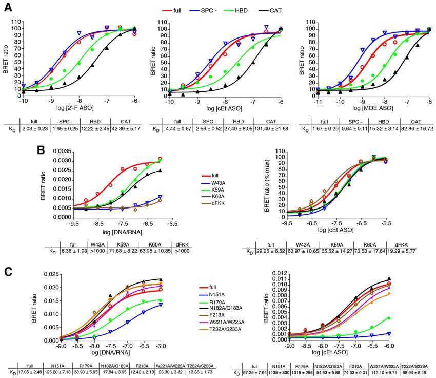
4 Nucleic Acids Research, 2019
1× PBS) at room temperature for 30 min, and incubated P54nrb is canonical, containing four aromatic residues at
in blocking buffer with primary antibody NanoLuc pAb, conserved positions that are typically essential for RNA
Promega; P54nrb pAb, ab70335 ABCAM) at room tem- binding (13), RRM2 is considered noncanonical with three
perature for 2 h. After washing three times with washing conserved residues substituted to Thr, Lys and Ile, implying
buffer (0.1% Tween-20 in 1× PBS; 5 min per wash), cells that either the RRM2s do not bind RNA, or that they bind
were incubated in blocking buffer with anti Rb Alexa-488 in an unexpected manner.
secondary antibody (Abcam ab 150073) at room tempera- To investigate the binding interaction of PS-ASOs with
ture for 1 h. Finally, cells were washed three times in wash- P54nrb, we utilized a previously described ASO/protein
Downloaded from https://academic.oup.com/nar/advance-article-abstract/doi/10.1093/nar/gkz771/5565286 by guest on 17 September 2019
ing buffer for 5 min each. Images were generated using an affinity assay (7) which relies on the transfer of light en-
LV200 Inverted Microscope (Olympus) fitted with ND10, ergy from a Nano luciferase (Nluc) tagged binding pro-
472/30 and 520/35 filters and analyzed using cellSens Di- tein acting as the BRET donor, to a fluorescently con-
mension v1.15 (Olympus). jugated ASO acting as the BRET acceptor. N-terminal
NLuc fusions were constructed for full-length P54nrb along
Live cell imaging of ASO/NanoBiT interactions with truncated proteins consisting of only the RNA bind-
ing domains (RRM1 or RRM2) or the core domains
Cells seeded on glass-bottom culture dishes (MatTek) were (NOPS and coiled-coil) (Figure 1A, Supplementary Fig-
transfected with LgBiT fusion plasmids using Effectene ure S2A). 106 RLU/well of the immunoprecipitated P54nrb
transfection reagent according to the manufacturer’s pro- fusion protein were incubated in a 96-well plate with PS-
tocol. The following day cells were transfected with Sm- modified, 5-10-5 ‘gap-mer’ PS-ASOs containing 10 de-
BiT conjugated ASO at 100 nM in 1 ml Leibovitz’s L-15 oxyribonucleotides in the middle flanked at both ends by
Medium, no phenol red (Thermo Fisher Scientific) with 6 five 2 -constrained ethyl (cEt), 2 -fluoro (2 -F), or 2 -O-
ug/ml Lipofectamine 2000 and 1 l Nano-Glo Live cell EX- methoxyethyl (MOE) conjugated at either the 3 end with
5455 substrate (Promega). Bright-field (ND1 and ND6 fil- Alexa 594 (Table 1) at concentrations ranging from 10 pM
ters) and luminescent images (460/60 filter) were collected to 1 M. NLuc substrate was then added and BRET ratios
from 0 to 2 h at 3-min intervals using an LV200 Luminoview determined as detailed in Materials and Methods. PS-ASOs
bioluminescent imaging system (Olympus) and processed were determined to bind the full-length protein with a KD of
using cellSens Dimension v1.15 software (Olympus). ∼3 nM, however binding to the full-length protein was not
strongly influenced by the wing chemistry of the PS-ASO
RIP-Seq gap-mer (Figure 1B/C, red lines). In contrast, binding to
the individual RRM domains was highly influenced by the
To construct RIP-seq libraries 5 × 10⁶ HeLa cells were chemistry of the PS-ASO. For the 2 -F PS-ASO (766638)
transfected with pHIS-SmBiT-H1 or with HIS-SmBiT- binding affinity to RRM1 (green lines) was approximately
RNaseH1 and LgBiT-P54nrb together using Effectene half that of the full-length protein, whereas binding of the
reagent. The following day cells were transfected with 100 cEt (766636) or MOE (766634) PS-ASO to RRM1 was 7–8-
nM PS-ASO 464917 in OptiMEM media with 6ug/l Lipo- fold weaker than to the full-length protein. The amplitude
fectamine 2000. Cells were collected by trypsinization and of the BRET signal for binding of RRM2 (blue lines) was
the pellet lysed in 250 l IP buffer for 30 min at 4◦ C. RNA– much lower than to the full-length protein or to the RRM1
protein complexes were purified by affinity pull-down with domain. However, BRET efficiency is a measure of distance
Ni-NTA magnetics beads then the RNA extracted using between the donor and the acceptor, which varies accord-
an RNeasy Mini kit (Qiagen). The purified RNA was then ing to an inverse sixth power of the distance between the
used to generate libraries using a NEXTflex Rapid Direc- two molecules (14). It is possible that the tertiary structure
tional qRNA-Seq kit according to the manufacturer’s pro- of RRM2 alone is such that the distance between the ASO
tocol (BIOO Scientific). Following 14 rounds of PCR am- binding site and the NLuc donor is greater than to RRM1
plification, libraries were size selected using 2% Pippin Prep alone or the full-length protein. When binding was normal-
gel collecting amplicons between 250 and 550 bp. The pu- ized for BRET amplitude (Supplementary Figure S2B), it
rified and size selected libraries were then sequenced on an was apparent that binding affinity to RRM2 was signifi-
Illumina NextSeq 550 System. cantly greater for the MOE and cEt gap-mers than for the
2 -F. Essentially no binding was observed to the core do-
RESULTS mains (black lines).
To evaluate the contribution of the PS linkage and sugar
Interaction of ASOs with P54nrb is highly dependent on
to the interaction of ASOs with P54nrb, a competitive
chemistry and binding environment
binding experiment was performed with full PS or full PO
P54nrb and PSF, are essential for multiple cellular pro- ASOs containing either ribose or deoxyribose sugar moi-
cesses, including transcription, splicing, polyadenylation, eties. Competitive binding to full length P54nrb was maxi-
nuclear retention, translation and DNA repair (10,11). The mal for the PS DNA ASO and ∼2 orders of magnitude less
structure of the human P54nrb/PSF heterodimer encom- for the PS RNA ASO (Figure 1D, dashed lines). No com-
passes two RNA binding domains: RRM1, RRM2, as well petitive binding was detected for either the PO DNA or PO
as NOPS, and coiled-coil core domains. P54nrb and PSF RNA ASOs (solid lines). The results were nearly identical
form homo- and heterodimers via their core domains (12) for binding to the RRM1 domain alone (Supplementary
and have been shown to interact with nucleic acids through Figure S3A) and imply that the PS linkage is essential for
their RRM domains (11). Although the RRM1 domain of ASO binding to P54nrb. In fact, binding affinity appears to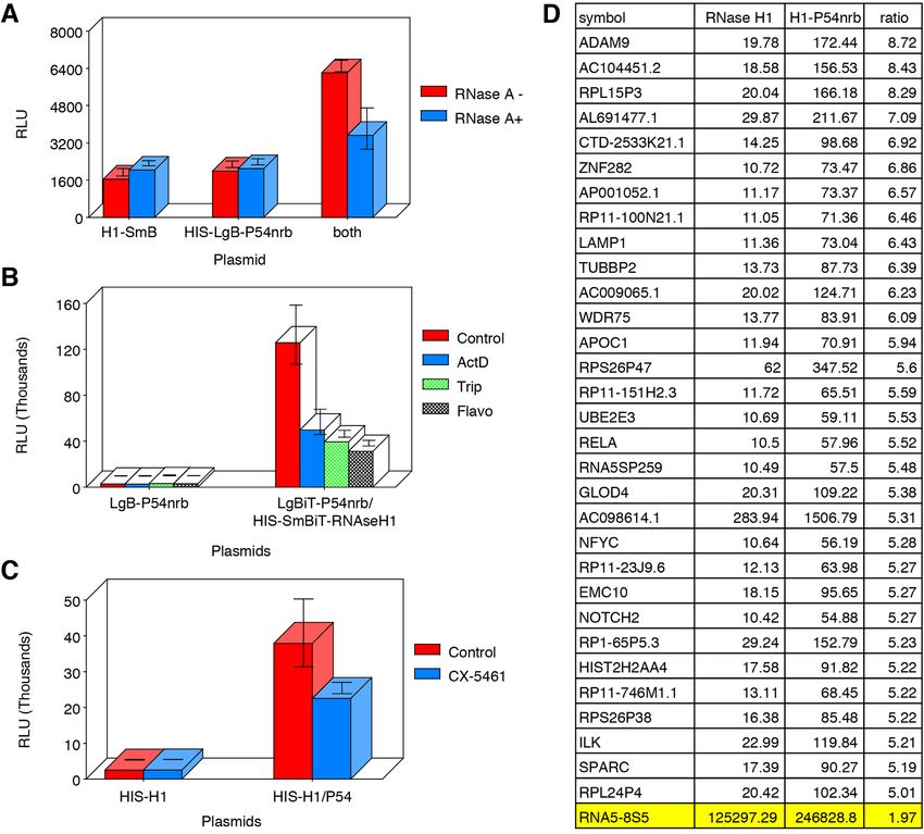
Nucleic Acids Research, 2019 5
Downloaded from https://academic.oup.com/nar/advance-article-abstract/doi/10.1093/nar/gkz771/5565286 by guest on 17 September 2019
Figure 1. (A) NLuc-P54nrb fusion constructs. NLuc was fused in frame to the amino-terminus of the P54 cDNA. RNA binding motifs RRM1 and RRM2,
and core domain were cloned separately beginning and ending at the indicated amino acid positions. Expression of the fusion protein was confirmed by
Western blot (Supplementary Figure S2A). (B) PS-ASOs bind P54nrb primarily via the RRM1 and RRM2 domains in a chemistry dependent manner.
NLuc-P54nrb fusion proteins were immunopurified as detailed in Online Methods and subsequently incubated with Alexa 594 conjugated 5-10-5 2 -F,
cEt, or MOE gap-mer PS-ASO at concentrations ranging from 10 pM to 1 M. BRET ratios were determined for full-length P54nrb-NLuc (red), RRM1
(green), RRM2 (blue), or NOPS/coiled-coil (black). Concentration response curves and KD ’s ± SEM (nM) are representative of two to three independent
experiments. Data normalized to maximum BRET signal may be found in Supplementary Figure S2B. (C) KD ’s (nM) are shown in tabular form with green
= highest affinity; red = lowest affinity. (D) Competitive binding of PS and PO RNA and DNA ASOs to P54nrb. Unconjugated competing ASOs at 3–10
000 nM were combined with 10 nM Alexa-linked cEt gapmer PS-ASO 766636, then binding to p54nrb evaluated by NanoBRET. (E) Salt dependence of
PS-ASO binding to P54nrb domains. KD ’s (nM) for binding of cEt gap-mer ASO to p54nrb in 25–400 mM NaCl. Concentration response curves can be
found in Supplementary Figure S4.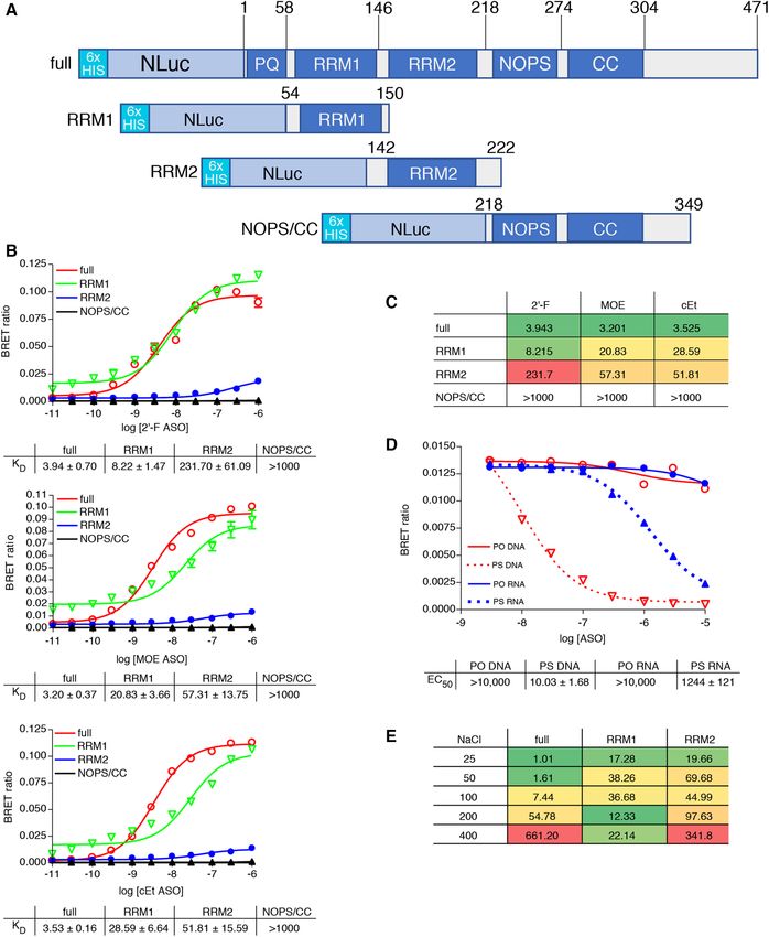
6 Nucleic Acids Research, 2019

be directly proportional to the number of PS linkages in the     tary Figure S5B, video). Fluorescent signal (red) from both
ASO (Supplementary Figure S3B).                                  of the unbound PS-ASOs was observable in the cytoplasm
   We next evaluated the effect of sodium concentration on       ∼60 min post-transfection. From the cytoplasm the PS-
the interaction of the cEt gap-mer PS-ASO with P54nrb.           ASOs traveled almost immediately to the nucleus where-
Binding of the cEt gap-mer PS-ASO 766636 was evaluated           upon association with the P54nrb/PSF complex was de-
to the full-length P54nrb protein as well as to the RRM1         tected by BRET emission (yellow). Consistent with previ-
and RRM2 domains alone in binding buffer with 25–400             ous observations (1), the toxic PS-ASO, 936532, was then
mM NaCl. For the full-length P54nrb and the RRM2 do-             localized in the nucleolus by 90 min (Figure 2B; Supplemen-

                                                                                                                                  Downloaded from https://academic.oup.com/nar/advance-article-abstract/doi/10.1093/nar/gkz771/5565286 by guest on 17 September 2019
main only, PS-ASO binding was strongly affected by salt          tary Figure S5C, video), while the safe PS-ASO, 936533, re-
concentration with the affinity decreasing as the salt con-      mained weakly associated with P54nrb in the nucleoplasm.
centration was increased (Figure 1E, Supplementary Figure           Since the substitution of a 2 -methoxy nucleotide at posi-
S4), whereas PS-ASO binding to RRM1 was only slightly            tion 2 of the DNA ‘gap’ portion of toxic PS-ASOs has been
affected by changes in salt concentration. These data sug-       demonstrated to greatly reduce toxicity without meaning-
gest that ionic interactions are a strong contributor to the     ful loss in potency (1), we also tested several pairs of un-
binding interaction between RRM2 and PS-ASOs, while              modified and 2 -methoxy substituted cEt gap-mers for lo-
affinity for RRM1 may involve other forces, such as base         calization of the PS-ASO interaction with the P54nrb/PSF
stacking or hydrophobic interactions.                            in live cells. Again, the LgBiT-P54nrb cell line was trans-
                                                                 fected with pSmBiT-PSF. The following day the cells were
                                                                 transfected with toxic cEt PS-ASOs with and without 2 -
Evaluation ASO/protein interactions and kinetics in live cells
                                                                 methoxy nucleotide at position 2 (Table 1) followed by ad-
Since binding to P54nrb and other proteins has been shown        dition of NLuc substrate and microscopy. Once again treat-
to significantly affect the toxic potentials of PS-ASOs we       ment with each of the known toxic unmodified PS-ASOs
sought to develop a NLuc-based structural complementa-           resulted localization of the LgBiT-P54nrb/SmBiT-PSF het-
tion reporter system which would enable elucidation of the       erodimer to the nucleoli (Figure 2C, upper panel), whereas
kinetics and localization of protein interactions interactions   in cells treated with 2 -methoxy-substituted ASOs of the
with toxic and safe cEt PS-ASOs, as well as protein–protein      same sequence the LgBiT-P54nrb/SmBiT-PSF heterodimer
interactions in real time in live cells (1). NanoLuc Binary      remained evenly distributed throughout the nucleus (lower
Technology (NanoBiT) is a two-subunit system based on            panel).
NLuc that was initially developed for intracellular detection       To monitor the interaction of ASOs with proteins more
of protein–protein interactions (15). In this assay, Large BiT   directly, PS-ASOs were synthesized with the SmBiT peptide
(LgBiT; 17.6 kDa) and Small BiT (SmBiT; 11 amino acids)          tethered to the 3 end. HeLa-LgBiT-P54nrb cells were trans-
NLuc subunits are fused to proteins of interest, and when        fected with safe (978671) or toxic (978780) SmBiT-linked
expressed, the protein–protein interaction brings the sub-       cEt gap-mer PS-ASOs and cells visualized for 1.5–2.5 h at
units into close proximity to form a functional enzyme that      3-min intervals. For the safe PS-ASO, luminescent associ-
generates a bright, luminescent signal. CRISPR was used          ation with P54nrb was detected in the nucleus as early as
to generate a stable cell line in which the LgBiT polypep-       60 min post-transfection (Figure 3A left panel, video Sup-
tide was fused in-frame at the amino-terminus of the en-         plementary Figure S6A). Association of the toxic PS-ASO
dogenous P54nrb gene. Expression and localization of the         was also not observed until ∼ 60 min post-transfection,
fusion protein was confirmed by immunofluorescent stain-         however the P54nrb complex with the toxic PS-ASO was
ing of the fixed cells with both NLuc and P54nrb antibod-        concentrated in the nucleoli rather than in the nucleo-
ies (Supplementary Figure S5A) and was consistent with the       plasm. (Figure 3A right panel, video Supplementary Fig-
previously reported nuclear distribution of this protein (11).   ure S6B). To confirm the nucleolar localization of the toxic
   To confirm the utility of the LgBiT-P54nrb fusion, we         PS-ASO, cells were transfected with a GSP fusion plasmid
constructed an amino-terminal SmBiT fusion to PSF which          overexpressing the nucleolar marker protein nucleophos-
is known to form a heterodimer with P54nrb (16). A con-          min 1 (NPM1), which was determined to be co-localized
trol HeLa cell line and the cell line harbouring the P54nrb-     with the PS-ASO/P54nrb complex (Supplementary Figure
LgBiT fusion were transfected with pSmBiT-PSF. The fol-          S6C). The kinetics and subcellular localization of several
lowing day Nluc signal was evaluated using a biolumines-         other PS-ASOs previously identified as safe or toxic (1) were
cent imaging microscope. No Nluc signal was detected in          also evaluated with essentially identical results (data not
cells expressing SmB-PSF or in the untransfected LgB-            shown).
P54nrb cell line however, a strong nuclear NLuc signal was
detected in the LgBiT-P54nrb cell line in which SmBiT-
                                                                 RNase H1 is required for the formation of the nucleolar
PSF was co-expressed (Figure 2A). The kinetics and local-
                                                                 P54nrb/ASO complex
ization of PS-ASO interaction with the P54nrb/PSF com-
plex were next evaluated. Again, the LgBiT-P54nrb cell line      HeLa-LgBiT-P54nrb cells were next treated 48 h with siR-
was transfected with pSmBiT-PSF. The following day the           NAs targeting RNase H1, P32, FUS and KPNB1 all pro-
cells were transfected with Alexa 594-linked safe (936533)       teins which have previously been demonstrated to bind and
or toxic (936532) cEt PS-ASOs, then visualized at 3-min in-      alter activity of PS-ASOs (1,17,18). Reduction of the tar-
tervals for a total of 2.5 h. Again, formation of the LgBiT-     geted protein was confirmed by qRT/PCR and western blot
P54nrb/SmBiT-PSF heterodimer resulted in a clear nuclear         (Supplementary Figure S7A). The cells were treated toxic
NLuc BRET donor signal (Figure 2B, green: Supplemen-             SmBiT-linked PS-ASO 978780 for 2 h then NLuc emission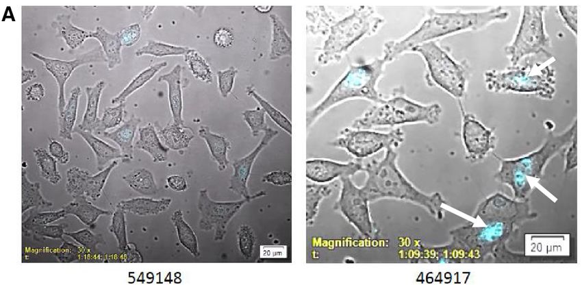
Nucleic Acids Research, 2019 7
Downloaded from https://academic.oup.com/nar/advance-article-abstract/doi/10.1093/nar/gkz771/5565286 by guest on 17 September 2019
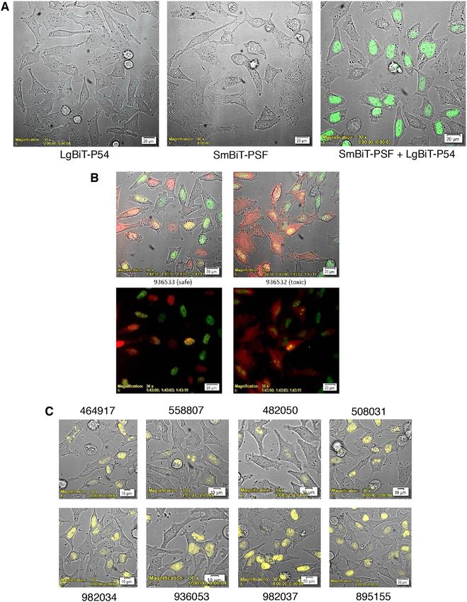
Figure 2. P54nrb-PSF NanoBiT assay. (A) Evaluation of LgBiT-P54nrb expression in CRISPR cell line. HeLa CRISPR LgBiT-P54nrb or HeLa control
cells seeded on glass-bottom culture dishes then transfected with a plasmid expressing a SmBiT-PSF fusion protein (pSmB-PSF). The following day
NanoGlo substrate was added to the cells and luminescence visualized using an Olympus LV200 Inverted microscope. Left panel, HeLa LgBiT–P54
fusion; center, control HeLa cells transfected with pSmB-PSF; right, HeLa LgB-P54 cells transfected with pSmB-PSF. (B) Safe/Toxic PS-ASO association
with P54nrb-PSF heterodimer in live cells. The LgBiT-P54nrb HeLa cell line was transfected with pSmBiT-PSF. The following day cells were treated with
Alexa595-linked safe ASO 936533 (left panels) or toxic ASO 936532(right panels) at 100 nM. Cells were then visualized using a bioluminescent imaging
microscope at 3-min intervals for a total of 2.5 h collecting brightfield, ASO fluorescence (red) and NLuc bioluminescence (green). Lower panels; brightfield
removed. This figure is representative of numerous fields and was repeated 3–4 times. For videos see Supplementary Figure S5B/C. (C) Substitution with
2 -methoxy nucleotide at position 2 of the DNA ‘gap’ portion of toxic PS-ASOs prevents nucleolar accumulation of P54nrb/PSF heterodimer. The LgBiT-
P54nrb HeLa cell line was transfected with pSmBiT-PSF. The following day cells were treated at 200 nM for 2.5 h with toxic PS-ASOs (upper panels)
or ASOs of the same sequence substituted at gap position 2 with 2 -methoxy (lower panels). Cells were then visualized using a bioluminescent imaging
microscope collecting brightfield and NLuc bioluminescence (yellow).8 Nucleic Acids Research, 2019
Downloaded from https://academic.oup.com/nar/advance-article-abstract/doi/10.1093/nar/gkz771/5565286 by guest on 17 September 2019
Figure 3. (A) Kinetics of cEt gap-mer ASO association with P54 in live cells. The LgBiT-P54nrb HeLa cell line was treated with 100 nM safe SmBiT-
conjugated cEt ASO 978671 or toxic SmBiT-conjugated cEt ASO 978780. Cells were then visualized using a bioluminescent imaging microscope at 3-
min intervals for a total of 2.25 h collecting brightfield and NLuc bioluminescence (blue). Arrows identify nucleolar localization of P54nrb–ASO complex.
For video see Supplementary Figure S6A/B. (B) Toxic cEt gap-mer ASO association with P54nrb in live cells treated with siRNAs targeting known
ASO interacting proteins. The LgBiT-P54nrb HeLa cell line was treated with an siRNAs targeting RNAse H1, P32, FUS or KPNB1 at 10 nM. Forty
eight hours later cells were treated with toxic SmBiT-conjugated cEt ASO 978780 at 200 nM for 2H. Following addition of NanoGlo cells were imaged
collecting substrate brightfield and NLuc. These figures are representative of numerous fields and were repeated 2–4 times. Kinetic imaging for ASO 978780
in RNAse H1 reduced cells from 0 to 2 H can be found in Supplementary Figure S7B.
form the ASO-P54nrb complex visualized. As observed pre- data suggest that PS-ASOs may enter the nucleus via a
viously, the toxic PS-ASO/P54nrb complex was localized nuclear receptor after which they may then associate with
in the nucleolus while the safe PS-ASO was associated with other proteins such as P54nrb. Interestingly, P54nrb–ASO
P54nrb throughout the nucleus of cells treated with the con- complex formation was observed after RNase H1 knock-
trol siRNA (Figure 3B). Reduction of P32 or FUS had lit- down, however, rather than the toxic PS-ASO complex be-
tle effect on the localization of the P54nrb–ASO complex ing localized in the nucleolus, it was dispersed throughout
for either PS-ASO. In contrast, when KPNB1 was reduced the nucleoplasm. The kinetics of toxic PS-ASO P54nrb in-
little P54nrb–ASO association was observed. KPNB1 func- teractions were also evaluated in RNase H1 reduced cells
tions in nuclear protein import, either in association with an and found to be similar to those of the safe PS-ASO (com-
adapter protein, such as importin-alpha subunit, or by act- pare videos, Supplementary Figures S6A/S7B).
ing as autonomous nuclear transport receptor (19). TheseNucleic Acids Research, 2019 9
These data suggest that RNase H1 is required for nu- rified from the complex and directional, strand-specific
cleolar accumulation of the P54nrb–ASO complex. To ex- libraries prepared for NEXT-Seq analysis as detailed in
plore this, we constructed a RNase H1-SmBiT fusion plas- Materials and Methods. Many RNAP II transcripts were
mid. HeLa-LgBiT-P54nrb cells were transfected with pH1- identified which generated 5X or more reads in the LgBiT-
SmBiT and treated with safe (549148) or toxic (464917) cEt P54nrb/SmBit H1 library relative to HIS-SmBiT-RNase
gap-mer PS-ASOs the following day (Table 1). NanoLuc H1 alone (Figure 5D). However, no individual RNAP II
luminescence from the association of P54nrb with RNase RNA was found to be predominantly associated with the
H1 was monitored for 90 min at 3-min intervals. Initially, P54nrb/RNase H1 complex. In contrast, 5.8S ribosomal
Downloaded from https://academic.oup.com/nar/advance-article-abstract/doi/10.1093/nar/gkz771/5565286 by guest on 17 September 2019
no NLuc signal was detected with either PS-ASO suggest- RNA (highlighted) was detected at very high levels and
ing that there is little association of P54nrb and RNase H1 increased ∼2-fold in the presence of both the p54nrb and
in the absence of PS-ASO (Figure 4, video Supplementary RNase H1 plasmid. The association of 5.8S RNA with the
Figure S8A). At ∼30 min, NLuc was detected in the nucleoli P54nrb–RNaseH1 complex is consistent with the results
with the toxic PS-ASO (464917) and maximal association of RNAP inhibition and the nucleolar association of toxic
of the PS-ASO with the P54nrb/RNase H1 was reached at PS-ASO/P54nrb/RNase H1 complex.
∼60 min. However, only a very slight nuclear signal was de- The association of RNase H1 with the P54nrb–toxic
tectable at the same time with the safe PS-ASO (549148). ASO complex was also evaluated by affinity pull-down.
An in vitro NanoBiT assay was used to further explore HeLa cells were co-transfected with plasmids expressing
the interaction between P54nrb and RNase H1. HeLa cells HIS tagged P54nrb-NLuc or RNaseH1. The following day
were transfected with HIS-LgBiT-P54nrb or with SmBiT- cells were transfected with safe or toxic PS-ASO for 2 h.
RNaseH1 and HIS-LgBiT-P54nrb together. The following Lysates of the cells were then prepared and protein com-
day cells were transfected with toxic cEt PS-ASO for 2 plexes pulled down via the 6X-HIS tagged P54nrb, sep-
h then lysates prepared and the P54nrb/RNase H1 com- arated by SDS-PAGE, and transferred to nitrocellulose.
plex purified using Ni-NTA beads. In this assay, NLuc Membranes were incubated then probed with antibodies
luminescence can only be generated when RNase H1 is to RNase H1 or P54nrb. A similar amount of P54nrb was
closely associated with the HIS-tagged P54nrb. As ex- pulled down with or without PS-ASO treatment (Supple-
pected, very little luciferase signal was detected with the mentary Figure S8B, left panel). In contrast, in the ab-
HIS-LgBiT-P54nrb or SmBiT-RNaseH1 plasmids, how- sence of PS-ASO treatment, a weak band corresponding
ever co-expression with of the HIS-LgBiT-P54nrb with to P54nrb-associated RNase H1 was detected (Supplemen-
SmBiT-RNaseH1 resulted in a significant increase in NLuc tary Figure S8B, right panel). In agreement with the live-
signal (Figure 5A, red bars). Furthermore, treatment of the cell NanoBiT assay, the intensity of the band was signifi-
lysates with RNase A reduced the NLuc signal substantially cantly increased with administration of the toxic PS-ASO.
(blue bars), suggesting that RNA may also be a component Interestingly, levels of P54nrb-associated RNase H1 pulled
of the P54nrb/RNaseH1 complex. Experiments in which down were slightly reduced in cells treated with the safe PS-
the HIS-tag was placed on the SmBiT-RNase H1 rather ASO as compared to the control. This may be the result of
than the LgBiT-P54nrb also produced a significant NLuc the safe ASO binding RNase H1 without promoting the as-
signal only when the plasmids were co-expressed (data not sociation with P54nrb, effectively reducing the amount of
shown). RNase H1 capable of interacting with P54nrb.
To identify the type of RNA associated with the Taken together, these data suggest that the nucleolar ac-
P54nrb/RNaseH1 complex, cells were treated with var- cumulation of the toxic PS-ASO may be mediated by in-
ious RNA polymerase (RNAP) inhibitors following creased interactions with RNaseH1 and P54nrb relative to
co-transfection with HIS-SmBiT-RNaseH1 and LgBiT- the safe PS-ASO. In fact, the in vitro binding affinity as de-
P54nrb. Once again, very little NLuc signal was detected termined by NanoBRET for the toxic PS-ASO 936532 was
in cells transfected with LgBiT-P54nrb only, whereas in 10-fold greater to P54nrb than the affinity for the safe PS-
the presence of SmBit-H1 a significant increase in signal ASO 936533, and ∼2-fold greater to RNase H1 (Supple-
was observed (Figure 5B, red). Treatment of the cells with mentary Figure S9A). However, no difference in RNase H1
1 ug/ml actinomycin D (blue), 1 uM triptolide (green) or binding affinity was observed between toxic PS-ASOs and
0.5 uM flavopiridol (gray) each significantly reduced the safe 2 -methoxy nucleotide substituted ASOs of the same
amount of P54nrb/RNase H1 association as measured by sequence although one ‘safe’ ASO (982037) did show a 2–3-
NLuc intensity. Since Act D and triptolide inhibit both fold reduction affinity to P54nrb relative to the unmodified
RNAP I and II transcription, and flavopiridol inhibits parent ASO (482050) (Supplementary Figure S9B).
RNAP II elongation and RNAP I processing (20), it is
possible that the RNA(s) associated with the complex are
P54nrb and RNAse H1 interact via specific domains
either RNAP I or II-dependent. We therefore repeated
the experiment, treating the cells with CX5461, a RNAP To determine which domains of RNase H1 are required
I-specific inhibitor which effectively reduced formation for the formation of the complex, RNase H1 deletion
of the LgBiT-P54nrb/SmBit H1 complex (Figure 5C). mutants were generated (Figure 6A). HeLa cells were
We then attempted to identify specifically associated then co-transfected with pLgBiT-P54nrb and the pSmBiT-
RNAs by RIP-Seq. Again, cells were transfected with H1 deletions. The following day cells were transfected
HIS-SmBiT-RNaseH1 or with HIS-SmBiT-RNaseH1 with toxic cEt PS-ASO 936532 and NLuc signal im-
and LgBiT-P54nrb together and the P54nrb/RNase H1 aged 2H post-transfection. As previously observed, the
complex isolated the following day. RNA was then pu- P54nrb/RNase H1 complex was localized to the nucleolus10 Nucleic Acids Research, 2019
Downloaded from https://academic.oup.com/nar/advance-article-abstract/doi/10.1093/nar/gkz771/5565286 by guest on 17 September 2019
Figure 4. Nucleolar P54nrb-RNAseH1 complex formation occurs in the presence of toxic, but not safe ASO. (A) LgBit-P54nrb HeLa cells were transfected
with SmBiT RNAse H1 plasmid. The following day cells were treated with safe (549148) or toxic (464917) ASOs at 200 nM capturing brightfield and
bioluminescent NLuc signal (blue) for 1.5 h at 3-min intervals. Arrows identify nucleolar localization of P54nrb/RNAseH1-ASO complex. For video, see
Supplementary Figure S8A.
upon treatment with the toxic PS-ASO (Figure 6B). Dele- overall PS-ASO affinity than do those at the CAT domain.
tion of the hybrid binding domain (dHBD) did not affect The observations also suggest that the binding isotherm for
P54nrb/RNaseH1 complex formation or localization, how- the full-length protein likely requires interactions with two
ever no P54nrb/RNase H1 association was detected with binding sites, one in the HBD and one in the catalytic site
deletion of the C-terminal region (dCTD) of RNase H1. and that affinities of the two sites are within 10-fold of each
A deletion mutant in which only the spacer domain was other as has been reported using other assays (21).
deleted (dSPCR) also was not able to form a complex with We next explored the effect of the binding environment on
P54nrb suggesting that the spacer region is the point of con- the interaction of the cEt gap-mer PS-ASO with RNase H1.
tact of RNase H1 with P54nrb. Binding of 766636 was evaluated to the full-length RNase
A similar set of experiments was performed by generating H1 protein as well as to the HBD and CAT domains alone
LgBiT-P54nrb deletion mutants which were co-expressed in binding buffer with 25–400 mM NaCl. For the full-length
in HeLa cells with the full length SmBiT RNase H1 then protein little effect of salt concentration was observed (Sup-
transfected with toxic cEt PS-ASO 936532. Once again, plementary Figure S10). Similarly, binding to the HBD was
full-length P54nrb was associated with RNase H1 and the largely unaffected by salt concentration except at 400 nM
PS-ASO in the nucleolus (Figure 6C). Deletion of RRM1, NaCl at which a loss of 4-fold in affinity was observed. In
RRM2 or the coiled-coil domains had no effect on the contrast, ASO binding to the catalytic domain was found to
formation or localization of the P54nrb/RNase H1 com- be highly affected by salt concentration with affinity varying
plex. However, deletion of the NOPS domain reduced the over 50-fold. These similarities in binding between the full-
number of P54nrb/RNase H1 complexes significantly and length protein and the HBD further suggest that PS-ASOs
when both the NOPS and coiled-coil domains were deleted, interact primarily with the HBD, but it is also likely that
no P54nrb/RNase H1 association was detected. Taken to- CAT domain interactions contribute to PS-ASO affinity as
gether these data imply that RNase H1 and P54nrb interact well.
with one another via the RNase H1 spacer and the P54nrb We also explored the binding interaction of PS-ASOs
core domains, while PS-ASOs appear to interact with this relative to the native PO DNA/RNA heteroduplex. As
complex via the RRM1 or RRM2 domain of P54nrb. it has previously been shown that the conserved Trp43,
Lys59 and Lys60 residues constitute the binding surface for
the HBD of human RNase H1 (22), three mutants were pre-
Evaluation of PS-ASO binding interactions with RNase H1 pared in which alanine was substituted for the conserved
The interaction of PS-ASOs with RNase H1 was also evalu- lysine residues at positions 59 and 60 (K59A, K60A), the
ated. ASO NanoBRET assays were performed with affinity conserved tryptophan at position 43 (W43A), and an ad-
purified NLuc RNaseH1fusion protein as well as the dele- ditional mutant were generated in which residues Phe58,
tion mutants depicted in Figure 6A. Affinity for the full- Lys59 and Lys60 were deleted (dFKK). Binding affinity of
length protein was similar for the 2 -F, MOE and cET gap- the PO heteroduplex as determined by NanoBRET of the
mers with KD ’s ranging between 2 and 4 nM (Figure 7A, red affinity purified mutant proteins, was significantly affected
lines). Deletion of the spacer domain had no effect on bind- the K59A and K60A mutations with a nearly 10-fold reduc-
ing of the 2 -F and increased affinity for cEt and MOE PS- tion in affinity relative to the native RNase H1 HBD, while
ASOs only slightly (blue lines). Binding to the HBD-only binding was completely ablated by the W43A substitution
(green lines) was ∼10-fold weaker than to the full-length and the FKK deletion (Figure 7B, left). In contrast, the
protein, whereas PS-ASO affinity for the catalytic domain FKK deletion had no effect on binding of the PS-ASO and
(black lines) was 4–5-fold less than for the HBD. These data only a 2-fold diminution in affinity was observed with the
suggest that PS-ASOs bind optimally to the full-length pro- Trp43, Lys59 and Lys60 to Ala substitutions (right panel).
tein, but that interactions at the HBD contribute more toNucleic Acids Research, 2019 11
Downloaded from https://academic.oup.com/nar/advance-article-abstract/doi/10.1093/nar/gkz771/5565286 by guest on 17 September 2019
Figure 5. RNA is associated with the P54nrb-RNaseH1-ASO complex. (A) Treatment with RNase A disrupts P54-RNase H1 association. HeLa cells
were transfected with HIS-LgBiT-P54nrb or with SmBiT-RNaseH1 and HIS-LgBiT-P54nrb together. The following day cell lysates were prepared. 1/2 of
each lysate was treated with RNase A at 1 g/ml for 30 min. Following RNase A treatment the P54nrb/RNase H1 complex purified using Ni-NTA beads.
Following 3 washes, bound material was eluted with 200 mM imidazole. NanoGlo substrate was then added and NLuc luminescence assayed. Red bars
control; blue bars +RNase A. (B) RNAP I/II inhibitors prevent formation of P54-RNase H1 complex. HeLa cells were transfected with LgBiT-P54nrb or
with HIS-SmBiT-RNaseH1 and HIS-LgBiT-P54nrb together. The following day cells were treated for 6 h with RNAP inhibitors. Following treatment cell
lysates were prepared and the P54nrb/RNase H1 complex purified and quantitated as above red bars, no inhibitor; blue bars, 1 g/ml Actinomycin D; green
bars, 1 M triptolide; grey bars, 0.5 M flavopiridol. (C) Treatment of cells with RNAP I inhibitor CX-5461 inhibits formation of P54nrb-RNaseH1-ASO
complex. Experiment was carried out as in B except following plasmid transfection cells were treated overnight with 300 nM CX-5461. (D) Identification of
P54/RNase H1 associated RNAs by RIP-Seq. HeLa cells were transfected with HIS-SmBiT-RNaseH1 or with HIS-SmBiT-RNaseH1 and LgBiT-P54nrb.
The P54nrb/RNase H1 complex was isolated the following day and RNA extracted. Directional, strand-specific libraries were prepared for NEXT-Seq
analysis. Genes represented by more than 5 times the number of reads in RNaseH1/P54nrb vs RNaseH1 only are shown.
Similarly, several amino acids have been identified as im- positions 221 and 225 (W221A,W225A), and Thr at posi-
portant contributors to the interaction of the RNase H1 tion 232 and Ser at position 233 (T232A, S233A). Again,
catalytic domain with the PO heteroduplex (23). C-terminal NanoBRET assays were performed to determine the ASO
domain only mutants were prepared in which alanine was binding affinity of the mutant proteins relative to the parent.
substituted for Phe at position 151 (N151A), Arg at posi- Mutations N151A And R179A had the greatest effect on
tion 179 (R179A), Asn at position 182 and Gln at position binding of the PO heteroduplex reducing affinity by more
183 (N182A, Q183A), Phe at position 213 (F213A), Trp at than 7-fold and 2-fold respectively (Figure 7C). Other sub-12 Nucleic Acids Research, 2019 Figure 6. (A) SmBiT/NLuc-RNAse H1 fusion constructs. SmBiT/NLuc was fused in frame to the amino-terminus of the RNAse H1 cDNA. Deletions Downloaded from https://academic.oup.com/nar/advance-article-abstract/doi/10.1093/nar/gkz771/5565286 by guest on 17 September 2019 were generated by site-directed mutagenesis as indicated in Supplementary Table S2. For the dCTD clone, amino acids 81–286 were deleted, for the spacer- clone amino acids 73–135 were deleted, and for the dHBD clone 1–80 were deleted. Expression of the fusion protein was confirmed by Western blot probed with NLuc pAB. Expected size of NLuc fusions are full length, 52kD; H1-dCTD, 29kD; H1-dHBD, 37.8kD; and dSPC-, 45.7kD. (B) RNAse H1 associates with P54nrb via the spacer domain. The LgBiT-P54nrb HeLa cell line was transfected with pSmBiT-RNAseH1 and with pSmBiT-RNAseH1 domain mutants as depicted in 5A. The following day cells were treated with toxic ASO 936532 at 200 nM for 2 h. Cells were then visualized collecting brightfield and NLuc bioluminescence (blue). (C) P54nrb associates with RNAse H1 via the NOPs/coiled-coil domains. HeLa cells were co-transfected with pSmBiT-RNAseH1 and with pLgBiT-P54nrb or with the indicated pLgBiT-P54nrb domain deletion mutants. The following day cells were treated with toxic ASO 936532 at 200 nM for 2 h. Cells were then visualized as in panel B. Images are representative of numerous fields and were repeated 3–4 times.
Nucleic Acids Research, 2019 13
Downloaded from https://academic.oup.com/nar/advance-article-abstract/doi/10.1093/nar/gkz771/5565286 by guest on 17 September 2019
Figure 7. (A) ASOs bind RNAse H1 at the HBD and catalytic domains, but do not interact with the spacer domain. NLuc-RNAse H1 fusion proteins
were immunopurified as detailed in Online Methods and subsequently incubated with Alexa 594 conjugated 5-10-5 cEt gap-mer ASO at concentrations
ranging from 10 pM to 1 M. BRET ratios were determined for full-length NLuc-RNAse H1 (red), Spacer- (blue), HBD-only (green) or CAT-only (black).
Concentration response curves and KD ’s ± SEM (nM) are representative of 2–3 three independent experiments. B) PS-ASOs interact with the RNAse H1
HBD differently than does the native RNA/DNA heteroduplex. ASO/BRET assays were carried out with purified NLuc-RNAse H1 HBD fusion protein
and with the indicated HBD mutant proteins. The purified fusion proteins were incubated with Alexa 594 conjugated 5-10-5 cEt gap-mer ASO or with
DNA/RNA heteroduplex of the same sequence with Alexa 594 on the DNA strand at concentrations ranging from 10 pM to 3 M. Concentration response
curves and KD ’s ± SEM (nM) are representative of two independent experiments. (C) PS-ASOs and PO heteroduplex interact with the RNase H1 catalytic
domain similarly. ASO/BRET assays were carried out with purified NLuc-RNase H1 CAT fusion protein and with the indicated CAT domain mutant
proteins. The purified fusion proteins were incubated with Alexa 594 conjugated 5-10-5 cEt gap-mer ASO or with DNA/RNA heteroduplex as above at
concentrations ranging from 10 pM to 1 M. Concentration response curves and KD ’s ± SEM (nM) are representative of 2 independent experiments.
stitutions had little effect on the binding of the heteroduplex DISCUSSION
to the catalytic domain. A similar effect was observed for
Recent work from our laboratory (17,24) has shown the PS-
binding of the PS gap-mer ASO which was strongly reduced
ASO’s interact with >50 cellular proteins and that interac-
by the N151A and R179A mutations, but not other substi-
tions with cellular proteins are critical to cellular uptake
tuted amino acids. Taken together, these data suggest that
and distribution as well as pharmacological activities and
the PS-ASO and PO heteroduplex interact similarly, with
toxicities. The interactions of PS-ASOs with many proteins
the catalytic domain, but that the interaction of PS-ASOs
are influenced by sequence, chemistry, length and where in a
in the HBD may involve additional interactions relative to
PS-ASO modifications are placed (7). We recently reported
the PO heteroduplex.14 Nucleic Acids Research, 2019
that protein-binding contributes profoundly to the toxic po- Evaluation ASO/protein interactions and kinetics in live cells
tentials of PS-ASOs, with toxic PS-ASOs binding more cel-
In an attempt to understand differences in the interaction of
lular proteins with higher affinity than non-toxic PS-ASOs
safe and toxic ASOs with proteins in real time in live cells
(1). These sequence and chemistry dependent ASO–protein
we used a NanoBiT complementation assay which relies on
interactions were shown to correlate with toxic potentials
the association of a SmBiT-linked ASO and LgBiT-linked
of cEt-modified PS-ASOs in vitro and in vivo altering the
protein to generate a detectable luminescent signal. A sta-
stability, function, or distribution of many cellular proteins
ble cell line was generated in which the LgBiT polypeptide
and resulting in significant toxicity. Toxic, but not safe cEt
Downloaded from https://academic.oup.com/nar/advance-article-abstract/doi/10.1093/nar/gkz771/5565286 by guest on 17 September 2019
was fused in-frame to P54nrb (Figure 2A). When these cells
PS-ASOs, caused rapid mislocalization of the paraspeckle
were treated with a SmBiT peptide conjugated PS-ASO, the
proteins P54nrb and PSF to nucleoli, resulting in nucleo-
binding interaction between the PS-ASO and P54nrb pro-
lar stress and fragmentation, upregulation of P21 mRNA
tein could be visualized using a bioluminescent imaging mi-
and activation of caspase activity, and ultimately, apoptotic
croscope (Figure 3, Supplementary Figure S6). For the safe
cell death. Given these observations, a more detailed under-
PS-ASO, association was observed in the nucleus, while for
standing of the sites of PS-ASOs interactions with proteins
the toxic PS-ASO the interaction was limited to the nucleo-
and the chemical characteristics of those interactions that
lus, consistent with previously reported observations (1,25).
meaningfully influence the behaviors of PS-ASO’s and the
Following the association in real time, we determined that
cellular responses to the PS-ASOs is essential. This is the
PS-ASOs can be observed binding P54nrb as early as 45
first report of our efforts to answer these important ques-
min after addition to the cells (Figure 3, Supplementary
tions.
Figure S6). The kinetics observed are consistent with our
previously reported data for PS-ASO activity (26).
Interaction of ASOs with P54nrb These observations were further confirmed by monitor-
ing the BRET interaction of fluorescently tagged PS-ASOs
Here we present an analysis of PS-ASO interactions with with the LgBit-P54nrb/SmBit-PSF complex. Again, the
P54nrb and associated proteins in an attempt to better un- safe ASO was found to interact with the P54nrb/PSF het-
derstand the contribution of PS-ASO/protein interactions erodimer throughout the nucleus beginning ∼45–60 min af-
to differences in cellular toxicity at the molecular level. Our ter addition to the cells (Supplementary Figure S5B). Sim-
data show that ASOs must contain PS linkages in order to ilarly, the toxic ASO was also observed to bind to the
bind P54nrb (Figure 1D, Supplementary Figure S3A) and P54nrb/PSF heterodimer throughout the nucleus 1 h after
that the affinity of an ASO for P54nrb is directly propor- addition to the cells, however, within 15 min the complex
tional to the number of PS linkages in in the ASO (Sup- began localizing to the nucleoli and by 2 h virtually all toxic
plementary Figure S3B). This is the first publication to PS-ASO bound heterodimer was contained within the nu-
show that there are two independent binding sites in P54nrb cleoli (Supplementary Figure S5C). These results were ex-
for PS-ASOs. Binding interactions occur primarily via the tended using with a series a series of unmodified and 2 -
RRM1 and RRM2 domains, but strikingly, the interaction methoxy substituted cEt gap-mers. Insertion of a single 2
can be influenced by the chemistry of the PS-ASO (Figure methoxy at position 2 in the oligodeoxynucleotide gap in
1C, Supplementary Figure S2B/C). Specifically, PS-ASOs PS-ASO’s has previously been demonstrated to ameliorate
with the 2 -F modification in the wings bind P54nrb pri- or ablate PS-ASO toxicity (1,25). Again, all of the toxic
marily via RRM1 interactions, while for PS-ASOs with ASOs were localized with the P54nrb/PSF heterodimer in
MOE or cEt modifications, RRM1 and RRM2 domain in- the nucleolus, while ASOs of the same sequence made safe
teractions contribute nearly equally to overall binding. The as a result of a single 2 -methoxy substitutions remained
strength of PS-ASO affinity for RRM2 was also found to bound throughout the nucleus (Figure 2C).
be highly influenced by the sodium concentration of the
binding buffer, while binding to RRM1 was not strongly
affected by changes in sodium concentration (Figure 1E, RNase H1 is specifically associated with the nucleolar toxic
Supplementary Figure S4). The salt-dependence of a bi- PS-ASOs complex with P54nrb/PSF
molecular association is indicative of the contribution of
charge–charge interactions to the free energy of binding. In an effort to understand the biochemical nature of the
Therefore, the increased effect of added sodium chloride on nucleolar complex of toxic PS-ASOs with P54nrb, HeLa-
binding to RRM2 as compared to RRM1 suggests that cEt LgBiT-P54nrb cells treated with siRNAs targeting a num-
PS-ASO interactions with RRM2 are primarily electrostatic ber of proteins previously demonstrated to bind and alter
in nature, involving contacts between basic residues on the activity of PS-ASOs (1,17,18). In cells with reduced RNAse
RRM2 domain and the phosphorothioate groups in the PS- H1, the association of the toxic PS-ASO with P54nrb was
ASO while interactions with RRM1 may be mediated by nuclear rather than concentrated in the nucleoli, resembling
hydrophobic or other forces. This is consistent with the ob- that of a safe PS-ASO (Figure 3A, Supplementary Figure
servation that the RRM1 interaction appears to dominate S7B). This suggests that toxic PS-ASOs may uniquely in-
the overall binding to the full-length protein for the more teract in a complex that includes RNase H1, P54nrb and
hydrophobic 2 -F gap-mer, while RRM1 and RRM2 inter- PSF. To determine nature of this interaction, RNase H1 was
actions contribute more equally to overall P54nrb binding fused with SmBiT peptide, which was co-transfected into
of the MOE and cEt gap-mers. HeLa cells with LgBiT P54nrb. Cells were then treated withYou can also read


