Competitive dewetting underlies site-specific binding of general anesthetics to GABA(A) receptors - bioRxiv
←
→
Page content transcription
If your browser does not render page correctly, please read the page content below
bioRxiv preprint first posted online Jul. 8, 2019; doi: http://dx.doi.org/10.1101/694612. The copyright holder for this preprint (which
 Biophysical
      was notJournal     Volume:is00theMonth
               peer-reviewed)                 Year 1–15
                                       author/funder,   who has granted bioRxiv a license to display the preprint in perpetuity.          1
                                It is made available under a CC-BY-NC-ND 4.0 International license.

            Competitive dewetting underlies site-specific binding of general
                          anesthetics to GABA(A) receptors
                             Sruthi Murlidaran1,2 , Jérôme Hénin3,4 , and Grace Brannigan*1,5
      1
        Center for Computational and Integrative Biology, Rutgers University-Camden, Camden, NJ, USA
  2
      Department of Anesthesiology and Critical Care, University of Pennsylvania, Philadelphia, PA, USA
            3
              CNRS, Université de Paris, UPR 9080 Laboratoire de Biochimie Théorique, Paris, France
     4
       Institut de Biologie Physico-Chimique–Fondation Edmond de Rothschild, PSL Research University,
                                                  Paris, France
                     5
                       Department of Physics, Rutgers University-Camden, Camden, NJ, USA

 Abstract
 GABA(A) receptors are pentameric ligand-gated ion channels playing a critical role in the modulation of neuronal excitabil-
 ity. These inhibitory receptors, gated by -aminobutyric acid (GABA), can be potentiated and even directly activated by
 intravenous and inhalational anesthetics. Intersubunit cavities in the transmembrane domain have been consistently identified
 as putative binding sites by numerous experiment and simulation results. Synaptic GABA(A) receptors are predominantly
 found in a 2↵:2 :1 stoichiometry, with four unique inter-subunit interfaces. Experimental and computational results have
 suggested a perplexing specificity, given that cavity-lining residues are highly conserved, and the functional effects of general
 anesthetics are only weakly sensitive to most mutations of cavity residues. Here we use Molecular Dynamics simulations and
 thermodynamically rigorous alchemical free energy perturbation (AFEP) techniques to calculate affinities of the intravenous
 anesthetic propofol and the inhaled anesthetic sevoflurane to all intersubunit sites in a heteromeric GABA(A) receptor. We
 find that the best predictor of general anesthetic affinity for the intersubunit cavity sites is water displacement: combinations
 of anesthetic and binding site that displace more water molecules have higher affinities than those that displace fewer. The
 amount of water displacement is, in turn, a function of size of the general anesthetic, successful competition of the general
 anesthetic with water for the few hydrogen bonding partners in the site, and inaccessibility of the site to lipid acyl chains. The
 latter explains the surprisingly low affinity of GAs for the      ↵ intersubunit site, which is missing a bulky methionine residue
 at the cavity entrance and can be occupied by acyl chains in the unbound state. Simulations also identify sevoflurane binding
 sites in the subunit centers and in the pore, but predict that these are lower affinity than the intersubunit sites.

 Significance
 After over a century of research, it is established that general anesthetics interact directly with hydrophobic cavities in proteins.
 We still do not know why not all small hydrophobic molecules can act as general anesthetics, or why not all hydrophobic cav-
 ities bind these molecules. General anesthetics can even select among homologous sites on one critical target, the GABA(A)
 heteropentamer, although the origins of selectivity are unknown. Here we used rigorous free energy calculations to find that
 binding affinity correlates with the number of released water molecules, which in turn depends upon the lipid content of the
 cavity without bound anesthetic. Results suggest a mechanism that reconciles lipid-centered and protein-centered theories,
 and which can directly inform design of new anesthetics.

 1        Introduction
 General anesthetics (GAs) are chemically diverse molecules that cause immobilization and amnesia with moderate potency.
 Their surprising structural diversity has motivated quantitative efforts to predict potency from molecular properties for over a

 © 2019 The Authors
 *Corresponding author: grace.brannigan@rutgers.edu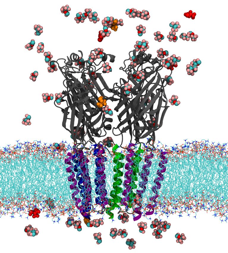
bioRxiv preprint first posted online Jul. 8, 2019; doi: http://dx.doi.org/10.1101/694612. The copyright holder for this preprint (which
2 was not peer-reviewed) is the author/funder, who has granted bioRxiv a license to display the preprint in perpetuity.1 INTRODUCTION
It is made available under a CC-BY-NC-ND 4.0 International license.
century. The classic Meyer-Overton trend indicated that potency of GAs could be predicted by their solubility in olive oil (1),
suggesting a lipid or membrane-mediated mechanism. It is now well established that direct binding of GAs to multiple pro-
teins in the central nervous system, including ligand-gated ion channels (2–4), is essential to the effects of GAs at clinical
concentrations. In particular, -aminobutyric acid receptors type A (GABAA ), which are critical for rapid inhibitory signaling,
have been identified as primary targets for widely-used general anesthetics like propofol and sevoflurane (5, 6).
GABAA Rs are members of the pentameric ligand-gated ion channel (pLGIC) superfamily, and most synaptic GABAA R
are heteropentamers with a 2↵:2 :1 stoichiometry. (7–9) Crystal structures of the 3 GABAA R homopentamer, (10) and
recent cryo-electron microscopy structures of the ↵1 2 2 heteropentamer (11, 12) and ↵1 3 2 heteropentamer (13, 14) have
confirmed that GABAA R share common structural features of the pLGIC family. Five subunits are arranged around a central
pore; each subunit consists of an extracellular domain (ECD) containing at least two orthosteric ligand binding sites, a trans-
membrane domain (TMD) containing 4 helices notated M1-M4, with the M2 helix lining the pore and the M4 helix facing
the lipid membrane, and a largely disordered intracellular domain.
General anesthetics (GAs) potentiate GABAA , but inhibit several other pLGICs, including some nAChRs and the prokary-
otic channels GLIC and ELIC. Our initial computational studies on the nAChR predicted GA binding to multiple sites,
including the pore lumen (15). Subsequently, the first crystal structure of GAs in complex with GLIC indicated binding in
the the subunit center, and inhibition was attributed to this intrasubunit site (16). More quantitative simulations confirmed
our pore binding hypothesis, and we argued that due to detergents in the crystallized GLIC pore, absence of crystallographic
evidence for pore block by GAs was not decisive (17). Using Molecular Dynamics Simulation and Alchemical Free Energy
Perturbation (AFEP) we predicted that, at half-saturating concentrations, propofol was as likely to bind to the GLIC pore as
to GLIC intrasubunit sites, while isoflurane was significantly more likely to bind to the pore. Subsequent experimental results
have increasingly supported a role for pore block in inhibition (18–20).
A pore binding site cannot, however, explain the gain of function that is observed in pLGICs responsible for the clinical
effects of GAs on humans, and in the present manuscript we predict that propofol and sevoflurane have lower affinity for
the pore binding sites of the GABAA receptor than for several other potential allosteric sites. The only structures of GAs in
complex with GABAA (or any other eukaryotic receptor) come from co-crystals of GABAA and several neurosteroids (21–
23). These structures reveal a common binding site that is external to the TMD, at subunit interfaces. Based on the body of
evidence from other co-crystal structures, photolabeling, mutageneis, and simulation, such a superficial site is not expected
to explain the strong modulatory effects of GAs on GABAA . It is likely that the intrasubunit binding mode detected in GLIC
causes gain of function, (20) as we originally suggested in (17), and possible that such an intrasubunit site could also cause
gain of function in GABAA . However, numerous photoaffinity labelling studies have pointed to anesthetic binding sites at
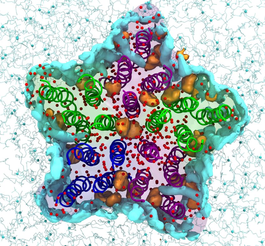
the interface between subunits in the GABAA transmembrane domain (TMD). (24–30) This is illustrated in Figure 1, which
represents data from (28, 29) using a propofol-based photolabel, and (30) using a sevoflurane-based photolabel. Perplexingly,
mutation of most intersubunit residues (with a few exceptions) yields only subtle quantitative shifts in EC50(26, 31, 32) but
affinity appears to be highly sensitive to subunit species. (29, 33, 34)
Propofol has been a widely used general anesthetic since its discovery in 1980; it has a higher potency and is better tol-
erated than older, volatile anesthetics like halothane or isoflurane. Propofol potentiates GABAA (26) at clinical (micromolar)
concentrations and even directly activates the channel at higher concentrations (35, 36). In contrast, most volatile anesthetics
potentiate GABAA at 100 µM concentrations (37).
The origins of the increased potency of propofol are not fully determined, despite studies involving a large number of
propofol analogs, (26) but some evidence supports a role for the strong hydrogen bonding tendency of the hydroxyl moiety.
A propofol analog with fluorine rather than hydroxyl (“fropofol”) has significantly reduced effects on GABAA receptors, (38)
and we previously reported (33) that propofol affinity for pseudosymmetric sites is an increasing function of hydrogen bond-
ing propensity. One surprising result from (33) was the low affinity of propofol for the intersubunit site with the most polar
residues (the ↵ + -↵- site). We report here that we previously underpredicted both hydrogen bonding and affinity for the
+ -
↵ -↵ site, as a result of a technical artifact, specific to flexible docking of propofol and corrected in the present work.
Several observations are inconsistent with affinity driven by hydrogen bonding, however. Foremost, it is not clear why
hydrogen bonding propensity would explain the strong correlation between potency and solubility in olive oil first observed
by Meyer and Overton. Although isoflurane and sevoflurane do not contain a hydroxyl group like propofol, they do contain
multiple weak hydrogen bond donors (39), and they are more soluble in water and less soluble in olive oil than propofol.
While direct substitution of the propofol hydroxyl with fluorine yields an inactive molecule(38), substitution with bromide
still leaves a general anesthetic as long as the solubility is maintained by modifying the isopropyl groups. (26) Furthermore,
the ↵+ - - site within the GABA(A) receptor is a so-called “orphan site”(40) that has not been identified using any anesthetic
photolabels, but contains the same number of hydrogen bonding residues(33) as the ↵+ - - site that does bind azipropofol. (40)
In the present manuscript we use Molecular Dynamics Simulation and Alchemical Free Energy Perturbation to predict
specific binding affinities across five subunit interfaces and two general anesthetics, and isolate the molecular origins of these
Biophysical Journal 00(00) 1–15
bioRxiv preprint first posted online Jul. 8, 2019; doi: http://dx.doi.org/10.1101/694612. The copyright holder for this preprint (which
      was not peer-reviewed) is the author/funder, who has granted bioRxiv a license to display the preprint in perpetuity.                     3
                                It is made available under a CC-BY-NC-ND 4.0 International license.

                       A                        ɣ                                B                                                        ECD

                                  ɣ+/β-
                                                          α+/γ-
                  βɣ                                                       αɣ
                                                                                                                                          TMD

     propofol           βγ+/α-                                    βα+/α-

                                                                                                                                          ICD
                                          α+/β-
                           αβ                                      βα

                       C                            M4                           D
                                           M3            M1

                                                    M2

     sevoflurane

 Figure 1: Experimentally-indicated contact residues for propofol and sevoflurane on the GABAA receptor. The trans-
 membrane domain of the GABAA R, viewed from the extracellular region (A,C) or from within the membrane (B,D), colored
 by subunit (↵-green, -purple, -blue). A-B) Propofol binding site residues indicated through photolabeling using AziPM(29)
 are shown in orange, o-PD(28) in red, and through mutagenesis(24) in yellow. The center of the intersubunit or pore binding
 cavities for propofol indicated by refinement simulations are marked with gray and cyan spheres, respectively. C-D) Sevoflu-
 rane binding site residues indicated through photolabeling with Azisevoflurane(30) are shown in orange. The center of the
 intersubunit or pore binding cavities for sevoflurane indicated by refinement simulations are marked with gray and cyan
 spheres, respectively. Both molecules were mobile in all intersubunit and pore sites during refinement simulation, and contact
 probabilities with individual residues are shown in Table 1.

 affinity differences. We also find that water molecule displacement, rather than hydrogen bonding, is a more universal predic-
 tor of affinities for both sevoflurane and propofol across all five intersubunit sites. Water displacement, in turn, is determined
 by size of the anesthetic, lipid-accessibility of the intersubunit binding site, and the ability of the anesthetic to replace water
 for a limited number of hydrogen bonding partners in the binding site.

 2    Methods
 We use three general types of MD simulations for finding and ranking GA binding sites: (1) Docking refinement simulations,
 (2) Flooding simulations, and (3) Binding affinity calculations. Refinement and flooding simulations share the goal of generat-
 ing poses and sampling interactions. Both use traditional equilibrium molecular dynamics approaches, but differ in the initial
                                                                                                                  Biophysical Journal 00(00) 1–15
bioRxiv preprint first posted online Jul. 8, 2019; doi: http://dx.doi.org/10.1101/694612. The copyright holder for this preprint (which
 4    was not peer-reviewed) is the author/funder, who has granted bioRxiv a license to display the preprint in perpetuity.          2 METHODS
                                It is made available under a CC-BY-NC-ND 4.0 International license.

 conditions and GA concentration. In refinement simulations, a small number of GA molecules begin in poses previously iden-
 tified by docking, and may then reorient or even unbind during the simulation. In flooding simulations, a large number of GA
 molecules are placed in the water and spontaneously partition to the membrane and sites on the protein during the course of
 the simulation. Neither of these methods provides any quantitative information to rank sites, requiring that we calculate bind-
 ing affinities using a different approach. We use the theoretically exact, alchemical free energy perturbation (AFEP) method
 to predict the GA concentration for which any specific site would be 50% occupied. Either refinement or flooding simulations
 can be used to provide initial coordinates for binding affinity calculations, but we primarily use refinement simulations. The
 workflow underlying our general approach for identifying and ranking binding sites is more explicitly detailed in Ref. (41).

                                                     2.1   Model systems for simulations
 For straightforward comparison with previous work(33), we use a model of the ↵1 3 2 GABAA receptor based on the nema-
 tode GluCl crystal structure (3RHW) with ivermectin, a positive modulator, bound to the intersubunit sites in the TMD (42).
 This structure represents the active state, and the crystallized GluCl chain posesses 33 to 39% sequence identity with the
 GABAA subunits studied here. Cryo-EM structures of human GABAA receptor heteromers have been released recently(11–
 14), yet none of them is representative of the active state that is best suited to study binding of positive allosteric modulators.
 The systems were prepared as in references 43 and 33, by embedding the protein in a lipid bilayer composed of phosphatidyl-
 choline (POPC), solvated and completed with 0.15 mol/L NaCl using CHARMM-GUI Membrane builder (44), with the final
 system containing 268 POPC molecules and about 180000 atoms.

                                                           2.2   Simulation details
 All simulations used the CHARMM36 model (45) for proteins, phospholipids, ions, and cholesterol molecules, and the TIP3P
 water model (46). The forcefield parameters for sevoflurane were those used in (47), based on the isoflurane parameters devel-
 oped in (39). Propofol parameters were those derived in (17), including the dihedral angle cross-terms (CMAP) required to
 adequately couple the hydroxyl and isopropyl groups of propofol. Energy minimization and MD simulations were conducted
 using the NAMD 2.12 package(48). All simulations employed periodic boundary conditions, long-ranged electrostatics were
 handled with a smooth particle mesh Ewald method, and a cutoff of 1.2 nm was used for Lennard-Jones potentials with a
 switching function starting at 1.0 nm. All simulations were run in the NPT ensemble with weak coupling to Langevin thermo-
 stat and a barostat at a respective 300 K and 1 atm. All bonds to hydrogen atoms were constrained using the SHAKE/RATTLE
 algorithm. A multiple time-step rRESPA method was used, with a high frequency time-step of 2 fs and a slow time-step of
 4 fs for the long-range force components. All standard MD simulations started with 10,000 steps of energy minimization.
 Harmonic restraints with force constant 0.5 kcal/mol/Å2 on the protein backbone positions prevented conformational drift
 during the AFEP calculations.

                                                     2.3   Docking and MD refinement
 General anesthetics were docked to intersubunit sites using the Autodock Vina software (49). The search space comprised
 the entire TM domain and intersubunit sites. Poses in each of the five intersubunit sites as well as the pore were chosen after
 performing multiple docking runs as described in Ref. (41). Although propofol bonds were rotatable during docking for our
 previous studies(17, 33), here they were made non-rotatable, and only the low-energy conformation of propofol was used for
 docking, as justified in Ref. (41).
     Unbiased docking refinement simulations involving propofol were run for about 600 ns following minimization. Propofol
 remained stable in the binding site throughout the course of the simulation. Autodock Vina initially docked sevoflurane to
 sites in the lower TMD, but during MD refinement for about 200 ns, sevoflurane reliably shifted higher in the TMD in three
 of the intersubunit sites, to a position more similar to that of propofol (41). Consequently, all initial sevoflurane configurations
 here use the “upper” site. In line with the faster dynamics of sevoflurane compared to propofol, these MD simulations of
 sevoflurane were run for about 150 ns, with sevoflurane remaining stably bound despite being highly mobile in the site.

                                               2.4     Free energy perturbation simulations
 Alchemical free energy perturbation simulations were performed on general-anesthetic bound GABAA R systems, with start-
 ing configurations obtained from standard MD simulations. Movement of the ligand was confined to 5 Å of the binding site
 center by flat-bottom restraints, which were spherical in the intersubunit sites and cylindrical in the pore, with a force constant
 Biophysical Journal 00(00) 1–15
bioRxiv preprint first posted online Jul. 8, 2019; doi: http://dx.doi.org/10.1101/694612. The copyright holder for this preprint (which
 2.5 Flooding   simulations
      was not peer-reviewed)     is the author/funder, who has granted bioRxiv a license to display the preprint in perpetuity.                5
                                It is made available under a CC-BY-NC-ND 4.0 International license.

 of 5 kcal/mol/Å2 . Decoupling of the ligand was performed over a series of 24 windows. The perturbation parameter was
 sampled with a step size equal to 0.025 between 0 < < 0.1 and 0.9 < < 1, and a step of 0.05 otherwise. Each window
 began with a 4 ps equilibration period followed by a 5 ns run for data collection. Lennard-Jones interactions at intermediate
   values were interpolated as a smooth, “separation-shifted” soft-core potential (50) as implemented in NAMD. Free energy
 of binding was calculated following Ref. (17), as:

                                                  Gbinding =     Gsolv +     Gvac!prot        Grestr                                         (1)

 d
     where Gsolv is the solvation free energy of the anesthetic, Gvac! prot is the free energy decoupling the ligand from
 the binding site and Grestr if the free energy cost of imposing restraints relative to the standard state volume. As these cal-
 culations deal with relatively small ligands with significant orientational mobility in the binding site, simple center-of-mass
 restraints are sufficient to ensure rapid convergence. This would not be the case for larger ligands with slow orientational and
 conformational fluctuations, which would benefit from more specialized restraints. (51)

                                                          2.5    Flooding simulations
 To identify additional candidate sites, we used an approach similar to that used for isoflurane in Ref. (15): sevoflurane was
 inserted randomly into the water surrounding the protein using tcl scripts within VMD (52), with a sevoflurane-to-lipid ratio
 of about 1:3. Flooding simulations were run for about 1.7 µs. Flooding with propofol was not pursued because the extremely
 low solubility causes aggregation of ligand in aqueous solution; sufficiently low concentrations prevent aggregation but extend
 the required time scales beyond those feasible for simulation. As discussed in Ref. (41), this approach is not straightforward
 for propofol, due to its low solubility.

                                                                  2.6   Analysis
 All molecular graphics was created using VMD (52). In flooding simulations, sevoflurane was determined to have partitioned
 to the membrane if there are at least 3 acyl chains within 5 Å of sevoflurane, in the protein if there are at least 3 protein
 residues within the 3 Å of sevoflurane, and in the aqueous phase otherwise. These criteria were chosen so that the sevoflurane
 partitioning status consistently matched visual inspection. Solvation of the binding cavities was determined by counting the
 number of lipid atoms or water molecules that met the following criteria per frame: coordinates within 5 Å from the center
 of the binding cavity. Hydrogen bonds were determined using geometric criteria, with a distance cutoff of 3.3 Å length and
 angle cutoff of 40 degrees. Solvation and hydrogen bonds were calculated for every ns, and the average was calculated after
 discarding the first 50 ns of simulations.

 3   Results
         3.1    Simulations show frequent contacts between general anesthetics and residues identified by photolabeling
 Various residues in the intersubunit sites of GABAA R have been identified using photoaffinity labeling as possible binding
 sites for propofol (29) and sevoflurane (30), as shown in Figure 1. For comparison, the contact frequency and mean distance
 measured from simulations are listed in Table 1. These values are extracted from refinement simulations following docking to
 the interface or pore sites.
     Propofol formed frequent contacts with three residues labeled by the photolabel azipropofol (29) and one residue indi-
 cated by mutagenesis (24), which line cavities between all subunit interfaces except ↵+ - - (Figure S1) . These residues are all
 located near the bulge in M1 at 180 and 190 . Two residues located farther from the bulge were not frequently contacted. One
 such residue ( M2 H267) was labeled by ortho-azipropofol (28), which does not compete with propofol. The other residue
 (↵ M1 I239) is on the intracellular end of the same interface as two residues (↵ M236 and M286) which we propose play a
 dominant role in forming a high-affinity site.
     Residues labeled by azisevofluorane (30) are concentrated in the intracellular half of the TMD (Figure 1D). They are
 primarily pore-lining residues on the M2 helix, with some on M1 facing the lipid membrane or the intrasubunit cavity. Since
 there are no residues labeled on M3, it is not possible to distinguish intersubunit vs intrasubunit binding modes, on the basis of
 residues labeled in Reference (30) alone. Interpretation of the photolabeling results in (30) relied heavily on complementary
 docking calculations based on the present structural model of the ↵1 3 2 GABA(A) receptor. As discussed in Ref. (41), dock-
 ing with volatile anesthetics is especially prone to false positives, and poses identified by docking may be found to be unstable
                                                                                                                  Biophysical Journal 00(00) 1–15
bioRxiv preprint first posted online Jul. 8, 2019; doi: http://dx.doi.org/10.1101/694612. The copyright holder for this preprint (which
 6    was not peer-reviewed) is the author/funder, who has granted bioRxiv a license to display the preprint in perpetuity.           3 RESULTS
                                It is made available under a CC-BY-NC-ND 4.0 International license.

 Table 1: Contacts between general anesthetics and experimentally-identified contact residues, sampled during refinement
 simulations in which a single general anesthetic molecule occupied all intersubunit cavities and the pore. Experimentally-
 identified residues are also shown in Figure 1. Contacts are characterized by both the mean distance throughout the simulation
 and the percentage of sampled frames in which the distance was below a 5 Å cutoff.

                    Anesthetic       Helix     Position    Expt. Residue       Mean distance (Å)       Contact freq. (%)
                                                  80        ↵ I239 (29)               6.9                      0
                                      M1         110       ↵ M236 (29)                2.6                     100
                                                 150          M227 (29)               3.0                     100
                      Propofol
                                                 150          N265 (24)               2.0                     100
                                      M2
                                                 170          H267 (28)               5.7                      13
                                      M3         220          M286 (29)               2.0                     100
                                                  10         W241 (30)               14.6                      0
                                      M1          60        ↵ S241 (30)               11                       0
                                                 130        ↵ C234 (30)               6.0                      2
                                                 -20        ↵ P253 (30)               2.8                     100
                                                 -20          A248 (30)               2.7                     100
                                                 -10          A249 (30)               4.1                      87
                                                  00        ↵ R255 (30)               7.1                      1
                    Sevoflurane
                                                  20       ↵ V257 (30)                2.6                     100
                                      M2          30          L268 (30)               6.3                      1
                                                  40          G269 (30)               8.1                      0
                                                  50       ↵ V260 (30)                5.9                      15
                                                  50          I255 (30)               6.5                      10
                                                  60        ↵ T261 (30)               7.1                      2
                                                 100        ↵ T265 (30)               6.4                      20

 or short-lived. To avoid this pitfall, we refined docking poses for sevoflurane docked to both intersubunit and pore sites using
 molecular dynamics simulation. The frequencies of sevoflurane contacts with photolabeled residues after 200 ns of docking
 refinement are shown in Table 1. In these refinement simulations, we rarely observed contacts between sevoflurane and the
 M1 photolabeled residues. If sevoflurane bound to an intersubunit site has little access to these M1 residues, the experimental
 results could instead be explained by the photolabel binding to the ↵ intrasubunit site rather than an intersubunit site. Most
 of the labeled residues in Reference (30) are on the M2 helix, and many of those face the pore in both the homology model
 and a very recent EM structure (14). In our view, these residues collectively suggest heavy labeling of the pore. Sevoflurane
 and the photolabel used in Ref. (30) both potentiate GABA(A) receptors, which is not consistent with pore block, but sevoflu-
 rane is a small, mobile molecule that may occupy the pore transiently without blocking it. We discuss the particular case of
 pore-binding in a subsequent section.

                         3.2   Sevoflurane spontaneously binds to intrasubunit, intersubunit, and pore sites.
 Flooding simulations involve introducing a high concentration of the ligand into the system (usually the water) and waiting
 for it to spontaneously partition into the membrane and/or bind to protein cavities. This approach can provide qualitative
 insight, by confirming that a reasonable pathway exists for putative sites, indicate sites where multiple occupancy should be
 considered, and suggest new putative sites. (15) Due to the limited number of observed binding and unbinding events, it is
 suitable for hypothesis generation but not quantitative calculations of binding affinities. Furthermore, flooding with molecules
 with very low solubility (such as propofol) is prohibitive, due to the high concentrations of ligand required. In the past, this
 approach has allowed us to accurately predict binding sites of isoflurane to the nAChR and GLIC, (15) while here we apply it
 to sevoflurane.
     The fraction of sevoflurane partitioning into the membrane, water, or protein sites was tracked throughout the flooding
 simulation (Figure 2 A,B) and all sevoflurane molecules partitioned into membrane or protein sites by the simulation con-
 clusion. Occupancy of protein sites was further broken down into intrasubunit, pore, and intersubunit, with ligand densities
 averaged over the final 1200 ns shown in Figure 2D. In addition to expected partitioning to intersubunit sites, sevoflurane was
 Biophysical Journal 00(00) 1–15
bioRxiv preprint first posted online Jul. 8, 2019; doi: http://dx.doi.org/10.1101/694612. The copyright holder for this preprint (which
 3.3 Both  Propofol
      was not          and sevoflurane
               peer-reviewed)           form hydrogenwho
                                 is the author/funder,  bondshaswith the M1
                                                                  granted    backbone
                                                                           bioRxiv      of and
                                                                                   a license       , but the
                                                                                             to display  not preprint
                                                                                                             ↵        in perpetuity.                                                                                              7
                                It is made available under a CC-BY-NC-ND 4.0 International license.

                        A                                   B

                  A               Before Equilibration
                                                           B         After 1.5µs
                                                                                              D                                                       E                Water molecules
        A                                                   B                             C                                                                                                 Sevoflurane atoms   Lipid molecules
                                                                                                      ɣ+/β-           ɣ-/α+
                                                                                                                                                              ɣ+-β-

                                                                                                                βα+-α-                   α+-β-
                                                                                              β+/α-                      β+/α-                                βɣ+-α-

                                                                                                              α+/β-

                                                                                                                                                     Number
                                                                                                                                                              α+-β-
            C
        Fraction of Sevoflurane

                                                                                                                                                              βα+-α-
                                                                                               α+-ɣ-
                                                                               Membrane
                                                                               Aqueous
                                                                               Protein
                                                                                                                                            βɣ+-α-
                                                                                                                                                              α+-ɣ-

                                                                                                                                 ɣ+-β-
                                                         Time (ns)                                                                                                                             Time (ns)

 Figure 2: Flooding simulation of GABAA R with sevoflurane. (A) GABAA R embedded in lipid membrane, flooded with
 sevoflurane (colored by element) with the outline of the simulation box. (B) GABAA R system after the sevoflurane partitions
 into the lipid membrane. (C) Fraction of sevoflurane molecules in membrane, protein and aqueous phase. (D) View of the
 TMD of GABAA R from the ECD at the final frame of the flooding simulation, displaying water molecules (red), average
 density of sevoflurane molecules (orange) and lipids atoms (cyan) bound to the TMD of the channel. (E) Number of water
 (red) molecules, lipid atoms (cyan) and sevoflurane (orange) atoms that enter each intersubunit cavity in the course of the
 simulation.

 observed partitioning to the intrasubunit cavity of both subunits (but not ↵ or subunits), consistent with the binding of
 propofol and desflurane observed in GLIC co-crystals. (16) In both subunits, sevoflurane binds between the M1 and M4
 helix, while a lipid acyl chain enters the subunit center from the M3 and M4 helix. Unlike the ↵ and subunits, the GABA(A)r
    subunit has a large cavity in the center that is free of protein density, similar to what we observed previously for multiple
 nAChR subtypes. (53, 54)
      Three sevoflurane molecules enter the pore through the ECD vestibule over 1.3 µs, consistent with multiple occupancy
 of the pore observed in flooding/equilibrium simulations of GLIC and nAChR exposed to isoflurane, (15, 55) as well as later
 crystal structures. (19, 20) Since one of the three pore-bound sevoflurane molecules spontaneously shifted from the pore to the
 ↵+ - - inter-subunit site, the pore occupancy was at most two sevoflurane molecules. Unlike GLIC, ELIC, or nAChR, GABAA
 receptor function is enhanced by sevoflurane, and this result is only consistent with functional results if sevoflurane has lower
 affinity for the pore than for other TMD binding sites. This is confirmed via free energy calculations in Table 2.
      Visual observation of sevoflurane molecules at the conclusion of the flooding simulations reveals multiply occupied cav-
 ities, particularly the ↵+ - - intersubunit cavity and the pore. (Figure 2E and F). In other inter and intrasubunit cavities,
 sevoflurane consistently interacts with lipids, which occupy ↵+ - - throughout the simulation and transiently bind + -↵- site
 as well. Multiple occupancy of one binding cavity can not be feasibly identified via photoaffinity labeling or mutagenesis.

                                  3.3                Both Propofol and sevoflurane form hydrogen bonds with the M1 backbone of                                                            and , but not ↵
 The fraction of frames in which the GA forms hydrogen bonds with each residue in the intersubunit cavities was calculated
 for the refinement MD simulations (Figure 3).
      The conserved proline at M1 14’ (56) creates a bulge in the helix at the 18’ and 19’ positions, freeing the carbonyl oxygens
 at those positions for hydrogen bonding with an intersubunit ligand (specific residues are shown in Figure 3). Similar to the
 results of our previous work (30, 57), we observed direct hydrogen bonding of the intersubunit-bound ligand with M1 180 and
 190 . Both propofol and sevoflurane form such hydrogen bonds with this bulge in either or M1 helices (i.e. in interfaces
 where the - face is either or ). This is consistent with the 62% sequence identity between the 3 and 2 M1 helices.
      In contrast, hydrogen bonding with the bulge on ↵ subunits in the +/↵- sites was infrequent, for both anesthetics. Propo-
 fol was more likely to form hydrogen bonds either 1 turn lower, at ↵P238 (M1 140 ) or reorient and form hydrogen bonds with
   N265 (M2 150 ) (Figure S3). Sevoflurane was also more likely to hydrogen bond with N265 than with other residues in the
                                                                                                                                                                                         Biophysical Journal 00(00) 1–15
bioRxiv preprint first posted online Jul. 8, 2019; doi: http://dx.doi.org/10.1101/694612. The copyright holder for this preprint (which
 8    was not peer-reviewed) is the author/funder, who has granted bioRxiv a license to display the preprint in perpetuity.           3 RESULTS
                                It is made available under a CC-BY-NC-ND 4.0 International license.

                                     A
                                                                     100                                                 M3 18’
                                                                                                                         M3 15’

                                      Probability of hydrogen bond
                                                                                                                         M2 15’
                                                                       75                                                M2 12’
                                                                                                                         M1 19’
                                                                                                                         M1 18’
                                                                                                                         M1 14’
                                                                       50

                                                                       25

                                                                        0

                                                                               pfl
                                                                             sevo

                                                                                       pfl
                                                                                     sevo

                                                                                                pfl
                                                                                              sevo

                                                                                                        pfl
                                                                                                      sevo

                                                                                                                 pfl
                                                                                                               sevo
                                                                             ɣ+-β-            α+-β-            α+-γ-
                                                                                     βɣ+-α-           βα+-α-
                                     B

                                                                     Helix      Prime           α          β             γ
                                                                                 14’          P233        P228         P243
                                                                     Μ1          18’          Q229        Q224         Q239
                                                                                 19’          I228        L223         I238
                                                                                 12’          T267        T262         T277
                                                                     Μ2
                                                                                 15’          S270        N265         S280
                                                                                 15’          A291        M286         S301
                                                                     Μ3
                                                                                 18’          D287        D282         D297

 Figure 3: Frequency and distribution of hydrogen-bonding interactions A) Fraction of refinement simulation frames in
 which a hydrogen-bond was formed between an intersubunit-bound GA and different residues facing the cavity. Each bar rep-
 resents a different intersubunit cavity and anesthetic, while the hydrogen-bonding partner residues are represented by stacks
 within the bar. Hydrogen bonds formed with M1 180 and M1 190 involve the backbone carbonyl oxygen. B) Prime numbering
 index for residues listed in (A), following (56).

 cavity (Figure S3). M2 150 has been the subject of multiple mutagenesis studies for numerous general anesthetics(6, 27, 58–
 60) and an extensive study on propofol suggesting a role for both affinity and efficacy. (61). The present results are consistent
 with favorable interactions between general anesthetics and polar sidechains at M2 150 , but suggest that hydrogen bonding
 with the backbone could compensate if these interactions were lost due to mutation.
      As shown in figure 3, although sevoflurane is a weak hydrogen bond donor and does hydrogen bond less frequently over-
 all than propofol within a given site, it has more hydrogen bonding partners. The large number of weak hydrogen-bonding
 partners is consistent with the presence of multiple donors in the ligand and high mobility of sevoflurane in the binding cavity.

             3.4   Full occupancy of the GABA(A) receptor pore requires high concentrations of general anesthetics
 Converging qualitative evidence indicates binding of anesthetics to the pore: previous photolabeling with azisevoflurane indi-
 cated a high number of pore-lining residues (Figure 1C and D), docking calculations report a GABA(A) receptor pore binding
 site for both propofol and sevoflurane, and sevoflurane spontaneously binds to the pore in flooding simulations (Figure 2). Yet,
 both molecules cause gain-of-function in GABA(A) receptors at low concentrations, indicating that the affinity for the pore
 must be lower than for the sites causing gain of function. We used AFEP to calculate affinities of sevoflurane and propofol for
 Biophysical Journal 00(00) 1–15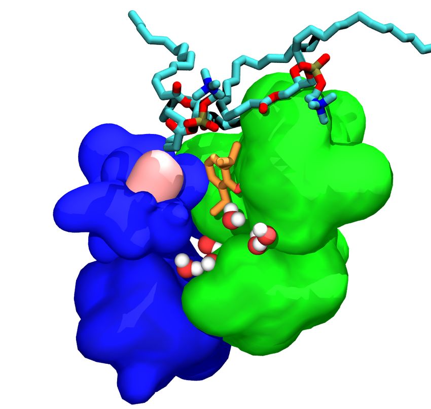
bioRxiv preprint first posted online Jul. 8, 2019; doi: http://dx.doi.org/10.1101/694612. The copyright holder for this preprint (which
 3.5 Methionine     residues determine
      was not peer-reviewed)             lipid accessibility
                                 is the author/funder,  whotohas
                                                               intersubunit cavitiesa license to display the preprint in perpetuity.
                                                                  granted bioRxiv                                                              9
                                It is made available under a CC-BY-NC-ND 4.0 International license.

                     A                                                          B

                                                                     α+/γ-          Lipid
                                                                                    Accessible
                Propofol KD (µM)

                                                        ∆Nwater
                                             βα+/α-

                                     ɣ+/β-                        ∆nlipid
                                   α+/β- βγ+/α-
                                                                                    Lipid
                                                                                    Inaccessible
                                                                                                    Apo         Propofol bound
                                                      ∆nlipid
 Figure 4: Solvation of lipid accessible and lipid inaccessible sites. (A) Correlation between average number of lipid
 molecules displaced in each intersubunit site: + - - (orange), + -↵- (blue), ↵+ - - (green), ↵ + -↵- (red), and ↵+ - - (white),
 and binding affinity of that site for propofol. Black error bars give the standard deviation for each site. Inset plot (in A)
 shows the correlation between average number of displaced water molecules ( Nwater ) and average number of displaced
 lipid atoms ( nlipid ) in each intersubunit site. (B) Snapshots of the intersubunit sites showing representative frames of the
 lipid accessible and inaccessible cavities in apo and propofol-bound receptor (propofol colored orange), viewed from the ECD
 along the pore axis. The M227 (M1-150 ) methionine residue forming the lipid-inaccessible cavity is colored in yellow; the
 homologous I242 (M1-150 ) isoleucine residue forming the lipid-accessible cavity is colored in pink.

 the pore as well as the intersubunit transmembrane binding sites (Table 2). For propofol, we calculate a KD for the pore of
 4 µM, which is weaker than three of the intersubunit sites, and a greater concentration than the typical EC50 of 0.5-5 µM (62)
 This result is a useful sanity check for our approach, and is consistent with gain-of-function caused by binding to one or more
 of these three sites (↵+ - - , ↵ + -↵- and + -↵- ).
      For particularly small molecules binding to the pore (including volatile anesthetics), it is critical to consider the possibility
 of multiple occupancy, for two reasons : 1) The binding site is so much larger than the ligand that multiple occupancy is not
 sterically unfavorable. In flooding simulations we observe double occupancy of the pore by sevoflurane, and the previously
 photolabeled residues line a much longer region of the channel lumen than a single sevoflurane molecule would contact.
 2) Such molecules are so small and fluid in the pore that a single such molecule may not block conduction. We presented
 a functional model accounting for a mixture of partially and fully blocked pores in Ref. (17), where the probability that a
 single molecule would block conduction was a tunable parameter. For the affinities we measured to be consistent with the
 experimentally measured IC50 , a single isoflurane molecule bound to the GLIC pore would block about 12% of the time.
      We calculated affinities for a single sevoflurane molecule binding to the pore, and a second molecule binding to a pore
 that was already occupied (Table 2). The single sevoflurane molecule had an affinity of 10 µM, comparable to the strongest
 intersubunit affinities and below the EC50 of 300 µM (63). The second sevoflurane molecule, however, had a significantly
 lower affinity of 200 µM, which is consistent with the pore unbinding incident we observed in flooding simulations. This
 negative cooperativity is consistent with what we measured for isoflurane binding to the closed GLIC pore (2.8 µ M for the
 first molecule, vs 80 µ for the second). In that case we also calculated a much lower affinity (2,800 µM) for the putative
 allosteric site, suggesting that GLIC is inhibited by general anesthetics because pore block requires a much lower concen-
 tration than occupancy of a gain-of-function site. Our results here suggest the reverse trend for GABA(A)r, consistent with
 gain-of-function observed at low concentrations.

                                    3.5      Methionine residues determine lipid accessibility to intersubunit cavities
 In the apo state, both water molecules and lipid acyl chains occupy the intersubunit cavities, with the relative distribution
 depending on the cavity sequence (Figure S4). The average number of water molecules and lipid atoms per intersubunit site
 was determined for the apo and bound configurations (Figure 4A), and also tracked over the course of the AFEP simulations.
                                                                                                                  Biophysical Journal 00(00) 1–15
bioRxiv preprint first posted online Jul. 8, 2019; doi: http://dx.doi.org/10.1101/694612. The copyright holder for this preprint (which
 10 was not peer-reviewed) is the author/funder, who has granted bioRxiv a license to display the preprint in perpetuity.             3 RESULTS
                                It is made available under a CC-BY-NC-ND 4.0 International license.

 We find that in the apo state, the sites containing ↵ and subunits have a similar number of water molecules, while the + - -
 site has slightly more due to an additional polar residue, as shown in our previous study (33). The number of water molecules
 decreases with binding for most sites, consistent with the observations of Tang and co-workers. (64)
     We also observe that the number of lipid atoms occupying the ↵+ - - site is significantly larger than for other sites. As
 depicted in figure 4 B, a bulky M286 at M3:22’ in sites ↵ + -↵- and + -↵- and M227 at M1:15’ block lipid access for all
 intersubunit cavities but the ↵+ - - interface: we refer to these cavities as “lipid-inaccessible”. The ↵+ - - interface contains an
 M1 helix from subunit, with the M1:15’ Met replaced with a slightly less bulky Ile. Acyl chains of the surrounding phos-
 pholipid membrane can more readily access this site, and thus we term it a “lipid-accessible” cavity. Other intersubunits sites
 have a methionine residue at the 15’ or 22’ position in either the M1 of a subunit or the M3 of the neighboring subunit forming
 the interface. As shown in the inset, lipid displacement by propofol is inversely correlated to water displacement; below, we
 show that water displacement is the best predictor of binding affinity for both sevoflurane and propofol. Displacement of water
 molecules by the ligand, however requires that water molecules (not lipids) occupy the cavity in the apo state.

                               3.6    Affinity directly correlates with water displacement upon binding.

 Table 2: Binding affinities of propofol and sevoflurane for multiple sites on the GABAA receptor, calculated using Alchemical
 free energy perturbation, including comparison to previous values we reported in (33). We discuss the substantial change
 in calculated affinity for propofol to the + - - site in the text. EC50 s for both propofol and sevoflurane reflect enhancement
 of GABA-activated chloride current in neurons incorporating several receptor subtypes. They indicate an upper bound on
 reasonable KD values, rather than an expected equality.

                                                             Propofol Kd (µM)                   Sevoflurane Kd (µM)
                           Binding site             Previous (Ref. 33)      Current
                               ↵+ - -                      0.1                0.2                          10
                                  + -
                                ↵ -↵                        2                 20                          150
                                  + -
                                   -↵                        -                0.2                          6
                                 + -
                                  -                        200                0.6                         220
                               ↵+ - -                       27                60                          120
                                pore                         -                 4                           10
                      pore(second molecule)                  -                 -                          200
                            intra-subunit                    -                 -                          370
                               EC50                                     0.5-5 (Ref. 62)              300 (Ref. 63)

     In Ref. (33), we found a direct correlation between the likelihood of hydrogen bonding and affinity of general anesthetics
 for binding sites. One surprising outcome was the low hydrogen bonding propensity and low affinity of propofol for the + - -
 site, despite a large number of hydrogen bonding partners. We now believe that those results were artefactual outcomes of
 designating propofol’s isopropyl groups as rotatable during the docking calculations. We have resolved this technical issue
 as described in Methods, and we present updated affinities in Table 2. We now predict that + - - will be among the high-
 est affinity cavities, whereas our previous calculations ranked it as the lowest affinity intersubunit cavity, with an affinity of
 200 µM.
     Other affinities remain similar to their previous values, which reflects the ability of the propofol to relax rapidly from
 the unfavorable conformation in all but the tightest binding sites. A second unexplained outcome in Ref. (33) was the low
 affinity and low hydrogen bonding propensity of propofol for the ↵+ - - site, which had the same number of hydrogen bonding
 partners as the higher affinity ↵+ - - site. In the present study, we find that propofol in the ↵+ - - site does form hydrogen
 bonds about as often as in the ↵+ - - site (Figure 3) but still measure a particularly low affinity for the ↵+ - - site. In general for
 lipid-inaccessible sites, KD for propofol does tend to decrease with increased hydrogen bonding, as we observed in Ref (33),
 but this trend does not extend to the lipid-accessible ↵+ - - site, or for sevoflurane.
     Given the unique lipid accessibility of ↵+ - - , we considered whether affinity was more directly correlated to displace-
 ment of water or lipid. As shown in Figure 5 A, propofol KD decreases monotonically with the number of displaced water
 molecules and increases monotonically with the number of displaced lipid atoms (Figure S5, row 1). This suggests that the
 intersubunit cavities generally prefer to be occupied by hydrophobic groups rather than water. For lipid-inaccessible cavities,
 Biophysical Journal 00(00) 1–15
bioRxiv preprint first posted online Jul. 8, 2019; doi: http://dx.doi.org/10.1101/694612. The copyright holder for this preprint (which
was not peer-reviewed) is the author/funder, who has granted bioRxiv a license to display the preprint in perpetuity. 11
It is made available under a CC-BY-NC-ND 4.0 International license.
A B
KD (µM)
∆Nwater Probability of hydrogen bond
Figure 5: Intersubunit binding affinity as a function of displaced water atoms and hydrogen bonding. Correlation
between binding site affinity (on a log scale) and (left) the average number of displaced water molecules Nwater or (right) the
probability of a hydrogen bond being formed between propofol (black) or sevoflurane (orange) and a side-chain residue. The
open and closed circles represent sites that are lipid-accessible and lipid-inaccessible respectively, as illustrated in Figure 4.
occupation by lipids is obviously not an option, but they can be occupied by small hydrophobic molecules like propofol. In all
high affinity (KD < 1µM) intersubunit propofol sites, more than five water molecules are displaced on average upon ligand
binding.
Very recent photolabeling protection studies(34) have indicated that propofol has a five-fold higher affinity for the two +
sites ( ↵ + -↵- and + -↵- ) than for the two sites (↵+ - - and + - - ). As shown in Table 2, we have measured a range of
values (0.2 - 20 µ M) across three calculations involving ↵+ / sites. Interestingly, we find that for individual cavities, the
variation in affinity from the mean also correlates with the variation in water displacement across replicas. As shown in Figure
4, an average of six water molecules is displaced from the + -↵- site, while an average of three water molecules is displaced
from the ↵ + -↵- site; the calculated affinities for the two sites are 0.2 µM and 20 µ M respectively. Differences in the initial
number of lipid atoms and water molecules across two sites with identical sequences can result from the initial lipid bilayer
packing process, as well as from thermal effects; we can not interpret these results as indicating an intrinsic difference based
on location within the pentamer. This observation suggests that endpoint solvation can serve as a primary source of statistical
noise in AFEP calculations of membrane proteins.
Results for sevoflurane are consistent with water displacement as a determinant of affinity (Figure 5 A). Sevoflurane is a
smaller molecule than propofol and does not displace more than five water molecules from any site, but the two sites with the
highest number of displaced water molecules are still the two sites with the strongest affinity (Figure S5, row 2). Observing
clearly monotonic trends across the sevoflurane data is challenging due to the large uncertainty in affinities and number of
displaced atoms for the lowest affinity sites.
Remarkably, however, we find that sevoflurane and propofol affinity data exhibit the same trend with respect to either the
number of water molecules or lipid atoms displaced, but not the number of hydrogen bonds formed (Figure 5). This suggests
that the increased potency of propofol relative to sevoflurane may be primarily due to its larger molecular volume, and the
resulting increased tendency to displace water from the cavity.
4 Conclusion
We predict the isolated binding affinities for sevoflurane and propofol of five transmembrane intersubunit cavities and the pore
of a heteromeric GABAA receptor, using MD simulation and the AFEP method. Our results exhibit mechanisms that answer
two significant questions regarding the interactions of general anesthetics and GABAA receptors.
The first longstanding question concerns the fundamental molecular interactions determining general anesthetic activity,
and in particular, potency at the GABAA receptor. We propose here that dewetting of hydrophobic cavities upon anesthetic
binding is the primary determinant of affinity for propofol and sevoflurane for intersubunit sites on GABAA receptors. This
result is consistent with a previous observation, using NMR, that a critical level of hydration was required for halothane to
Biophysical Journal 00(00) 1–15bioRxiv preprint first posted online Jul. 8, 2019; doi: http://dx.doi.org/10.1101/694612. The copyright holder for this preprint (which
12 was not peer-reviewed) is the author/funder, who has granted bioRxiv a license to display the preprint in perpetuity. 4 CONCLUSION
It is made available under a CC-BY-NC-ND 4.0 International license.
bind to proteins (65). To our knowledge, this is the first time the number of displaced water molecules has been correlated
with affinity to explain GA specificity and selectivity.
In general, the proposed mechanism follows in a straightforward manner from the fundamental origin of the hydrophobic
effect: water molecules able to hydrogen bond with only a few partners, as at an interface or within a cavity, can reduce their
free energy by moving to bulk water. (Hydrophobicity is a slightly misleading term for this effect, since it is water that is
“lipophobic” rather than the reverse). A broader study of ligand binding (66) identified a crucial role for water displacement
in determining binding affinity to hydrophobic cavities, yielding the WaterMap docking corrections.
The number of displaced water molecules will increase with ligand-size and hydration of the cavity, suggesting that these
factors will underlie affinity for the ligand and site respectively. This does not mandate that there is no role for hydrogen bond-
ing between the general anesthetic and the protein in determining affinity. Cavities that are both hydrated and hydrophobic
must balance the particular residue-residue interactions that favor the cavity with the surface tension of the water droplet that
fills it. Polar side-chains can reduce that surface tension, especially for a small droplet.
In order to separate the role of dewetting from the role of hydrogen bonding between GA and sidechains, it was critical that
we were able to compare binding to different homologous sites without varying the ligand. In principle our findings do not rule
out a secondary contribution of hydrogen binding to polar side-chains to strengthening affinity: this would be enthalpically
favorable, while still maintaining the entropic benefit of releasing cavity water. In practice, typical structure-activity experi-
ments are unable to change ligand hydrogen bonding propensity without simultaneously introducing steric clashes, removing
solubility, increasing pore block, and/or affecting dewetting. These conflicting contributions may explain the complex depen-
dence observed by Harrison and colleagues for potency of a series of propofol analogs that potentiate GABAA receptors, (67)
or the fact that hydroxyl substitution with fluorine(38), but not bromine(67), removes general anesthetic function.
It is likely that the affinities we calculated may be particularly sensitive to the choice of initial structure and sampling of
endpoint solvation. This could affect the overall predicted rank order of the four higher-affinity intersubunit propofol sites,
and introduce the modest quantitative discrepancies with the results of Ref. (34). We would not expect these changes to affect
the observed relationship between solvation and affinity. Consequently, our results suggest an intriguing possible origin for
state-dependence of affinities: conformational changes that alter cavity solvation may directly affect ligand binding affinity
for the cavity.
The second ongoing question we have addressed is the origin of the “orphan” site at the ↵+ - - interface. We propose here
that the orphan site indirectly also reflects water displacement as a driver of affinity, because this lipid-accessible site has
uniquely low hydration in the apo state. The M1 150 contains an isoleucine residue ( I242), which allows lipids to access
the binding cavity, while the ↵ and subunits contain a methionine at the same location, which closes off the intersubunit
site. This hypothesis could be tested experimentally by mutation of M1 150 (I242M).
Overall, our results are consistent with results collected via mutagenesis and photolabeling involving GABAA receptors
over the past two decades, particularly those reported in Refs. (26, 29, 30, 34, 40, 58, 59, 67–70). These studies have focused
exclusively on protein-mediated mechanisms of anesthetic binding. Nonetheless, membrane-mediated mechanisms underly-
ing general anesthesia have remained intriguing to many researchers in part because of the compelling correlation of general
anesthetic potency with lipophilicity first observed by Meyer and Overton (1), followed by numerous observations of the
physical effects of anesthetics on lipid bilayers (71) at supertherapeutic concentrations. (72) Our results here suggest that
this framing (lipid-mediated vs. protein-mediated) is misleading; general anesthetics don’t act via lipids, they act like lipids:
amphipathic molecules that displace water from the protein surface and (in pLGICs) transmembrane cavities. This interpreta-
tion is consistent with the established sensitivity of the pLGIC family to lipids (73–78) and implies that lipid-specificity and
general anesthetic specificity are deeply related questions.
Author Contributions
SM: designed research; performed research; analyzed data; interpreted results; wrote the paper
JH: designed research; wrote the paper
GB: designed research; interpreted results; wrote the paper
Acknowledgments
SM and GB were supported by research grant NIH P01GM55876-14A1. JH acknowledges support by the French govern-
ment under grants DYNAMO (ANR-11-LABX-0011) and CACSICE (ANR-11-EQPX-0008). This project was supported
with computational resources from the National Science Foundation XSEDE program through allocation NSF-MCB110149,
as well as through the Rutgers Discovery Informatics Institute, which is supported by Rutgers and the State of New Jersey.
Biophysical Journal 00(00) 1–15bioRxiv preprint first posted online Jul. 8, 2019; doi: http://dx.doi.org/10.1101/694612. The copyright holder for this preprint (which
was not peer-reviewed) is the author/funder, who has granted bioRxiv a license to display the preprint in perpetuity. 13
It is made available under a CC-BY-NC-ND 4.0 International license.
References
1. Overton, C. E., 1901. Studien ber die Narkose, zugleich ein Beitrag zur allgemeinen Pharmakologie. Gustav Fischer Verlag, Jena.
2. Harris, R. A., S. J. Mihic, J. E. Dildy-Mayfield, and T. K. Machu, 1995. Actions of anesthetics on ligand-gated ion channels: role of
receptor subunit composition. FASEB J 9:1454–1462.
3. Belelli, D., M. Pistis, J. A. Peters, and J. J. Lambert, 1999. The interaction of general anaesthetics and neurosteroids with GABA(A)
and glycine receptors. Neurochem. Int. 34:447–452.
4. Belelli, D., M. Pistis, J. A. Peters, and J. J. Lambert, 1999. General anaesthetic action at transmitter-gated inhibitory amino acid
receptors.
5. Tanelian, D. L., P. Kosek, I. Mody, and M. B. MacIver, 1993. The role of the GABAA receptor complex in anesthesia. Anesthesiology
78:757–776.
6. Krasowski, M. D., and N. L. Harrison, 1999. General anaesthetic actions on ligand-gated ion channels. Cell. Mol. Life Sci.
55:1278–1303.
7. Chang, Y., R. Wang, S. Barot, and D. S. Weiss, 1996. Stoichiometry of a recombinant GABAA receptor. J. Neurosci. 16:5415–24.
8. Farrar, S. J., P. J. Whiting, T. P. Bonnert, and R. M. McKernan, 1999. Stoichiometry of a Ligand-gated Ion Channel Determined by
Fluorescence Energy Transfer. J. Biol. Chem. 274:10100–10104.
9. Baumann, S. W., R. Baur, and E. Sigel, 2002. Forced subunit assembly in alpha1beta2gamma2 GABA(A) receptors. Insight into the
absolute arrangement. J Biol Chem 277:46020–46025.
10. Miller, P. S., and A. R. Aricescu, 2014. Crystal structure of a human GABAA receptor. Nature 512:270–275.
11. Zhu, S., C. M. Noviello, J. Teng, R. M. Walsh, J. J. Kim, and R. E. Hibbs, 2018. Structure of a human synaptic GABAA receptor.
Nature 559:67–72.
12. Phulera, S., H. Zhu, J. Yu, D. P. Claxton, N. Yoder, C. Yoshioka, and E. Gouaux, 2018. Cryo-EM structure of the benzodiazepine-
sensitive 112S tri-heteromeric GABA, javax.xml.bind.JAXBElement@fc05a49, receptor in complex with GABA. Elife 7.
13. Masiulis, S., R. Desai, T. Uchaski, I. Serna Martin, D. Laverty, D. Karia, T. Malinauskas, J. Zivanov, E. Pardon, A. Kotecha, J. Steyaert,
K. W. Miller, and A. R. Aricescu, 2019. GABAA receptor signalling mechanisms revealed by structural pharmacology. Nature
565:454–459.
14. Laverty, D., R. Desai, T. Uchaski, S. Masiulis, W. J. Stec, T. Malinauskas, J. Zivanov, E. Pardon, J. Steyaert, K. W. Miller, and A. R.
Aricescu, 2019. Cryo-EM structure of the human 132 GABA, javax.xml.bind.JAXBElement@18520d8a, receptor in a lipid bilayer.
Nature 565:516–520.
15. Brannigan, G., D. N. LeBard, J. . Hnin, R. G. Eckenhoff, and M. L. Klein, 2010. Multiple binding sites for the general anesthetic
isoflurane identified in the nicotinic acetylcholine receptor transmembrane domain. Proc Natl Acad Sci U S A 107:14122–14127.
16. Nury, H., C. Van Renterghem, Y. Weng, A. Tran, M. Baaden, V. Dufresne, J.-P. Changeux, J. M. Sonner, M. Delarue, and P.-J. Corringer,
2011. X-ray structures of general anaesthetics bound to a pentameric ligand-gated ion channel. Nature 469:428–31.
17. LeBard, D. N., J. Hnin, R. G. Eckenhoff, M. L. Klein, and G. Brannigan, 2012. General anesthetics predicted to block the GLIC pore
with micromolar affinity. PLoS Comput. Biol. 8:e1002532.
18. Spurny, R., B. Billen, R. J. Howard, M. Brams, S. Debaveye, K. L. Price, D. A. Weston, S. V. Strelkov, J. Tytgat, S. Bertrand,
D. Bertrand, S. C. R. Lummis, and C. Ulens, 2013. Multisite binding of a general anesthetic to the prokaryotic pentameric Erwinia
chrysanthemi ligand-gated ion channel (ELIC). J. Biol. Chem. 288:8355–64.
19. Chen, Q., M. N. Kinde, P. Arjunan, M. M. Wells, A. E. Cohen, Y. Xu, and P. Tang, 2015. Direct Pore Binding as a Mechanism for
Isoflurane Inhibition of the Pentameric Ligand-gated Ion Channel ELIC. Sci. Rep. 5:13833.
20. Fourati, Z., R. J. Howard, S. A. Heusser, H. Hu, R. R. Ruza, L. Sauguet, E. Lindahl, and M. Delarue, 2018. Structural Basis for a
Bimodal Allosteric Mechanism of General Anesthetic Modulation in Pentameric Ligand-Gated Ion Channels.
21. Miller, P. S., S. Scott, S. Masiulis, L. De Colibus, E. Pardon, J. Steyaert, and A. R. Aricescu, 2017. Structural basis for GABAA
receptor potentiation by neurosteroids. Nat. Struct. Mol. Biol. 24:986–992.
22. Laverty, D., P. Thomas, M. Field, O. J. Andersen, M. G. Gold, P. C. Biggin, M. Gielen, and T. G. Smart, 2017. Crystal structures of a
GABAA-receptor chimera reveal new endogenous neurosteroid-binding sites. Nat. Struct. Mol. Biol. 1–14.
23. Chen, Q., M. M. Wells, P. Arjunan, T. S. Tillman, A. E. Cohen, Y. Xu, and P. Tang, 2018. Structural basis of neurosteroid anesthetic
action on GABAA receptors. Nat. Commun. 9:3972.
24. Jurd, R., M. Arras, S. Lambert, B. Drexler, R. Siegwart, F. Crestani, M. Zaugg, K. E. Vogt, B. Ledermann, B. Antkowiak, and
U. Rudolph, 2003. General anesthetic actions in vivo strongly attenuated by a point mutation in the GABAA receptor beta3 subunit.
FASEB J. 17:250–252.
25. Siegwart, R., K. Krhenbhl, S. Lambert, and U. Rudolph, 2003. Mutational analysis of molecular requirements for the actions of general
anaesthetics at the gamma-aminobutyric acidA receptor subtype, alpha1beta2gamma2. BMC Pharmacol. 3:13.
26. Krasowski, M. D., K. Nishikawa, N. Nikolaeva, A. Lin, and N. L. Harrison, 2001. Methionine 286 in transmembrane domain 3 of the
GABA(A) receptor beta subunit controls a binding cavity for propofol and other alkylphenol general anesthetics. Neuropharmacology
41:952–964.
27. Bali, M., and M. H. Akabas, 2004. Defining the propofol binding site location on the GABAA receptor. Mol. Pharmacol. 65:68–76.
28. Yip, G. M. S., Z. W. Chen, C. J. Edge, E. H. Smith, R. Dickinson, E. Hohenester, R. R. Townsend, K. Fuchs, W. Sieghart, A. S. Evers,
and N. P. Franks, 2013. A propofol binding site on mammalian GABA A receptors identified by photolabeling. Nat. Chem. Biol.
9:715–720.
Biophysical Journal 00(00) 1–15You can also read