MRNA TRANSFECTION-INDUCED ACTIVATION OF PRIMARY HUMAN MONOCYTES AND MACROPHAGES: DEPENDENCE ON CARRIER SYSTEM AND NUCLEOTIDE MODIFICATION - NATURE
←
→
Page content transcription
If your browser does not render page correctly, please read the page content below
www.nature.com/scientificreports

            OPEN           mRNA Transfection-Induced
                           Activation of Primary Human
                           Monocytes and Macrophages:
                           Dependence on Carrier System and
                           Nucleotide Modification
                           Hanieh Moradian1,2,3, Toralf Roch1,4,5, Andreas Lendlein1,2,3 & Manfred Gossen1,2*

                           Monocytes and macrophages are key players in maintaining immune homeostasis. Identifying
                           strategies to manipulate their functions via gene delivery is thus of great interest for immunological
                           research and biomedical applications. We set out to establish conditions for mRNA transfection in
                           hard-to-transfect primary human monocytes and monocyte-derived macrophages due to the great
                           potential of gene expression from in vitro transcribed mRNA for modulating cell phenotypes. mRNA
                           doses, nucleotide modifications, and different carriers were systematically explored in order to
                           optimize high mRNA transfer rates while minimizing cell stress and immune activation. We selected
                           three commercially available mRNA transfection reagents including liposome and polymer-based
                           formulations, covering different application spectra. Our results demonstrate that liposomal reagents
                           can particularly combine high gene transfer rates with only moderate immune cell activation. For
                           the latter, use of specific nucleotide modifications proved essential. In addition to improving efficacy
                           of gene transfer, our findings address discrete aspects of innate immune activation using cytokine
                           and surface marker expression, as well as cell viability as key readouts to judge overall transfection
                           efficiency. The impact of this study goes beyond optimizing transfection conditions for immune cells, by
                           providing a framework for assessing new gene carrier systems for monocyte and macrophage, tailored
                           to specific applications.

                           Innate immune cells play an important role in response to pathological conditions and maintaining immune
                           homeostasis1. Among them, monocytes and monocyte-derived macrophages have remarkable properties, includ-
                           ing their immunomodulatory capacities. Monocytes with various distinct phenotypes are key players of early
                           inflammation. During inflammation, monocytes can dynamically repolarize to different phenotypes in response
                           to local signals, which is thought to be more efficient at resolving tissue homeostasis than the recruitment of other
                           anti-inflammatory and pro-regenerative subsets of monocytes or macrophages2–4.
                               Macrophages themselves have special functions such as phagocytosis of invading pathogens or apoptotic
                           cells, antigen presentation to T cells, elimination of pathogens via releasing reactive oxygen species or proteo-
                           lytic enzymes, and secretion of pro- or anti-inflammatory signaling molecules to recruit various types of other
                           immune cells5–9. Elucidation and manipulation of monocyte and macrophage phenotypes is therefore essential
                           to fully explore their role in immunoregulation. This will benefit not only basic immunological research but also
                           clinical and translational studies3,10,11.
                               For innate immune cell manipulation, transfections have been commonly used to introduce nucleic acids,
                           such as plasmid DNA (pDNA) or small interfering RNA, to induce or inhibit the expression of a target protein,

                           1
                            Institute of Biomaterial Science, Helmholtz-Zentrum Geesthacht, 14513, Teltow, Germany. 2Berlin-Brandenburg
                           Center for Regenerative Therapies (BCRT), 13353, Berlin, Germany. 3Institute of Biochemistry and Biology, University
                           of Potsdam, 14476, Potsdam, Germany. 4Charité – Universitätsmedizin Berlin, corporate member of Freie Universität
                           Berlin, Humboldt-Universität zu Berlin, and Berlin Institute of Health, Berlin-Brandenburg Center for Regenerative
                           Therapies, Berlin, Germany. 5Center for Translational Medicine, Medical Department I, Marien Hospital Herne,
                           University Hospital of the Ruhr-University Bochum, Herne, Germany. *email: manfred.gossen@hzg.de

Scientific Reports |   (2020) 10:4181 | https://doi.org/10.1038/s41598-020-60506-4                                                            1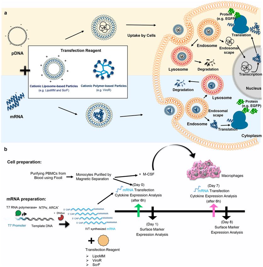
www.nature.com/scientificreports/                                                             www.nature.com/scientificreports

                           Figure 1. (a) Different paths to protein expression upon cells transfection with mRNA or pDNA (b)
                           Experimental work flow to isolate and generate primary human monocytes and macrophages and to compare
                           the transfection efficiency of mRNA transfection reagents. LipoMM: Lipofectamine MessengerMax, ScrF:
                           ScreenFect, ViroR: Viromer RED, IVT-mRNA: in vitro transcribed mRNA, ARCA: anti–reverse cap analog,
                           EGFP: enhanced green fluorescent protein.

                           respectively12–14. However, initial studies revealed that, particularly for macrophages, transfection is more chal-
                           lenging in comparison to most other primary mammalian cells15. The low transfection efficiency in monocytes/
                           macrophages can be attributed to the following reasons. Firstly, there is a very limited chance for pDNA to freely
                           reach the nucleus due to nuclear envelope breakdown during mitosis, since macrophages do not, or hardly, prolif-
                           erate16,17. Secondly, these immune cells are equipped with pattern recognition receptors, which can detect nucleic
                           acids as potential foreign and dangerous viral invaders, and initiate the inflammatory signaling cascade leading to
                           pDNA degeneration or macrophage apoptosis18. Hence, finding a robust transfection approach to address these
                           issues is highly demanded.
                               Transfection of messenger RNA (mRNA) is a promising alternative to pDNA or viral vector to achieve target
                           protein expression, particularly in non-proliferative cells such as primary human cells19,20. One advantage of
                           mRNA transfection is that there is neither the need for mRNA to enter the cell nucleus, nor the possibility to
                           integrate into the host genomic DNA (Fig. 1a)21. Thus, this method can be a proper alternative for transfection of
                           non-proliferative cells22, including primary human macrophages and monocytes. Moreover, it will avoid genotox-
                           icity issues associated with chromosomal insertion of DNA vectors in clinical gene transfer applications. In con-
                           trast to most pDNA transfections and viral transduction protocols, mRNA transfection will result in a transient,
                           non-stable gene expression. However, transient expression is beneficial for several “hit-and-run” applications,
                           including current differentiation protocols23,24.
                               The mRNA gene delivery technology made significant progress after overcoming commonly known issues
                           related to mRNA, such as susceptibility to degradation by RNases in surrounding media before reaching the

Scientific Reports |   (2020) 10:4181 | https://doi.org/10.1038/s41598-020-60506-4                                                          2
www.nature.com/scientificreports/                                                                        www.nature.com/scientificreports

                            Features provided by
                            the manufacturer       LipoMM                          ViroR                               ScrF
                                                                                   Polymeric carrier, based on
                                                                                   polycationic PEI core, highly       Cationic thioether lipids, containing
                            Material’s chemistry   Lipid-based nanoparticle
                                                                                   substituted with hydrophobic and    hydrophobic alkyl groups
                                                                                   anionic side chains
                            Specific structural    Cationic lipids optimized for   Polymers mimicking viral (influenza Biomimetic lipid-like molecules made by
                            features               mRNA delivery application;      hemagglutinin) biophysics           thiol-yne click chemistry
                                                   Primary cell types such as
                                                                                    Primary adherent and suspension    Many different human and mouse cell lines
                                                   neurons, fibroblast, hepatocytes
                            Tested cell types                                       cells including monocytes and      such as HEK293, NIH3T3, RAW 264.7 and
                                                   and Keratinocytes, specifically
                                                                                    macrophages and stem cells         mouse embryonic stem cells (mESC)
                                                   tested for mRNA CRISPRs

                           Table 1. Characteristics of the three commercially available mRNA transfection reagents provided by the
                           manufacturer. LipoMM: Lipofectamine MessengerMax, ScrF: ScreenFect, ViroR: Viromer RED.

                           target cell25. Many parameters of in vitro transcription technology have been evaluated and optimized to prevent
                           mRNA degradation, improve translation efficiency, and reduce unspecific immunogenicity upon transfection19.
                               Despite 5′ and 3′-end modifications mimicking natural mRNAs, in vitro transcribed mRNA (IVT-mRNA)
                           can activate immune responses in transfected cells. This effect is often more dramatic for macrophages, which are
                           highly specialized cells for defense against RNA-based viruses and are equipped with numerous receptors includ-
                           ing pattern recognition receptors. Toll-like receptors (TLR), particularly endosomal TLR3, 7, and 8, can recog-
                           nize single- and double-stranded nucleic acids21. Therefore, when passing through the endosome, transfected
                           IVT-mRNA could be recognized by immune cells as foreign. However, the immune response can be significantly
                           diminished by utilizing modified nucleotides, as was initially reported by Kariko et al. for pseudouridine (Ψ) and
                           5′-methyl cytidine26.
                               The aim of this study was to set up a robust method for IVT-mRNA transfection in primary human mono-
                           cytes and monocyte-derived macrophages, while minimizing pleiotropic effects, in particular immune cell activa-
                           tion. We tested three commercially available transfection reagents for mRNA delivery. These included liposomal
                           and polymer-based formulations, as cationic lipid based carriers have different physicochemical properties such
                           as size, shape, and chemical structure compared with polyplexes. These key features not only affect and determine
                           the way they condense and transport their cargo, but also uptake mechanism and subsequent endosomal release,
                           and eventually transfection efficiency. The effects of mRNA modification as well as mRNA concentrations were
                           systematically investigated, using cell viability, transfection efficiency, and the monocyte and macrophage acti-
                           vation as critical readouts. The results of this study provide guidelines for choosing proper mRNA transfection
                           carriers for monocytes and macrophages and highlights the need for not only focusing on gene transfer rates, but
                           also for analyzing cell stress and activation in parallel.

                           Results
                           Experimental setup.          In order to evaluate the effect of different mRNA transfection protocols on mono-
                           cytes and macrophages, experiments were designed as follows. mRNAs were synthesized using the in vitro tran-
                           scription method, with non-modified or modified nucleotides. Either of three commercially available mRNA
                           transfection reagents, namely the liposomal reagents Lipofectamine MessengerMax (LipoMM) and ScreenFect
                           mRNA (ScrF) as well as the polymeric reagent Viromer RED (ViroR) were compared for transfection efficiency;
                           see also Table 1 provided in Methods. CD14 positive monocytes purified from peripheral blood mononuclear cells
                           (PBMCs) using magnetic cell sorting, were immediately transfected. A fraction of the CD14 positive monocytes
                           were differentiated into macrophages by cultivation for seven days in the presence of macrophage colony-stim-
                           ulating factor (M-CSF) and transfected at day 7. Supernatants from monocyte and macrophage cultures were
                           harvested 6 h post transfection and analyzed for the expression of tumor necrosis factor alpha (TNF-α) and inter-
                           feron beta (IFN-β). One day after transfection, the reporter gene and the CD80 expression was analyzed by flow
                           cytometry. The schematic overview of the experimental workflow is depicted in Fig. 1b.

                           Viability of monocytes and macrophages upon mRNA transfection. The forced introduction of
                           nucleic acids in cells often causes substantial stress that might ultimately effect viability. Crucial parameters are
                           type and purity of nucleic acid, the transfection protocol followed and the type of transfection reagent used27.
                           The meaningful cell type-dependent optimization of transfection protocols requires that post-transfection phe-
                           notypes that are caused by the genetic payload-dependent alteration in the target cells’ transcriptome can be
                           distinguished from side effects of the chosen transfection method or the potential innate immune response of
                           cells upon uptake of exogenous nucleic acids. To this end, we comparatively analysed primary human mono-
                           cytes and macrophages, treated with different transfection reagents for the introduction of IVT-mRNA encoding
                           enhanced green fluorescent protein (EGFP) to allow for single cell analysis of transfected and non-transfected
                           cells. To address post transfection viability, cells were treated with 4′,6-Diamidino-2-Phenylindole, dihydrochlo-
                           ride (DAPI) immediately prior to the flow cytometric analysis to discriminate live from dead cells. To identify
                           live single cells, a gating strategy as illustrated in Fig. S1 was applied. Cells were first discriminated from debris
                           using forward versus side scatter (FSC vs. SSC) parameters (Fig. S1a). Then, aggregated cells were excluded using
                           FSC-area (FSC-A) against FSC-height (FSC-H) (Fig. S1b) followed by identification of DAPI positive cells, which
                           are considered as dead or apoptotic (Fig. S1c). Among live cells, the amount of EGFP positive cell populations was
                           determined by using untransfected cells as gating control (Fig. S1d).

Scientific Reports |   (2020) 10:4181 | https://doi.org/10.1038/s41598-020-60506-4                                                                               3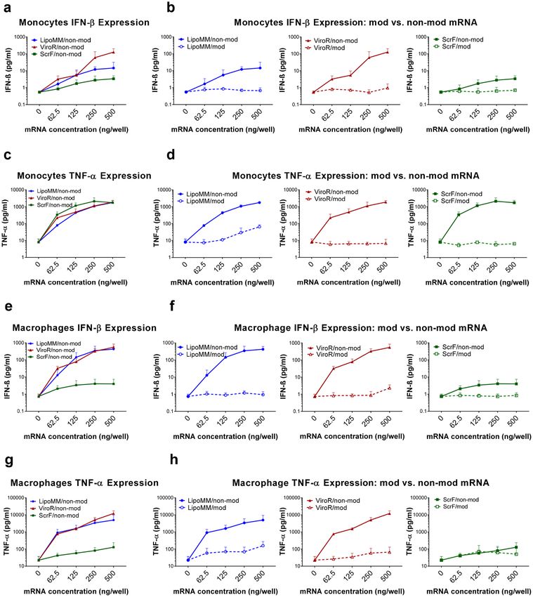
www.nature.com/scientificreports/ www.nature.com/scientificreports
Figure 2. Viability of monocytes and macrophages after mRNA transfection with different mRNA transfection
reagents; (a) Viability of monocytes transfected with non-modified mRNA using LipoMM (blue), ViroR
(red), and ScrF (green); (b) monocytes viability after transfection with modified or non-modified mRNA
using LipoMM, ViroR, and ScrF. (c) Evaluation of macrophages viability after transfection with non-modified
mRNA comparing LipoMM, ViroR, and ScrF; viability of macrophages transfected with modified or non-
modified mRNA by LipoMM, ViroR, and ScrF. (d) Values for no mRNA (0 ng/well) refer to untransfected cells
throughout. Values are presented as mean ± standard deviation (SD), n = 3. Error bars indicate SD. Statistical
differences in viability is depicted with * for LipoMM vs. ViroR, # for LipoMM vs. ScrF and + for ViroR vs.
ScrF. *, #p < 0.05, **p < 0.01.
Monocyte viability was measured after transfection of the three chosen reagents complexed with either modi-
fied or non-modified IVT-mRNA. For all transfection reagents, viability decreased with increasing mRNA doses
used. LipoMM resulted in significantly higher monocyte viability for most mRNA concentrations, as indicated
(Fig. 2a). However, there was no difference between viability of monocytes transfected with ScrF and ViroR
(Fig. 2a).
For the macrophage viability, no statistically significant difference between the different transfection reagents
was observed when comparing within each dose or average of all doses (Fig. 2c).
The viability of monocytes and macrophages after transfection with mRNA specifically modified with pseu-
douridine and 5-methyl-cytidine was compared to their viability after transfection with non-modified mRNA
for each transfection reagent. In most conditions modified mRNA fails to demonstrate a convincing decrease in
cell death when compared to non-modified mRNA for both monocytes and macrophages (Fig. 2b,d), resulting
in a lack of statistical significant difference. Surprisingly, monocytes seemed to be more vulnerable to increasing
mRNA doses than macrophages, when comparing the overall decrease of viability between monocytes and mac-
rophages (shown in Fig. 2a,c, respectively). For instance, the significant viability decrease was observed in mono-
cytes at 250 ng/well for LipoMM, 62.5 ng/well for ViroR and 125 ng/well for ScrF; whereas, a significant drop in
viability of macrophages was only seen above 500 ng/well for all transfection reagents.
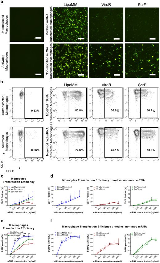
Transfection efficiency in primary human monocytes and macrophages. EGFP was used as a
reporter protein to monitor mRNAs transfection efficiency in monocytes and macrophages, which was initially
visualized by fluorescent microscopy (Fig. 3a) and subsequently quantified by flow cytometry (Fig. 3b–f). A sub-
stantial number of EGFP expressing macrophages could be microscopically observed already for 125 ng mRNA
per well, especially when the transfection was performed with LipoMM (Fig. 3a). The numbers of EGFP express-
ing macrophages after transfection with ViroR and ScrF were clearly lower in comparison to LipoMM. The use
of modified mRNA seems to decrease the number of EGFP expressing macrophages, at least for ViroR and ScrF
(Fig. 3a). Besides, morphology of transfected macrophages was evaluated using phase contrast microscopy, which
indicated morphologically heterogeneous population of cells within various groups compared to untransfected
and activated cells (Fig. S2). Moreover, non-modified mRNA resulted in higher EGFP intensity, when compared
to modified mRNA for all three transfection reagents (Fig. 3b).
Quantification by flow cytometry over all mRNA doses confirmed a higher frequency of EGFP positive cells
among macrophages transfected with mRNA complex with LipoMM comparing to ViroR and ScrF (Fig. 3b,e).
Scientific Reports | (2020) 10:4181 | https://doi.org/10.1038/s41598-020-60506-4 4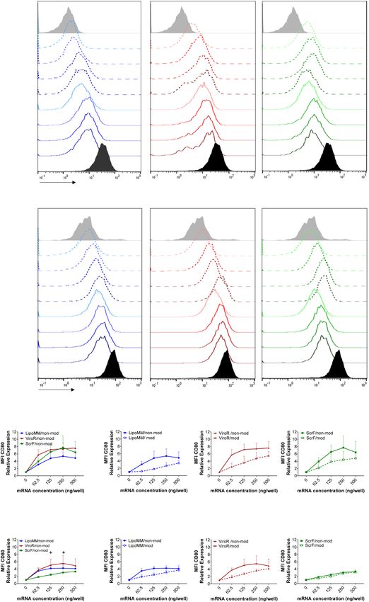
www.nature.com/scientificreports/                                                            www.nature.com/scientificreports

                           Figure 3. Transfection efficiency for monocytes and macrophages comparing LipoMM, ViroR, and ScrF; (a)
                           Representative fluorescent microscopy images (bar = 100 µm) and (b) contour plots indicating EGFP expression
                           of macrophages transfected with 125 ng/well of modified and non-modified mRNA via LipoMM, ViroR, and
                           ScrF. (c) Quantification of EGFP positive cells as indicator of transfection efficiency for non-modified mRNA
                           transfected monocytes and comparison of monocytes transfected with modified or non-modified mRNA using
                           LipoMM, ViroR, and ScrF (d). The same comparison was performed for macrophages transfected with non-
                           modified mRNA (e) and for macrophages transfected with modified or non-modified mRNA using LipoMM,
                           ViroR, and ScrF (f). Values for no mRNA (0 ng/well) refer to untransfected cells throughout. “Activated” refers
                           to cells treated with lipopolysaccharide (LPS) (2 µg·mL−1) and interferon gamma (IFN-γ) (10 ng·mL−1) for 24 h.
                           Statistical differences in transfection efficiency is depicted with * for LipoMM vs. ViroR, # for LipoMM vs. ScrF
                           and + for ViroR vs. ScrF. +p < 0.05, ##p < 0.01, ***p < 0.001; Values are presented as mean ± SD, n = 3. Error
                           bars indicate SD.

Scientific Reports |   (2020) 10:4181 | https://doi.org/10.1038/s41598-020-60506-4                                                        5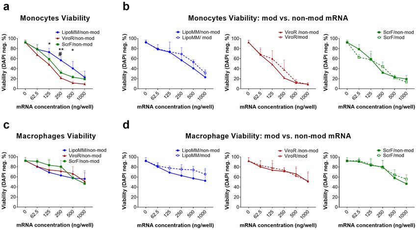
www.nature.com/scientificreports/                                                              www.nature.com/scientificreports

                           No statistical differences were observed when the transfection efficiency was compared for modified and
                           non-modified mRNA (Fig. 3f).
                               To investigate the dose-dependence of the transfection efficiency for monocytes, the frequency of EGFP pos-
                           itive cells was determined after transfection with increasing amounts of mRNA. The flow cytometry quantifica-
                           tion of EGFP expressing monocytes and macrophages revealed increasing transfection efficiency as measured by
                           percentage of EGFP positive cells when increasing amounts of mRNA were applied to the cells. Non-modified
                           mRNA transfection via LipoMM resulted in consistently highest efficiency for both types of immune cells, even
                           more than 80% for macrophages, followed by ScrF and ViroR (Fig. 3c,e). As for the microscopic analysis, only
                           minor differences between the use of modified versus non-modified mRNA was obvious for both monocytes
                           and macrophages (Fig. 3d,f, respectively), at least when quantifying the percentage of EGFP positive cells (see
                           above). Noteworthy, the mRNA dose/response mostly points at saturation effects, i.e. expression plateaued out
                           at high mRNA doses. Overall, it is apparent that macrophages are more efficiently to transfect with mRNA when
                           compared to monocytes.

                           Monocytes and macrophages activation. Activation of immune cells such as monocyte or macrophages
                           is a natural process, in which cells acquire pro-inflammatory functions associated with the expression of charac-
                           teristic cell surface molecules such as CD80 and releasing inflammatory mediators such as TNF-α. Monocytes
                           and macrophages can be activated by lipopolysaccharides (LPS, also known as endotoxin), which can be further
                           potentiated by interferon gamma (IFNγ)28. Cell stimulation also occurs in artificial in vitro culture settings, such
                           as introduction of external synthetic nucleic acids. Here we investigated whether mRNA transfection can trigger
                           monocyte and macrophage activation. Part of the cell activation by exogenous nucleic acids is based on antiviral
                           response mechanisms of innate immune cells, resulting in the expression of antiviral response molecules, such as
                           cytokine IFN-β. Accordingly, LPS + INF-γ-triggered activation was selected as positive control due to its capacity
                           to induce CD80 and TNF-α expression. Cells treated with the three different transfection reagents were analysed
                           for CD80 expression by flow cytometry. The level of CD80 expression of cells transfected with increasing amounts
                           of modified and non-modified mRNA doses using the three different transfection reagents, LipoMM, ViroR,
                           and ScrF, is illustrated as histograms for monocytes and macrophages (Fig. 4a,b). In general, higher amounts of
                           mRNA resulted in increasing activation levels for all three transfection reagents. However, modified mRNAs trig-
                           gered less CD80 expression in monocytes and macrophages compared to non-modified mRNA in all conditions
                           (Fig. 4a,b).
                               Monocytes transfected with LipoMM induced the lowest activation compared to ViroR and ScrF (Fig. 4c).
                           Whereas, the minimum CD80 expression was observed in macrophages transfected with ScrF (Fig. 4e). The
                           polyplex transfection reagent, ViroR, turned out to elicit the highest monocyte and macrophage activation for
                           all mRNA doses (Fig. 4c,e). For monocytes and macrophages, the results also indicated that mRNA modifica-
                           tion resulted in substantially less cell CD80 expression when compared to non-modified mRNA (Fig. 4d,f). One
                           exception was the low-level activation of macrophages transfected with ScrF (see above), which was similar for
                           modified and non-modified mRNA (Fig. 4f). Comparing the slope of the CD80 dose/response in monocyte
                           versus macrophages revealed that monocytes activation was more responsive to increasing mRNA doses than in
                           macrophages (Fig. 4c,e).
                               When analyzed under the same transfection conditions for the secretion of TNF-α and IFN-β, both cell types
                           overall responded to increasing concentrations of lipo-/polyplex in a dose-dependent manner (Fig. 5). However,
                           while this activation was very modest for modified IVT-mRNA, the use of non-modified IVT-mRNA led to a
                           dramatic increase in cytokine levels, up to two orders of magnitude.
                               Lastly, we addressed the question if the contact with, or uptake of carriers alone (i.e., without being com-
                           plexed with mRNA) by cells might contribute to their activation. For most of the conditions analyzed, neither
                           monocytes nor macrophages were activated by “transfection reagent-only”, as assessed by CD80 expression and
                           TNF-α secretion, and no effect on viability could be detected. Only for ScrF reagent added to monocytes, CD80
                           and TNF-α were slightly increased (Fig. 6). We noted a substantial, but transient acidification of the medium after
                           addition of this transfection reagent, and monocytes might be particular sensitive to these conditions. Overall,
                           increasing mRNA doses always increased cell activation both for monocytes and macrophages, particularly for
                           non-modified mRNA.

                           Activation of macrophages transfected with mRNA coding mCherry vs. EGFP. Certain levels
                           of cell stimulation persisted, despite reduced cell activation for modified mRNA, even for cells transfected with
                           the lowest amount of mRNA for all three transfection reagents. Some studies attribute immune stimulation to
                           the expression of EGFP protein29–32. To elucidate if EGFP protein contributes to cell activation, another fluo-
                           rescent protein with substantially different amino acid sequence was selected. The homology value calculated
                           is only 28.8%. To this end, macrophages were transfected with modified and non-modified mRNA coding for
                           either EGFP or mCherry and the expression of CD80 and TNF-α were evaluated as the marker of cell activation.
                           mCherry expression was validated using fluorescent microscopy (Fig. 7a) and flow cytometry analysis (Fig. 7b,c),
                           which indicated higher mCherry intensity for non-modified mRNA compared with modified mRNA similar to
                           what was observed for EGFP. However, there was no significant decrease in cell viability within different groups
                           (Fig. 7d). The CD80 expression analyzed via flow cytometry illustrated no significant difference in activation of
                           macrophages transfected with mRNA coding EGFP and mCherry within both lower and higher concentrations
                           (Fig. 7e). Consistently, there was no significant difference in TNF-α expression of cells transfected with either of
                           the two evaluated fluorescent proteins (Fig. 7f). Remarkably, the minimum level of CD80 expression and TNF-α
                           secretion was observed for macrophages transfected with lower dose (62.5 ng) of modified mRNA coding EGFP
                           (Fig. 7e,f).

Scientific Reports |   (2020) 10:4181 | https://doi.org/10.1038/s41598-020-60506-4                                                           6
www.nature.com/scientificreports/ www.nature.com/scientificreports
a LipoMM ViroR ScrF
Untransfected
62.5 ng
Modified mRNA
125 ng
250 ng
Monocytes
500 ng
62.5 ng
Non-modified mRNA
125 ng
250 ng
500 ng
Activated
CD80
b LipoMM ViroR ScrF
Untransfected
62.5 ng
Modified mRNA
125 ng
250 ng
Macrophages
500 ng
62.5 ng
Non-modified mRNA
125 ng
250 ng
500 ng
Activated
CD80
c d
Monocytes CD80 Expression Monocytes CD80 Expression: mod vs. non-mod mRNA
e f
Macrophages CD80 Expression Macrophage CD80 Expression: mod vs. non-mod mRNA
Figure 4. Activation of monocytes and macrophages assessed via CD80 expression; Histograms of CD80
expression in (a) monocytes and (b) macrophages transfected with modified (dashed lines) and non-modified
mRNA (solid line) using LipoMM, ViroR, and ScrF. (c) Mean fluorescent intensity (MFI) of CD80 normalized
to untransfected cells in monocytes transfected with non-modified mRNA using three transfection reagents.
CD80 expression in monocytes transfected with either modified or non-modified mRNA using LipoMM,
ViroR, and ScrF (d). The same assessment performed for macrophages to compare activation caused by three
transfection methods (e), or modified versus non-modified mRNA transfected by LipoMM, ViroR and ScrF (f).
Values for no mRNA (0 ng/well) refer to untransfected cells throughout. “Activated” refers to cells treated with
LPS (2 µg·mL−1) and IFN-γ (10 ng·mL−1) for 24 h. Statistical differences in activation levels are depicted with
+ for ViroR vs. ScrF. +p < 0.05, **p < 0.01, ***p < 0.001; Values are presented as mean ± SD, n = 3. Error bars
indicate SD.
Scientific Reports | (2020) 10:4181 | https://doi.org/10.1038/s41598-020-60506-4 7www.nature.com/scientificreports/ www.nature.com/scientificreports
Figure 5. Evaluation of cytokine secretion in transfected monocytes and macrophages; Quantification of IFN-β
secreted by monocytes transfected with non-modified mRNA using the three different transfection reagents (a) and
comparison of IFN-β secretion in monocytes between modified (dashed lines) and non-modified mRNA (solid line)
transfected by LipoMM, ViroR, and ScrF (b). Quantification of TNF-α secreted by monocytes transfected with non-
modified mRNA using three transfection reagents (c) and comparison of TNF-α secretion in monocytes between
modified (dashed lines) and non-modified mRNA (solid line) (d). The analogous assessment as shown in (a,b) for
monocytes performed for macrophage IFN-β secretion secreted in response to the three transfection methods (e,f).
The analogous assessment as shown in (c,d) for monocytes performed for macrophage TNF-α secretion induced via
three transfection reagents (g,h). All cytokines were measured 6 h after transfection; Values for no mRNA (0 ng/well)
refer to untransfected cells throughout. Values are presented as mean ± SD, n = 3. Error bars indicate SD.
Discussion
Developing transfection methods for manipulation of macrophage functions would be of utmost interest for
basic as well as applied translational researches. In this regard, many studies have investigated different nucleic
acid transfection strategies for macrophages, such as gene gun33, nucleofection14,34, magnetofection35, and
Scientific Reports | (2020) 10:4181 | https://doi.org/10.1038/s41598-020-60506-4 8www.nature.com/scientificreports/ www.nature.com/scientificreports
Figure 6. The effect of mRNA and carrier reagent only on viability (a), CD80 expression (b), and TNF-α
secretion (c) in monocytes transfected with low dose equivalent to 62.5 ng/well and high dose equivalent to
250 ng/well mRNA condition. In the same way, viability (d), CD80 expression (e), and TNF-α secretion (f)
measured in macrophages. Cells were evaluated 24 h after transfection. Values are presented as mean ± SD,
n = 3. Error bars indicate SD.
lipofection36,37. However, all of these transfection methods have their limitations due to the resulting poor trans-
fection efficiency, low cell survival, and high rates of immune stimulation of transfected cells16,17.
Most of the existing knowledge about monocytes and macrophages comes from either mouse models or cell
lines such as murine RAW 264.7 and human THP-138. Neither of these cells represents human monocytes and
macrophages properly. However, isolation and culture of primary human monocytes from blood and subsequent
in vitro differentiation to macrophages is a valuable tool, providing a more precise model for studies focused on
macrophages13. Thus, to develop a transfection method for primary human cells, which are more relevant for
clinical applications, we investigated monocytes isolated from PBMCs and monocyte-derived macrophages.
In vitro transcribed mRNA was selected as cargo for this gene transfer study, as it promotes a high level but
transient expression of transgenes and lacks genotoxicity. Despite the very high transfection efficiency for higher
mRNA doses, up to 90% for macrophages transfected with LipoMM, cell viability under these conditions was
low. However, we could achieve over 70% EGFP positive macrophages with no significant impact on their via-
bility. In contrast to pDNA, this high transfection efficiency for the non-proliferative macrophages can at least
be partially attributed to the fact that reaching to the cytoplasm is sufficient for mRNA to be expressed. pDNA
has to either actively pass the nuclear envelope, or wait for its breakdown during mitosis, a process restricted to
proliferating cells. This explanation is consistent with a former study, in which mRNA transfection resulted in
over 45% of EGFP expressing cells using two different cell lines with chemically inhibited-proliferation and in
non-dividing primary human neurons22. In a comparative study, Van De Parre et al. also reported a significant
difference between mRNA and pDNA transfection efficiency for a murine macrophage cell line39. In another
study, EGFP coding mRNA nanoparticles, made of Stemfect mRNA transfection reagent, were used for transfec-
tion of the JAWSII cell line, primary human and mouse dendritic cells (DCs). This resulted in over 97% transfec-
tion efficiency for the cell line, but only around 50% and 60% in human and mouse primary cells40. This great gap
between the established cell line and the primary cells highlights the importance of using a model, which is more
relevant for therapeutic and clinical applications.
Another advantage of using IVT-mRNA is that its in vitro production provides the opportunity to incorpo-
rate various nucleoside modifications to the transcribed mRNA, and to investigate the impact of these modi-
fications along with non-modified mRNA. Among different modifications, we have chosen the substitution of
cytidine and uridine with 5-methyl-cytidine and pseudouridine. Our results showed that the nucleotide modifi-
cation initiated in general lower activation of monocytes and macrophages upon mRNA transfection in almost
Scientific Reports | (2020) 10:4181 | https://doi.org/10.1038/s41598-020-60506-4 9www.nature.com/scientificreports/ www.nature.com/scientificreports
Figure 7. Macrophages transfected with modified or non-modified mRNA encoding for either EGFP or
mCherry. mCherry expression was evaluated via fluorescent microscopy (a), and flow cytometry (b) which was
measured in parallel with similar doses (62.5 and 250 ng/well) for EGFP (c). Viability of transfected cells was
measured side by side (d), which shows no significant reduction compared with untransfected cells. Immune
activation was assessed via CD80 expression normalized to untransfected cells (e) and TNF-α secretion also
normalized to untransfected cells (f). No difference in CD80 expression was observed between EGFP and
mCherry neither for modified nor for non-modified mRNA at lower dose (62.5 ng/well). All parameters have
been measured 24 h after transfection. Values are presented as mean ± SD, n=3. Error bars indicate SD.
all different conditions when compared to non-modified mRNA. The extent of differential immune activation,
non-modified vs. modified IVT-mRNA, depended on the chosen readout. Among activation markers, upregula-
tion of CD80 was much less pronounced than that of TNF-α secretion. IFN-β secretion, as an antiviral response
marker, was increased by up to two orders of magnitude when using non-modified IVT-mRNA compared to
the modified RNA. While cell type and carrier system-dependent differences emerged, they did not consolidate
to a clear picture pointing at either lipoplexes or polyplexes as preferred carriers for minimizing immune cell
activation. A study by Kariko et al., upon evaluation of various modifications, concluded that complete substi-
tution of non-modified nucleosides with 5-methyl-cytidine and pseudouridine, could remarkably reduce DCs
cell activation26. These DCs were differentiated from primary-human monocytes, the same progenitor cells as
for macrophages used in this study. Given the importance of using modified IVT-mRNA to dampen immune
cell activation, it is not surprising that efforts continue to find better modifications as well as other approaches to
improve the quality of IVT-mRNA41. For instance, in a more recent study, Vaidyanathan et al. screened various
chemical modifications of 5′-CAP and nucleotides as well as transcript sequence optimization for expression of
Cas9 protein, as a gene editing tool in CRISPR/Cas9 technology42.
At least for the carrier systems and cell types analyzed here, cell activation triggered by intrinsic properties of
transfection reagents alone seems to be minor. With one notable exception (ScrF for monocytes), the reagents in
absence of mRNA neither decreased viability nor did they upregulate CD80 or TNF-α expression. However, it is
noteworthy to point out the limitations of such controls, as “transfection reagent-only” differs in size, and other
physical properties such as net charge of the particles, when compared to mRNA-containing lipo- or polyplexes.
The other important factor determining an efficient gene transfection is the type of carrier. Various non-viral
gene delivery vehicles made of cationic polymers or lipids with different modifications have been developed27,43.
However, beyond the transfection of established cell lines, only few of them achieved robust gene delivery in
primary cells22,44. The number of successful gene transfer systems is even more limited, when delivery of specific
cargo such as mRNA is demanded45.
Scientific Reports | (2020) 10:4181 | https://doi.org/10.1038/s41598-020-60506-4 10www.nature.com/scientificreports/ www.nature.com/scientificreports
Optimized transfection protocols are often a compromise between different requirements, which do not nec-
essarily correlate with each other, including transfection efficiency, viability, and absence of pleiotropic effects46.
In that sense, LipoMM was distinguished for mRNA transfection of macrophages, in terms of higher transfection
efficiency in most transfection conditions in comparison with the two others. This major difference in transfec-
tion efficiency could be due to the intrinsic different nature of these particles. Lipoplexes and polyplexes differ
in physicochemical characteristics and therefore their function as nucleic acid carrier. Liposomal-based carriers
consist of a hydrophilic cationic head group and a hydrophobic tail. The size ratio of these two groups determines
the final structure of lipoplex particles upon electrostatic interaction with negatively charged nucleic acid mol-
ecules, which can be micellar, vesicle-like bilayer or multilamellar47. However, polyplexes can form branched
spherical shape, or tubular structure depending of molecular weight, geometry of the cationic polymer, and num-
ber of primary amines available at polymer surface47,48.
Another important carrier property, which can affect cellular uptake is the surface charge49. Lipoplexes are
known to have overall positive charge even after complexation with nucleic acids50, whereas the polyplex used in
this study, ViroR, was reported to have overall neutral charge on the surface upon nucleic acid complexation due
to its special chemical structure (information provided by supplier). Complexes with overall positive charge were
reported to result in higher transfection efficiency, due to increase in cellular uptake mediated and augmented
by initial electrostatic interaction with negatively charged cell membrane proteoglycans48,51. However, the dis-
advantage of having positive surface charge could be the potential aggregate formation in presence of negatively
charged serum proteins. Endosomal release is the next important step, which is very crucial for a successful
gene transfection. Lipoplexes can escape from endosome by fusion to endosomal membrane and subsequently
release their cargo to cytoplasm, due to the presence of hydrophobic tail47,48. However, polyplexes cannot harness
this mechanism, due to the lack of hydrophobic tail. Instead, the “proton sponge” effect is suggested and widely
accepted mechanism to explain their endosomal escape48. Another critical step for polyplexes is the release of
cargo from the carrier upon successful release to cytosolic space, which can be the limiting factor and hinder
successful transfection in case of high molecular weight polycations with high charge density52.
Fluorescent proteins have been widely used in gene transfection studies due to their convenient traceability
with single cell resolution. Therefore, to ensure that the measured cell activation is primarily attributed to the
mRNA-carrier complex and not specific to (over)expression of the chosen reporter protein, another fluorescent
marker with different amino acid sequence, namely mCherry, was compared to EGFP. There was no consistent
pattern of reporter-specific CD80 or TNF-α stimulation and, most importantly, no differences in viability when
comparing EGFP with mCherry. In other words, our results suggest that the observed macrophage activation was
not influenced by the type of reporter protein as has been speculated for EGFP, causing cell stimulation by itself
in certain experimental settings31.
In summary, we systematically investigated responses of primary human monocytes and monocyte-derived
macrophages to three widely available mRNA transfection reagents as well as the necessity of mRNA modifica-
tion. A crucial parameter in our study was the immune activation of cells, which was evaluated and considered
as a key factor aside from cells viability and transfection efficiency. Overall, LipoMM turned out to be superior
to ViroR and ScrF in terms of higher transfection efficiency and in most cases resulted in higher viability. With
regard to immune cell activation we conclude that the use of non-modified IVT-mRNA is the only consistent
parameter resulting in low-level activation. By contrast, no clear picture emerged in this study whether the use of
lipoplexes or polyplexes would be of principle advantage. Preferences would have to be established depending on
the specific cell type and actual reagent considered. Despite the success of lipoplex-based transfection reagents,
the future perspective on exploiting mRNA technology in the biomedical and translational researches, like the
emergence of transcript-activated matrixes (TAMs)53, highlights the need for further development of IVT-mRNA
polyplex nanoparticles. For instance, ViroR as a commercial polyplex was outperformed here with regard to cell
viability and transfection efficiency by the widely used LipoMM, leaving room for further improvement of poly-
plex carriers, especially given their potential for in vivo delivery. Thus, the results presented in this study might
serve as a blueprint for the evaluation of any new mRNA carrier system, in particular highlighting the need for a
comprehensive evaluation of cellular immune response mechanism.
Methods
mRNA synthesis by in vitro transcription (IVT). A plasmid vector, pRNA2-(A)12854, was used as a tem-
plate for in vitro transcription of mRNA coding EGFP. This plasmid contains a T7 promoter, 5′UTR, coding
region for EGFP, tandem of human β-globin 3′-UTRs, and a 128-base polyadenine [poly(A)] sequence facilitating
the generation of mRNA encoded with poly(A) tail without a post-transcriptional in vitro tailing reaction. The
plasmids were first digested downstream of the poly(A) site using BspMI enzyme (New England Biolabs, Ipswich,
MA). The digested plasmids were analysed and simultaneously purified by agarose gel electrophoreses and iso-
lation of the IVT template band using a gel extraction kit (MN, Germany). The concentration of the purified
fragment was measured using UV/Vis-spectroscopy (NanoDrop 1000 Spectrophotometer; PEQLAB). mRNAs
were synthesized using a TranscriptAid T7 High Yield Transcription Kit (K0441, Thermo Scientific) following the
manufacturer’s instruction. The 5′ end of mRNA was modified co-transcriptionally with anti–reverse cap analog
(ARCA) (Jena Bioscience, Germany)55. Chemically modified mRNAs were also generated by complete substi-
tution of uridine and cytidine with 100 mM pseudouridine (Jena Bioscience, Germany) and 5-methyl-cytidine
(Jena Bioscience, Germany), respectively. DEPC treated RNase free water and lithium chloride were added to the
mRNA products to the final concentration of 2.5 M and the reaction was incubated at −20 °C overnight followed
by centrifugation at 13000 g at 4 °C for 30 minutes. Further washing was done using 70 vol% cold ethanol, and
™
final mRNA products were resuspended in UltraPure nuclease-free sterile water (Merck Millipore, Germany).
All IVT-mRNAs were analysed by denaturing agarose gel electrophoresis for integrity (Fig. S3) and homogeneity
and the concentration was determined photospectroscopically.
Scientific Reports | (2020) 10:4181 | https://doi.org/10.1038/s41598-020-60506-4 11www.nature.com/scientificreports/ www.nature.com/scientificreports
Preparation of primary human monocyte and monocyte-derived macrophages. PBMC were
isolated from buffy coats (Deutsche Rote Kreuz, Berlin; ethics vote EA2/018/16; Charité University Medicine
Berlin) using Ficoll (L6115, Biochrom, Germany) density gradient centrifugation. Monocytes were purified from
PBMCs by negative selection using Monocyte Isolation Kit II (Miltenyi Biotec, Germany) according to the man-
ufacturer’s instruction. Monocytes express high levels of CD1456. Therefore, the purity of isolated monocytes was
evaluated through CD14 expression, measured by flow cytometry (MACSQuant VYB, Miltenyi Biotec) using
previously published protocol57,58, which in most cases was at least 80%. Upon purification, cells were suspended
in pre-warmed very low endotoxin (VLE) RPMI 1640 (FG 1415, Biochrom) supplemented with 10 vol% FBS
(Biochrom) and were seeded in 24 well plates (TPP Techno Plastic Product AG, Switzerland) at a density of
5 × 10⁵ cells per well. To avoid unspecific endotoxin mediated cell activation, all solutions used in the monocyte
®
and macrophage assays were evaluated for endotoxin levels using EndoLISA test (Hyglos, Germany) and only
used when the amount of detected endotoxins was below 0.5 EU/mL. Monocytes were either used directly for
transfection or further cultivated at 37 °C and 5 vol% CO2 for 6–7 days in medium supplemented with 50 ng·mL−1
human M-CSF (Miltenyi Biotec) to generate monocyte-derived macrophages. The medium was changed every
third day, upon washing vigorously with pre-warmed complete medium to remove non-adherent cells. Cells were
cultured in antibiotic-free medium.
mRNA transfection using three different transfection reagents. The transfection of mRNA was
performed using three commercially available transfection reagents including Lipofectamine MessengerMAX
(Thermo scientific), Viromer RED (Lipocalyx, Germany), and ScreenFect mRNA (InCella, Germany), all of
which are, according to the manufacturers, specifically formulated for mRNA transfection. To the extent avail-
able through the suppliers, physical and chemical characteristics as well as validated target cells are provided in
Table 1.
A premixed concentrated solution containing carrier-mRNA complexes was prepared for each transfec-
tion reagent according to the detailed protocol describes as follows. MessengerMAX reagent was diluted in
®
Opti-MEM medium (Gibco by life technologiesTM, Germany) at 1:50 volume ratio, and incubated for 10 min-
utes at room temperature. The equal volume of diluted modified or non-modified IVT-mRNAs in Opti-MEM
medium (4 ng·µL−1) were subsequently added to MessengerMAX solution. LipoMM-mRNA complex mixtures
were incubated for 5 minutes at room temperature and the corresponding volumes to deliver various mRNA
doses (62.5, 125, 250, 500, 1000 ng per well) were added to each 24-well.
To prepare ViroR-mRNA polyplexes, mRNA was diluted in 225 µL of provided Viromer RED buffer at
®
11 ng·µL−1. In another tube, a 0.75 µL droplet of Viromer was placed on the tubes’ wall and immediately mixed
with 18 µl of buffer and vortexed for 5 seconds. The mRNA solution was then added to the diluted Viromer RED ,
mixed swiftly, and incubated for 15 min at room temperature.
®
®
The ScrF-mRNA master solution was prepared as follows. The concentrated ScreenFect mRNA was mixed
with the provided dilution buffer (1:20 volume ratio) then combined immediately with the equal volume of
mRNA diluted in the same buffer (8 ng·µL−1). The resulting solution was mixed by pipetting thoroughly, and
incubated for 20 min at room temperature to allow complex formation.
To transfect monocytes immediately after cell purification, upon formation of an mRNA-transfection reagent
master mixes, the corresponding amount of mRNA complexes were added to the respective empty well (24-well
plate) followed by adding 500 µl of cell suspension. Accordingly, at the end of the differentiation period at day 7,
the medium was replaced with warm VLE RPMI supplemented with 10 vol% FBS. After 4 h, the proper amounts
of transfection reagent-mRNA complexes were added dropwise to each well of monocyte-derived macrophages.
As positive control for immune stimulation, LPS (2 µg·mL−1) (Enzo life science, USA) and IFN-γ (10 ng·mL−1)
(Miltenyi Biotec) were added to the medium of untransfected cells.
Evaluation of transfection efficiency by fluorescent microscopy. To evaluate the EGFP expression
of adherent human macrophages, cells were imaged 24 h after transfection, using a Nikon inverted microscope
ELIPSE Ti-U equipped with long-life mercury light source, Intensilight C-HGFI. The NIS-Elements imaging
software package (version 4.51) was used to analyze microscopic images.
Measurement of cell viability, transfection efficiency, and activation of monocytes and
macrophages by flow cytometry. Cells were harvested 24 h after transfection for further staining and
analyzed by flow cytometry. Whereas monocytes, could be harvest by pipetting, macrophages had to be dissoci-
®
ated using TrypLE Select (Gibco by life technologiesTM, Germany) according to manufacturer’s instruction. To
avoid unspecific antibody binding, cells were blocked by incubation with FcR Blocking Reagent (Miltenyi Biotec)
for 10 min at 4 °C after washing with flow cytometry washing solution (PBS pH 7.2, BSA, EDTA). Subsequently,
cells were stained with antibodies including anti-human CD14-PE-Vio770 (clone TÜK4) (Miltenyi Biotec), and
™
CD80-PE (clone L307.4) (BD Pharmingen , San Jose, USA) for 10 min at 4 °C using the recommended dilution
factor 1:100 (5 µg·mL−1 final concentration). After a final washing step with cold flow cytometry washing solu-
®
tion, cells were acquired with MACSQuant VYB (Miltenyi Biotec). DAPI at a final concentration of 1 µg·mL−1,
was added to each sample immediately prior to flow cytometric analysis, to discriminate DAPI-negative live cells
from DAPI-positive dead cells. All flow cytometric data were analysed using FlowJo software V10.
Cytokine detection in monocyte and macrophage cell culture supernatants. Monocyte and
macrophage culture supernatants were harvested and stored at −20 °C until further usage. The secretion of
®
IFN-β and TNF-α was quantified in thawed supernatants using Bio-Plex Multiplex Immunoassay System
(BioRad, Geramny) according to the manufacturer’s instructions. Briefly, to prepare the standard curves, 50 µL
of the reconstituted cytokine standards were added to 150 μL culture medium (the same batch as samples were
Scientific Reports | (2020) 10:4181 | https://doi.org/10.1038/s41598-020-60506-4 12www.nature.com/scientificreports/ www.nature.com/scientificreports
collected) and eight 4-fold serial dilutions were made. Anti-cytokine coupled beads were diluted in assay buffer
and 50 μL were added into each well of the plate. Plates were washed twice, before 50 μL of standard solution or
sample supernatants were added. After incubation at 900 rpm for 30 min at room temperature, plates were washed
three times with 1x washing buffer. The detection antibodies (20 × stock) were diluted in detection antibody dil-
uent HB, and 25 μL were added into each well followed by an incubation at 900 rpm for 30 min at room tempera-
ture. After plates had been washed three times, 50 μL of PE conjugated streptavidin diluted 1:200 were added into
each well and incubated at 900 rpm for 10 min at room temperature. After three final washing steps, beads were
resuspended in 125 μL assay buffer, shaken at 900 rpm for 30 s and the plates were analyzed using the Bio-Plex
200 system (BioRad, Germany).
®
Comparison of EGFP and mCherry in terms of macrophages activation. The PCR-amplified
DNA fragment encoding mCherry was cloned into pRNA2-(A)128. Briefly, EGFP coding sequence was replaced
with mCherry by digestion of plasmid with HindIII and NotI restriction enzymes (New England Biolabs), and
insertion of mCherry fragment in the vector. mRNA synthesis was performed using the new plasmid, pRNA2-
(A)128-mCherry, exactly as described for pRNA2-(A)128; see Methods section “mRNA synthesis by in vitro
transcription”. Macrophages were transfected with 62.5 ng and 250 ng mRNA coding EGFP or mCherry by
Lipofectamine MessengerMAX (Thermo scientific). Moreover, the homology value of the two proteins amino
acid sequence was calculated by NCBI online blast tool.
Statistics. Data are presented as means± standard deviation (SD) of at least three independent experiments.
Normally distributed data of multiple groups were statistically analysed by Two-Way ANOVA, Tukey’s multiple
comparison test using GraphPad Prism 7.00 (La Jolla, CA 92037, USA). Statistical significance is considered as
p < 0.05.
Data availability
The datasets generated during and/or analysed during the current study are available from the corresponding
author on reasonable request.
Received: 13 September 2019; Accepted: 4 February 2020;
Published: xx xx xxxx
References
1. Zhou, D. et al. Macrophage polarization and function with emphasis on the evolving roles of coordinated regulation of cellular
signaling pathways. Cell Signal. 26, 192–197, https://doi.org/10.1016/j.cellsig.2013.11.004 (2014).
2. Mitchell, A. J., Roediger, B. & Weninger, W. Monocyte homeostasis and the plasticity of inflammatory monocytes. Cell Immunol.
291, 22–31, https://doi.org/10.1016/j.cellimm.2014.05.010 (2014).
3. Ogle, M. E., Segar, C. E., Sridhar, S. & Botchwey, E. A. Monocytes and macrophages in tissue repair: Implications for
immunoregenerative biomaterial design. Exp. Biol. Med. 241, 1084–1097, https://doi.org/10.1177/1535370216650293 (2016).
4. Kratofil, R. M., Kubes, P. & Deniset, J. F. Monocyte Conversion During Inflammation and Injury. Arterioscler. Thromb. Vasc. Biol. 37,
35–42, https://doi.org/10.1161/atvbaha.116.308198 (2017).
5. Anders, C. B., Lawton, T. M. W. & Ammons, M. C. B. Metabolic immunomodulation of macrophage functional plasticity in
nonhealing wounds. Curr. Opin. Infect. Dis. 32, 204–209, https://doi.org/10.1097/qco.0000000000000550 (2019).
6. Ginhoux, F. & Jung, S. Monocytes and macrophages: developmental pathways and tissue homeostasis. Nat. Rev. Immunol. 14,
392–404, https://doi.org/10.1038/nri3671 (2014).
7. Varol, C., Mildner, A. & Jung, S. Macrophages: development and tissue specialization. Annu. Rev. Immunol. 33, 643–675, https://doi.
org/10.1146/annurev-immunol-032414-112220 (2015).
8. DeNardo, D. G. & Ruffell, B. Macrophages as regulators of tumour immunity and immunotherapy. Nat. Rev. Immunol. 19, 369–382,
https://doi.org/10.1038/s41577-019-0127-6 (2019).
9. Murray, P. J. Macrophage Polarization. Annu. Rev. Physiol. 79, 541–566, https://doi.org/10.1146/annurev-physiol-022516-034339
(2017).
10. Brown, B. N., Ratner, B. D., Goodman, S. B., Amar, S. & Badylak, S. F. Macrophage polarization: an opportunity for improved
outcomes in biomaterials and regenerative medicine. Biomater. 33, 3792–3802, https://doi.org/10.1016/j.biomaterials.2012.02.034
(2012).
11. Kim, Y. K., Chen, E. Y. & Liu, W. F. Biomolecular strategies to modulate the macrophage response to implanted materials. J. Mater.
Chem. B 4, 1600–1609, https://doi.org/10.1039/C5TB01605C (2016).
12. Troegeler, A. et al. An efficient siRNA-mediated gene silencing in primary human monocytes, dendritic cells and macrophages.
Immunol. Cell Biol. 92, 699–708, https://doi.org/10.1038/icb.2014.39 (2014).
13. Warwick, C. A. & Usachev, Y. M. Culture, Transfection, and Immunocytochemical Analysis of Primary Macrophages. Methods Mol.
Biol. 1554, 161–173, https://doi.org/10.1007/978-1-4939-6759-9_9 (2017).
14. Scherer, O. et al. A procedure for efficient non-viral siRNA transfection of primary human monocytes using nucleofection. J.
Immunol. Methods 422, 118–124, https://doi.org/10.1016/j.jim.2015.04.007 (2015).
15. Keller, A. A., Maess, M. B., Schnoor, M., Scheiding, B. & Lorkowski, S. Transfecting Macrophages. Methods Mol. Biol. 1784, 187–195,
https://doi.org/10.1007/978-1-4939-7837-3_18 (2018).
16. Maess, M. B., Wittig, B. & Lorkowski, S. Highly efficient transfection of human THP-1 macrophages by nucleofection. J Vis Exp,
e51960, https://doi.org/10.3791/51960 (2014).
17. Maess, M. B., Keller, A. A., Rennert, K., Mosig, A. & Lorkowski, S. Optimization of the transfection of human THP-1 macrophages
by application of Nunc UpCell technology. Anal. Biochem. 479, 40–42, https://doi.org/10.1016/j.ab.2014.12.023 (2015).
18. Zhang, X. & Mosser, D. M. Macrophage activation by endogenous danger signals. J. Pathol. 214, 161–178, https://doi.org/10.1002/
path.2284 (2008).
19. Oh, S. & Kessler, J. A. Design, Assembly, Production, and Transfection of Synthetic Modified mRNA. Methods 133, 29–43, https://
doi.org/10.1016/j.ymeth.2017.10.008 (2018).
20. Weissman, D. & Kariko, K. mRNA: Fulfilling the Promise of Gene Therapy. Mol. Ther. 23, 1416–1417, https://doi.org/10.1038/
mt.2015.138 (2015).
21. Sahin, U., Kariko, K. & Tureci, O. mRNA-based therapeutics–developing a new class of drugs. Nat. Rev. Drug. Discov. 13, 759–780,
https://doi.org/10.1038/nrd4278 (2014).
Scientific Reports | (2020) 10:4181 | https://doi.org/10.1038/s41598-020-60506-4 13You can also read