Cardiac myofibroblast and fibrosis - Preprints.org
←
→
Page content transcription
If your browser does not render page correctly, please read the page content below
Preprints (www.preprints.org) | NOT PEER-REVIEWED | Posted: 19 May 2021                   doi:10.20944/preprints202105.0432.v1

                    Cardiac myofibroblast and fibrosis
                    Hitoshi Kurose

                    Graduate School of Pharmaceutical Sciences, Kyushu University, Fukuoka 812-8582,
                    Japan

                    Abstract: Fibroblasts are differentiated to myofibroblasts and produce collagen and other
                    extracellular matrix when the heart is exposed to stresses. Myocardial infarction and
                    pressure overload-induced hypertrophy are major stresses to induce differentiation of
                    fibroblasts. Since collagen can compensate the missing tissue due to injury, appropriate
                    production of collagen is beneficial for the injured heart against rupture. However,
                    excessive deposition of collagen is called fibrosis and causes cardiac dysfunction. After
                    fibroblasts are differentiated to myofibroblasts, myofibroblasts can further change their
                    phenotypes. In addition, myofibroblasts are found to have a new function other than
                    collagen production. Myofibroblasts have macrophage-like functions that engulf dead
                    cells and secrete anti-inflammatory cytokines. So far, research on fibroblasts has been
                    delayed due to the lack of available markers for selective isolation of fibroblasts. In recent
                    years, it has become possible to genetically label fibroblasts, sequence the cells at single
                    cell levels, and manipulate function or the number of cells. Based on new technologies,
                    the origin of fibroblasts and myofibroblasts, time-dependent changes of fibroblast states
                    after injury, and heterogeneity have been demonstrated. Here, I will introduce recent
                    advances in fibroblasts and myofibroblasts.

                    Keywords: myofibroblasts; fibrosis; heart failure

                    Introduction

                    Cardiac fibrosis is defined as the state with excess extracellular deposition of collagens
                    and extracellular matrix [1]. It occurs when the heart is exposed to stresses such as
                    ischemic injury and chronic high blood pressure. Since fibrosis causes cardiac
                    dysfunction, it is a target for treatment with drugs, medical devices or tissue
                    transplantation. Collagen and extracellular matrix are produced by myofibroblasts that
                    are differentiated mainly from resident fibroblasts. Manipulation of activity and number
                                                                   1

           © 2021 by the author(s). Distributed under a Creative Commons CC BY license.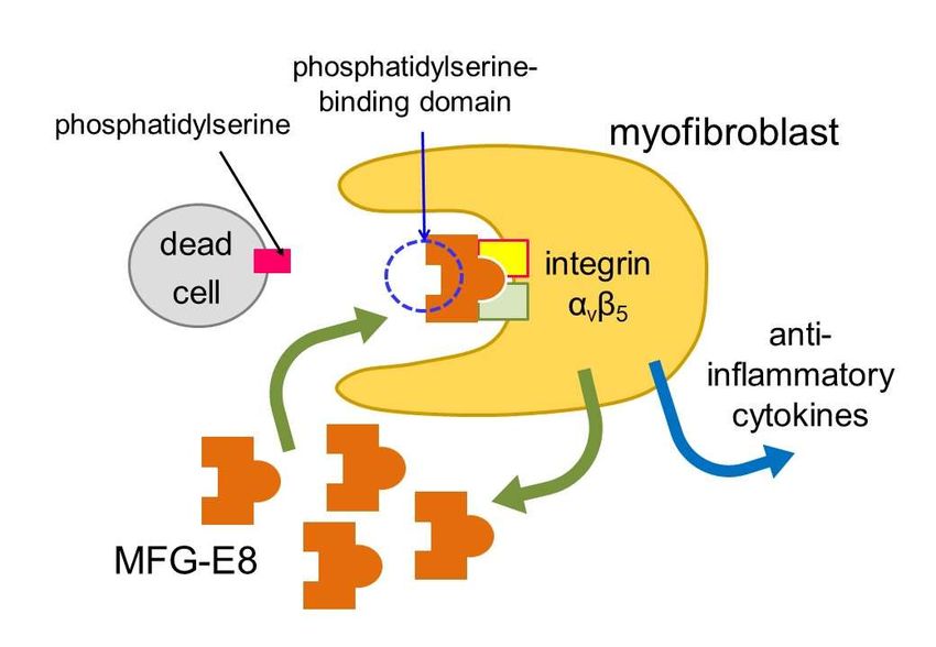
Preprints (www.preprints.org) | NOT PEER-REVIEWED | Posted: 19 May 2021               doi:10.20944/preprints202105.0432.v1

                    of myofibroblasts is proposed to be important for inhibition of progression to more severe
                    fibrotic states or recovery from fibrotic state [2]. Since myofibroblasts are an important
                    player in inflammation and fibrosis after cardiac injury [3], it is an urgent need to
                    understand the origin, function and fate of myofibroblasts. Recent technological advances
                    reveal some of these issues. progress of research topics for fibroblasts, myofibroblasts
                    and fibrosis will be reviewed.

                    1. Classification of fibroblasts

                    There are several types of cells in the heart such as cardiomyocytes and immune cells.
                    They interact with each other to regulate homeostasis in healthy and diseased conditions
                    [4]. Among several types of cells, fibroblasts are unique, since they produce extracellular
                    matrix that supports morphological integrity at resting state. Recent histology-based and
                    flow cytometric methods have demonstrated that fibroblasts account for about 13% of
                    cells in the mouse heart [5, 6]. When the heart is exposed to injury such as myocardial
                    infarction and hypertrophy, fibroblasts differentiate into myofibroblasts and produce
                    extracellular matrix. Myofibroblasts are only the cells that produce extracellular matrix.
                    Excess deposition of extracellular matrix causes fibrosis leading to tissue dysfunction. It
                    has been recognized that management of the number or function of myofibroblasts is
                    important for treatment of fibrosis.

                    Origin of myofibroblasts is analyzed by labeling the various types of cells with reporter
                    genes under cell-specific promoter (lineage-tracing experiment) [7]. Genetic labeling has
                    advantage over immunological detection of marker proteins, since maker proteins
                    sometimes disappear during development or differentiation. In addition, marker proteins
                    are expressed in not only cells that are analyzed but also functionally irrelevant cells.
                    Lineage tracking experiment is to express reporter gene under the control of cell-type
                    specific promoter. It labels the cells permanently even after the promoter activity turns
                    off and then allows to trace the cells that promoter is once activated.

                    Lineage-tracing experiments showed that about fibroblasts present in the left ventricle
                    and ventricular septum are derived from endocardial cells via endothelial-mesenchymal
                    transition (EndoMT) and epicardial cells through epithelial-mesenchymal transition
                    (EMT) [8]. A small number of fibroblasts were generated by differentiation of neural crest
                    cells. However, mature endothelial cells, epicardial cells, or bone marrow-derived cells

                                                                 2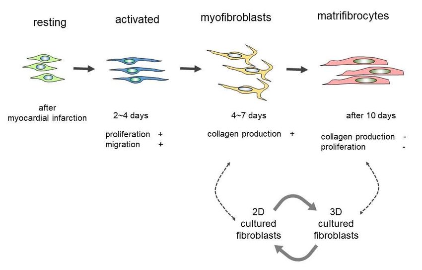
Preprints (www.preprints.org) | NOT PEER-REVIEWED | Posted: 19 May 2021                          doi:10.20944/preprints202105.0432.v1

                    did not contribute to population of fibroblasts. After labeling fibroblasts using cell-type
                    specific promoter-reporter gene, it was investigated whether fibroblasts exhibit different
                    functions depending on their origin. After pressure overload, epicardial-derived
                    fibroblasts (labeled with Tbx18 promoter-GFP) and endocardial-derived cells (labeled
                    with Tie2 promoter-GFP) were isolated. Analysis of RNAs showed similar expression
                    profiles between epicardial-derived and endocardial-derived cells [9-11]. In addition,
                    these two groups of cells had similar proliferative activity [12]. Therefore, it was
                    concluded that there is no significant relationship between function of fibroblasts and
                    origin of fibroblasts.

                    Molkentin's group used genetic labeling technique to trace the changes in the
                    characteristics of fibroblasts over time after myocardial infarction [13]. They found that
                    fibroblasts change their properties four different states after myocardial infarction, that is,
                    resting fibroblasts, active fibroblasts, myofibroblasts and matrifibrocytes (Figure 1).

                        Figure 1: Differentiation of fibroblasts to myofibroblast and matrifibrocytes. Fibroblasts of each
                        state have different proliferating activity and function. Dotted line is not established pathway.

                    Tcf21 promoter can label fibroblasts of resting conditions, which is tissue-resident
                    fibroblasts. When proliferative activity of fibroblasts was measured by feeding mice with

                                                                        3
Preprints (www.preprints.org) | NOT PEER-REVIEWED | Posted: 19 May 2021 doi:10.20944/preprints202105.0432.v1
5-ethynyl-2'-deoxyuridine (EdU) or immunodetection of Ki-67 after myocardial
infarction, they could detect actively proliferating fibroblasts 2 to 4 days after myocardial
infarction (active fibroblasts), and also found that fibroblasts convert to myofibroblasts 4
to 7 days after myocardial infarction. Myofibroblasts were derived from tissue-resident
fibroblasts. Their findings are consistent with other studies. Myofibroblasts were
transformed to new type of cells, which is called matrifibrocytes, 10 days after myocardial
infarct. Analysis of the expressing mRNAs suggested that each cell has different
properties. Fibroblasts of active state had high proliferative and migration activities.
Myofibroblasts produced collagen and α-smooth muscle actin (α-SMA). Matrifibrocytes
are unique, since they localized at scar and express the genes detected in tendons, bones
and cartilage. The physiological implications of these bone-related genes in the heart are
not known. Since matrifibrocytes localize only at scar, it suggests that they play a special
role in scar formation or maintenance. Matrifibroblasts have several unique properties.
When diphtheria toxin was administrated to mice with the toxin receptor expressing in
myofibroblasts, matrifibrocytes were resistant to killing by the toxin [13]. Susceptibility
to diphtheria toxin treatment was different from resting and active fibroblasts. In addition,
matrifibrocytes did not show proliferative activity when mice were treated with
angiotensin II and phenylephrine. In this context, Kim’s group reported an interesting
finding. In diabetes, inter-α-trypsin inhibitor heavy chain 1 (ITIH1) secreted from liver
was found to be responsible for systemic glucose intolerance. It blocked insulin action on
adipose tissue and skeletal muscle (14). ITIH1 works as a glue to tighten the binding
between extracelluar matrix. Anti-ITIH1 neutralizing antibody releases the inhibition of
ITIH1 and recovered insulin sensitivity. Inaccessibility of diphtheria toxin to periostin-
expressing cells at the late stage of fibrosis may be caused by ITIH1 or ITIH1-like
molecule. Future studies are waited to reveal the roles of matrifibrocytes in cardiac
fibrosis.
2. Heterogeneity of fibroblasts
Single-cell RNA sequencing (scRNAseq) is a relatively new but rapidly developing
technology [15]. It allows to comprehensively characterize gene expression and
relationships between individual cells. Single-cell analysis of 11,492 cells revealed
heterogeneity of fibroblasts and cardiomyocytes during pressure overload-induced
cardiac hypertrophy [16]. In their report, fibroblasts were grouped into 6 clusters: FB1 to
4Preprints (www.preprints.org) | NOT PEER-REVIEWED | Posted: 19 May 2021 doi:10.20944/preprints202105.0432.v1
FB6. FB1 corresponds to active fibroblasts in previous reports and FB6 is myofibroblast-
like cells that highly express extracellular matrix and periostin. It is unknown whether the
cells of each group differently contribute to fibrosis and which group corresponds to the
cells of the previous classification. In contrast to fibroblasts, cardiomyocytes were
divided into four groups (FC1~FC4) based on their expressing proteins. The cells of each
group expressed different combination of proteins that are involved in muscle
development, metabolism and contraction. Among them, FC3 and FC4 are interesting due
to expression of endothelial or fibroblast markers such as cadherin 5, von Willebrand
factor, vimentin and decorin. FC3 and FC4 were not fibroblast origin, since they did not
express marker proteins that label fibroblasts such as transcription factor Tcf21 and
PDGFα receptor. Correlation analysis suggested that the changes in FC3 and FC4 groups
are highly correlated to cardiomyocyte pathology at late stage. However, it remains to be
determined whether FC3 and FC4 have specific function in the progression of myocardial
infarction-induced heart failure.
There are several studies that use scRNAseq for analyzing cellular states. Skelly et al.
reported new cardiac fibroblast states with the cells isolated from healthy hearts [17]. A
new fibrocyte population of cells was identified, expressing markers of both fibroblasts
and immune cells. However, the functional role of these cells in the heart at baseline or
injury was not investigated. Farbehi et al. used lineage tracing to isolate the cells
expressing PDGF receptor α and sequenced them [18]. They identified novel
myofibroblast subtypes expressing both profibrotic and antifibrotic signatures. McLellan
et al. studied fibroblast populations present after angiotensin II infusion by scRNAseq
[19]. They found that myofibroblasts expressing αSMA are not detected. Instead, they
identified two fibroblast subpopulations expressing the matricellular proteins Cilp and
thrombospondin 4.
These results demonstrate the heterogeneity of fibroblasts but the relationship and identity
between the cells assigned by different groups are unknown. Furthermore, their function
and contribution to cardiac fibrosis remain to be determined in future.
5Preprints (www.preprints.org) | NOT PEER-REVIEWED | Posted: 19 May 2021 doi:10.20944/preprints202105.0432.v1
3. Differentiation of fibroblasts to other cells
The possibility that fibroblasts convert to other cells or vice versa after maturation has
been investigated [20]. Prolonged culture of macrophages resulted in the cells that express
various fibroblast markers such as type I collagen, prolyl-4-hydroxylase, fibroblast
specific protein-1, and fibroblast activation protein [20]. Next, the animals that express
yellow fluorescent protein (YFP) only in the cells of myeloid lineage were created. These
marker fibroblast proteins were detected in infiltrating YFP-positive macrophages after
myocardial infarction. Chlodronate liposome treatment to deplete macrophages reduced
the number of collagen positive fibroblast marker-expressing cells. These results
suggestsed that fibroblasts are derived from macrophages. It is interesting to examine the
contribution of macrophage-fibroblast transition to cardiac fibrosis. Inhibition of the
transition to fibroblasts may help reduce fibrosis after myocardial infarction.
It was reported that endothelial cells are not a major source of fibroblasts in adult mouse
heart [10]. However, there is a controversy on conversion of endothelial cells to
myofibroblasts. There is a report that fibroblasts acquire the endothelial cell-like
phenotype during ischemia-reperfusion [21, 22]. A series of experiments using mice with
genetically labeled fibroblasts demonstrated that 20-40% of fibroblasts express various
markers of endothelial cells, and the isolated cells can form a capillary network.
Expression of p53 was essential for the process of conversion from fibroblasts to
endothelial cells [21]. In addition, stimulation of p53 signaling improved cardiac
dysfunction during ischemia-reperfusion. However, an opposite result was reported by
different group, in which resident fibroblasts did not contribute to neovascularization after
cardiac injury [22]. In the report, pulse chase labeling of fibroblasts after ischemia-
reperfusion showed that resident fibroblasts do not express the genes involved in
angiogenesis, which are characteristics of endothelial cells. Origin of almost all
endothelial cells was resident endothelial cells. Different approaches resulted in distinct
conclusion. Thus, it may be necessary to confirm the findings with other techniques such
as scRNAseq and proteomic analysis of isolated cells.
4. Myofibroblasts as phagocytes
Myofibroblasts have been recognized only as the cells that produce extracellular matrix
such as collagen and fibronectin. They also interact with the inflammatory cells through
6Preprints (www.preprints.org) | NOT PEER-REVIEWED | Posted: 19 May 2021 doi:10.20944/preprints202105.0432.v1
secreted factors. New role of myofibroblasts in inflammation induced by myocardial
infarction was reported. Myofibroblasts phagocytose apoptotic cells and secret cytokines
that suppress the inflammatory responses [23]. This activity is similar to that of
macrophages which induce immuno-suppressive responses by engulfment of apoptotic
cells.
Myocardial infarction induces necrosis of cardiomyocytes. Phosphatidylserine is
presented on the cellular surface of the cells that has undergone necrosis as observed at
apoptosis. Therefore, terminal deoxynucleotidyl transferase-mediated dUTP nick end
labeling (TUNEL) staining is conveniently used as a marker of dead cells. Apoptotic cells
are thought to be engulfed by phagocytes such as macrophages. Nakaya et al. examined
expression of various molecules involving in engulfment after myocardial infarction [23]
(Figure 2). The expression of a factor called milk fat globule-EGF factor 8 (MFG-E8)
was increased. MFG-E8 binds both phosphatidylserine expressing on the membrane of
apoptotic cells and integrin being present at the surface of phagocytic cells [24]. Since
integrin does not directly bind phosphatidylserine, MFG-E8 functions as a bridge between
apoptotic cells and phagocytes. MFG-E8 was found to be produced by myofibroblasts
and was used by myofibroblasts to phagocytose dead cells. By phagocytosing dead cells,
myofibroblasts release anti-inflammatory cytokines and prevent excessive inflammation.
Myofibroblasts are similar to macrophages in phagocytosis of dead cells and subsequent
secretion of anti-inflammatory cytokines. It indicates that myofibroblasts behave like
macrophages at myocardial infarction. The activity of myofibroblasts to engulf dead cells
is weaker than that of macrophages. However, fibroblasts are easily differentiated to
myofibroblasts at the injury sites and the number of myofibroblasts is supposed to be high
at ischemic area during myocardial infarction. Thus, myofibroblasts compensate low
ability of engulfment with the number of cells that engulf. However, there is a big
difference between myofibroblasts and macrophages. Unlike macrophages,
myofibroblasts dis not have antigen presentation activity [23]. Myofibroblast-mediated
phagocytosis of apoptotic cells is considered to be an efficient way to prevent excess
inflammation at the injured site. It is interesting that not all myofibroblasts phagocytose
dead cells in vitro. Thus, distinct group members of myofibroblasts may have different
functions such as phagocytosis and differentiation to other types of cells.
7Preprints (www.preprints.org) | NOT PEER-REVIEWED | Posted: 19 May 2021 doi:10.20944/preprints202105.0432.v1
Figure 2: Engulfment of dead cells by myofibroblasts. Myofibroblasts engulf dead cells with the
help of MFG-E8 secreted by themselves and then secrete anti-inflammatory cytokines.
5. Signaling that controls differentiation to myofibroblasts
TGF-β is a strong inducer of differentiation of fibroblasts to myofibroblasts [25].
Inflammatory cells recruiting to the injury sites release cytokines including TGF-β. Injury
cells also release alarmins and damage-associated molecular patterns (DAMPs) [26].
These molecules cause inflammation leading to differentiation of fibroblasts to
myofibroblasts. Thus, any inhibition of inflammatory responses will block appearance of
myofibroblasts, which eventually suppresses fibrosis. At early stage of myocardial
infarction, neutrophils are firstly recruited to the injury sites [27, 28]. Leukotriene B4 is
a powerful attractant of neutrophils by binding to Leukotriene B4 receptor (BLT1).
Inhibition of neutrophil recruitment by BLT1 gene knockout or BLT1 blocker decreased
cardiac fibrosis by inhibition of inflammation [29, 30].
Interleukin (IL) is an important group of inflammatory cytokines. Multiple IL receptors
are expressed in cardiac fibroblasts, which regulates fibroblast states and function [27].
The effects of proinflammatory ILs on cardiac fibroblasts ae blocked by cardiac
fibroblast-specific deletion of IL receptors. Knockout of IL11 receptor or IL17 receptor
genes reduced injury-induced cardiac fibrosis and cardiac dysfunction [31, 32]. These
results reveal that cardiac fibroblast-specific deletion of IL receptor genes decreases the
8Preprints (www.preprints.org) | NOT PEER-REVIEWED | Posted: 19 May 2021 doi:10.20944/preprints202105.0432.v1
infiltration to or activity of immune cells in the injury area. It also suggests that cardiac
fibroblasts play an important role in the regulation of injury-induced inflammation that is
mediated by IL signaling.
After myocardial infarction, monocytes are mobilized from bone marrow and
differentiated to macrophages at the injury sites [33]. Macrophages can be depleted by
the treatment of mice with chlodronate-liposomes [34]. Macrophage-depleted mice
showed decreased fibrosis and improved cardiac functions. These results show that single
step of inflammation subsequent to myocardial infarction blocks fibrosis possibly through
inhibition of differentiation to myofibroblasts.
TGF-β stimulation activates both canonical Smad2/3 and non-canonical MAP kinase
signaling pathways [25]. Fibrotic responses were inhibited by knockout of fibroblast-
specific TGF-β receptor 1/2 or knockout of transcription factor Smad3 that is activated
downstream of TGF-β receptor [35]. It demonstrated that TGF-β-Smad2/3 signaling of
fibroblasts is a major factor of cardiac fibrosis induced by pressure overload. It was
interesting that knockout of TGF-β receptor 1/2 in fibroblasts also inhibited cardiac
hypertrophy by pressure overload [35]. It indicates that myofibroblasts interact with
cardiac myocytes through direct cell-cell communication or indirect mediator-mediated
interaction, which is secreted from myofibroblasts.
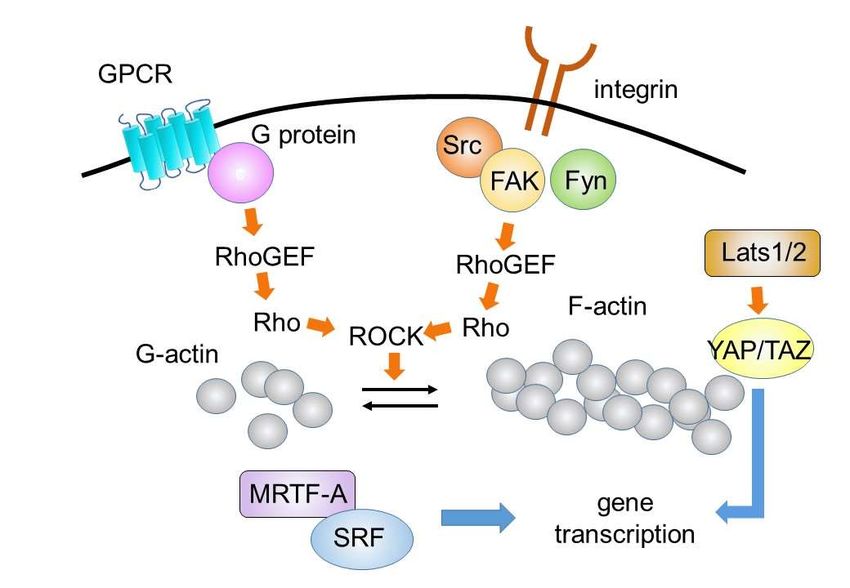
In addition to TGF-β, lysophosphatidic acid (LPA) stimulation induces differentiation of
fibroblasts to myofibroblasts [36, 37]. LPA binds their own GPCRs to activate cellular
responses [38]. In vitro, LPA stimulation induced fibrosis by activation of myocardin-
related transcription factor-serum responsive factor (MRTF-SRF) pathway. Rho regulated
actin oligomerization that is regulated by phosphorylation of actin by Rho kinase (ROCK).
ROCK-mediated phosphorylation of actin increased monomeric form of actin that results
in release of SRF inhibition. SRF together with MRTF activated transcription of various
genes including profibrotic genes [39]. Compound CCG-203971 is a small-molecule
inhibitor of the Rho-mediated MRTF-SRF pathway [40]. Administration of CCG-203971
inhibited bleomycin-induced lung fibrosis. Rho is signaling molecule that is activated
downstream of various receptors including GPCRs and TGF-β receptors. Inhibitors of
Rho-Rock pathway may suppress fibroblast activation and fibrosis more efficiently than
receptor inhibition.
9Preprints (www.preprints.org) | NOT PEER-REVIEWED | Posted: 19 May 2021 doi:10.20944/preprints202105.0432.v1
G protein-coupled Receptor Kinase 2 (GRK2) is known as a regulator of G protein-
coupled receptors (GPCRs) by phosphorylating agonist-bound GPCRs [41].
Cardiomyocyte-specific knockout of GRK2 demonstrated that GRK2 ablation protects
the heart against cardiac dysfunction and fibrosis by myocardial infarction [42]. These
mice also showed the reduction of the development of heart failure by myocardial
infarction. Fibroblast-specific knockout of GRK2 using the collagen 1α2 promoter
reduced the secretion of TNFα and suppressed gene expression of profibrotic factors after
ischemia-reperfusion [43]. Inhibition of fibroblast function improved cardiac dysfunction.
These results show that inhibition of GRK2 protects the heart against cardiac stresses in
cardiomyocytes as well as in fibroblasts.
Transient receptor potential channel canonical 6 (TRPC6) is a voltage-independent cation
channel and mediates angiotensin II-stimulated hypertrophic responses [44]. The
increased intracellular Ca2+ also plays an important role in conversion of fibroblasts to
myofibroblasts. It activates cellular signaling that is sufficient for promoting conversion
to myofibroblast and resulting fibrosis [45]. TGF-β-induced upregulation of TRPC6 was
inhibited by blockade of p38-MAPK-mediated signaling [46]. It is interesting that TRPC6
knockout fibroblasts did not show changes in Ca2+ signaling and did not promote
conversion of fibroblasts to myofibroblasts when the cells are treated with angiotensin II
and TGF-β. These results demonstrate that TRPC6-Ca2+ signaling is essential for
induction of myofibroblasts in cardiac fibroblasts.
There are other signaling molecules involved in induction of fibrosis. Deletion of β-
catenin gene in cardiac fibroblasts improves cardiac function and reduces fibrosis, which
is due to decreased production of extracellular matrix proteins by cardiac fibroblasts [47].
It was recently found that functional primary cilia in cardiac fibroblasts is required for
canonical TGFβ signaling-induced differentiation of cardiac fibroblasts to myofibroblasts
[48]. Primary cilia express polycystin-1 that is known as regulator of cell proliferation,
cell migration, and interactions with other cells. Polycystin-1 is required for maintaining
cellular structures of primary cilia. When primary cilia of cardiac fibroblasts were
disrupted specifically by deletion of polycystin-1 gene, TGFβ-Smad3 signaling-induced
extracellular matrix protein production and fibroblast differentiation were impaired.
Deletion of polycystin-1 gene enhanced pathological cardiac remodeling after myocardial
infarction [49], suggesting the important role of primary cilia in hypertrophy and fibrosis.
10Preprints (www.preprints.org) | NOT PEER-REVIEWED | Posted: 19 May 2021 doi:10.20944/preprints202105.0432.v1
It also suggested that primary cilia are functional and participate in TGFβ-induced fibrosis
and myofibroblast differentiation. Heat shock protein (Hsp) is chaperon molecule for
conversion of fibroblasts to myofibroblasts and resulting fibrosis. Among various Hsp
proteins, Hsp47 is known as collagen-specific chaperone. Cardiac fibroblast-specific
deletion of Hsp47 significantly reduced cardiac fibrosis and improved cardiac diastolic
dysfunction after pressure overload [50]. However, the reduced collagen production in
these mice increased lethality after myocardial infarction due to the insufficient scar
formation.
Collectively, these results suggest that manipulation of expression and activity of various
signaling molecules involved in fibrotic pathway modulates fibroblast states leading to
alteration of fibrosis.
6. Control of differentiation of fibroblasts by extracellular environment
Fibroblasts exist in interstitial spaces between cardiomyocytes in healthy condition.
When the heart is exposed to stresses such as myocardial infarction and hypertrophy,
fibroblasts differentiate to myofibroblasts and produce extracellular matrix such as
collagen. Fibroblasts generated at the injury site actively proliferate and form aggregate.
An in vitro three-dimensional culture system that mimics several states of fibroblasts was
developed [51]. When fibroblasts were isolated and cultured in two or three dimensions,
the properties of fibroblasts were changed. Standard polystyrene-coated culture plates
(not coated with collagen or other extracellular matrix) were used for two-dimensional
(2D) cultures, and ultra-low adhesion plates (coated with any extracellular matrix) for 3D
cultures (Figure 1). Fibroblasts cultured with 3D structure plates formed spheres
(spherical masses) within 24 hours. The morphology of fibroblasts formed by 2D and 3D
cultures was reversible but did not depend on the tension or rigidity of the extracellular
environment. Interestingly, a correlation of the expressing genes was found between 3D
culture fibroblasts and the remodeling heart (treatment with isoprenaline for 3 weeks or
cryo-injury treatment). However, the expression of α-smooth muscle actin (α-SMA) was
decreased in the aggregates. Since the expression of α-SMA, a marker of myofibroblasts,
is decreased, it cannot be said that fibroblasts obtained by 3D culture is conventional
myofibroblasts. However, expression pattern of mRNA of 3D culture fibroblasts is similar
to that of matrifibrocytes reported by Fu et al. [52]. Analysis of fibroblasts with 3D culture
may elucidate function and fate of matrifibrocytes. Interestingly, 3D cultured fibroblasts
11Preprints (www.preprints.org) | NOT PEER-REVIEWED | Posted: 19 May 2021 doi:10.20944/preprints202105.0432.v1
reversibly changed their morphology and expressing genes by transferring the cells to 2D
culture. Thus, 3D culture but not normal 2D culture provides a sufficient signal to trigger
remodeling. Since in vitro system is essential for analyzing mechanism of differentiation
and function of fibroblasts and myofibroblasts, the exchange of culture conditions may
be a promising technique to analyze complex behavior of fibroblasts.
Fibroblast fate is also regulated by stiffness of extracellular matrix [53]. The stiffness
around fibroblasts increases during progression of fibrosis. Increased stiffness was sensed
by integrin receptors and actin cytoskeleton that promote translocation of p38-MAPK to
the nucleus and stimulates remodeling [54]. Integrin-actin cytoskeleton signaling
complex also activates tyrosine kinases such as focal adhesion kinase (FAK), Src and Fyn.
These kinases stimulate GDP-GTP exchange of Rho through guanine nucleotide
exchange factors (RhoGEFs). It leads to activation of Rho-ROCK-MRTF-A pathway that
increases gene transcription in concerted action with SRF [55]. This pathway will be
described in more detail in YAP-TAZ signaling.
7. YAP-TAZ signal in fibroblasts
Hippo pathway is known for its inhibitory activity on cardiomyocyte proliferation [56].
Yes-associated protein (YAP) is a transcriptional coactivator in Hippo pathway that is
negatively regulated by large tumor suppressor kinase 1 (Lats1) and Lats 2 (Figure 3).
12Preprints (www.preprints.org) | NOT PEER-REVIEWED | Posted: 19 May 2021 doi:10.20944/preprints202105.0432.v1
Figure 3: Rho- and YAP/TAZ-mediated signaling pathways.
Deletion of YAP from transcription factor 21 (Tcf21) and Col1a1-expressing fibroblasts
decreased their collagen deposition, proliferation, and activation after myocardial
infarction [57]. Similar decreases in angiotensin II/phenylephrine-treated fibrosis were
seen when YAP was knocked down from fibroblasts [58]. Myocardin-related transcription
factor A (MRTF-A) levels were decreased in YAP knockout mice, suggesting that MRTF-
A expression was regulated by YAP function. These results demonstrate the importance
of YAP-MRTF-A signaling for determination of myofibroblast states in response to
ischemic and chronic stresses. Interestingly, cardiomyocyte specific deletion of YAP
decreased hypertrophy and significantly increased fibrosis [59]. Thus, YAP protects the
heart against ischemic stress or pressure overload.
Lats1 and Lats2 phosphorylate YAP and inhibit YAP-mediated transcriptional activation
of Hippo pathway [60]. Deletion of Lats1 and Lats2 increased YAP activity. Cardiac
fibroblast-specific deletion of Lats1 and Lats2 induced spontaneous myofibroblast
differentiation [61]. Lats1 and Lats2 knockout mice showed increased fibrosis both at
baseline and after myocardial infarction. Mechanistic analysis revealed that YAP directly
activates transcriptional machinery of myofibroblasts leading to fibrosis. These results
13Preprints (www.preprints.org) | NOT PEER-REVIEWED | Posted: 19 May 2021 doi:10.20944/preprints202105.0432.v1
suggest that Lats1/2-dependent YAP inhibition plays an essential role for maintaining the
resting state of fibroblasts.
Lats1 and Lats2 activities are regulated by actin cytoskeleton that are regulated by Rho.
Rho are also involved in MRTF-A- and MRTF-B-mediated fibrotic pathway. MRTF-A
and -MRTF-B help serum response factor (SRF) to bind a promotor sequence known as
the serum response element (also known as the CArG box). Rho activates transcriptional
machinery leading to fibrotic responses. Thus, Rho, Lats1/2, YAP and MRTF-A/B form
complex network of induction of fibrotic responses.
8. Development of treatment of fibrosis and heart failure
GPCR stimulation dissociates heterotrimeric G protein in Gα and Gβγ. Gallein is a small
molecule that inhibits Gβγ-dependent GRK2 activation [62, 63]. When administered in
vivo, gallein suppressed cardiac injury after ischemia-reperfusion, activation of
fibroblasts, and the onset of heart failure possibly by GRK2 inhibition [62]. Gallein could
attenuate the activation responses of fibroblasts isolated from heart failure patients.
However, the affinity of gallein for Gβγ is low (Ki is about 0.2μM), non-specific effects
of gallein should be examined by the experiments that gallein-mediated protective
responses disappear in GRK2 knockout mice. From high throughput screen, paroxetine
was identified as a GRK2 inhibitor that attenuated the development of heart failure by
myocardial infarction [64, 65]. Although paroxetine is a selective serotonin reuptake
inhibitor (SSRI), another SSRI fluvoxamine did not inhibit GRK2 and did not attenuate
heart failure [65]. These results shows that new GRK2-selective inhibitor is a therapeutic
option for treatment of heart failure.
Cyclic nucleotides (cAMP and cGMP) play important roles in cellular signaling. Cyclic
AMP and cGMP are generated by adenylyl cyclase or guanylyl cyclase, respectively.
Various drugs that increase cAMP or cGMP are already used in clinic for treatment of
cardiovascular diseases. These nucleotides are degraded by phosphodiesterases (PDEs).
PDE comprises superfamily and is categorized into 11 isozymes (PDE1~PDE11) [66].
Each PDE has different selectivity for cAMP or cGMP, although selectivity depends on
cellular environment. It should be noted that cGMP selective PDE degrades cAMP when
the concentration of cAMP is very high. There are PDEs that degrade both cAMP and
cGMP. PDE2 is a PDE that is activated by cGMP and degrades cGMP and cAMP [67].
14Preprints (www.preprints.org) | NOT PEER-REVIEWED | Posted: 19 May 2021 doi:10.20944/preprints202105.0432.v1
Inhibition of PDE2 suppressed the onset of heart failure by inhibiting cGMP degradation
[68]. The elevated cGMP activated protein kinase G leading to activation of transcription
factors (Nuclear Factor of Activated T cells, NFAT) and inhibition of TRPCs. PDE2
inhibitor such as BAY 60-7550 preferentially promoted NO-guanylyl cyclase-cGMP
signaling and suppressed the onset of heart failure [69]. However, it should be careful to
use PDE inhibitor for the treatment of heart failure, since cAMP causes harmful effects
on heart failure.
Bcl2 family is involved in apoptosis and is categorized into three groups based on the
homology of amino acid sequence, that is Bcl2-like, Bax-like and BH3-only. Bcl2-like
protein has anti-apoptotic activity [70]. In contrast to Bcl2-like protein, Bax-like and
BH3-only proteins promote apoptosis. BH3 domains of these apoptosis-promoting
proteins bind hydrophobic BH3-binding pocket of anti-apoptotic Bcl2 to form
heterodimer and inhibit anti-apoptotic activity by Bcl-2 [71]. Navitoclax (also known as
ABT-263) is a BH3-mimetic antagonist of the Bcl2-anti-apoptotic protein [72]. It
promoted apoptosis of myofibroblasts isolated from the patients of scleroderma [73].
Since enhanced apoptosis of myofibroblasts is effective in recovery from fibrosis,
navitoclax will be a promising drug for treatment of scleroderma. Venetoclax that has
excellent selectivity for BCL-2 is currently in clinical trials as an anticancer drug [74]. In
the future, venetoclax may be also applied to treatment for cardiac fibrosis.
Many studies report that fibrosis is inhibited by knockout of gene in myofibroblasts or
cardiomyocytes. However, inhibition of ongoing fibrosis by compounds does not always
recover the heart from fibrotic states. Metformin activates AMP-activated protein kinase
(AMPK). It was reported that metformin recovers the lung from bleomycin-induced
fibrosis [75]. The effects of metformin were proposed to promote apoptosis of
myofibroblasts or inactivate myofibroblasts. Activation of AMPK with metformin or
compound A769662 also suppressed hypertrophic responses of neonatal ventricular
myocytes by phenylephrine stimulation [76]. These AMPK activators inhibited ERK
activation and NFAT nuclear translocation. Mechanistic analysis showed that inhibition
of cardiac hypertrophy by AMPK was associated with increase of protein O-linked
acetylglucosamine (O-GlcNAcylation) and decrease of O-GlcNAcylation by inhibition
of glutamine: fructose-6-phosphate aminotransferase (GFAT). AMPK activators
decreased O-GlcNAcylation of troponin T that is associated with development of adverse
15Preprints (www.preprints.org) | NOT PEER-REVIEWED | Posted: 19 May 2021 doi:10.20944/preprints202105.0432.v1
cardiac remodeling. Since the effects of AMPK activators were not observed in AMPKα2-
deficient mice, the effects of AMPK activators are mediated by AMPKα2. Cardiac
fibrosis is always accompanied by hypertrophy. Inhibition of hypertrophy will indirectly
suppress fibrosis.
In the heart, aquaporin transfers hydrogen peroxide (H2O2) from extracellular space to
cytosol [77]. H2O2 entering into the cells modifies proteins and changes protein function
that cause detrimental effects. Several isoforms of aquaporins are expressed in the mouse
heart. Since H2O2 are generated from superoxide anion (O2-) at extracellular space
produced by NADPH oxidases [78, 79], aquaporin-mediated transfer of H 2O2 to cytosol
is critical for induction of hypertrophy and other responses. Treatment of mice with a
clinically approved aquaporin 1 inhibitor, Bacopaside, attenuated cardiac hypertrophy
[80]. These results suggest that aquaporin is a promising target to treat hypertrophy.
However, aquaporins are expressed in whole body, and inhibition of water transfer
activity of aquaporins by drug or antibody may cause side effects in other tissues. It has
been reported that anti-aquaporin-4 antibody is main pathogen of human neuromyelitis
optica spectrum disorders and level of the antibody in the plasma links to poor visual
prognosis in human [81].
9. Immunotherapy
Immunological strategy to target cardiac fibrosis was recently reported [82]. Chimeric
antigen receptor (CAR) T cells were engineered to specifically recognize myofibroblasts
and induced ablation of myofibroblasts leading to improved cardiac function. CAR
consists of antigen-recognizing regions of single chain Fv fragment, transmembrane
domain, intracellular domains of T cell activation receptor (CD3ζ) and co-stimulation
receptor (CD28). After binding of CAR to myofibroblast-specific protein, in this case
mouse fibroblast activation protein, CAR T cells causes cytotoxic killing to decrease the
number of myofibroblasts. CAR T cells are already used for cancer therapy and are
reported to have beneficial effects on the treatment. Strategy to eliminate myofibroblasts
is supported by the following experiments. When diphtheria toxin receptor specifically
expressing in myofibroblasts binds diphtheria toxin, the toxin decreases the number of
myofibroblasts and reduces cardiac fibrosis by myocardial infarction [83]. However,
several concerns are raised before application of CAR T cells to heart failure patients who
are seriously suffered from fibrosis [84]. CAR T cells release cytokines for attacking
16Preprints (www.preprints.org) | NOT PEER-REVIEWED | Posted: 19 May 2021 doi:10.20944/preprints202105.0432.v1
myofibroblasts and heart failure patients are in advanced inflammatory state. Released
cytokines will complicate the conditions of heart failure. It may cause detrimental effects
on heart failure patients. Authenticity of elimination of myofibroblasts is also questioned.
Myofibroblasts produce extracellular matrix such as collagen and fibronectin that are in
part protective against cardiac rupture. Another point is the lack of information of
myofibroblast-specific marker proteins in human. Specific antigen is essential for
development of CAR T cells-dependent treatment. Although there are several concerns,
proper management of number and appearance of myofibroblasts is expected to lead to
the treatment of cardiac fibrosis with low side effects.
CAR T cell strategy utilizes myofibroblast-specific antigen and antibody-mediated
cytotoxic actions. CAR T cell strategy requires dual recognition protein. A protein with
dual inhibitory activities of programmed death lighand-1 (PD-L1) and TGF-β (M7824)
was successively used for the treatment of tumor growth and metastasis [85]. Dual
inhibitor was more effective than treatment with TGF-β inhibitor alone. It is expected to
increase the specificity of TGF-β inhibitor. Thus, to combine TGF-β inhibitor and
antibody that recognizes myofibroblast-specific protein into single molecule may be
another strategy to treat cardiac fibrosis. Identification of myofibroblast-specific antigen
helps increase specificity of TGF-β inhibitor against cardiac fibrosis and restricts the
action of TGF-β inhibitor at the localized area.
10. Conclusion
Myofibroblasts are the cells that mainly differentiate from fibroblasts and are responsible
for production of extracellular matrix in injury. Relationship between fibroblasts and
myofibroblasts is more complex than previously thought. Single cell RNA sequencing
and lineage tracing techniques demonstrate heterogeneity of fibroblasts, although
functional differences are remained to be determined. Treatment of cardiac fibrosis is
eagerly waited, signaling analysis of various fibroblast states will provide a therapeutic
target that is suitable for drug development. Although direct reprogramming of cardiac
fibroblasts to cardiomyocytes with chemical compounds is not mentioned in this review,
technologies of direct reprogramming are currently in progress [86] and may be another
option for the treatment of cardiac fibrosis in future.
References
17Preprints (www.preprints.org) | NOT PEER-REVIEWED | Posted: 19 May 2021 doi:10.20944/preprints202105.0432.v1
1. Rockey, D. C.; Bell, P. D.; Hill, J. A. Fibrosis -A common pathway to organ injury and
failure. New England Journal of Medicine 2015, 372, 1138-1145.
2. Gourdie, R. G.; Dimmeler, S.; Kohl, P. Novel therapeutic strategies targeting fibroblasts
and fibrosis in heart disease. Nature Review Drug Discovery 2016, 15, 620-638.
3. Kurose, H.; Mangmool, S. Myofibroblasts and inflammatory cells as players of cardiac
fibrosis. Archives of Pharmacal Research 2016, 39, 1100-1113.
4. Howard, C.M.; Baudino, T. A. Dynamic cell-cell and cell-ECM interactions in the heart.
Journal of Molecular and Cellular Cardiology 2014, 70, 19-26.
5. Pinto, A. R.; Ilinykh, A.; Ivey, M. J.; Kuwabara, J. T.; D'Antoni, M. L.; Debuque, R.;
Chandran, A.; Wang, L.; Arora, K.; Rosenthal, N. A.; Tallquist, M. D. Revisiting cardiac
cellular composition. Circulation Research 2016, 118, 400-409.
6. Zhou, P.; Pu, W. Recounting cardiac cellular composition. Circulation Research 2016,
118, 368-370.
7. Kebschull, J. M.; Zador, A. M. Cellular barcoding: lineage tracing, screening and
beyond. Nature Methods 2018, 15, 871-879.
8. Doppler, S. A; Carvalho, C.; Lahm, H.; Deutsch, M. A.; Dreßen, M.; Puluca, N.; Lange,
R.; Krane, M. Cardiac fibroblasts: more than mechanical support. Journal of Thoracic
Disease 2017, 9, S36-S51.
9. Onur Kanisicak, O.; Khalil, H.; Ivey, M. J.; Karch, J.; Maliken, B. D.; Correll, R. N.;
Brody, M. L.; Lin, S-C. J.; Aronow, B. J.; allquist, M. D.; Molkentin, J. D. Genetic lineage
tracing defines myofibroblast origin and function in the injured heart. Nature
Communications 2016, 7, 12260.
10. Moore-Morris, T.; Guimarães-Camboa, N.; Banerjee, I.; Zambon, A. C.; Kisseleva,
T.; Velayoudon, A.; Stallcup, W. B.; Gu, Y.; Dalton, N. D.; Cedenilla, M.; Gomez-Amaro,
R.; Zhou, B.; Brenner, D. A.; Peterson, K. I.; Chen, J.; Evans, S. M. Resident fibroblast
lineages mediate pressure overload-induced cardiac fibrosis Journal of Clinical
Investigation 2014, 124, 2921-2934.
11. Ali, S. R.; Ranjbarvaziri, S.; Talkhabi, M.; Zhao, P.; Subat, A.; Hojjat, A.; Kamran, P.;
Müller, A. M. S.; Volz, K. S.; Tang, Z.; Red-Horse, K.; Ardehali, R. Developmental
18Preprints (www.preprints.org) | NOT PEER-REVIEWED | Posted: 19 May 2021 doi:10.20944/preprints202105.0432.v1
heterogeneity of cardiac fibroblasts does not predict pathological proliferation and
activation. Circulation Research 2014, 115, 625-635.
12. Wu, R.; Ma, F.; Tosevska, A.; Farrell, C.; Pellegrini, M.; Deb, A. Cardiac fibroblast
proliferation rates and collagen expression mature early and are unaltered with advancing
age. JCI insight 2020, 17, e140628.
13. Fu, X.; Khalil, H.; Kanisicak, O.; Boyer, J. G.; Vagnozzi, R. J.; Maliken, B. D.; Sargent,
M. A.; Prasad, V.; Valiente-Alandi, I.; Blaxall, B. C.; Molkentin, J. D. Specialized
fibroblast differentiated states underlie scar formation in the infarcted mouse heart.
Journal of Clinical Investigation 2018, 128, 2127-2143.
14. Kim, T. H.; Koo, J. H.; Heo, M. J.; Han, C. Y.; Kim, Y-I.; Park, S-Y.; Cho, I. J.; Lee,
C. H.; Choi, C. S.; Lee, J. W.; Kim, W.; Cho, J-Y.; Kim, S. G. Overproduction of inter-α-
trypsin inhibitor heavy chain 1 after loss of Gα13 in liver exacerbates systemic insulin
resistance in mice. Science Translational Medicine 2019, 11, eaan4735.
15. Yamada, S.; Nomura, S. Review of Single-Cell RNA Sequencing in the Heart.
Internatioal Journal of Molecular Sciences 2020, 21, 8345.
16. Ren, Z.; Yu, P.; Li, D.; Li, Z.; Liao, Y.; Wang, Y.; Zhou, B.; Wang, L. Single-Cell
Reconstruction of Progression Trajectory Reveals Intervention Principles in Pathological
Cardiac Hypertrophy. Circulation 2020, 141, 1704-1719.
17. Skelly, D. A.; Squiers, G. T.; McLellan, M. A.; Bolisetty, M. T.; Robson, P.; Rosenthal,
N. A.; Pinto, A. R. Single-cell transcriptional profiling reveals cellular diversity and
intercommunication in the mouse heart. Cell Reports 2018, 22, 600-610.
18. Farbehi, N.; Patrick, R.; Dorison, A.; Xaymardan, M.; Janbandhu, V.; Wystub-Lis, K.;
Ho, J. W.; Nordon, R.E.; Harvey, R. P. Single-cell expression profiling reveals dynamic
flux of cardiac stromal, vascular and immune cells in health and injury. eLife 2019, 8,
e43882.
19. McLellan, M. A.; Skelly, D. A.; Dona,M. S. I.; Squiers, G. T.; Farrugia , G.E.; Gaynor,
T. L.; Cohen, C. D.; Pandey, R.; Diep, H.; Vinh, A.; Rosenthal, N. A.; Pinto, A. R. High-
resolution transcriptomic profiling of the heart during chronic stress reveals cellular
drivers of cardiac fibrosis and hypertrophy. Circulation 2020, 142, 1448-1463.
19Preprints (www.preprints.org) | NOT PEER-REVIEWED | Posted: 19 May 2021 doi:10.20944/preprints202105.0432.v1
20. Haider, N.; Boscá, L.; Zandbergen, H. R.; Kovacic, J. C.; Narula, N.; González-Ramos,
S.; Fernandez-Velasco, M.; Agrawal, S.; Paz-García, M.; Gupta, S.; DeLeon-Pennell, K.;
Fuster, V.; Ibañez, B.; Narula, J. Transition of macrophages to fibroblast-like cells in
healing myocardial infarction. Journal of the American College of Cardiology 2019, 74,
3124-3135.
21. Ubil, E.; Duan, J.; Pillai, I. C. L.; Rosa-Garrido, M.; Wu, Y.; Bargiacchi, F.; Lu, Y.;
Stanbouly, S.; Huang, J.; Rojas, M; Vondriska, T. M.; Stefani, E.; Deb, A. Mesenchymal-
endothelial transition contributes to cardiac neovascularization. Nature 2014, 514, 585-
590.
22. He, L.; Huang, X.; Kanisicak, O.; Li, Y.; Wang, Y.; Li, Y.; Pu, W.; Liu, Q.; Zhang, H.;
Tian, X.; Zhao, H.; Liu, X.; Zhang, S.; Nie, Y.; Hu, S.; Miao, X.; Wang, Q-D.; Wang, F.;
Chen, T.; Xu, Q.; Lui, K. O.; Molkentin, J. D.; Zhou, B. Preexisting endothelial cells
mediate cardiac neovascularization after injury. Journal of Clinical Investigation 127,
2968-2981.
23. Nakaya, M.; Watari, K.; Tajima, M.; Nakaya, T.; Matsuda, S.; Ohara, H.; Nishihara,
H.; Yamaguchi, H.; Hashimoto, A.; Nishida, M.; Nagasaka, A.; Horii, Y.; Ono, H.; Iribe,
G.; Inoue, R.; Tsuda, M.; Inoue, K.; Tanaka, A.; Kuroda, M.; Nagata, S.; Kurose, H.
Cardiac myofibroblast engulfment of dead cells facilitates recovery after myocardial
infarction. Journal of Clinical Investigation 127, 383-401.
24. Hanayama, R.; Tanaka, M.; Miwa, K.; Shinohara, A.; Iwamatsu, A.; Nagata, S.
Identification of a factor that links apoptotic cells to phagocytes. Nature 2002, 417, 182-
187.
25. Parichatikanond, W.; Luangmonkong, T.; Mangmool, S.; Kurose, H. Therapeutic
targets for the treatment of cardiac fibrosis and cancer: Focusing on TGF-β signaling.
Frontier in Cardiovascular Medicine 2020, 7, 34.
26. Turner, N. A. Inflammatory and fibrotic responses of cardiac fibroblasts to myocardial
damage associated molecular patterns (DAMPs). Journal of Molecular and Cellular
Cardiology 2016, 94, 189-200.
27. Prabhu, S. D.; Frangogiannis, N. G. The Biological Basis for Cardiac Repair After
Myocardial Infarction: From Inflammation to Fibrosis. Circulation Research 119, 91-112.
20Preprints (www.preprints.org) | NOT PEER-REVIEWED | Posted: 19 May 2021 doi:10.20944/preprints202105.0432.v1
28. Daseke 2nd, M. J.; Chalise, U.; Becirovic-Agic, M.; Salomon, J. D.; Cook, L. M.;
Case, A. J.; Lindsey, M. L. Neutrophil signaling during myocardial infarction wound
repair. Cellular Signaling 77, 109816.
29. de Hoog, V. C.; Bovens, S. N.; de Jager, S. C. A.; van Middelaar, B. J.; van
Duijvenvoorde, A.; Doevendans, P. A.; Pasterkamp, G.; de Kleijn, D. P. V.; Timmers, L.
BLT1 antagonist LSN2792613 reduces infarct size in a mouse model of myocardial
ischaemia-reperfusion injury. Cardiovascular Research 2015, 108, 367-376.
30. Horii, Y.; Nakaya, M.; Ohara, H.; Nishihara, H.; Watari, K.; Nagasaka, A.; Nakaya,
T.; Sugiura, Y.; Okuno, T.; Koga, T.; Tanaka, A.; Yokomizo, T.; Kurose, H. Leukotriene
B4 receptor 1 exacerbates inflammation following myocardial infarction. FASEB Journal
2020, 34, 8749–8763.
31. Schafer, S.; Viswanathan, S.; Widjaja, A. A.; Lim, W-W.; Moreno-Moral, A.;
DeLaughter, D. M.; Ng, B.; Patone, G.; Chow, K.; Khin, E.; Tan, J.; Chothani, S. P.; Ye,
L.; Rackham, O. J. L.; Ko, N. S. J.; Sahib, N. E.; Pua, C. J.; Zhen, N. T. G.; Xie, C.; Wang,
M.; Maatz, H.; Lim, S.; Saar, K.; Blachut, S.; Petretto, E.; Schmidt, S.; Putoczki, T.;
Guimarães-Camboa, N.; Wakimoto, H.; van Heesch, S.; Sigmundsson, K.; Lim, S. L.;
Soon, J. L.; Chao, V. T. T.; Chua, Y. L.; Tan, T. E.; Evans, S. M.; Loh, Y. J.; Jamal, M. H.;
Ong, K. K.; Chua, K. C.; Ong, B-H.; Chakaramakkil, M. J.; Seidman, J. G.; Seidman, C.
E.; Hubner,N.; Sin, K. Y. K.; Cook, S. A. IL-11 is a crucial determinant of cardiovascular
fibrosis. Nature 2017, 552, 110-115.
32. Zhang, Y.; Zhang, Y-Y.; Li, T-T.; Wang, J.; Jiang, Y.; Zhao, Y.; Jin, X-X.; Xue, G-L.;
Yang, Y.; Zhang, X-F.; Sun, Y-Y.; Zhang, Z-R.; Gao, X.; Du, Z-M.; Lu, Y-J.; Yang, B-F.;
Pan, Z-W. Ablation of interleukin-17 alleviated cardiac interstitial fibrosis and improved
cardiac function via inhibiting long non-coding RNA-AK081284 in diabetic mice.
Journal of Molecular and Cellular Cardiology 2018, 115, 64-72.
33. Honold and Nahrendorf, Circ Res, 2018
Honold, L.; Nahrendorf, M. Resident and monocyte-derived macrophages in
cardiovascular disease. Circulation Research 122, 113-127.
34. Frantz, S.; Hofmann, U.; Fraccarollo, D.; Schäfer, A.; Kranepuhl, S.; Hagedorn, I.;
Nieswandt, B.; Nahrendorf, M.; Wagner, H.; Bayer, B.; Pachel, C.; Schön, M. P.; Kneitz,
21Preprints (www.preprints.org) | NOT PEER-REVIEWED | Posted: 19 May 2021 doi:10.20944/preprints202105.0432.v1
S.; Bobinger, T.; Weidemann, F.; Ertl, G.; Bauersachs, J. Monocytes/macrophages prevent
healing defects and left ventricular thrombus formation after myocardial infarction.
FASEB Journal 2013, 27, 871-881.
35. Khalil, H.; Kanisicak, O.; Prasad, V.; Correll, R. N.; Fu, X.; Schips, T.; Vagnozzi, R.
J.; Liu, R.; Huynh, T.; Lee, S-J.; Karch, J.; Molkentin, J. D. Fibroblast-specific TGF-β-
Smad2/3 signaling underlies cardiac fibrosis. Journal of Clinical Investigation 2017, 127,
3770-3783.
36. Wang, J.; Gareri, C.; Rockman, H. A. G-protein-coupled receptors in heart disease.
Circulation Research 2018, 123, 716-735.
37. Zmajkovicova, K.; Bauer, Y.; Menyhart, K.; Schnoebelen, M.; Freti, D.; Boucher, M.;
Renault, B.; Studer, R.; Birker-Robaczewska, M.; Klenk, A.; Nayler, O.; Gatfield, J.
GPCR-induced YAP activation sensitizes fibroblasts to profibrotic activity of TGFβ1.
PLoS One 2020, 15, e0228195.
38. Choi, J. W.; Herr, D. R.; Noguchi, K.; Yung, Y. C.; Lee, C-W.; Mutoh, T.; Lin, M-E.;
Teo, S. T.; Park, K. E.; Mosley, A. N.; Chun, J. LPA receptors: subtypes and biological
actions. Annual Review of Pharmacology and Toxicology 2010, 50, 157-186.
39. Olson, E. N.; Nordheim, A. Linking actin dynamics and gene transcription to drive
cellular motile functions. Nature Reviews Molecular Cell Biology 2010, 11, 353-365.
40. Lisabeth, E. M.; Kahl, D.; Gopallawa, I.; Haynes, S. E.; Misek, S. A.; Campbell, P.
L.; Dexheimer, T. S.; Khanna, D.; Fox, D. A.; Jin, X.; Martin, B. R.; Larsen, S. D.; Neubig,
R. R. Identification of pirin as a molecular target of the CCG-1423/CCG-203971 series
of antifibrotic and antimetastatic compounds. ACS Pharmacology and Translational
Science 2019, 12, 92-100.
41. Murga, C.; Arcones, A. C.; Cruces-Sande, M.; Briones, A. M.; Salaices, M.; Mayor
Jr, F. G protein-coupled receptor kinase 2 (GRK2) as a potential therapeutic target in
cardiovascular and metabolic diseases. Frontiers in Pharmacology 2019, 10, 112.
42. Raake, P. W.; Vinge, J. E.; Gao, E.; Boucher, M.; Rengo, G.; Chen, X.; DeGeorge Jr,
B. R.; Matkovich, S.; Houser, S. R.; Most, P.; Eckhart, A. D.; Dorn II, G. W.; Koch, W. J.
22Preprints (www.preprints.org) | NOT PEER-REVIEWED | Posted: 19 May 2021 doi:10.20944/preprints202105.0432.v1
G protein-coupled receptor kinase 2 ablation in cardiac myocytes before or after
myocardial infarction prevents heart failure. Circulation Research 2008, 103, 413-422.
43. Woodall, M. C.; Woodall, B. P.; Gao, E.; Yuan, A.; Koch, W. J. Cardiac fibroblast
GRK2 deletion enhances contractility and remodeling following ischemia/reperfusion
injury. Circulation Research 2016, 119, 1116-1127.
44. Onohara, N.; Nishida, M.; Inoue, R.; Kobayashi, H.; Sumimoto, H.; Sato, Y.; Mori,
Y.; Nagao, T.; Kurose, H. TRPC3 and TRPC6 are essential for angiotensin II-induced
cardiac hypertrophy. EMBO Journal 2006, 25, 5305-5316.
45. Davis, J.; Burr, A. R.; Davis, G. F.; Birnbaumer, L.; Molkentin, J. D. A TRPC6-
dependent pathway for myofibroblast transdifferentiation and wound healing in vivo.
Developmental Cell 2012, 23, 705-715.
46. Molkentin, J. D.; Bugg, D.; Ghearing, N.; Dorn, L. E.; Kim, P.; Sargent, M. A.; Gunaje,
J.; Otsu, K.; Davis, J. Fibroblast-specific genetic manipulation of p38 mitogen-activated
protein kinase in vivo reveals its central regulatory role in fibrosis. Circulation 2017, 136,
549-561.
47. Xiang, F-L.; Fang, M.; Yutzey, K. E. Loss of β-catenin in resident cardiac fibroblasts
attenuates fibrosis induced by pressure overload in mice. Nature Communications 2017,
8, 712.
48. Ana S Leal, A. S.; Misek, S. A.; Lisabeth, E. M.; Neubig, R. R.; Liby, K. T. The
Rho/MRTF pathway inhibitor CCG-222740 reduces stellate cell activation and modulates
immune cell populations in Kras G12D; Pdx1-Cre (KC) mice. Scientific Reports 2019, 9,
7072.
49. Villalobos, E.; Criollo, A.; Schiattarella, G. G.; Altamirano, F.; French, K. M.; May,
H. I.; Jiang, N.; Nguyen, N. U. N.; Romero, D.; Roa, J. C.; García, L.; Diaz-Araya, G.;
Morselli, E.; Ferdous, A.; Conway, S. J.; Sadek, H. A.; Gillette, T. G.; Lavandero, S.; Hill,
J. A. Fibroblast primary cilia are required for cardiac fibrosis. Circulation 2019, 139,
2342-2357.
50. Khalil, H.; Kanisicak, O.; Vagnozzi, R. J.; Johansen, A. K.; Maliken, B. D.; Prasad,
V.; Boyer, J. G.; Brody, M. J.; Schips, T.; Kilian, K. K.; Correll, R. N.; Kawasaki, K.;
23Preprints (www.preprints.org) | NOT PEER-REVIEWED | Posted: 19 May 2021 doi:10.20944/preprints202105.0432.v1
Nagata, K.; Molkentin, J. D. Cell-specific ablation of Hsp47 defines the collagen-
producing cells in the injured heart. JCI Insight 2019, 4, e128722.
51. Yu, J.; Seldin, M. M.; Fu, K.; Li, S.; Lam, L.; Wang, P.; Wang, Y.; Huang, D.; Nguyen,
T. L.; Wei, B.; Kulkarni, R. P.; Di Carlo, D.; Teitell, M.; Pellegrini, M.; Lusis, A. J.; Deb,
A. Topological arrangement of cardiac fibroblasts regulates cellular plasticity. Circulation
Research 2018, 123, 73-85.
52. Fu, X.; Khalil, H.; Kanisicak, O.; Boyer, J. G.; Vagnozzi, R. J.; Maliken, B. D.; Sargent,
M. A.; Prasad, V.; Valiente-Alandi, I.; Blaxall, B. C.; Molkentin, J. D. Specialized
fibroblast differentiated states underlie scar formation in the infarcted mouse heart.
Journal of Clinical Investigation 2018, 128, 2127-2143.
53. van Putten, S.; Shafieyan, Y.; Hinz, B. Mechanical control of cardiac myofibroblasts.
Journal of Molecular and Cellular Cardiology 2016, 93, 133-142.
54. Aikawa, R.; Nagai, T.; Kudoh, S.; Zou, Y.; Tanaka, M.; Tamura, M.; Akazawa, H.;
Takano, H.; Nagai, R.; Komuro, I. Integrins play a critical role in mechanical stress-
induced p38 MAPK activation. Hypertension 2002, 39, 233-238.
55. Herum, K. M.; Lunde, I. G.; McCulloch, A. D.; Christensen, G. The soft- and hard-
heartedness of cardiac fibroblasts: Mechanotransduction signaling pathways in fibrosis
of the heart. Journal of Clinical Medecine 2017, 6, 53.
56. Zhou, Q.; Li, L.; Zhao, B.; Guan, K-L. The hippo pathway in heart development,
regeneration, and diseases. Circulation Research 2015, 116, 1431-1447.
57. Francisco, J.; Zhang, Y.; Jeong, J. I.; Mizushima, W.; Ikeda, S.; Ivessa, A.; Oka, S.;
Zhai, P.; Tallquist, M. D.; Del Re, D. P. Blockade of fibroblast YAP attenuates cardiac
fibrosis and dysfunction through MRTF-A inhibition. JACC: Basic to Translational
Science 2020, 5. 931-945.
58. Niu, L.; Jia, Y.; Wu, M.; Liu, H.; Feng, Y.; Hu, Y.; Zhang, X.; Gao, D.; Xu, F.; Huang,
G. Matrix stiffness controls cardiac fibroblast activation through regulating YAP via
AT1R. Journal of Cellular Physiology 2020, 235, 8345-8357.
59. Byun, J.; Del Re, D. P.; Zhai, P.; Ikeda, S.; Shirakabe, A.; Mizushima, W.; Miyamoto,
S.; Brown, J. H.; Sadoshima, J. Yes-associated protein (YAP) mediates adaptive cardiac
24You can also read