International Journal of Biochemistry and Cell Biology
←
→
Page content transcription
If your browser does not render page correctly, please read the page content below
International Journal of Biochemistry and Cell Biology 138 (2021) 106050
Contents lists available at ScienceDirect
International Journal of Biochemistry and Cell Biology
journal homepage: www.elsevier.com/locate/biocel
The ketogenic diet as a therapeutic intervention strategy in
mitochondrial disease
Changbo Qu a, b, c, Jaap Keijer d, Merel J.W. Adjobo-Hermans b, c, Melissa van de Wal b, c,
Tom Schirris c, e, Clara van Karnebeek c, f, Yihang Pan a, **, Werner J.H. Koopman b, c, *
a
Tomas Lindahl Nobel Laureate Laboratory, Precision Medicine Research Center, The Seventh Affiliated Hospital of Sun Yat-sen University (SAHSYSU), Shenzhen,
518107, China
b
Department of Biochemistry (286), Radboud Institute for Molecular Life Sciences, Radboud University Medical Center, Nijmegen, the Netherlands
c
Radboud Center for Mitochondrial Medicine, Radboud University Medical Center, Nijmegen, the Netherlands
d
Human and Animal Physiology, Wageningen University & Research, Wageningen, the Netherlands
e
Department of Pharmacology and Toxicology, Radboud Institute for Molecular Life Sciences, Radboud University Medical Center, Nijmegen, the Netherlands
f
Department of Pediatrics, Radboud University Medical Center, Nijmegen, the Netherlands
A R T I C L E I N F O A B S T R A C T
Keywords: Classical mitochondrial disease (MD) represents a group of complex metabolic syndromes primarily linked to
Mitochondria dysfunction of the mitochondrial ATP-generating oxidative phosphorylation (OXPHOS) system. To date, effective
Mitochondrial disease therapies for these diseases are lacking. Here we discuss the ketogenic diet (KD), being a high-fat, moderate
Ketone bodies
protein, and low carbohydrate diet, as a potential intervention strategy. We concisely review the impact of the
Bioenergetics
KD on bioenergetics, ROS/redox metabolism, mitochondrial dynamics and mitophagy. Next, the consequences of
the KD in (models of) MD, as well as KD adverse effects, are described. It is concluded that the current exper
imental evidence suggests that the KD can positively impact on mitochondrial bioenergetics, mitochondrial ROS/
redox metabolism and mitochondrial dynamics. However, more information is required on the bioenergetic
consequences and mechanistic mode-of-action aspects of the KD at the cellular level and in MD patients.
1. Introduction cytosol of virtually every eukaryotic cell. Functionally, mitochondria are
well-known with respect to their role in ATP generation, although they
Mitochondria are double-membrane organelles residing in the are also involved in fatty acid oxidation (FAO), reactive oxygen species
Abbreviations: ACA, acetoacetate; AcCoA, acetyl coenzyme A; AMPK, AMP-activated protein kinase; β-HAD, β-hydroxyacyl-CoA dehydrogenase; BHB,
β-hydroxybutyrate; C10, decanoic acid; CI-CV, complex I to complex V; CS, citrate synthase; CAT, catalase; ETC, electron transport chain; ETF, electron-transferring
flavoprotein; FA, fatty acid; FADH2, flavin adenine dinucleotide; FAO, fatty acid oxidation; FATP, fatty acid transfer protein; FGF21, Fibroblast growth factor 21;
GCL, glutamate cysteine ligase; GLUTs, glucose transporters; GPX, glutathione peroxidase; GR, glutathione reductase; GSH, reduced glutathione; GS, glutathione
synthase; GSSG, oxidized glutathione; HDACs, histone deacetylases; KD, ketogenic diet; LCAD, long-chain acyl-CoA dehydrogenase; LS, Leigh syndrome; LHON,
Leber’s hereditary optic neuropathy; LKB1, liver kinase B1; mAD, modified Atkins Diet; MCAD, medium-chain acyl-CoA dehydrogenase; MCT-KD, medium-chain
triglyceride ketogenic diet; Mfn2, mitofusin 2; MD, mitochondrial disease; MELAS, Mitochondrial encephalopathy lactic acidosis, and stroke-like episodes; MIM,
mitochondrial inner membrane; mPT, mitochondrial permeability transition; mPTP, mitochondrial permeability transition pore; mTORC1, mammalian target of
rapamycin complex 1; mtDNA, mitochondrial DNA; NAD+, oxidized nicotinamide adenine dinucleotide; NADH, reduced nicotinamide adenine dinucleotide; NAMPT,
nicotinamide phosphoribosyltransferase; nDNA, nuclear DNA; NNT, nicotinamide nucleotide transhydrogenase; NRF2, nuclear factor erythroid-derived 2-related
factor 2; OXPHOS, oxidative phosphorylation; PARP, polyADP ribose polymerase; PEO, Progressive external ophthalmoplegia; PI3K, Phosphatidylinositol 3-ki
nase; PPP, pentose phosphate pathway; PRDX3, peroxiredoxin 3; ROS, reactive oxygen species; SCFAs, short chain fatty acids; SOD, superoxide dismutase; TCA,
tricarboxylic acid; TRXR2, thioredoxin reductase 2; TRXS2, oxidized thioredoxin; TRX(SH)2, reduced thioredoxin; UCPs, uncoupling proteins; VLCAD, very-long-
chain acyl-CoA dehydrogenase.
* Corresponding author at: Department of Biochemistry (286), Radboud Institute for Molecular Life Sciences (RIMLS), Radboud University Medical Center
(Radboudumc), P.O. Box 9101, NL-6500 HB, Nijmegen, the Netherlands.
** Corresponding author at: The Seventh Affiliated Hospital of SunYat-senUniversity, Shenzhen, 518107, China.
E-mail addresses: panyih@mail.sysu.edu.cn (Y. Pan), Werner.Koopman@radboudumc.nl (W.J.H. Koopman).
https://doi.org/10.1016/j.biocel.2021.106050
Received 1 June 2021; Received in revised form 2 July 2021; Accepted 16 July 2021
Available online 21 July 2021
1357-2725/© 2021 The Author(s). Published by Elsevier Ltd. This is an open access article under the CC BY license (http://creativecommons.org/licenses/by/4.0/).
C. Qu et al. International Journal of Biochemistry and Cell Biology 138 (2021) 106050
(ROS) generation, calcium (Ca2+) homeostasis and the induction of glycolysis pathway to form pyruvate, albeit at a much slower rate
apoptosis and inflammatory responses (Weinberg et al., 2015; Bulthuis (Iannetti et al., 2018). Importantly, glycolysis is also interfaced with
et al., 2019) Glucose often represents the initial substrate for mito other key pathways including amino acid metabolism and redox ho
chondrial ATP generation (Fig. 1A,B). Following entry into the cell via meostasis (via the pentose phosphate pathway; PPP). When glycolysis is
glucose transporters (GLUTs), glucose enters the glycolysis pathway to highly active, for instance during mitochondrial dysfunction, excess
generate ATP, reduced nicotinamide adenine dinucleotide (NADH) and amounts of pyruvate can be converted into lactate, which is transported
pyruvate. Also, other monosaccharides like galactose can enter the out of the cell thereby acidifying the extracellular environment (Fig. 1B).
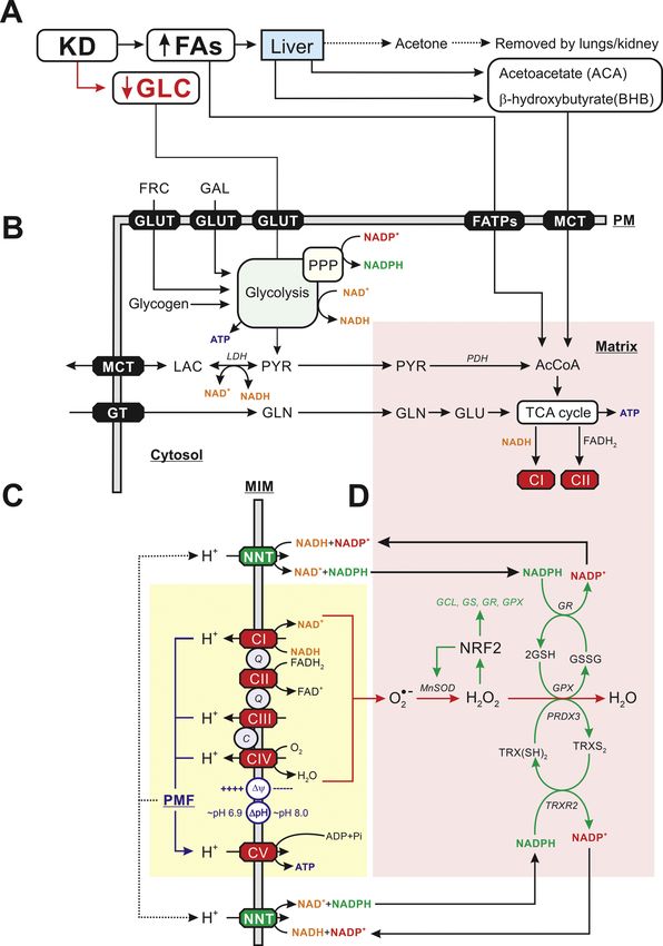
Fig. 1. Impact of the ketogenic diet on cellular
energy metabolism and redox homeostasis. (A)
The ketogenic diet (KD) contains high and low
levels of fatty acids (FAs) and glucose (GLC),
respectively. FA uptake stimulates hepatic pro
duction of acetoacetate (ACA) and β-hydrox
ybutyrate (BHB). Mitochondria in extrahepatic
cells can take up FAs, ACA and BHB to generate
acetyl coenzyme A (AcCoA) and fuel the
tricarboxylic acid (TCA) cycle. The latter gen
erates substrates for electron transport chain
(ETC) complex I (CI; NADH) and complex II
(CII; FADH2). (B) Galactose (GLC) and fructose
(FRC) enter the cell via glucose transporters
(GLUTs) after which they are converted by the
glycolysis pathway into pyruvate (PYR), ATP,
NAD+ and NADH. Glycogen also can reversibly
be converted into GLC. Glycolysis is linked to
redox metabolism (NADPH, NADP+) via the
pentose phosphate pathway (PPP). PYR is
either reversibly converted into lactate (LAC),
which is released from the cell, or enter mito
chondria to be converted into AcCoA. Alterna
tively, glutamine (GLN) can be taken up by the
cell, converted into glutamate (GLU) and enter
the TCA cycle. (C) CI and CII accept electrons
from NADH and FADH2, respectively, and
donate them to Coenzyme Q10 (“Q’’). The latter
molecule transports electrons to CIII, from
where they are conveyed to CIV by cytochrome-
c (“C’’). At CIV, the electrons are donated to
molecular oxygen (O2) to form water. During
electron transport, energy is gradually released
and used (at CI, CIII, and CIV) to expel protons
(H+) from the mitochondrial matrix across the
MIM. As a consequence, an inward-directed
trans-MIM proton-motive force (PMF) is gener
ated, consisting of an electrical (Δψ) and
chemical (ΔpH) component. The PMF is utilized
by CV to catalyze the formation of ATP from
adenosine diphosphate (ADP) and inorganic
phosphate (Pi) by allowing the controlled re-
entry of protons into the matrix. (D) Electrons
escaping from the ETC complexes can form su
peroxide (O⋅-2 ), which is subsequently converted
into hydrogen peroxide (H2O2) and water
(H2O). This involves the action of various ROS-
detoxifying systems. ROS levels can stimulate
gene expression of Nuclear factor erythroid 2-
related factor 2 (NRF2), which increases
expression of superoxide dismutase 2 (SOD2 or
MnSOD), glutamate cysteine ligase (GCL),
glutathione synthase (GS), glutathione reduc
tase (GR) and glutathione peroxidase (GPX).
This allows a more efficient removal of O⋅-2 and
H2O2. Nicotinamide nucleotide trans
hydrogenase (NNT), provides a functional link
between OXPHOS (yellow) and mitochondrial
redox homeostasis (pink) by converting
NADH + NADP+ into NAD++NADPH. See main
text for details. This figure was compiled using
information from: Branco et al., 2016; Koop
man et al., 2013; Bulthuis et al., 2019; Morris et al., 2020; Teixeira et al., 2021; Huang et al., 2006.
2
C. Qu et al. International Journal of Biochemistry and Cell Biology 138 (2021) 106050
Normally, the majority of pyruvate enters mitochondria, where it is represents the major circulating form of ketone bodies (Jensen et al.,
converted into acetyl coenzyme A (AcCoA) to fuel ATP production by 2020). In extrahepatic cells, FAs, ACA and BHB enter the mitochondrial
the integrated action of the tricarboxylic acid (TCA) cycle and the matrix, where they are converted into AcCoA that fuels the TCA cycle
oxidative phosphorylation (OXPHOS) system (see below). ATP can also (Yang et al., 2019). The KD alters gut microbiome composition (Paoli
be produced from other substrates like glutamine and FAs entering the et al., 2019) by increasing the amount of “good bacteria” (Akkermansia,
TCA cycle (Goodpaster and Sparks, 2017). Importantly, the conversion Bacteroidetes, Firmicutes, Muciniphila, Lactobacillus), which generate
of pyruvate into AcCoA by pyruvate dehydrogenase (PDH) is irreversible short-chain FAs (SCFAs) like acetate, propionate and butyrate. In par
and therefore carbohydrates can be converted into fats but not vice versa allel, the KD decreases pro-inflammatory “bad bacteria” (Desulfovibrio,
(Frayn and Evans, 2019). Action of the electron transport chain (ETC) is Turicibacter). With respect to mitochondrial energy metabolism, gut
central to mitochondrial ATP generation (Fig. 1C). The ETC consists of microbiota can release metabolites that directly affect ETC function and
five multi-subunit complexes (CI–CV) and two electron transport mol mitochondrial ATP production. For instance, hydrogen sulfide produced
ecules, Coenzyme Q10 and cytochrome-c, which are embedded in the by intestinal bacteria can inhibit mitochondrial CIV or serve as an
mitochondrial inner membrane (MIM). Collectively, the ETC catalyzes alternative ETC electron donor (Szabo et al., 2014). In addition,
electron transfer from NADH (at CI) and reduced flavin adenine dinu microbiota-produced butyrate significantly increased mitochondrial
cleotide (FADH2; at CII) via Coenzyme Q10 to CIII (Fig. 1C). From oxygen consumption during oxidative stress conditions (Clark and
thereon, cytochrome-c transports electrons to CIV where they are Mach, 2017). The KD and low-carbohydrate diet also functionally
donated to molecular oxygen (O2). ETC action is linked to ATP pro upregulate bioenergetic pathways (Miller et al., 2018): (1) the OXPHOS
duction by the FoF1-ATPase (CV) by a chemiosmotic coupling mecha system (CI, CII, CIII, CIV, CV and cytochrome-c), (2) the TCA cycle
nism (Mitchell and Moyle, 1967). This involves the ETC-mediated (citrate synthase, isocitrate dehydrogenase, malate dehydrogenase), (3)
creation of an inward-directed trans-MIM proton motive force. The FAO (carnitine palmitoyltransferase, medium-chain acyl-CoA dehydro
latter consists of a chemical (ΔpH) and electrical component (Δψ), genase/MCAD, long-chain acyl-CoA dehydrogenase/LCAD,
which drive CV-mediated proton reentry into the mitochondrial matrix very-long-chain acyl-CoA dehydrogenase/VLCAD, β-hydroxyacyl-CoA
and thereby ATP generation. Importantly, electrons can also enter the dehydrogenase/β-HAD), and (4) ketolysis (β-hydroxybutyrate dehy
ETC via alternative pathways that are often tissue-specific. These path drogenase). In a neuronal context, the MCT-KD (i.e. decanoic acid or
ways include electron donation to Coenzyme Q10 by C10) stimulated the astrocyte-neuron lactate shuttle (Augustin et al.,
glycerol-3-phosphate dehydrogenase, dihydroorotate dehydrogenase 2018). The latter mediates neuronal lactate import (produced in astro
and the electron-transferring flavoprotein (ETF)-ubiquinone oxidore cytes), to drive the formation of pyruvate for mitochondrial ATP gen
ductase (Koopman et al., 2010). Cellular ATP generation is highly eration (Magistretti and Allaman, 2018). In addition, BHB has been
flexible and can rapidly switch from mitochondrial OXPHOS- to shown to suppress microglial activation, which is a hallmark of brain
glycolysis-mediated ATP production when the former pathway is pathology (Dheen et al., 2007; Ghosh et al., 2018).
impaired (Liemburg-Apers et al., 2015, 2016). In addition to ATP gen
eration, ETC action also sustains virtually all other mitochondrial 3. The KD impacts on mitochondrial redox metabolism
functions including metabolite/ion exchange and protein import
(Bulthuis et al., 2019). Moreover, mitochondria can produce reactive Evidence was provided that the KD reduces oxidative stress, poten
oxygen species (ROS) as “by-products” of ETC action, in particular tially mediated by activation of nuclear factor erythroid-derived 2-
during pathological conditions (Murphy, 2009). These ROS can act as related factor 2 (NRF2; Yarar-Fisher et al., 2021). The latter acts as a
messenger molecules in physiological cell control and induce antioxi key controlling factor in antioxidant responses (Fig. 1D) and mito
dant signaling (Fig. 1D), but, when reaching too high levels, can induce hormesis (Wallace et al., 2010; Achanta and Rae, 2017; Teixeira et al.,
oxidative stress (Halliwell and Gutteridge, 2015). Under 2021). Alternatively, KD-fed mice displayed lower ROS levels in brain
non-pathological conditions too high ROS levels are prevented by the relative to standard diet-fed controls. Mechanistically, it is suggested
action of various interlocked antioxidant pathways including the nico that KD may diminish ROS production by increasing the expression and
tinamide nucleotide transhydrogenase (NNT), glutathione (GSH), activity of mitochondrial uncoupling proteins (UCPs; Sullivan et al.,
glutathione peroxidase (GPX) and superoxide dismutase (SOD) enzymes 2004). In a mouse model of ischemic stroke, combined ACA/BHB in
(Fig. 1D). jection increased the NAD+/NADH ratio relative to vehicle-treated
control animals (Yin et al., 2015). Relevant in the context of this re
2. The KD impacts on mitochondrial energy metabolism view, NAD+ is generated by CI and the NNT and functionally links
mitochondrial bioenergetics with ROS/redox homeostasis (Fig. 1C,D).
The ketogenic diet (KD) is a high-fat, moderate protein, and low- NAD+ is a substrate of various other enzymes including polyADP ribose
carbohydrate diet that exists in various formulations including: (1) the polymerase (PARP), cyclic ADP ribose synthetases, and Sirtuin (SIRT)
“classical” KD, (2) the “medium-chain triglyceride” (MCT-KD), and (3) deacetylases, involved in mitochondrial redox metabolism (Cantó et al.,
the less restrictive “modified Atkins diet” (mAD). For the classical KD, 2015; Katsyuba and Auwerx, 2017). Moreover, BHB treatment activates
the ratio of fat to carbohydrate and protein is 4:1 (4 g of fat for every 1 g AMP-activated protein kinase (AMPK), which is a major regulator of
of protein plus carbohydrate), which reduces carbohydrate intake cellular bioenergetics and the activity/abundance of nicotinamide
(Dhamija et al., 2013). Relative to the classical KD, the MCT-KD results phosphoribosyltransferase (NAMPT), a key enzyme in the NAD+ salvage
in a more efficient generation of ketone bodies, which provide an pathway (Bae et al., 2016; Han et al., 2016).
additional energy source for extrahepatic tissues, (Augustin et al., 2018).
The mAD mimics the classical KD but is a more palatable diet with a 1:1 4. The KD impacts on mitochondrial dynamics and mitophagy
ratio of fat to carbohydrates and protein (Olgac et al., 2020). Func
tionally, the KD mimics a metabolic state of fasting and/or caloric To maintain a healthy mitochondrial population and network
restriction/starvation, during which the body shifts from carbohydrate structure, mitochondria are motile and undergo continuous cycles of
metabolism (e.g. glucose, galactose) to fat metabolism, leading to fission and fusion (Archer, 2013). Aberrations in mitochondrial struc
enhanced FAO, gluconeogenesis and ketogenesis. These processes ture were observed in a large variety of human diseases including MD
mainly occur in the liver (Fig. 1A) and lead to production of ketone (Bulthuis et al., 2019). It was found that expression of the mitochondrial
bodies, which enter the blood stream (Morris et al., 2020). The gener fusion protein Mitofusin 2 (Mfn2) is required for adaption to a high-fat
ated ketone bodies consist of acetone (largely removed by lung/kidney diet (Liesa and Shirihai, 2013). Moreover, in vivo evidence suggests that
action), acetoacetate (ACA) and β-hydroxybutyrate (BHB). The latter the KD reduces mitochondrial fission and thereby improves
3C. Qu et al. International Journal of Biochemistry and Cell Biology 138 (2021) 106050
mitochondrial function in the heart of Type 2 diabetic mice (Guo et al., Table 1
2020). It was also demonstrated that BHB stimulates mitochondrial Effect of the ketogenic diet in selected models of mitochondrial dysfunction.
elongation in cultured cells (Santra et al., 2004) and ameliorates mito Model Treatment and effects Reference
chondrial morphology aberrations in mouse brain and muscle (Li et al.,
Patients
2018; Ahn et al., 2020). Mitochondrial dynamics is crucial for the se CASE REPORT: 7-year-old mAD with 30− 40 g/day Della Marina
lective removal of damaged mitochondria by mitochondria-specific female with Björnstad carbohydrates [1500 kcal/ et al., 2020
autophagy “mitophagy” (Kluge et al., 2013). With respect to MD, evi syndrome and BCS1L- day], 10% of energy from
dence was provided that defective mitochondria with high levels of related mitochondrial carbohydrates, 25% from
disease (BCS1L is protein and 65% from fat. Blood
mutated mtDNA can be selectively removed by mitophagy (Suen et al., implicated in CIII ketone levels of 2− 4 mM were
2010), and that the expression level of the mitophagy regulator gene biogenesis). achieved. mAD improved hair
BNIP3 is significantly increased in the liver of KD-fed mice, suggesting growth. After 4 months no
potential mitophagy activation (Newell et al., 2016). improvement of hearing. Hair
was lost again 6 months after
cessation of the diet.
5. Application of the KD in MD patients, animal models and MD CASE REPORT: 3-year-old High-fat KD in combination Deberles et al.,
patient-derived cells female with MD (mutation with antioxidant 2020
m.5559A > G in the mt- supplementation improved
Current evidence suggests that 1136 human gene products are tRNATrp gene) neurologic status and heart
parameters.
mitochondrially localized (Rath et al., 2021). A previous inventory CASE REPORT: 3-year-old Seizure frequency was reduced Di Pisa et al.,
demonstrated that mutations in 265 of these genes were linked to human boy with pyruvate and psychomotor development 2012
disease (Koopman et al., 2012), which was increased to more than 340 dehydrogenase deficiency improved.
during the last decade (Gusic and Prokisch, 2021). For the sake of CASE REPORT: 31-month- KD containing 4 parts fat:1 part Joshi et al.,
old female with Alpers- each of carbohydrate and 2009
brevity, we here will primarily focus on mutations in genes encoding
Huttenlocher syndrome protein. Clinical improvement,
mitochondrial OXPHOS subunits. Genetically, 92 different genes dramatic electroencephalogram
encoding these subunits have been described (Koopman et al., 2013). improvement
Mutations in 81 of these genes have been linked to primary OXPHOS CASE REPORT: 7-year-old Seemingly improved the Laugel et al.,
disorder (Frazier et al., 2019). In case of CI, CIII, CIV and CV, these male who presented at the oculomotor palsy but did not 2007
age of 7 months with correct other neurologic
mutations can be either nuclear DNA (nDNA)- or mitochondrial DNA progressive symptoms.
(mtDNA)-encoded, whereas for CII these mutations all originate from ophthalmoplegia and later
the nDNA. Dietary intervention is frequently used for symptomatic developed cerebellar
management of MD patients, as these diets are readily available and ataxia, spasticity, and
dystonia (NDUFV1
their (patient-specific) application diminishes the requirement for
mutation).
pharmaceutical prescription (Gorman et al., 2016). The KD was origi CASE REPORT: 3-month-old Partial control of seizures after Seo et al., 2010
nally developed in the 1920s for treating epilepsy (Kang et al., 2006). female with Ohtahara consuming classic KD with
Although limited experimental data is available, the effect of the KD has syndrome and CI mitochondrial cocktail
been evaluated in various MD and MD-related (OXPHOS deficiency) deficiency. (coenzyme Q10 5 mg/kg/day,
thiamine 5 mg/kg/day, L-
models (Table 1). Using diverse readouts, positive effects of the KD were carnitine 100 mg/kg/day,
described in case reports of human patients with Björnstad syndrome, rivoflavin 5 mg/kg/day,
MD, PDH deficiency, Alpers-Huttenlocher syndrome, progressive oph vitamin C 50 mg/kg/day,
thalmoplegia, Ohtahara syndrome and mitochondrial encephalopathy, vitamin E 200 IU/day, and
vitamin B complex)
lactic acidosis, and stroke-like episodes (MELAS). In addition, (tran
supplementation.
sient) positive effects were reported in patients with epilepsy and ETC CASE REPORT: 22-year-old A modified KD and magnesium Steriade et al.,
defects. The latter included, children with glucose transporter type 1 female with MELAS were introduced, leading to 2014
deficiency syndrome, children with myoclonic-astatic epilepsy and (m.3260A > G) seizure freedom despite
adults with mitochondrial myophathy and progressive external oph development of a new stroke-
like lesion. A decrease in
thalmoplegia (PEO). In animal models, phenotypic improvement by the frequency of stroke-like
KD was demonstrated in mice with mitochondrial myopathry, CI defi episodes was observed.
ciency, CIII deficiency and mitochondrial pyruvate carrier deficiency, as Fourteen children with KD with 4:1 lipid to nonlipid Kang et al.,
well as in flies with mitochondrial encephalomyopathy. Finally, studies epilepsy and ETC defects ratio (% by weight), but 2007
without nitial fasting and fluid
with cell models of MELAS, Leigh syndrome (LS), Kearns-Sayre syn
restriction. Seven patients
drome (KSS) and Leber’s hereditary optic neuropathy (LHON), revealed became seizure-free after
positive effects of the KD on mtDNA copy number (MELAS), CI stability commencing the classic KD, 3 of
(MELAS), citrate synthase activity (LS), FA metabolism (LS), catalase whom successfully completed
(CAT) activity, oxidative stress (LS), and mtDNA mutational load the diet without relapse.
Five adults with MM/PEO mAD induced progressive Ahola et al.,
(LHON).
muscle pain, leakage of muscle 2016
enzymes leading to premature
6. Adverse effects of the KD discontinuation of the diet.
Follow-up after 2 years suggests
activation of muscle
In addition to the potentially positive effects described in the pre
regeneration.
vious sections the KD can also display detrimental effects. For instance, Eleven children with KD induced a > 50 % reduction Caraballo et al.,
the effect of ketone bodies appears to be concentration dependent, as myoclonic-astatic epilepsy in seizures in over half of the 2006
exemplified by the observation in mice that high BHB levels can induce children.
minor damage to blood-brain barrier integrity (Orhan et al., 2016). A Six Japanese males (7–16 mAD markedly decreased Ito et al., 2011
years old) with glucose epileptic seizures and other
recent study showed that feeding KD exacerbated spongiosis and gliosis paroxysmal events in all
in a mouse model of mitochondrial spongiotic brain disease (Ignatenko
et al., 2020). Furthermore, due to the chemical properties of ketone
(continued on next page)
bodies, the KD induced blood acidosis in rats after 60 days (Arsyad et al.,
4C. Qu et al. International Journal of Biochemistry and Cell Biology 138 (2021) 106050
Table 1 (continued ) muscle enzymes. These effects lead to premature termination of the diet.
Model Treatment and effects Reference However, follow-up analysis after 2 years suggested that muscle
regeneration occurred in the MM/PEO patients (Ahola et al., 2016).
transporter type 1 patients. In addition, an
deficiency syndrome improvement in motivation and
cognitive function was 7. Summary and future perspectives
observed.
Animal models Evidence obtained in various cellular and organismal models, sup
Mouse with MM An ad libitum KD slowed down Ahola-Erkkilä
ports the conclusion that the KD affects mitochondrial bioenergetics,
disease progression. et al., 2010
Harlequin mouse with CI High fat diet improved Schiff et al., mitochondrial ROS/redox metabolism, and mitochondrial dynamics.
deficiency neurodegenerative symptoms. 2011 Previous analysis of LS patient cells suggests that several of these pa
Mice with CIII deficiency Classic KD delayed liver fibrosis Purhonen et al., rameters are disturbed in MD and therefore might constitute potential
(Bcs1l mutant) and inhibits stellate cell 2017 targets for intervention (Koopman et al., 2012, 2013; Koopman et al.,
activation and hepatic
progenitor cell response.
2016). However, the information in the previous section illustrates that
Drosophila model of KD reduced seizure recovery Fogle et al., our current understanding of the bioenergetic consequences and
mitochondrial time and severity of seizure 2016 mechanistic aspects of the KD in MD patients is still insufficient. As a first
encephalomyopathy phenotype. step towards addressing this problem, MD patient-derived cells consti
Mice with mitochondrial Progressively developing McCommis
tute an easily accessible model system that can be analyzed with respect
pyruvate carrier deficiency cardiac dilation and contractile et al., 2020
dysfunction was completely to the above and additional parameters (e.g., oxygen consumption,
reversed. ROS/redox state, mitochondrial dynamics, cell viability). To allow
proper analysis of these cell models, it is crucial to apply KD-mimicking
Cell models culture media with respect to incubation time, refreshment regimen,
SH-SY5Y cybrid cells DMEM medium containing: Frey et al., 2017
substrate concentrations (e.g., glucose, galactose, pyruvate, glutamine,
modeling MELAS “low” glucose (0.5 g/L), 1%
(m.3260A > G; 98.6 % glutamine, 50 μg/ml uridine,
lactate, FAs), and FA and/or ketone body composition (e.g., C8, C10,
mutant load). 5 mM ACA and 5 mM BHB. BHB). This requires quantitative information on glucose, FA, and BHB
mtDNA copy number was blood levels in human subjects during KD treatment. The serum level of
increased but the mutant load fibroblast growth factor 21 (FGF21) has been proposed as a biomarker of
was not changed (no
muscle-manifesting mitochondrial respiratory chain deficiency (Suo
heteroplasmy shift) after
treatment with ketone bodies. malainen et al., 2011), as well as mitochondrial translation and mtDNA
Primary fibroblasts from six DMEM medium containing Kanabus et al., maintenance disorders (Lehtonen et al., 2016). Importantly, FGF21 is a
LS patients (NDUFV1, 25 mM glucose, 1 mM 2016 regulator of energy homeostasis (Fisher and Maratos-Flier, 2016) and
NDUFV2, NDUFS3, pyruvate, 4 mM glutamine and promotes ketone body utilization in neurons through AMPK activation
NDUFS4 mutation). 50 mg/l uridine, to which was
Rotenone treatment (CI added: 250 μM C10, 5 mM BHB
(Katsu-Jiménez and Giménez-Cassina, 2019). Though still controversial
inhibitor) was also used. or 5 mM ACA. 6 days treatment. (Badakhsi and Jin, 2020), FGF21 might constitute a biomarker in MD
C10 increased CS activity in 50 patients to evaluate KD effects. In addition, analysis of serum acylcar
% of patients in a PPAR- nitine and amino acid profiles at different time point during KD treat
γ-mediated manner. C10
ment could be useful to predict the effectiveness of KD (Hung et al.,
increased FA metabolism, CAT
activity and decreased 2021). We conclude that the potential benefits of the KD were observed
oxidative stress. Not all cell under specific conditions in various models. This suggests that different
lines responded to C10. mitochondrial diseases might require different ketogenic diets. There
Cybrid cell lines modeling Treatment with ketone bodies Emperador fore, integrated efforts at the clinical and mechanistic level are required
LHON reduced the percentage of the et al., 2019
m.13094 T > C heteroplasmic
to better understand KD mode-of-action.
mutation. This treatment also
increased the mtDNA levels of Declaration of Competing Interest
the m.11778 G > A
mitochondrial genotype.
WJHK is a scientific advisor of Khondrion B.V. (Nijmegen, The
A cloned heteroplasmic cell Treatment with KD for 5 days Santra et al.,
line modeling increased the proportion of 2004 Netherlands). This company was not involved in the writing of the
Kearns–Sayre syndrome wild-type mtDNA from 13 % to manuscript nor in the decision to submit the manuscript for publication.
~22 % and improved
mitochondrial protein Acknowledgements
synthesis.
The project of Dr. Qu at the Radboud University Medical Center is
2020). In this context, too high ketone body plasma concentrations were supported by The International Exchange Fellowship Program of the Sun
associated with adverse outcomes, whereas lower levels were beneficial Yat-sen University, China, the “Sanming Project of Medicine” in
(Nasser et al., 2020). Moreover, gastrointestinal disorders, such as Shenzhen to Y. Pan (No. SZSM201911003), and the Research Start-up
diarrhea, were also frequently observed (Kang et al., 2004). Although it Fund of the Seventh Affiliated Hospital of Sun Yat-sen University to C.
is unclear whether application of the KD directly increases the incidence Qu (ZSQYRSFPD0028). We apologize for not including various studies
of cardiovascular disease, a 25-year follow-up study in a large cohort, due to space limitations.
suggested that a low-carbohydrate diet is associated with increased
mortality (Nasser et al., 2020). The KD was also demonstrated to in References
crease cardiac fibrosis potentially by BHB-induced effects on the Sirtuin
Achanta, L.B., Rae, C.D., 2017. β-Hydroxybutyrate in the brain: One Molecule, multiple
7 promotor (Xu et al., 2021). Moreover, KD treatment was associated
mechanisms. Neurochem. Res. 42 (1), 35–49.
with mild carnitine depletion (Berry-Kravis et al., 2001), progressive Ahn, Y., Sabouny, R., Villa, B.R., Yee, N.C., Mychasiuk, R., Uddin, G.M., et al., 2020.
bone mineral content loss (Bergqvist et al., 2008) and kidney stone Aberrant mitochondrial morphology and function in the BTBR mouse model of
autism is improved by two weeks of ketogenic diet. Int. J. Mol. Sci. 21 (9), 3266.
formation (Kielb et al., 2000). In case of MD, treatment of MM/PEO
patients with the mAD induced progressive muscle pain and leakage of
5C. Qu et al. International Journal of Biochemistry and Cell Biology 138 (2021) 106050
Ahola, S., Auranen, M., Isohanni, P., Niemisalo, S., Urho, N., Buzkova, J., et al., 2016. Huang, T.-T., Naeemuddin, M., Elchuri, S., Yamaguchi, M., Kozy, H.M., Carlson, E.J.,
Modified Atkins diet induces subacute selective ragged-red-fiber lysis in et al., 2006. Genetic modifiers of the phenotype of mice deficient in mitochondrial
mitochondrial myopathy patients. EMBO Mol. Med. 8 (11), 1234–1247. superoxide dismutase. Hum. Mol. Genet. 15 (7), 1187–1194.
Ahola-Erkkilä, S., Carroll, C.J., Peltola-Mjösund, K., Tulkki, V., Mattila, I., Seppänen- Hung, P.L., Lin, J.L., Chen, C., Hung, K.Y., Hsieh, T.Y., Hsu, M.H., et al., 2021. An
Laakso, T., Oresic, M., et al., 2010. Ketogenic diet slows down mitochondrial examination of serum acylcarnitine and amino acid profiles at different time point of
myopathy progression in mice. Hum. Mol. Genet. 19 (10), 1974–1984. ketogenic diet therapy and their association of ketogenic diet effectiveness. Nutrients
Archer, S.L., 2013. Mitochondrial dynamics - mitochondrial fission and fusion in human 13 (1), 21.
diseases. N. Engl. J. Med. 369 (23), 2236–2251. Iannetti, E.F., Smeitink, J.A.M., Willems, P., Beyrath, J., Koopman, W.J.H., 2018. Rescue
Arsyad, A., Idris, I., Rasyid, A.A., Usman, R.A., Faradillah, K.R., Latif, W.O.U., et al., from galactose-induced death of Leigh Syndrome patient cells by pyruvate and NAD
2020. Long-term ketogenic diet induces metabolic acidosis, anemia, and oxidative +
. Cell Death Dis. 9 (11), 1135.
stress in healthy wistar rats. J. Nutr. Metab. 2020, 3642035. Ignatenko, O., Nikkanen, J., Kononov, A., Zamboni, N., Ince-Dunn, G., Suomalainen, A.,
Augustin, K., Khabbush, A., Williams, S., Eaton, S., Orford, M., Cross, J.H., et al., 2018. 2020. Mitochondrial spongiotic brain disease: astrocytic stress and harmful
Mechanisms of action for the medium-chain triglyceride ketogenic diet in rapamycin and ketosis effect. Life Sci. Alliance 3 (9) e202000797.
neurological and metabolic disorders. Lancet Neurol. 17 (1), 84–93. Ito, Y., Oguni, H., Ito, S., Oguni, M., Osawa, M., 2011. A modified Atkins diet is
Badakhsi, Y., Jin, T., 2020. Current understanding and controversies on the clinical promising as a treatment for glucose transporter type 1 deficiency syndrome. Dev.
implications of fibroblast growth factor 21. Crit. Rev. Lab Sci. 31, 1–30. Med. Child Neurol. 53 (7), 658–663.
Bae, H.R., Kim, D.H., Park, M.H., Lee, B., Kim, M.J., Lee, E.K., et al., 2016. Jensen, N.J., Wodschow, H.Z., Nilsson, M., Rungby, J., 2020. Effects of ketone bodies on
β-Hydroxybutyrate suppresses inflammasome formation by ameliorating brain metabolism and function in neurodegenerative diseases. Int. J. Mol. Sci. 21
endoplasmic reticulum stress via AMPK activation. Oncotarget 7 (41), 66444–66454. (22), 8767.
Bergqvist, A.G., Schall, J.I., Stallings, V.A., Zemel, B.S., 2008. Progressive bone mineral Joshi, C.N., Greenberg, C.R., Mhanni, A.A., Salman, M.S., 2009. Ketogenic diet in Alpers-
content loss in children with intractable epilepsy treated with the ketogenic diet. Huttenlocher syndrome. Pediatr. Neurol. 40 (4), 314–316.
Am. J. Clin. Nutr. 88 (6), 1678–1684. Kanabus, M., Fassone, E., Hughes, S.D., Bilooei, S.F., Rutherford, T., Donnell, M.O., et al.,
Berry-Kravis, E., Booth, G., Sanchez, A.C., Woodbury-Kolb, J., 2001. Carnitine levels and 2016. The pleiotropic effects of decanoic acid treatment on mitochondrial function
the ketogenic diet. Epilepsia 42 (11), 1445–1451. in fibroblasts from patients with complex I deficient Leigh syndrome. J. Inherit.
Branco, A.F., Ferreira, A., Simões, R.F., Magalhães-Novais, S., Zehowski, C., Cope, E., Metab. Dis. 39 (3), 415–426.
Silva, et al., 2016. Ketogenic diets: from cancer to mitochondrial diseases and Kang, H.C., Chung, D.E., Kim, D.W., Kim, H.D., 2004. Early- and late-onset complications
beyond. Eur. J. Clin. Invest. 46 (3), 285–298. of the ketogenic diet for intractable epilepsy. Epilepsia 45 (9), 1116–1123.
Bulthuis, E.P., Adjobo-Hermans, M.J.W., Willems, P., Koopman, W.J.H., 2019. Kang, H.C., Kim, H.D., Lee, Y.M., Han, S.H., 2006. Landau-Kleffner syndrome with
Mitochondrial morphofunction in mammalian Cells. Antioxid. Redox Signal. 30 (18), mitochondrial respiratory chain-complex I deficiency. Pediatr. Neurol. 35 (2),
2066–2109. 158–161.
Cantó, C., Menzies, K.J., Auwerx, J., 2015. NAD+ Metabolism and the control of energy Kang, H.C., Lee, Y.M., Kim, H.D., Lee, J.S., Slama, A., 2007. Safe and effective use of the
homeostasis: a balancing act between mitochondria and the nucleus. Cell Metab. 22 ketogenic diet in children with epilepsy and mitochondrial respiratory chain
(1), 31–53. complex defects. Epilepsia 48 (1), 82–88.
Caraballo, R.H., Cersósimo, R.O., Sakr, D., Cresta, A., Escobal, N., Fejerman, N., 2006. Katsu-Jiménez, Y., Giménez-Cassina, A., 2019. Fibroblast growth Factor-21 promotes
Ketogenic diet in patients with myoclonic-astatic epilepsy. Epileptic Disord. 8 (2), ketone body utilization in neurons through activation of AMP-dependent kinase.
151–155. Mol. Cell. Neurosci. 101, 103415.
Clark, A., Mach, N., 2017. The crosstalk between the gut microbiota and mitochondria Katsyuba, E., Auwerx, J., 2017. Modulating NAD+ metabolism, from bench to bedside.
during exercise. Front. Physiol. 8, 319. EMBO J. 36 (18), 2670–2683.
Deberles, E., Maragnes, P., Penniello-Valette, M.J., Allouche, S., Joubert, M., 2020. Kielb, S., Koo, H.P., Bloom, D.A., Faerber, G.J., 2000. Nephrolithiasis associated with the
Reversal of cardiac hypertrophy with a ketogenic diet in a child with mitochondrial ketogenic diet. J. Urol. 164 (2), 464–466.
disease and hypertrophic cardiomyopathy. Can. J. Cardiol. 36 (10), 1690. Kluge, M.A., Fetterman, J.L., Vita, J.A., 2013. Mitochondria and endothelial function.
e1691–1690.e1693. Circ. Res. 112 (8), 1171–1188.
Della Marina, A., Leiendecker, B., Roesch, S., Wortmann, S.B., 2020. Ketogenic diet for Koopman, W.J.H., Nijtmans, L.G., Dieteren, C.E., Roestenberg, P., Valsecchi, F.,
treating alopecia in BCS1l-related mitochondrial disease (Bjornstad syndrome). Smeitink, J.A., et al., 2010. Mammalian mitochondrial complex I: biogenesis,
JIMD Rep. 53 (1), 10–11. regulation, and reactive oxygen species generation. Antioxid. Redox Signal. 12 (12),
Dhamija, R., Eckert, S., Wirrell, E., 2013. Ketogenic diet. Can. J. Neurol. Sci. 40 (2), 1431–1470.
158–167. Koopman, W.J.H., Willems, P.H.G.M., Smeitink, J.A.M., 2012. Monogenic mitochondiral
Dheen, S.T., Kaur, C., Ling, E.A., 2007. Microglial activation and its implications in the disorders. N. Engl. J. Med. 366 (12), 1132–1141.
brain diseases. Curr. Med. Chem. 14 (11), 1189–1197. Koopman, W.J.H., Distelmaier, F., Smeitink, J.A., Willems, P.H., 2013. OXPHOS
Di Pisa, V., Cecconi, I., Gentile, V., Di Pietro, E., Marchiani, V., Verrotti, A., et al., 2012. mutations and neurodegeneration. EMBO J. 32 (1), 9–29.
Case report of pyruvate dehydrogenase deficiency with unusual increase of fats Koopman, W.J.H., Beyrath, J., Fung, C.W., Koene, S., Rodenburg, R.J., Willems, P.H.,
during ketogenic diet treatment. J. Child Neurol. 27 (12), 1593–1596. et al., 2016. Mitochondrial disorders in children: toward development of small-
Emperador, S., López-Gallardo, E., Hernández-Ainsa, C., Habbane, M., Montoya, J., molecule treatment strategies. EMBO Mol. Med. 8 (4), 311–327.
Bayona-Bafaluy, M.P., et al., 2019. Ketogenic treatment reduces the percentage of a Laugel, V., This-Bernd, V., Cormier-Daire, V., Speeg-Schatz, C., de Saint-Martin, A.,
LHON heteroplasmic mutation and increases mtDNA amount of a LHON Fischbach, M., 2007. Early-onset ophthalmoplegia in Leigh-like syndrome due to
homoplasmic mutation. Orphanet J. Rare Dis. 14 (1), 1–6. NDUFV1 mutations. Pediatr. Neurol. 36 (1), 54–57.
Fisher, F.M., Maratos-Flier, E., 2016. Understanding the physiology of FGF21. Annu. Rev. Lehtonen, J.M., Forsström, S., Bottani, E., Viscomi, C., Baris, O.R., Isoniemi, H., et al.,
Physiol. 78, 223–241. 2016. FGF21 is a biomarker for mitochondrial translation and mtDNA maintenance
Fogle, K.J., Herzler, J.I., Shon, J.H., Palladino, M.J., 2016. The ATP-sensitive K channel disorders. Neurology 87 (22), 2290–2299.
is seizure protective and required for effective dietary therapy in a model of Li, J., Kanasaki, M., Xu, L., Kitada, M., Nagao, K., Adachi, Y., et al., 2018. A ketogenic
mitochondrial encephalomyopathy. J. Neurogenet. 30 (3), 247–258. amino acid rich diet benefits mitochondrial homeostasis by altering the AKT/4EBP1
Frayn, K.N., Evans, R., 2019. Human Metabolism: a Regulatory Perspective, 4th ed. and autophagy signaling pathways in the gastrocnemius and soleus. Biochim.
Wiley-Blackwell, USA. Biophys. Acta Gen. Subj. 1862 (7), 1547–1555.
Frazier, A.E., Thorburn, D.R., Compton, A.G., 2019. Mitochondrial energy generation Liemburg-Apers, D.C., Schirris, T.J., Russel, F.G., Willems, P.H., Koopman, W.J., 2015.
disorders: genes, mechanisms, and clues to pathology. J. Biol. Chem. 294 (14), Mitoenergetic dysfunction triggers a rapid compensatory increase in steady-state
5386–5395. glucose flux. Biophys. J. 109 (7), 1372–1386.
Frey, S., Geffroy, G., Desquiret-Dumas, V., Gueguen, N., Bris, C., Belal, S., et al., 2017. Liemburg-Apers, D.C., Wagenaars, J.A., Smeitink, J.A., Willems, P.H., Koopman, W.J.,
The addition of ketone bodies alleviates mitochondrial dysfunction by restoring 2016. Acute stimulation of glucose influx upon mitoenergetic dysfunction requires
complex I assembly in a MELAS cellular model. Biochim. Biophys. Acta. Mol. Bas. LKB1, AMPK, Sirt2 and mTOR-RAPTOR. J. Cell. Sci. 129 (23), 4411–4423.
Dis. 1863 (1), 284–291. Liesa, M., Shirihai, O.S., 2013. Mitochondrial dynamics in the regulation of nutrient
Ghosh, S., Castillo, E., Frias, E.S., Swanson, R.A., 2018. Bioenergetic regulation of utilization and energy expenditure. Cell Metab. 17 (4), 491–506.
microglia. Glia 66 (6), 1200–1212. Magistretti, P.J., Allaman, I., 2018. Lactate in the brain: from metabolic end-product to
Goodpaster, B.H., Sparks, L.M., 2017. Metabolic flexibility in health and disease. Cell signalling molecule. Nat. Rev. Neurosci. 19 (4), 235–249.
Metab. 25 (5), 1027–1036. McCommis, K.S., Kovacs, A., Weinheimer, C.J., Shew, T.M., Koves, T.R., Ilkayeva, O.R.,
Gorman, G.S., Chinnery, P.F., DiMauro, S., Hirano, M., Koga, Y., McFarland, R., et al., et al., 2020. Nutritional modulation of heart failure in mitochondrial pyruvate
2016. Mitochondrial diseases. Nat. Rev. Dis. Prim. 2, 16080. carrier-deficient mice. Nat. Metab. 2 (11), 1232–1247.
Guo, Y., Zhang, C., Shang, F.F., Luo, M., You, Y., Zhai, Q., et al., 2020. Ketogenic diet Miller, V.J., Villamena, F.A., Volek, J.S., 2018. Nutritional ketosis and mitohormesis:
ameliorates cardiac dysfunction via balancing mitochondrial dynamics and potential implications for mitochondrial function and human health. J. Nutr. Metab.
inhibiting apoptosis in Type 2 diabetic mice. Aging Dis. 11 (2), 229–240. 2018, 5157645.
Gusic, M., Prokisch, H., 2021. Genetic basis of mitochondrial diseases. FEBS Lett. 595 (8), Mitchell, P., Moyle, J., 1967. Chemiosmotic hypothesis of oxidative phosphorylation.
1132–1158. Nature 213 (5072), 137–139.
Halliwell, B., Gutteridge, J.M., 2015. Free Radicals in Biology and Medicine. Oxford Morris, G., Maes, M., Berk, M., Carvalho, A.F., Puri, B.K., 2020. Nutritional ketosis as an
University Press, USA. intervention to relieve astrogliosis: possible therapeutic applications in the treatment
Han, X., Tai, H., Wang, X., Wang, Z., Zhou, J., Wei, X., et al., 2016. AMPK activation of neurodegenerative and neuroprogressive disorders. Eur. Psych. 63 (1), e8.
protects cells from oxidative stress-induced senescence via autophagic flux Murphy, M.P., 2009. How mitochondria produce reactive oxygen species. Biochem. J.
restoration and intracellular NAD+ elevation. Aging Cell 15 (3), 416–427. 417 (1), 1–13.
6C. Qu et al. International Journal of Biochemistry and Cell Biology 138 (2021) 106050
Nasser, S., Vialichka, V., Biesiekierska, M., Balcerczyk, A., Pirola, L., 2020. Effects of Suen, D.F., Narendra, D.P., Tanaka, A., Manfredi, G., Youle, R.J., 2010. Parkin
ketogenic diet and ketone bodies on the cardiovascular system: concentration overexpression selects against a deleterious mtDNA mutation in heteroplasmic
matters. World J. Diabet. 11 (12), 584–595. cybrid cells. Proc. Natl. Acad. Sci. U.S.A. 107 (26), 11835–11840.
Newell, C., Shutt, T.E., Ahn, Y., Hittel, D., Khan, A., Rho, J.M., et al., 2016. Tissue Sullivan, P.G., Rippy, N.A., Dorenbos, K., Concepcion, R.C., Agarwal, A.K., Rho, J.M.,
specific impacts of a ketogenic diet on mitochondrial dynamics in the BTBRT+tf/j 2004. The ketogenic diet increases mitochondrial uncoupling protein levels and
mouse. Front. Physiol. 7, 654. activity. Ann. Neurol. 55 (4), 576–580.
Olgac, A., İnci, A., Okur, İ., Biberoğlu, G., Oğuz, D., Ezgü, F.S., et al., 2020. Beneficial Suomalainen, A., Elo, J.M., Pietiläinen, K.H., Hakonen, A.H., Sevastianova, K.,
effects of modified Atkins diet in glycogen storage disease type IIIa. Ann. Nutr. Korpela, M., et al., 2011. FGF-21 as a biomarker for muscle-manifesting
Metab. 76 (4), 233–241. mitochondrial respiratory chain deficiencies: a diagnostic study. Lancet Neurol. 10
Orhan, N., Ugur Yilmaz, C., Ekizoglu, O., Ahishali, B., Kucuk, M., Arican, N., et al., 2016. (9), 806–818.
Effects of beta-hydroxybutyrate on brain vascular permeability in rats with Szabo, C., Ransy, C., Módis, K., Andriamihaja, M., Murghes, B., Coletta, C., et al., 2014.
traumatic brain injury. Brain Res. 1631, 113–126. Regulation of mitochondrial bioenergetic function by hydrogen sulfide. Part I.
Paoli, A., Mancin, L., Bianco, A., Thomas, E., Mota, J.F., Piccini, F., 2019. Ketogenic diet Biochemical and physiological mechanisms. Br. J. Pharmacol. 171 (8), 2099–2122.
and microbiota: Friends or enemies? Genes (Basel) 10 (7), 534. Teixeira, J., Basit, F., Willems, P., Wagenaars, J.A., van de Westerlo, E., Amorim, R.,
Purhonen, J., Rajendran, J., Mörgelin, M., Uusi-Rauva, K., Katayama, S., Krjutskov, K., et al., 2021. Mitochondria-targeted phenolic antioxidants induce ROS-protective
et al., 2017. Ketogenic diet attenuates hepatopathy in mouse model of respiratory pathways in primary human skin fibroblasts. Free Rad. Biol. Med. 163, 314–324.
chain complex III deficiency caused by a Bcs1l mutation. Sci. Rep. 7 (1), 957. Wallace, D.C., Fan, W., Procaccio, V., 2010. Mitochondrial energetics and therapeutics.
Rath, S., Sharma, R., Gupta, R., Ast, T., Chan, C., Durham, T.J., et al., 2021. Annu. Rev. Pathol. Mech. Dis. 5, 297–348.
MitoCarta3.0: an updated mitochondrial proteome now with sub-organelle Weinberg, S.E., Sena, L.A., Chandel, N.S., 2015. Mitochondria in the regulation of innate
localization and pathway annotations. Nucl. Acids Res. 49 (D1), D1541–d1547. and adaptive immunity. Immunity 42 (3), 406–417.
Santra, S., Gilkerson, R.W., Davidson, M., Schon, E.A., 2004. Ketogenic treatment Xu, S., Tao, H., Cao, W., Cao, L., Lin, Y., Zhao, S.M., et al., 2021. Ketogenic diets inhibit
reduces deleted mitochondrial DNAs in cultured human cells. Ann. Neurol. 56 (5), mitochondrial biogenesis and induce cardiac fibrosis. Signal Trans. Targ. Therap. 6
662–669. (1), 54.
Schiff, M., Bénit, P., El-Khoury, R., Schlemmer, D., Benoist, J.F., Rustin, P., 2011. Mouse Yang, H., Shan, W., Zhu, F., Wu, J., Wang, Q., 2019. Ketone bodies in neurological
studies to shape clinical trials for mitochondrial diseases: high fat diet in Harlequin diseases: focus on neuroprotection and underlying mechanisms. Front. Neurol. 10,
mice. PLoS One 6 (12), e28823. 585.
Seo, J.H., Lee, Y.M., Lee, J.S., Kim, S.H., Kim, H.D., 2010. A case of Ohtahara syndrome Yarar-Fisher, C., Li, J., Womack, E.D., Alharbi, A., Seira, O., Kolehmainen, K.L., et al.,
with mitochondrial respiratory chain complex I deficiency. Brain Dev. 32 (3), 2021. Ketogenic regimens for acute neurotraumatic events. Curr. Opin. Biotechnol.
253–257. 70, 68–74.
Steriade, C., Andrade, D.M., Faghfoury, H., Tarnopolsky, M.A., Tai, P., 2014. Yin, J., Han, P., Tang, Z., Liu, Q., Shi, J., 2015. Sirtuin 3 mediates neuroprotection of
Mitochondrial encephalopathy with lactic acidosis and stroke-like episodes (MELAS) ketones against ischemic stroke. J. Cereb. Blood Flow Metab. 35 (11), 1783–1789.
may respond to adjunctive ketogenic diet. Pediatr. Neurol. 50 (5), 498–502.
7You can also read