Extraction of short chain chitooligosaccharides from fungal biomass and their use as promoters of arbuscular mycorrhizal symbiosis
←
→
Page content transcription
If your browser does not render page correctly, please read the page content below
www.nature.com/scientificreports
OPEN Extraction of short chain
chitooligosaccharides from fungal
biomass and their use as promoters
of arbuscular mycorrhizal symbiosis
Andrea Crosino1, Elisa Moscato1, Marco Blangetti2, Gennaro Carotenuto1, Federica Spina1,
Simone Bordignon2, Virginie Puech‑Pagès3, Laura Anfossi2, Veronica Volpe1, Cristina Prandi2,
Roberto Gobetto2, Giovanna Cristina Varese1 & Andrea Genre1*
Short chain chitooligosaccharides (COs) are chitin derivative molecules involved in plant-fungus
signaling during arbuscular mycorrhizal (AM) interactions. In host plants, COs activate a symbiotic
signalling pathway that regulates AM-related gene expression. Furthermore, exogenous CO
application was shown to promote AM establishment, with a major interest for agricultural
applications of AM fungi as biofertilizers. Currently, the main source of commercial COs is from the
shrimp processing industry, but purification costs and environmental concerns limit the convenience
of this approach. In an attempt to find a low cost and low impact alternative, this work aimed to
isolate, characterize and test the bioactivity of COs from selected strains of phylogenetically distant
filamentous fungi: Pleurotus ostreatus, Cunninghamella bertholletiae and Trichoderma viride. Our
optimized protocol successfully isolated short chain COs from lyophilized fungal biomass. Fungal COs
were more acetylated and displayed a higher biological activity compared to shrimp-derived COs, a
feature that—alongside low production costs—opens promising perspectives for the large scale use of
COs in agriculture.
Agriculture is experiencing an urgent need to shift toward low-input practices that can be integrated with the
environment and aim at food safety for the growing human population. Arbuscular mycorrhizal (AM) fungi can
play a key role in this scenario4. AM is the most widespread plant symbiosis, in terms of geographical distribu-
tion and phylogenetic coverage in the plant k ingdom1. In this mutualistic symbiosis, part of the plant sugars and
lipids are fed to the f ungus2, while soil mineral nutrients and water are scavenged by the extraradical mycelium
and transferred to the plant, improving its fitness1.
AM symbiosis plays a central ecological role in the functioning of natural ecosystems, but most crop plants
establish this beneficial interaction t oo4. In this context, the potential use of AM fungi in sustainable produc-
tion under low chemical input conditions is currently raising a broad interest in agronomic context3. Promot-
ing and maintaining functional and persistent mycorrhizal symbioses in crop fields to improve soil quality and
productivity requires proper management of agroecosystems with strategies that include shallow tillage, low
fertilizer input, perennial crop development, as well as AM promotion with fungal inoculation and treatments
that stimulate symbiosis e stablishment4,5.
Plants interact with a multitude of microbes and their ability to recognize them and deploy appropriate
responses is largely based on the recognition of microbe-specific molecules known as microbe-associated molecu-
lar patterns6. Among them, long-chain oligomers of chitin—which is the main fibrillar component of fungal cell
wall—are strong elicitors of plant defense responses7–9. The perception of long-chain chito-oligomers such as
chitooctaose (composed of 8 N-acetyl-glucosamine residues) activates powerful defense strategies of the plant,
such as the release of chitinases, phytoalexins, reactive oxygen species and callose deposition in the cell w all10.
Other chitin-related molecules are known to play a signalling role in symbiosis as so-called mycorrhizal
factors, or Myc-factors. Among them, tetra/penta-chito-oligosaccharides (CO4-5) activate symbiotic signalling
in all tested dicot and monocot AM h osts11,12. The plant signal transduction pathway mediating Myc-factor
1
Department of Life Science and Systems Biology, University of Turin, 10125 Turin, Italy. 2Department of
Chemistry, University of Turin, 10125 Turin, Italy. 3Laboratoire de Recherche en Sciences Végétales, Université de
Toulouse, CNRS, UPS, 31320 Castanet‑Tolosan, France. *email: andrea.genre@unito.it
Scientific Reports | (2021) 11:3798 | https://doi.org/10.1038/s41598-021-83299-6 1
Vol.:(0123456789)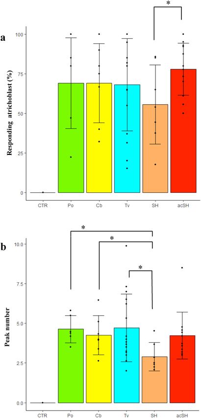
www.nature.com/scientificreports/
Chitin Chitosan COs
g % g % g %
Pleurotus ostreatus 3.7 24.7 0.006 0.04 0.202 6.70
Cunninghamella bertholletiae 1.2 8.30 0.027 0.18 0.362 29.20
Trichoderma viride 2.6 17.30 0.039 0.26 0.042 1.70
Table 1. Chitin, chitosan and CO yield from each of the three fungi. Yields are expressed as both dry mass
(g) and percentage values (%) of the starting fungal dry biomass (for chitin and chitosan) or the starting chitin
amount (for COs).
recognition includes the activation of intense oscillations in Ca2+ concentration in the nuclei of root epidermal
cells13, which is now commonly used as a hallmark of Myc-factor perception13,14.
Myc-factors activate plant symbiotic responses ranging from gene regulation to metabolic and develop-
mental changes15,16 that have been defined as part of an anticipation program preparing the host to a successful
association17. Furthermore, the application of exogenous COs was recently demonstrated to stimulate lateral root
development and branching in Oryza sativa13 and AM establishment in the model legume Medicago truncatula4,
paving the way to possible use of CO treatments to promote AM in agricultural applications.
Currently, commercial COs of different length are obtained from fishing waste processing industries, mainly
through hydrolysis of shrimp shell-derived chitin and c hitosan18,19. Seasonal availability, environmental concerns
and purification costs are the main drawbacks of the use of shrimp-derived COs in large-scale applications. An
alternative, more sustainable and cheaper source of COs are fungal biomass wastes from fermentation industries.
In fact, in spite of having lower chitin content than crustaceans (10–26% as a chitin-glucan complex), fungal
biomass does not need aggressive acid treatments—normally required for the removal of calcium carbonate and
other minerals from crustacean shells—prior to CO extraction. Furthermore fungal biomass production is not
subject to seasonal and regional limitations19.
Here we present a protocol (see Supplementary Fig. S1 online) to efficiently extract COs from fungal biomass
of different origin (Pleurotus ostreatus, Cunninghamella bertholletiae and Trichoderma viride); we confirm the
chemical properties of the extracted COs and demonstrate their stronger biological activity as promoters of AM
symbiosis when compared with commercial COs.
Results
Extraction yields. When grown on a standard medium (liquid malt extract), P. ostreatus provided the high-
est amount of mycelium with a yield of 10 g/L. The quantity of products obtained after the extraction of chitosan
and chitin and after chitin acid hydrolysis are shown in Table 1. The amount of chitin extracted varied among
species, with P. ostreatus biomass displaying the highest yield in chitin (24.7%). All tested fungi presented a low
yield in chitosan, ranging between 0.26 and 0.04% of the starting biomass.
Due to the very limited amount of extracted chitosan, only chitin was treated by acid hydrolysis for CO
production. The yield in putative COs was 6.70% of the starting chitin amount for P. ostreatus, 29.20% for C.
bertholletiae and 1.70% for T. viride.
Altogether, putative CO yield resulted to be 1.65% of the original mycelium dry weight for P. ostreatus, 3.69%
for C. bertholletiae and 0.30% for T. viride (Table 1). By combining growth speed, biomass, chitin and CO yield
and lower CO polymerization (max CO6), we decided to use COs from P. ostreatus, which is also considered as
a GRAS (Generally Recognized As Safe) organism, for all subsequent pot culture analyses.
Extracted samples contain COs of the expected size and acetylation degree. Direct infusion
mass spectrometry (DIMS) indicated that the extracted samples contained acetylated and de-acetylated COs
with length comprised between 2 and 7 residues (see Supplementary Fig. S2 online). Pseudo-molecular ions
[M + H+] of COs composed of 2 to 7 N-acetyl-glucosamine residues were detected, while double-charged ions
and longer chains, if existing, were below the limit of detection of the system.
High-performance liquid chromatography-Mass spectometry (HPLC–MS/MS) analysis of the CO mixture
(see Supplementary Fig. S3 online) confirmed that our extracted samples contained fully acetylated, mono-
deacetylated and di-deacetylated CO molecules composed of 2 to 5 N-acetyl-glucosamine residues.
Solid-State Nuclear Magnetic Resonance (13C CPMAS SSNMR) was used to characterize the materials under
study, while solution 1H NMR was applied for the determination of percent acetylation for each CO sample20,21.
Figure 1 shows the 13C CPMAS SSNMR spectra of COs from C. bertholletiae, P. ostreatus, T. viride and shrimps.
Both C. bertholletiae and shrimp CO spectra exhibited wider and less sharp spikes than those present in P. ostrea-
tus and T. viride spectra. These differences indicated that C. bertholletiae and shrimp COs were composed of a
heterogeneous mix of COs containing a different number of N-acetyl-d-glucosamine units, whereas P. ostreatus
and T. viride COs appeared to have a more uniform composition in terms of molecule length. C. bertholletiae
COs and shrimp shell COs only differed in the acetylation degree, which resulted to be higher in C. bertholletiae
COs. Overall, SSNMR spectra, albeit intrinsically not quantitative, suggested rather diverse degrees of acetylation
for the four samples, with the lowest acetyl peaks for shrimp-derived and the tallest for T. viride-derived COs.
This prompted us to investigate CO acetylation in more detail through solution 1H NMR analyses. To this aim
we also prepared a batch of peracetylated shrimp COs by increasing their acetylation degree with a treatment in
Scientific Reports | (2021) 11:3798 | https://doi.org/10.1038/s41598-021-83299-6 2
Vol:.(1234567890)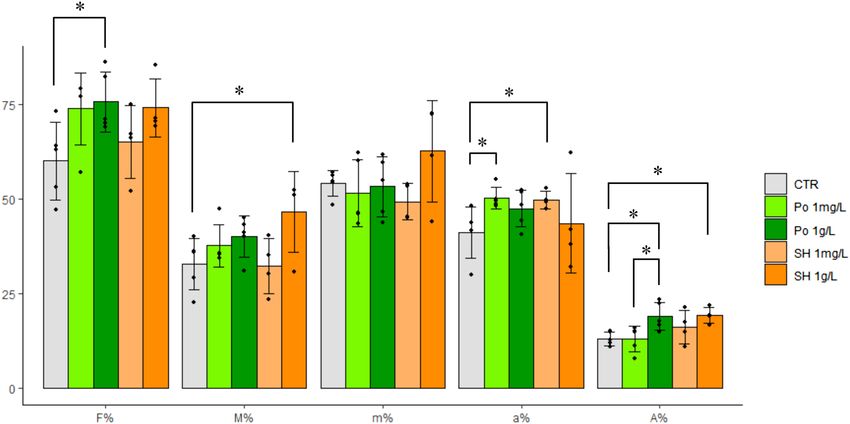
www.nature.com/scientificreports/
Figure 1. 13C CPMAS SSNMR spectra of chitooligosaccharides from T. viride, P. ostreatus, C. bertholletiae and
shrimps, acquired at room temperature at a spinning speed of 20 kHz. The labels of carbon atoms (C1-C6) refer
to the scheme on the left. The spectra indicate that the four samples are characterized by different degrees of
acetylation; in particular, this is lowest in shrimp-derived COs and highest in T. viride-derived COs.
acetic acid (see “Methods” section). Figure 2 and Supplementary Fig. S4 display the 1H spectra of the analyzed
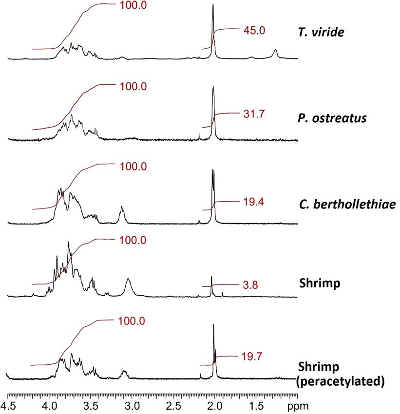
samples. In order to quantify the acetylation degree (AD) and compare it among samples, the signals ascribable
to protons in the polymeric chain were normalized at 100.0, so that the integrals for acetyl groups could directly
represent percent values of acetylation, as reported in Table 2. Solution NMR results confirmed the assump-
tion from the solid-state analysis: T. viride resulted to provide the most acetylated COs (AD = 45%), followed
by P. ostreatus (AD = 31.7%) and C. bertholletiae (AD = 19.4%). The analysis also confirmed the efficiency of
N-acetylation on shrimp COs, indicating a significant increase in their AD from 3.8 to 19.7%. In conclusion, a
combination of chemical analyses confirmed the presence of short chain COs in the extracted samples and the
efficiency of our N-acetylation protocol on the otherwise poorly acetylated shrimp-derived commercial COs.
Extracted COs are active as symbiotic signals. Each CO sample was tested for its ability to elicit sym-
biotic signalling in an AM host plant. To this aim, 1 mg/L CO solutions were applied to M. truncatula root
organ cultures (ROCs) expressing the NUP-YC2.1 probe (see “Methods” section), and nuclear Ca2+ signals were
recorded in epidermal cells by live confocal imaging. As expected, control treatments with sterile distilled water
did not activate any variation in nuclear Ca2+ concentration. On the contrary, repeated nuclear Ca2+ oscillations
(spiking) were observed in atrichoblasts upon treatment with all CO solutions (Fig. 3).
In order to compare the spiking response elicited by each CO solution, we used the method developed by
Russo et al.22 for quantifying a few characteristic features of the Ca2+ spiking response: the percentage of respond-
ing epidermal cells (atrichoblasts) and the average number of peaks per responding cell. This quantitative analysis
did not reveal any statistically significant difference in the percent of responding cells between shrimp- and
fungal-derived COs. However, fungal COs induced a significantly higher number of peaks, on average, compared
to shrimp-derived COs. No significant difference could be observed between fungal samples. Lastly, both the
percent of responding atrichoblasts and the average peak number in response to shrimp CO treatment were
significantly increased following CO peracetylation.
In short, all tested COs were able to activate symbiotic signalling in M. truncatula ROCs, but fungal COs and
peracetylated shrimp COs elicited a stronger response than commercial shrimp-derived COs.
Plant treatment with fungal COs enhances arbuscular mycorrhizal colonization. A precise
quantification of root colonization by the AM fungus was done according to Trouvelot et al.23. As summarized in
Fig. 4, the main effect of CO treatment was a general increase in all parameters describing root colonization, in
agreement with Volpe et al.4. In more detail, a 1 g/L solution of P. ostreatus COs produced a significant increase
in colonization frequency (F%) and arbuscule abundance in the whole root system (A%). A more diluted treat-
ment (1 mg/L) with P. ostreatus COs generated an analogous (albeit not statistically confirmed) trend on F%,
and had a major impact on arbuscule abundance within the colonized areas (a%). Concentrated shrimp COs
(1 g/L) significantly increased M% (average extension of fungal colonization in the root system) and A%—but
not F%—and never significantly outperformed concentrated (nor diluted) fungal COs. Importantly, diluted
shrimp COs (1 mg/L) achieved the overall lowest efficiency in AM promotion, with poor increases in F%, M%,
m% (intensity of mycorrhization in colonized parts of the root) and A% and a significant increase compared to
Scientific Reports | (2021) 11:3798 | https://doi.org/10.1038/s41598-021-83299-6 3
Vol.:(0123456789)www.nature.com/scientificreports/
Figure 2. Detail of solution 1H NMR spectra shown in Supplementary Fig. S4. Red lines and numbers represent
integrals for both chain protons (between 3.2 and 4.2 ppm, normalized at 100.0) and methylic protons of the
acetyl groups (at ~ 2.0 ppm).
T. viride P. ostreatus C. bertholletiae Shrimp Peracetyl. shrimp
Acetylation (%) 45.0 31.7 19.4 3.8 19.7
Table 2. Percent degree of acetylation of the extracted COs, as resulting from 1H NMR spectra.
controls only for a%. Altogether, fungal COs promoted AM development more efficiently than shrimp COs, with
a dose-dependent response.
Discussion
The modifications we have introduced to the method by Di Mario et al.24, in order to reduce reagent use and pro-
cessing time, provided a 61% increase in chitin recovery from P. ostreatus dry biomass, compared to the original
article where the chitin recovery was 15.3% ± 2.2%. However, this value remains within the range described by
Jones et al.19. Also chitin recovery from C. bertholletiae did not differ significantly from literature results25. Since
literature data report a high chitosan/chitin ratio in mucoromycetes26,27, we were not surprised to extract a large
amount of chitosan from C. bertholletiae, but—unexpectedly—this fungus also provided a relevant major amount
of chitin, which was indeed about 46 times more abundant than chitosan in terms of percent yield. Lastly, a very
high chitosan yield was also surprisingly obtained from T. viride, even if ascomycota cell walls are reported to
be richer in c hitin26,27. Such differences between expected and actual yields may be ascribed to several causes:
growth conditions and strain-specific features may have a major role, but we cannot exclude that the changes we
introduced to the extraction protocol differently affect chitin and chitosan extraction efficiency.
In addition to a considerable improvement in chitin extraction efficiency, our protocol also provided a very
high yield in COs: a + 65% increase was achieved from acid hydrolysis of C. bertholletiae chitin, compared to the
chitin acid hydrolysis from crab shell and squid pen performed by Kazami et al.28, while short chain CO recovery
Scientific Reports | (2021) 11:3798 | https://doi.org/10.1038/s41598-021-83299-6 4
Vol:.(1234567890)www.nature.com/scientificreports/
Figure 3. Calcium spiking response to 1 mg/L CO treatment in M. truncatula atrichoblasts. All CO solutions
activated nuclear calcium spiking, in a comparable number of cells (expressed as percentage in a). Nevertheless,
the average number of peaks recorded over 30 min (b) indicated a significantly stronger response to fungal
compared to shrimp COs. Remarkably, this difference was canceled when shrimp COs acetylation degree
was increased by peracetylation. CTR = water-treated control; Po = P. ostreatus COs; Cb = C. berthollethiae
COs; Tv = T. viride COs; SH = shrimp-derived COs; acSH = peracetylated shrimp-derived COs. A minimum
of six biological replicates were evaluated for each treatment. Significant differences are marked by an asterisk
(Student’s t test: P < 0.05); the difference between CTR and each CO treatment was highly significant in both
analyses (Student’s t test: P < 0.005).
from P. ostreatus did not differ significantly from the expected y ield28. The presence of different amounts of lipid,
glucan or protein traces in the chitin powder may have affected CO hydrolysis and solubilisation efficiency, and
account for such differences.
NMR analysis of extracted COs generated spectra with relatively broad and low peaks. Such features are often
associated with polymers, whereas NMR spectra of oligomers generally have narrower and more defined spikes29.
However, this consideration applies to purified solutions of homogeneous oligomers (e.g. tetramers); in our case,
fungal chitin was randomly hydrolysed, producing a mix of COs of different length. This range of variability can
Scientific Reports | (2021) 11:3798 | https://doi.org/10.1038/s41598-021-83299-6 5
Vol.:(0123456789)www.nature.com/scientificreports/
Figure 4. Quantitative analysis of fungal colonization in mycorrhizal M. truncatula roots treated with
different CO solutions. A general promotion of fungal colonization was induced by all treatments. In more
detail, the application of 1 g/L COs from P. ostreatus (Po 1 g/L) induced a significant increase in colonization
frequency (F%) and arbuscule abundance in the whole root system (A%) compared to water-treated controls.
A comparable increase in F% was also obtained using a 1 mg/L solution (Po 1 mg/L), which also produced
a significant increase in arbuscule abundance in the colonized areas (a%). By contrast, shrimp-derived COs
produced a significant increase in A% and M% (representing the average extension of fungal colonization in
the root system), as well as a relevant but statistically non significant elevation of F%, when applied at high
concentration (SH 1 g/L), but SH 1 mg/L produced the lowest increase in F% and only a significant increase in
arbuscule abundance in the colonized areas (a%). No significant differences were observed in the intensity of
the mycorrhization in colonized parts of the root system (m%). In short, P. ostreatus COs resulted to be more
efficient promoters of AM colonization than shrimp COs, especially when used at low dosage. A minimum of
four biological replicates were evaluated for each treatment. Asterisks indicate significant differences (Student’s t
test: P < 0.05).
explain the observed spectral features, as suggested by the analogous results of Kazami et al.28, who identified six
peaks in GPC chromatography of water-extracted COs as N-acetyl-glucosamine monomers, di-chitooligosac-
charides (CO2), tri-chitooligosaccharides ( CO3), tetra-chitooligosaccharides ( CO4), penta-chitooligosaccharides
(CO5) and hexa-chitooligosaccharides ( CO6) based on their retention times: the amounts of C O4, CO5 and C
O6
recovered exceeded those of N-acetyl-glucosamine and C O2. The presence of C O4–CO5 oligomers in all our CO
samples was also confirmed by HPLC–MS/MS analyses as described. However, considering that the samples
were not subjected to further purification, we cannot completely exclude the presence of longer chain oligomers.
Beside glomeromycetes, other fungi have been reported to produce small amounts of L COs41. Their presence
in our extracts appears unlikely for several reasons: the relatively harsh treatments included in the extraction
process are not supposed to preserve this type of molecules; furthermore, LCOs are poorly soluble in water
and have only been detected using specific butanol extraction41. In this context, the small traces of acyl chains
detected only in T. viride extracts by NMR analysis (peaks at 1.2 and 0.9 ppm in Supplementary Fig. S4 online)
appears surprising and could be related to sample contamination (e.g. by paraffins used to grease the glassware
junctions). Confirming the presence and origin of this anomalous signal goes beyond the scope of this work,
also considering the low yield of T. viride (Table 1), which makes the use of this strain unpractical for low-cost
production of chitinic molecules to be used for field-scale applications.
Among hydrolysed products, the highest acetylation degree (45%) was observed in T. viride COs. In addi-
tion, T. viride and P. ostreatus CO spectra showed narrower and clearer spikes than those of C. bertholletiae and
shrimp COs, a possible clue of higher homogeneity in oligomer length in the former two samples. By contrast,
shrimp-derived COs displayed the lowest acetylation degree (3.8%), which raised to 19.7% following our pera-
cetylation treatment.
NMR analysis showed acetylation degree as the only major difference between fungal and shrimp COs.
This is not surprising because chitin in arthropods and fungi presents the same α polymorphic form (the most
abundant in nature), despite their very distant phylogenetic positions18; β-chitin occurring only in squid pen,
sea tube worms, and some a lgae30,31. Significantly, the higher acetylation degree of fungal COs was associated
with their higher activity as elicitors symbiotic signalling in the AM host plant. In line with this observation, our
tests demonstrated that shrimp CO bioactivity was boosted upon peracetylation, with a significant enhancement
of the nuclear Ca2+ spiking response (average number of peaks and percent of activated atrichoblasts). These
converging results hint at acetylation as a fundamental feature for CO bioactivity in symbiotic signalling, shed-
ding new light on the molecular basis of plant-fungus recognition in AM.
Scientific Reports | (2021) 11:3798 | https://doi.org/10.1038/s41598-021-83299-6 6
Vol:.(1234567890)www.nature.com/scientificreports/
Plants have evolved refined receptorial mechanisms to perceive and discriminate between COs, with impor-
tant differences between plant lineages. In Arabidopsis, long chain molecules (CO8) elicit defence responses when
recognized by a homodimer of the plasma membrane-associated receptors-like kinase Chitin Elicitor Receptor
Kinase 1 (AtCERK132); in rice, OsCERK1 has lost the ability to directly bind COs, but this function in defence
elicitation is played by Chitin Elicitor Binding Protein (OsCEBiP7), which interacts with OsCERK1 in a hetero-
tetrameric receptor c omplex33,34. Recently, Feng et al.16 showed the activation of defence and symbiotic signalling
as a result of short chain COs perception by the MtCERK1 and the AtLYK5 (A. thaliana chitin receptor) homolog
LYR4 in M. truncatula. They also observed that diverse combinations of stimuli and receptors, including the
receptor-kinase MtDMI2, might contribute to inhibit plant defence responses and to promote symbiosis signal-
ling after CO p erception34. A role in both symbiosis and immunity regulation has also been shown for the LysM
receptor-like kinase MtLYK9 by Gibelin-Viala et al.35. On the same line, a putative CO receptor with a strong
affinity for short chain but not for longer chain COs has recently been characterized in rice as OsMYR136. The
OsMYR1/OsCERK1 heterodimer assembles and activates upon CO4 binding to OsMYR1, triggering symbiotic
signalling through nuclear C a2+ spiking36.
All such CO-binding proteins have extracellular domains containing three tandem LysM motifs (LysM1-3)
that bind N-acetyl-glucosamine residues37. Detailed structural analyses revealed a crucial role for the second
motif, LysM2, which discriminates chitin from other polysaccharides by interacting with N-acetyl groups38.
Furthermore, the structure of chitin-bound AtCERK1-ECD shows that saccharide units exhibit an alternation
of 180° flipping along the chitin chain, with the acetyl groups from one side of the chitin chain being solvent-
exposed, suggesting that full acetylation may not be necessary for maximal chitin interaction with AtCERK139,
and possibly for all LysM domain receptors.
This scenario is fully consistent with our observation that all short chain COs tested were able to elicit sym-
biotic Ca2+ signals, but stronger bioactivity was recorded for COs with higher degrees of acetylation, in line with
the key role of the N-acetyl group in receptor-ligand interaction. The relevance of acetylation degree of chitin,
chitosan and their derivatives is also highlighted in other biological processes, such as the ability of wound
recovery in animals, as described by Jones et al.19.
Chitin-derived molecules have previously been used in laboratory conditions, with beneficial effects on
plant growth and stress tolerance4. As shown in the Results section, our CO treatments generally increase fungal
colonization, in agreement with Volpe et al.4.
Our results provide a more detailed set of data on dose–response correlation regarding AM colonization on
M. truncatula. Evidence shows that COs dose–response correlation is also demonstrated in the pre-symbiotic
stage, as described by O ldroyd13, who compared Ca2+ spiking triggered by different molecules, including rhizobial
Nod factors, LCOs (lipo-chitooligosaccharides) and short COs.
Rush et al.41 observed that COs slightly enhance spore germination in Aspergillus fumigatus and pseudohy-
phae formation in Candida glabrata, albeit to a significantly more limited extent, compared to LCOs40. Further
analyses will be necessary to investigate if analogous effects are also present in AM fungi.
According to our results, the progressive enhancement of AM colonization in M. truncatula correlates with
increasing concentrations of P. ostreatus-derived COs from 1 mg/L to 1 g/L. Further investigations will be needed
to define the optimal concentrations for biologically relevant improvements in AM development for agricultural
applications.
Perspectives. Despite the positive results in protocol optimization, both extraction and hydrolysis protocols
still appear to be considerably time-consuming and complex: further simplifications will have to be tested in the
next experiments to shorten the total processing time, improve extraction yields and reduce costs. To this aim,
the protocol could significantly be improved by reusing media and reagents, skipping a few redundant steps in
the purification phase, combined with the possibility to use organic waste from industrial food processing activi-
ties as growth media.
Water-soluble chitin oligomers, in addition to their acknowledged role in stimulating the development of AM
symbiosis4, have many additional applications, including lowering of blood cholesterol and blood p ressure41,42,
43
inducing protective effects against infections and enhancing antitumor properties and anti-adhesion
activity44–46. They are also used in food and biomedical industries47–49. Furthermore, they are considered to be
functional foods because of their non-digestibility by intestinal enzymes, which allows their use as prebiotics.
They stimulate beneficial bacteria in the gastrointestinal tract (Bifidobacterium and Lactobacillus sp.)50,51. They
can also act as thickeners and stabilizing a gents30.
Consequently, large-scale and low-cost CO production is of great interest for industrial and agricultural
application.
Methods
Fungi. The mucoromycete Cunninghamella bertholletiae MUT 2861, the basidiomycete Pleurotus ostreatus
MUT 2976 and the ascomycete Trichoderma viride MUT 3170 were grown on malt extract broth (ME), con-
taining 20 g/L glucose, 20 g/L malt extract, and 2 g/L peptone, as described by Spina et al.52. In more detail,
fungi were inoculated as conidial suspension in 500 mL flasks containing 350 mL of medium and incubated at
24 ± 2 °C in the dark on an orbital shaker set at 130 rpm. After 12 days, the biomass was filtered, washed and
lyophilized.
Chitin purification. Chitin was purified from lyophilized biomass of P. ostreatus, C. bertholletiae and T.
viride, using a modified version of the method described by Di Mario et al.24. Briefly, fungal biomass was depro-
teinated by treatment of 15 g of lyophilized mycelium with 500 mL of 1 N NaOH under vigorous stirring over-
Scientific Reports | (2021) 11:3798 | https://doi.org/10.1038/s41598-021-83299-6 7
Vol.:(0123456789)www.nature.com/scientificreports/
night at 40 °C. The suspension was centrifuged at 4500×g for 45 min and the supernatant, containing proteins
and other impurities, was discarded. This treatment was repeated three times. The pellet, containing fungal wall
polysaccharides, was suspended in 500 mL of boiling distilled water in a round-bottom flask equipped with a
reflux condenser and stirred overnight at reflux in order to remove glucans. The suspension was then centrifuged
at 4500×g for 45 min. The supernatant containing wall glucans was discarded. The pellet was washed three times
with distilled water at 100 °C. In a round-bottom flask equipped with a reflux condenser, the pellet was treated
with 500 mL of aqueous acetic acid (5%) and stirred for 3 h at 90 °C. After centrifugation at 4500×g for 45 min,
the chitin pellet was separated from the supernatant (containing chitosan), washed three times with distilled
water and lyophilized. Chitosan was precipitated by adding 1 N NaOH until pH reached 12 and centrifuged at
4500×g for 45 min. The resulting pellet was washed twice with distilled water and lyophilized. Fresh and dry
weight of both extracted chitin and chitosan were measured before and after lyophilization.
Chitooligosaccharide production. Chitin hydrolysis under acidic conditions was performed following a
modified version of the method described by Kazami et al.28. Briefly, 1 g of lyophilized chitin for each fungus was
hydrolysed in 30 mL of 37% HCl at 40 °C for 1 h. After cooling back to room temperature, the reaction mixture
was then poured into 160 mL of acetone and stirred overnight at 4 °C. The solution was then centrifuged at
10,000×g for 10 min (4 °C) and the precipitate was washed with acetone until pH reached 5. Finally, the pellet
was re-suspended in cold diethyl ether, centrifuged at 10,000×g for 10 min at 4 °C and dried under vacuum over
P2O5.
The dried precipitate was soaked in 25 mL of distilled water and stirred overnight at 20 °C. Subsequently, the
pellet was centrifuged at 15,000 g for 10 min (20 °C). Centrifugation produced a precipitate (which was soaked
in 10 mL of distilled water and stirred overnight at 20 °C) and a supernatant (which was stored). The suspended
precipitate was further centrifuged at 15,000 g for 10 min at 20 °C. The combined supernatant obtained for each
fungus—expected to contain water-soluble chitin oligomers—was completely lyophilized to obtain a powder of
purified chitooligomers.
Shrimp CO peracetylation. In order to check for differences in the biological activity of COs with differ-
ent acetylation degree, shrimp shell-derived COs (Zhengzhou Sigma Chemical Co., Ltd.—Zhengzhou, Henan,
China) were N-acetylated following the protocol described by Trombotto et al.53.
100 mg of shrimp CO mixture were dissolved in 25 mL of methanol/water (50/50, v/v) solution and add-
ing 50.3 mL of acetic anhydride. After stirring at room temperature for 5 min the solution was concentrated to
dryness, the solid residue was re-dissolved in distilled water and evaporated under vacuum. This treatment was
repeated twice, then the CO sample was dissolved in 50 mL of 0.01 M HCl and, after freeze-drying, isolated as
a white powder.
Chemical analyses. Extracted COs were initially analysed by DIMS on an LCQFleet Ion trap mass spec-
trometer (Thermo Fisher, USA) equipped with an electrospray source. Extracts were dissolved in MilliQ water
(10 mg L−1) added with 20% of acetic acid:methanol (1:100, v/v) and filtered by 0.2 µm cellulose acetate syringe
filter. The syringe pump was used at a flow rate of 10 mL min−1 to infuse samples directly into the mass spec-
trometer. The electrospray source was operated in positive ion mode, and the following source conditions were
set: spray voltage at 3.5 kV, sheat gas flow at 20 arbitrary units, auxiliary and sweep gas flow at 0 arbitrary units,
ion transfer temperature at 280 °C. The mass spectrometer was operated in full scan mode; exploring the scan
range m/z 200–2000 and splitting the scan range in six scans (m/z 200–600, 500–900, 800–1200, 1100–1500,
1400–1800, 1700–2000).
COs were subsequently characterized by HPLC using an Ultimate 3000 (Dionex). Separation was performed
using a hypercarb column (5 µm, 2 × 100 mm; Hypercarb, Thermo). Solutions of acetic acid:water (1/1000, v/v)
and acetonitrile were pumped at 0.4 mL min−1. The gradient used was 100% acetic acid:water for 1 min, then
100% to 50% acetic acid:water in 30 min then 50% to 0% acetic acid:water in 3 min.
COs were identified in the multiple reaction monitoring (MRM) mode using a 4500 Q Trap mass spectrom-
eter (Applied Biosystems, Foster City, CA, USA) with an electrospray ionization in the positive ion mode by
monitoring the transitions from parent ion > common daughter ions (m/z: 204, 407, 610), and for quantification
using the MRM 425 > 204 m/z (CO2), 628 > 204 m/z (CO3), 831 > 204 m/z (CO4), 1034 > 204 m/z (CO5). Mono-
deacetylated (deAc) COs were identified in MRM mode by monitoring the transitions from parent ion > com-
mon daughter ions (m/z: 162, 365, 568), and for quantification using the MRM 383 > 162 m/z (deAc-CO2),
586 > 162 m/z (deAc-CO3), 789 > 162 m/z (deAc-CO4), 992 > 162 m/z (deAc-CO5). The capillary voltage was
fixed at 5500 V, source temperature at 400 °C. Fragmentation was performed by collision-induced dissociation
(CID) with nitrogen, collision energy 20 to 54 V, declustering potential 90 to 130 V depending on m olecules11.
13 54–57 1 58
Furthermore, C CPMAS S SNMR and solution H NMR , were used to obtain detailed structural infor-
mation of both chitin and COs.
SSNMR spectra were acquired with a Bruker Avance II 400 Ultra Shield instrument, operating at 400.23 and
100.63 MHz, respectively for 1H and 13C nuclei. Samples were packed into cylindrical zirconia rotors with a 4 mm
o.d. and an 80 µL volume. A small amount of sample (40 to 150 mg, depending on the sample) was collected
from each batch and shredded into pieces, small enough to fill the rotor. 13C CPMAS spectra were acquired at
a spinning rate of 12 kHz, using a ramp cross-polarization pulse sequence with a contact time of 3 ms, a 90°
1
H pulse of 3.60 µs, optimized recycle delays between 1 and 2.1 s, for a number of scans between 400 and 3170,
depending on the sample. For every spectrum, a two-pulse phase modulation (TPPM) decoupling scheme was
used, with a radiofrequency field of 69.4 kHz. The 13C chemical shift scale was calibrated through the methylenic
signal of external standard glycine (at 43.7 ppm).
Scientific Reports | (2021) 11:3798 | https://doi.org/10.1038/s41598-021-83299-6 8
Vol:.(1234567890)www.nature.com/scientificreports/
Solution (D2O) 1H NMR spectra were acquired on a Jeol Eclipse 400 instrument, operating at 400.23 MHz
for 1H nuclei. About 5 mg of each sample was dissolved in 600 µL of D 2O and the solution was transferred in
an NMR tube. The spectra were collected at room temperature. In order to ensure a complete relaxation of the
magnetization, a relaxation delay of 60 s for 256 scans was employed for each spectrum.
Bioactivity assay. The bioactivity of purified COs was analysed using an established test for the activa-
tion of the common symbiotic signalling pathway, based on live imaging of root epidermal cells in confocal
microscopy59. Biological analyses were done using Agrobacterium rhizogenes-transformed ROCs of wild-type
Medicago truncatula (genotype Jemalong A17) expressing the 35S:NupYC2.1 c onstruct60. The ROC line, avail-
able in the lab11, was propagated in vertically-oriented Petri dishes containing M medium 61 and incubated at
25 °C in the dark for 10–14 days. Explants with consistent morphology and an identical number of lateral roots
were chosen for all technical replicates.
Excised 1–2 cm-long lateral M. truncatula roots were treated with 1 mg/L solution of COs, extracted from
each fungus (P. ostreatus, C. bertholletiae and T. viride); sterile distilled water was used as negative control and
1 mg/L solutions of short chain COs purified from shrimp shells (acetylated and non-acetylated) were used as
positive control, based on previous studies11,62. A Leica TCS SP2 AOBS confocal laser-scanning microscope,
equipped with a 40 × water-immersion objective, was used for live cell imaging of NupYC2.1 fluorescence in
root atrichoblasts. Ratiometric analysis of NupYC2.1 FRET intensity over time was used to visualize variations
in calcium concentration for each imaged nucleus during 30 min following the treatments. A minimum of 20
atrichoblasts from at least 6 independent root segments were analysed for each treatment.
Pot cultures. The efficiency of P. ostreatus COs as stimulants of AM colonization was tested on pot-grown
M. truncatula plants. Seeds were first scarified on sand paper in order to break the seed coat. They were steri-
lized using 5% sodium hypochlorite in water and washed several times in sterile distilled water. Seeds were then
imbibed on 0.6% agar plates at 4 °C in the dark for 48 h to break dormancy, then moved at 23 °C for 5 days to
allow germination. 10 days-old seedlings were transferred to 10 × 10 × 12 cm flowerpots with sterile quartz sand
and grown for two weeks before further treatment.
1 g/L and 1 mg/L solutions were prepared in sterile distilled water using P. ostreatus COs (Po 1 g/L and Po
1 mg/L, respectively) or shrimp-derived COs (SH 1 g/L and SH 1 mg/L). Sterile distilled water was used for
control treatments (CTR). All test solutions were added with 0.005% Tween 20 as a surfactant. Each plant was
sprayed with 5 mL of test solution and the treatment was repeated 2 days later, prior to inoculation with the AM
fungus Funnelliformis mosseae, (strain BEG 12) using a commercial inoculum (MycAgroLab, Bretenièr, France)
mixed at 5% with sand.
Ten replicates were done for each of the following lines: CTR, Po 1 mg/L, Po 1 g/L, SH 1 mg/L and SH 1 g/L.
During the first week, a plastic bag was placed on the flowerpots to prevent excessive desiccation and allow pro-
gressive plant acclimatisation. Plants were grown for 28 days in phytochamber under photoperiod of 16 h/day
(23 °C) and 8 h/night (21 °C) and each plant was fertilized once a week with 20 mL of a modified Long Asthon
nutrient solution containing 3.2 μM K H2PO4 as P source63.
Plants were harvested 28 days post inoculation and washed from sand before fresh weight of roots and shoots
was determined. The shoots of 5 plants for each line were then dried at 60 °C until their weight was stabilised,
to determine their dry weight (see Supplementary Fig. S5 online).
To determine the extension of fungal colonization, the remaining 5 plants for each test line were stained
following the ’lactic blue’ protocol developed by Trouvelot et al. (1986): root systems were excised, placed in
50 mL-tubes and submerged in the staining solution (0.1% cotton blue in lactic acid) for 12 h, then rinsed 2 times
in water and 2 times in lactic acid (2 h for each rinsing) until all the excessive dye was removed. For microscope
observation, each stained root system was cut randomly in 1 cm length pieces and distributed on 5 microscope
slides (20 pieces for slide), until 1 m of root was collected for each plant.
The staining of intraradical fungal structures with lactic blue allowed both a morphological characterisa-
tion and a statistical quantification of root colonization according to the method by Trouvelot et al. (1986).
Four quantitative parameters were calculated and used to compare the intensity of root colonization between
samples. F%, colonization frequency in the root system, represents the percentage of segments containing intra-
radical fungal structures and is considered as a general indicator of the plant mycorrhizal status; M%, intensity
of mycorrhizal colonization in the root system, reports on the average volume occupied by the fungus in each
fragment, providing more detailed information on the extension of single infection units; m%, intensity of the
mycorrhization in colonized parts of the root; a%, arbuscule abundance in colonized areas, indicates the average
percentage of arbusculated cells within colonized segments and represents a reporter of symbiosis efficiency;
A%, arbuscule abundance in the root system, estimates the average abundance of arbuscules in the whole root
system, including non-colonized parts.
Data availability
The datasets generated during and/or analysed during the current study are available from the corresponding
author on reasonable request.
Received: 3 November 2020; Accepted: 29 January 2021
Scientific Reports | (2021) 11:3798 | https://doi.org/10.1038/s41598-021-83299-6 9
Vol.:(0123456789)www.nature.com/scientificreports/
References
1. Smith, S. & Read, D. Mycorrhizal Symbiosis (Elsevier Ltd, Amsterdam, 2008).
2. Rich, M. K., Nouri, E., Courty, P. E. & Reinhardt, D. Diet of arbuscular mycorrhizal fungi: Bread and butter?. Trends Plant Sci. 22,
652–660 (2017).
3. Lanfranco, L., Bonfante, P. & Genre, A. The mutualistic interaction between plants and arbuscular mycorrhizal fungi. Microbiol.
Spect. 4, 727–747 (2016).
4. Volpe, V. et al. Short chain chito-oligosaccharides promote arbuscular mycorrhizal colonization in Medicago truncatula. Carbohydr.
Polym. 229, 115505 (2020).
5. Lanfranco, L., Fiorilli, V., Venice, F. & Bonfante, P. Strigolactones cross the kingdoms: Plants, fungi, and bacteria in the arbuscular
mycorrhizal symbiosis. J. Exp. Bot. 69, 2175–2188 (2018).
6. Schmitz, A. M. & Harrison, M. J. Signaling events during initiation of arbuscular mycorrhizal symbiosis. J. Integr. Plant Biol. 56,
250–261 (2014).
7. Kaku, H. et al. Plant cells recognize chitin fragments for defense signaling through a plasma membrane receptor. Proc. Natl. Acad.
Sci. USA 103, 11086–11091 (2006).
8. Wan, J. et al. LYK4, a lysin motif receptor-like kinase, is important for chitin signaling and plant innate immunity in Arabidopsis.
Plant Physiol. 160, 396–406 (2012).
9. Cao, Y. et al. The kinase LYK5 is a major chitin receptor in Arabidopsis and forms a chitin-induced complex with related kinase
CERK1. eLife 3, 3766 (2014).
10. Hamel, L. P. & Beaudoin, N. Chitooligosaccharide sensing and downstream signaling: Contrasted outcomes in pathogenic and
beneficial plant-microbe interactions. Planta 232, 787–806 (2010).
11. Genre, A. et al. Short chain chitin oligomers from arbuscular mycorrhizal fungi trigger nuclear Ca2+ spiking in Medicago truncatula
roots and their production is enhanced by strigolactone. New Phytol. 198, 190–202 (2013).
12. Sun, J. et al. Activation of symbiosis signaling by arbuscular mycorrhizal fungi in legumes and rice. Plant Cell 27, 823–838 (2015).
13. Oldroyd, G. E. D. Speak, friend, and enter: Signalling systems that promote beneficial symbiotic associations in plants. Nat. Rev.
Microbiol. 11, 252–263 (2013).
14. Barker, D. G., Chabaud, M., Russo, G. & Genre, A. Nuclear Ca 2+ signalling in arbuscular mycorrhizal and actinorhizal endosym-
bioses: On the trail of novel underground signals. New Phytol. 214, 533–538 (2017).
15. Bonfante, P. & Genre, A. Arbuscular mycorrhizal dialogues: Do you speak “plantish” or “fungish”?. Trends Plant Sci. 20, 150–154
(2015).
16. Feng, F. et al. A combination of chitooligosaccharide and lipochitooligosaccharide recognition promotes arbuscular mycorrhizal
associations in Medicago truncatula. Nat. Commun. https://doi.org/10.1038/s41467-019-12999-52 (2019).
17. Gutjahr, C. & Parniske, M. Cell and developmental biology of arbuscular mycorrhiza symbiosis. Annu. Rev. Cell Dev. Biol. 29,
593–617 (2013).
18. Hamed, I., Özogul, F. & Regenstein, J. M. Industrial applications of crustacean by-products (chitin, chitosan, and chitooligosac-
charides): A review. Trends Food Sci. Technol. 48, 40–50 (2016).
19. Jones, M., Kujundzic, M., John, S. & Bismarck, A. Crab vs. Mushroom: A review of crustacean and fungal chitin in wound treat-
ment. Mar. Drugs https://doi.org/10.3390/md18010064 (2020).
20. Heux, L., Brugnerotto, J., Desbrières, J., Versali, M. F. & Rinaudo, M. Solid state NMR for determination of degree of acetylation
of chitin and chitosan. Biomacromol 1, 746–751 (2000).
21. Mourya, V. K., Inamdar, N. N. & Choudhari, Y. M. Chitooligosaccharides: Synthesis, characterization and applications. Polym.
Sci. A 53, 583–612 (2011).
22. Russo, G., Spinella, S., Sciacca, E., Bonfante, P. & Genre, A. Automated analysis of calcium spiking profiles with CaSA software:
Two case studies from root-microbe symbioses. BMC Plant Biol. 13, 224 (2013).
23. Trouvelot, A., Kough, J.L. & Gianinazzi-Pearson, V. Mesure du taux de mycorhization VA d’un système radiculaire. Recherche de
méthodes d’estimation ayant une signification fonctionnelle. in Physiological and Genetical Aspects of Mycorrhizae (eds Gianinazzi-
Pearson, V. & Gianinazzi, S.), INRA edition, Paris (1986).
24. Di Mario, F., Rapanà, P., Tomati, U. & Galli, E. Chitin and chitosan from basidiomycetes. Int. J. Biol. Macromol. 43, 8–12 (2008).
25. Berger, L. R. R. et al. Green conversion of agroindustrial wastes into chitin and chitosan by rhizopus arrhizus and cunninghamella
elegans strains. Int. J. Mol. Sci. 15, 9082–9102 (2014).
26. Raghukumar, S. & Raghukumar, S. Fungi: Characteristics and Classification. Fungi in Coastal and Oceanic Marine Ecosystems 1–15
(Springer International Publishing, New York, 2017).
27. Lecointe, K., Cornu, M., Leroy, J., Coulon, P. & Sendid, B. Polysaccharides cell wall architecture of mucorales. Front. Microbiol.
https://doi.org/10.3389/fmicb.2019.00469 (2019).
28. Kazami, N. et al. A simple procedure for preparing chitin oligomers through acetone precipitation after hydrolysis in concentrated
hydrochloric acid. Carbohyd. Polym. 132, 304–310 (2015).
29. Saito, H., Mamizuka, T. & Tabeta, R. High resolution 13C NMR spectra of chitin oligomers in aqueous solution. Chem. Lett. 10,
1483–1484 (1981).
30. Rinaudo, M. Chitin and chitosan: Properties and applications. Progress Polym. Sci. 31, 603–632 (2006).
31. Abo Elsoud, M. M. & el Kady, E. M. Current trends in fungal biosynthesis of chitin and chitosan. Bull. Natl. Res. Centre https://
doi.org/10.1186/s42269-019-0105-y (2019).
32. Wan, J. et al. A LysM receptor-like kinase plays a critical role in chitin signaling and fungal resistance in Arabidopsis. Plant Cell
20, 471–481 (2008).
33. Antolín-Llovera, M. et al. Knowing your friends and foes: Plant receptor-like kinases as initiators of symbiosis or defence. New
Phytol. 204, 791–802 (2014).
34. Zipfel, C. & Oldroyd, G. E. D. Plant signalling in symbiosis and immunity. Nature 543, 328–336 (2017).
35. Gibelin-Viala, C. et al. The Medicago truncatula LysM receptor-like kinase LYK9 plays a dual role in immunity and the arbuscular
mycorrhizal symbiosis. New Phytol. 223, 1516–1529 (2019).
36. He, J. et al. A LysM receptor heteromer mediates perception of arbuscular mycorrhizal symbiotic signal in rice. Mol. Plant 12,
1561–1576 (2019).
37. Petutschnig, E. K., Jones, A. M. E., Serazetdinova, L., Lipka, U. & Lipka, V. The lysin motif receptor-like kinase (LysM-RLK)
CERK1 is a major chitin-binding protein in Arabidopsis thaliana and subject to chitin-induced phosphorylation. J. Biol. Chem.
285, 28902–28911 (2010).
38. Hayafune, M. et al. Chitin-induced activation of immune signaling by the rice receptor CEBiP relies on a unique sandwich-type
dimerization. Proc. Natl. Acad. Sci. USA 111, E404–E413 (2014).
39. Liu, T. et al. Chitin-induced dimerization activates a plant immune receptor. Science 336, 1160–1164 (2012).
40. Rush, T. et al. Lipo-chitooligosaccharides as regulatory signals of fungal growth and development. Nat. Commun. https://doi.
org/10.1038/s41467-020-17615-5 (2020).
41. Choi, C. R. et al. Chitooligosaccharides decreases plasma lipid levels in healthy men. Int. J. Food Sci. Nutr. 63, 103–106 (2012).
42. Giustina, A. & Ventura, P. Weight-reducing regimens in obese subjects: Effects of a new dietary fibre integrator. Acta Toxicol. Ther.
16, 199–214 (1995).
Scientific Reports | (2021) 11:3798 | https://doi.org/10.1038/s41598-021-83299-6 10
Vol:.(1234567890)www.nature.com/scientificreports/
43. Tokoro, A. et al. Protective effect of N-acetyl chitohexaose on Listeria monocytogenes infection in mice. Microbiol. Immunol. 33,
357–367 (1989).
44. Xu, W. et al. Chitooligosaccharides and N-acetyl-D-glucosamine stimulate peripheral blood mononuclear cell-mediated antitumor
immune responses. Mol. Med. Rep. 6, 385–390 (2012).
45. Xu, C., Lei, C., Meng, L., Wang, C. & Song, Y. Chitosan as a barrier membrane material in periodontal tissue regeneration. J.
Biomed. Mater. Res. B 100, 1435–1443 (2012).
46. Nishimura, K. et al. Macrophage activation with multi-porous beads prepared from partially deacetylated chitin. J. Biomed. Mater.
Res. 20, 1359–1372 (1986).
47. Kim, S. K. & Rajapakse, N. Enzymatic production and biological activities of chitosan oligosaccharides (COS): A review. Carbohyd.
Polym. 62, 357–368 (2005).
48. Seyfarth, F., Schliemann, S., Elsner, P. & Hipler, U. C. Antifungal effect of high- and low-molecular-weight chitosan hydrochloride,
carboxymethyl chitosan, chitosan oligosaccharide and N-acetyl-d-glucosamine against Candida albicans, Candida krusei and
Candida glabrata. Int. J. Pharm. 353, 139–148 (2008).
49. Xia, W., Liu, P., Zhang, J. & Chen, J. Biological activities of chitosan and chitooligosaccharides. Food Hydrocoll. 25, 170–179 (2011).
50. Morganti, P., Morganti, G. & Morganti, A. Transforming nanostructured chitin from crustacean waste into beneficial health
products: A must for our society. Nanotechnol. Sci. Appl. 4, 123–129 (2011).
51. Harish Prashanth, K. V. & Tharanathan, R. N. Chitin/chitosan: modifications and their unlimited application potential-an overview.
Trends Food Sci. Technol. 18, 117–131 (2007).
52. Spina, F., Tigini, V., Romagnolo, A. & Varese, G. C. Bioremediation of landfill leachate with fungi: Autochthonous vs. allochthonous
strains. Life 8, 27 (2018).
53. Trombotto, S., Ladavière, C., Delolme, F. & Domard, A. Chemical preparation and structural characterization of a homogeneous
series of chitin/chitosan oligomers. Biomacromol 9, 1731–1738 (2008).
54. FernandezDiaz-Rullo, F. et al. Synthesis and hyperpolarisation of eNOS substrates for quantification of NO production by 1H NMR
spectroscopy. Bioorg. Med. Chem. 25, 2730–2742 (2017).
55. Pauli, G. F., Gödecke, T., Jaki, B. U. & Lankin, D. C. Quantitative 1H NMR Development and potential of an analytical method:
An update. J. Nat/ Prod. 75, 834–851 (2012).
56. Bharti, S. K. & Roy, R. Quantitative 1H NMR spectroscopy. Trends Anal. Chem. 35, 5–26 (2012).
57. Zuriarrain, A., Zuriarrain, J., Villar, M. & Berregi, I. Quantitative determination of ethanol in cider by 1H NMR spectrometry.
Food Control 50, 758–762 (2015).
58. Schanda, P. & Ernst, M. Studying dynamics by magic-angle spinning solid-state NMR spectroscopy: Principles and applications
to biomolecules. Prog. Nucl. Magn. Reson. Spectrosc. 96, 1–46 (2016).
59. Chabaud, M. et al. Arbuscular mycorrhizal hyphopodia and germinated spore exudates trigger Ca2+ spiking in the legume and
nonlegume root epidermis. New Phytol. 189, 347–355 (2011).
60. Sieberer, B. J. et al. A nuclear-targeted cameleon demonstrates intranuclear Ca2+ spiking in Medicago truncatula root hairs in
response to rhizobial nodulation factors. Plant Physiol. 151, 1197–1206 (2009).
61. Becard, G. & Fortin, J. A. Early events of vesicular-arbuscular mycorrhiza formation on Ri T-DNA transformed roots. New Phytol.
108, 211–218 (1988).
62. Carotenuto, G. et al. The rice LysM receptor-like kinase OsCERK1 is required for the perception of short-chain chitin oligomers
in arbuscular mycorrhizal signaling. New Phytol. 214, 1440–1446 (2017).
63. Hewitt, E. J. Sand and Water Culture Methods Used in the Study of Plant Nutrition (Springer, New York, 1952).
Author contributions
A.C. performed the fungal cultures, mycelia production, chitin, chitosan and CO extraction, bioactivity analyses,
contributed to the pot cultures experiments and wrote the manuscript’s original draft. E.M. contributed to the
mycelia production, chitin and chitosan extraction, CO purification, bioactivity analyses and performed the
pot cultures experiments. M.B. supervised chitin and CO extraction, reviewed and edited the manuscript. G.C.
contributed to chitin and CO extraction and bioactivity analyses. F.S. supervised the fungal culture, reviewed and
edited the manuscript. S.B. performed the NMR analyses and contributed to the original draft. V.P.P. performed
the HPLC analyses and contributed editing the manuscript. L.A. performed the DIMS analyses and contributed
editing the text. V.V. supervised the pot culture experiments and reviewed and edited the text. C.P. contributed
to the experimental set up and revised the text. R.G. contributed to the experimental set up and revised the text.
G.C.V. contributed to experimental design and text writing. A.G. designed the research and experiments and
wrote the text.
Funding
This work was supported by MIUR, through a PhD fellowship to AC and FFABR 2017 grant to AG; University
of Torino grants to AG (Ricerca Locale 2018–2019). Support for mass spectrometry analyses was provided by
the MetaboHUB (Grant ANRINBS-0010)-Metatoul- AgromiX Facilities.
Competing interests
The authors declare no competing interests.
Additional information
Supplementary Information The online version contains supplementary material available at https://doi.
org/10.1038/s41598-021-83299-6.
Correspondence and requests for materials should be addressed to A.G.
Reprints and permissions information is available at www.nature.com/reprints.
Publisher’s note Springer Nature remains neutral with regard to jurisdictional claims in published maps and
institutional affiliations.
Scientific Reports | (2021) 11:3798 | https://doi.org/10.1038/s41598-021-83299-6 11
Vol.:(0123456789)www.nature.com/scientificreports/
Open Access This article is licensed under a Creative Commons Attribution 4.0 International
License, which permits use, sharing, adaptation, distribution and reproduction in any medium or
format, as long as you give appropriate credit to the original author(s) and the source, provide a link to the
Creative Commons licence, and indicate if changes were made. The images or other third party material in this
article are included in the article’s Creative Commons licence, unless indicated otherwise in a credit line to the
material. If material is not included in the article’s Creative Commons licence and your intended use is not
permitted by statutory regulation or exceeds the permitted use, you will need to obtain permission directly from
the copyright holder. To view a copy of this licence, visit http://creativecommons.org/licenses/by/4.0/.
© The Author(s) 2021
Scientific Reports | (2021) 11:3798 | https://doi.org/10.1038/s41598-021-83299-6 12
Vol:.(1234567890)You can also read