Cupid, a cell permeable peptide derived from amoeba, capable of delivering Gfp into a diverse range of species - Nature
←
→
Page content transcription
If your browser does not render page correctly, please read the page content below
www.nature.com/scientificreports
OPEN Cupid, a cell permeable peptide
derived from amoeba, capable
of delivering GFP into a diverse
range of species
Daniel Fenton1, Dylan Phillips1, Anne Maddison1, Christopher H. George2,
Jonathan Ryves3* & Huw D. Jones1*
Cell permeating peptides (CPPs) are attracting great interest for use as molecular delivery vehicles
for the transport of biologically active cargo across the cell membrane. The sequence of a novel CPP
sequence, termed ‘Cupid’, was identified from the genome of Dictyostelium discoideum. A Cupid-Green
Fluorescent Protein (Cupid-GFP) fusion protein was tested on mammalian, whole plant cells, plant
leaf protoplast and fungal cell cultures and observed using confocal microscopy. GFP fluorescence
builds up within the cell cytosol in 60 min, demonstrating Cupid-GFP has permeated them and folded
correctly into its fluorescent form. Our combined data suggest Cupid can act as a molecular vehicle
capable of delivering proteins, such as GFP, into the cytosol of a variety of cells.
The cell membrane or plasma membrane surrounding the cytoplasm of living cells is selectively permeable to
ions and organic molecules, and acts as a barrier to regulate what enters and exits the cell. It is made mostly from
a bilayer of phospholipid molecules with a variety of protein molecules embedded within it and, depending on
the type of organism, it may be further bounded externally by a cell wall1,2.
Cell-permeable or cell-penetrating peptides (CPPs) (also known as a protein transduction domain or mem-
brane translocation sequence) are increasingly being used to overcome the impermeability of the plasma mem-
brane. Their ability to traverse cell membranes and access the cell interior allows them to be exploited to internal-
ize molecules (generically termed ‘cargo’) such as proteins, oligonucleotides, peptides, nucleic acids, and other
pharmacologically active compounds into a range of living systems. Thus, CPPs provide a promising strategy
for cellular delivery of otherwise membrane-impermeable molecules for research and therapeutic p urposes3–10.
Understanding how CPPs interact with membranes and achieve access to the cell interior is an on-going area
of research.
CPPs may be classified based upon their binding properties to lipids linked to their peptide sequences: pri-
mary amphipathic; secondary amphipathic; and non-amphipathic11–13. They can also be classified according to
the physical mechanism of cellular uptake, for example whether it is endocytosis-mediated or direct permeation
of the membrane.
For endocytosis-mediated delivery, CPPs are thought to first interact with the outer surface of the cell
membrane via molecular charge or binding to surface proteoglycans, glycosaminoglycans or other receptor
before being internalized by the endocytotic machinery. Macropinocytosis, clathrin-coated pits and caveolae/
lipid-raft-mediated endocytosis mechanisms have been implicated through their differing sensitivity to drug
applications14–19. These processes involve energetic membrane folding and employ internalization proteins that
require cellular ATP to function. Direct permeation of the membrane involves a mechanism that is independent
of classic endocytosis, requiring no cell surface receptor or cell-derived ATP energy to do so. It is thought to
occur without the requirement of the cell internalization machinery and has been proposed to involve sequential
steps of binding (localization) then insertion into the bilayer followed by translocation across the membrane
by ‘flipping’ or inverted lipid micelle formation. Interactions between CPPs and the phosphate groups on both
1
Institute of Biological, Environmental and Rural Sciences (IBERS), Aberystwyth University, Penglais, Aberystwyth,
Ceredigion SY23 3DA, Wales, UK. 2Institute of Life Sciences, Swansea University Medical School, Singleton Park
Campus, Swansea SA2 8PP, Wales, UK. 3Cupid Peptides, Cardiff Medicentre, Heath Park, Cardiff CF14 4UJ, Wales,
UK. *email: jonnyryves@cupidpeptides.com; huw.jones@aber.ac.uk
Scientific Reports | (2020) 10:13725 | https://doi.org/10.1038/s41598-020-70532-x 1
Vol.:(0123456789)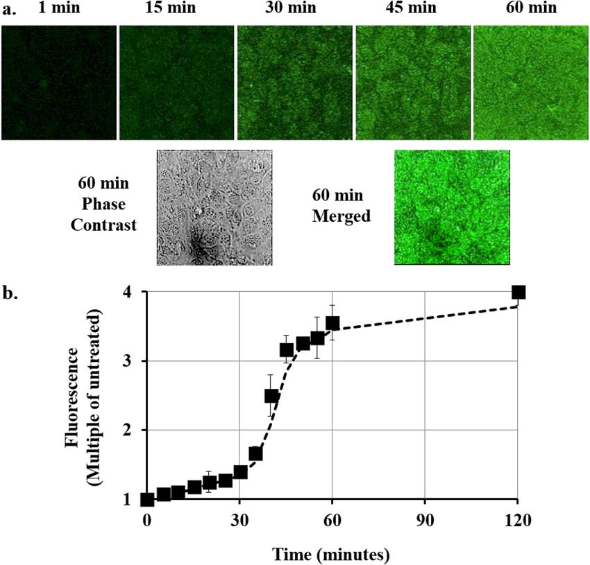
www.nature.com/scientificreports/
sides of the lipid bilayer, the strength of the transbilayer potential and the formation of a transient pore have
been suggested to play roles in these steps20–25. However this process, especially with respect to permeation of
CPPs with large cargoes, remains to be fully elucidated at the molecular level. Additionally, the mode of cell
permeation may be influenced by other factors such as the CPP concentration and the size and type of the cargo
attached to the CPP. It has been suggested that small cargoes are capable of utilizing a non-endocytic pathway,
whereas larger ones tend to use the endocytic delivery26–28.
Finally, the route of permeation will directly affect the bioavailability of cargoes within the target cell since
endosomal trapping can lead to lysosomal degradation hence limiting cargo access to other intracellular
targets29–31 unless further protein engineering can facilitate subsequent endosomal e scape32.
It is well accepted that fluorescent proteins such as GFP, whether denatured or folded into their functional
fluorescent form, do not permeate cell membranes unaided33. This paper introduces a novel cell permeating
peptide sequence identified in the genome of the free-living amoeba, Dictyostelium discoideum, termed ‘Cupid’,
(Cellular Permeability factor in Dictyostelium). Using confocal fluorescence microscopy we report here on the
capability of Cupid to deliver the 29 kD protein, GFP, into the cytosol of cultured eukaryotic, whole plant cells,
plant leaf protoplasts and fungal cells.
Materials and methods
Identification of Cupid peptide sequence. The Dictyostelium discoideum genome34 was searched for
possible penetratin peptide sequence (RQIKIWFQNRRAKWKK)3. After failing to find any identical matches,
a degenerative BLAST search identified a putative CPP sequence in the hypothetical protein DDB_G0284293
(NCBI Reference Sequence: XP_638704.1). The 16-mer peptide (RRVQIWFQNKRAKVKR), which we termed
CUPID, was tested for cell penetrating activity.
Generating Cupid peptides. N terminal FITC-Cupid (RRVQIWFQNKRAKVKR) was synthesized by
Severn Biotech Ltd, UK and diluted in buffer (10 mM Tris). For Cupid-GFP, a pBR322-based plasmid was con-
structed to encode GFP from Aequorea victoria (Jellyfish) (UniProtKB/Swiss-Prot: P42212.1). The GFP cas-
sette incorporated sequence encoding amino acids 2–239 of GFP (ie missing the initial methionine) a Hisx6
N-terminal tag and the Cupid sequence (RRVQIWFQNKRAKVKR). This plasmid was used to transform the
E. coli strain BL21(DE3) which was grown overnight to mid-log phase at 37 °C in LB broth supplemented with
Ampicillin (100 µg/mL). Cupid-GFP peptide production was induced with 0.5 mM IPTG for 3 h and the bac-
teria harvested by centrifugation. Bugbuster Reagent (Novagen, UK) was used to lyse the pelleted cells before
passing the lysate through Ni–NTA His Bind Resin (Novagen, UK). Bound peptide was washed and eluted under
either native or denaturing conditions (according to manufacturers recommendations) then dialyzed overnight.
Peptides were dried and stored at − 20 °C until use, when they were resuspended at a stock concentration of
100 µM in 10 mM Tris buffer (at the pH of the buffer system of the cells undergoing experimentation) or alter-
natively distilled sterile water. Peptide purity was assessed by SDS-PAGE analysis and protein concentration was
determined using a Pierce BCA Protein Assay Kit (Life Technologies, UK). The fluorescent spectrum of native
or denatured Cupid-GFP peptide in 10 mM Tris buffer solution pH 8 was assessed using a NanoDrop spectro-
photometer (Thermo Scientific, Rockford, USA). For control experiments, His-tagged-GFP (H-GFP) lacking
the Cupid CPP, was constructed and purified, stored and resuspended in a similar manner. H-GFP migrated as
a single band at the expected mass of 29.2 kD on SDS gels (Fig. 1 and Supplemental Fig. 4).
Spectrophotometric absorbance of native and denatured Cupid‑GFP. Native and denatured
Cupid-GFP were incubated for 24 h at 37 °C and room temperature in the following cell-culture media: DMEM,
Claycomb, phosphate-buffered saline and W5. After 24 h the fluorescence spectrum of native or denatured
Cupid-GFP peptide in 10 mM Tris buffer solution pH 8 was assessed using a NanoDrop spectrophotometer
(Thermo Scientific, Rockford, USA). For each treatment, the machine was blanked, as appropriate, against the
relevant media used.
Amoebae cell preparation and imaging. Dictyostelium discoideum AX2 strain cells were grown on
Klebsiella aerogenes plates35. For experimentation, cells at the growth zone were collected and washed 5 times
with 10 mM potassium phosphate buffer, pH 7.2 and seeded in slide chambers at a density of 105 cells/mL. Cells
were incubated with FITC-labelled Cupid, Cupid-GFP or H-GFP at the doses indicated for 60 min, washed 3
times in buffer and fixed in situ using Fluorsave (Calbiochem, UK) under a coverslip. Cell microscopy was per-
formed on an upright fluorescence microscope (Olympus BX61) with the green fluorescent filter set, coupled
with a digital camera.
Mouse cell cultures and imaging. Murine HL-1 cardiomyocytes were cultured on a gelatin (0.02% w/v)/
fibronectin (10 µg/ml) (GF) matrix in Claycomb medium supplemented with 10% fetal bovine serum (FBS),
norepinephrine (0.1 mM), L-glutamine (2 mM) and penicillin/streptomycin antibiotics (100 units/ml, 100 µg/
ml, respectively). Cells were maintained in a humidified 37 °C incubator in 5.0% CO2 atmosphere. For imag-
ing work HL-1 cells were seeded on 30 mm GF-coated glass-bottomed dishes (Mattek) at 80% confluency and
cultured to confluence (96 h). Prior to the start of experiments, media containing peptide/recombinant protein
was added to the cells.
The confocal microscopy on live cultured mouse cells was performed using a Leica SP5 TCS system equipped
with a HCX PL APO CS 63× objective (numerical aperture 1.4). For excitation of GFP signal a 488 nm laser line
of an Argon laser set at 30% maximum power, further attenuated to 6% power using an acousto-optical tuning
filter (AOTF) was used. Scanning at 8000 Hz GFP fluorescence emission between 510–550 nm was collected over
Scientific Reports | (2020) 10:13725 | https://doi.org/10.1038/s41598-020-70532-x 2
Vol:.(1234567890)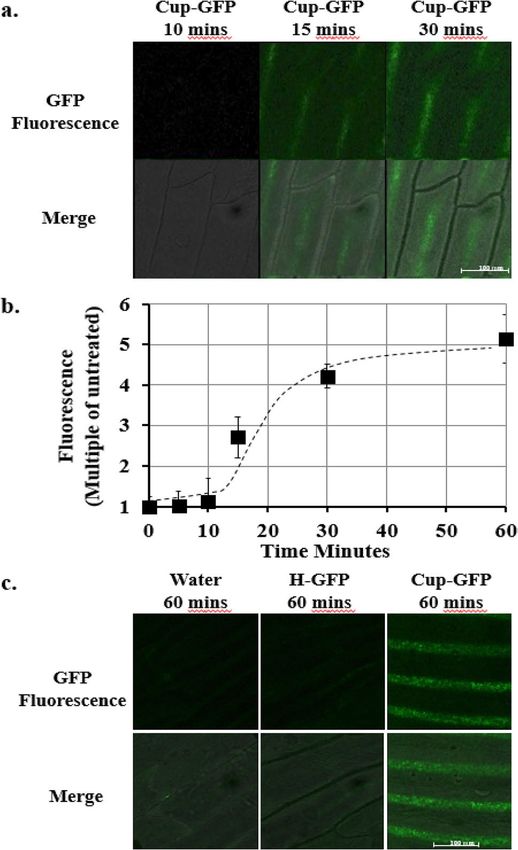
www.nature.com/scientificreports/

                            Figure 1.  (a) Cupid sequence. (b) Fluorescein labelled Cupid (F-Cupid). (c) Cupid-GFP. (d) Cupid-GFP (4 µg)
                            and H-GFP (4 µg) after SDS-PAGE gel stained with Instant blue. Molecular weight markers (Mr) shown in
                            kD. (e) Absorbance Spectrum wavelength in nanometres of native (fluorescent) GFP (red) and Cupid-GFP
                            after resolubilisation (denatured /non-fluorescent) (black). (f) Dictyostelium attached to chambered slides were
                            treated with: no peptide, 10 µM F-Cupid, 10 µM H-GFP or 10 µM Cupid-GFP for 1 h. Following washing steps,
                            cells were fixed and observed with phase (Upper Panel) or green fluorescent (Lower Panel) filters.

                            a 512 × 512 pixel array 145 × 145 µm2 with images produced by averaging 4 times per line and 4 frames. Z-stacks
                            were collected over approximately 32 µm depth with each Z section being separated by 1.09 µm. Data were col-
                            lected using photomultiplier tubes (PMT) set at 8-bit resolution (i.e. pixel intensity ranged from 0–255). Leica
                            LAS software was used to process the raw image files. Bleed through between the channels was avoided using
                            sequential scanning with photomultiplier and laser settings remaining constant within each experiment. Opti-
                            mization of PMT offset and gain was performed to the same extent within each experiment making the image
                            panels comparable.

Scientific Reports |   (2020) 10:13725 |              https://doi.org/10.1038/s41598-020-70532-x                                           3

                                                                                                                                      Vol.:(0123456789)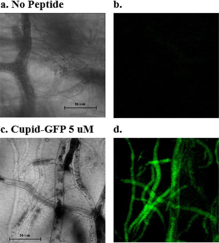
www.nature.com/scientificreports/
Plant cell preparation and imaging. A 1 × 1 cm square was cut out from an onion bulb (Allium cepa L.)
and scales carefully separated into individual layers with forceps. The epidermal cells comprising a thin film on
the inside of each layer, was peeled off and flattened onto glass slides. To this layer was added 20 µL of either pep-
tides (5 µM) or water, drop by drop, and then a coverslip was firmly pressed onto the film, flattening it as much
as possible, before inverting the glass slide and placing it under the microscope. Because the film of cells is very
thin, approximately 1 or 2 cells thick, dropping the media on top of it and then adding a cover slip was enough
to immerse the whole epidermal layer in fluid.
Protoplasts were isolated from the leaf tissue of 25-day-old Brachypodium distachyon plants by the enzymatic
methodology of Jung et al.36,37. The final preparation of purified protoplasts was resuspended in 5 mL of filter-
sterilized W5 solution (0.037% (w/v) KCl, 0.9% (w/v) NaCl, 1.84% (w/v) CaCl2, 2 mM MES-KOH pH 5.7).
Confocal live plant and fungal cell microscopy was performed on a Leica TCS SP5 II confocal system using
a Leica DMI6000 inverted microscope stand. For this study we used a DPSS 561 nm laser, an Argon laser, PL
APO 40x/1.25—0.7 and HCX PL APO 63x/1.40—0.6 (oil) objectives, GFP (green) fluorescent filters, and LAS
AF version 2.7.3.9723 software to control the microscope, scanning, laser module, tools and the image acquisi-
tion and processing.
Fungal cell preparation and imaging. The wild-type strain of zygomycetous fungus Phycomyces
blakesleeanus (Burgeff) NRRL 1555(–) was used in this study on solid agar medium supplemented with yeast
extract (0.1%) and bacto-casitone (0.1%), with agar at 1.5% (w/v) in 10 cm petri d ishes38. A 0.5 × 0.5 cm block
of agar from a plate infused with a growing P. blakesleeanus was excised and placed in contact at the side of
a sheet of agar 2 cm × 2 cm × 0.5 cm. After 4 days the fungal mycelia network had invaded this sheet, and a
0.5 × 0.5 × 0.5 cm cube was selected, excised and placed on a microscope slide. 70 µL of either water or peptide
(5 µM, non-fluorescent) was added drop by drop, allowing it to soak into the agar. After waiting 30 min, a cover-
slip was firmly pressed onto the agar. After 90 min the slide was inverted and imaged using the system above (see
Plant Cell work). Three time lapse videos were created to show the Cupid-GFP interacting with the cytoplasmic
flow within the mycelia network (4 min time lapse, 8 s imaging interval) (see Supplemental Videos 1, 2 and 3).
Image data analysis. Raw images were assessed for increase of GFP fluorescence using Image J software
(NIH Image, Version 10.2) and average values + /− SEM were compared with the untreated controls to produce
time-course graphs of Cupid-GFP uptake.
Results
The 16 amino acid Cupid sequence (Fig. 1a) was synthesized with a fluorescein molecule attached at the N
terminal (F-Cupid, Fig. 1b). When added to Dictyostelium amoeba cells in buffer at 10 µM, F-Cupid caused the
cells to become fluorescent within 60 min while fluorescein-labelled BSA did not (data not shown), indicating
cell-penetrating capability (Fig. 1f).
To investigate the nature of this capability, an expression plasmid was constructed to link the carboxy-ter-
minus of the Cupid peptide to the Aequorea victorea Green Fluorescent Protein (GFP) sequence, and the fusion
protein was purified using an N terminal polyhistidine motif (see “Materials and methods” section, Fig. 1c).
This Cupid-GFP protein migrated as a single band at the expected mass of 32.3 kD on SDS gels (Fig. 1d and
Supplemental Fig. 4) and under the conditions of purification was non-fluorescent, indicating a denatured state
(Fig. 1e). However when we tested the native, fluorescent Cupid-GFP, it aggregated in the culture medium and
the fluorescent peptide did not penetrate the cells (Supplemental Fig. 2). Therefore, all subsequent experiments
were conducted with denatured, non-fluorescent peptides. Furthermore, when the dried denatured peptides
were solubilised (in either water, phosphate-buffered saline, DMEM, Claycomb or W5 media) they remained
non-fluorescent for at least 24 h at room temperature or at 37 °C (Supplemental Fig. 3). When amoeba were
treated with Cupid-GFP (10 µM, 1 h), they became fluorescent (Fig. 1f) demonstrating the GFP cargo refolds to
its fluorescent form under the physiological conditions within the cell. Denatured H-GFP failed to demonstrate
the same permeability/fluorescence profile.
These initial experiments in amoeba used the fixative reagent Fluorsave prior to imaging which could have
altered the subcellular distribution of Cupid-GFP as has previously been shown for other C PPs39. We therefore
turned to confocal fluorescence microscopy of living, unfixed cells to examine the extent of cupid GFP perme-
ability and localization.
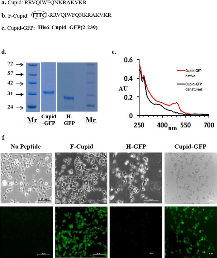
GFP fluorescence was monitored in confluent cultures of mouse cardiomyocytes using Cupid-GFP at a dose
of 5 µM and imaging total GFP fluorescence over time (Fig. 2). As the added Cupid-GFP was initially non-
fluorescent, it was possible to image the cells at 5 min intervals without disturbing the plate by washing. The data
were used to plot the overall increase in fluorescence with time, relative to the background (Fig. 2b). The data
indicated fluorescence accumulation progressed through 3 phases: an initial slow phase up to 30 min followed
by a rapid phase to 60 min and a final slow phase to saturation at 2 h. Confocal z-slices through the cells at the
60 min time point found GFP to be evenly distributed throughout the internal cell architecture and not confined
to discrete vesicles (Supplemental Fig. 1).
In the next set of experiments, the penetration of Cupid-GFP into plant and fungal cells was assayed. For
intact plant cells, onion epidermis was prepared on a glass slide and treated with peptide before a cover slip
was applied. After a lag phase, the onion epidermal cells developed strong fluorescence over background which
increased with time (Fig. 3a) to a maximum at 60 min (approx. sixfold, Fig. 3b). Onion cells treated with the
H-GFP protein (His tagged GFP, identical to Cupid-GFP but lacking the Cupid motif) failed to develop fluores-
cence above the level seen in water treated cells (Fig. 3c).
Scientific Reports | (2020) 10:13725 | https://doi.org/10.1038/s41598-020-70532-x 4
Vol:.(1234567890)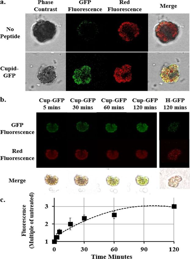
www.nature.com/scientificreports/
Figure 2. (a) Total Z-Stack confocal imaging of confluent mouse cardiomyocytes. Top panel: Culture treated
with 5 µM Cupid-GFP and imaged for Total green fluorescence at times indicated. Lower Panel: Phase contrast
image and merged image at the 60 min time point. (b) Increase in GFP fluorescence in mouse cardiomyocytes
after addition of 5 µM Cupid-GFP. The Z Stack green fluorescence at each time point was plotted as a multiple of
Z Stack green fluorescence of untreated cells. Error bars + /− SEM N = 4.
Brachypodium was selected as the test system for plant protoplasts, where the outer cellulose layer of plant
cells are enzymatically removed and the resultant protoplasts are recovered through density sedimentation and
sieving, since these have been established as a model system for studying and manipulating plant physiology.
Here protoplasts were prepared from Brachypodium leaves and treated with Cupid-GFP at a dose of 5 µM. Proto-
plasts treated with Cupid-GFP quickly developed GFP fluorescence over background and compared to the auto-
fluorescence captured in the red channel observed in all protoplasts (Fig. 4a). This GFP fluorescence increased
with time (Fig. 4b), whereas protoplasts treated with the H-GFP protein failed to develop any fluorescence above
control. The total fluorescence increase was calculated relative to untreated control and data compiled into a
graph (Fig. 4c), showing Cupid-GFP caused a rapid increase in fluorescence tending toward a plateau after 2 h.
To observe the effects of Cupid-GFP in fungi, the model organism P. Blakesleeanus was chosen. For experi-
ments to monitor cell penetration, a slab of agar in which P. Blakesleeanus was growing was soaked in non-
fluorescent Cupid-GFP (5 µM, 70 µL) and left for 90 min. The slab was then mounted for microscopy and the
mycelia network was observed to be strongly fluorescent (Fig. 5). A 1 min 42 s time-lapse movie (8 s imaging
interval) with both phase and fluorescent filters showed GFP fluorescence to be moving rapidly through the
mycelia network in the living fungus (Supplemental Video 1, 2 and 3).
Discussion
Although natural cell permeable peptides (CPPs) are present in diverse species, the potential for sequences to
npredictable40 and none have yet been shown in Dictyostelium amoeba. This suggests either the
act as CPPs is u
amoebae have no requirement for a protein transduction mechanism via a CPP, or it might achieve this end with
a cryptic CPP sequence. The present study reports the identification and characterization of a novel CPP derived
from a homeobox transcription factor in the slime mould Dictyostelium discoideum, termed Cupid (Cellular
Permeability factor in Dictyostelium) to denote its origin.
The Fluorescein-labelled 16-mer Cupid peptide was found to be efficiently taken up by amoeba cells sug-
gesting that the Cupid sequence is a CPP. It is known that the attachment of dye can influence cell penetration
Scientific Reports | (2020) 10:13725 | https://doi.org/10.1038/s41598-020-70532-x 5
Vol.:(0123456789)www.nature.com/scientificreports/
Figure 3. (a) Total Z-Slice confocal imaging of Onion epidermal cells at 10, 15, 30 min after addition of 5 µM
Cupid-GFP. Living plant cells were observed with green fluorescent (Upper Row) filter set and this was merged
with phase images (Lower Row). (b) Increase in GFP fluorescence in Onion epidermal cells after addition of
5 µM Cupid-GFP. The Total Z Stack green fluorescence at each time point was plotted as a multiple of green
fluorescence of untreated cells. Error bars +/− SEM N = 4. (c) Total Z-Slice confocal imaging of in Onion
epidermal cells at 60 min after addition of Water, 5 µM H-GFP or 5 µM Cupid-GFP. Living plant cells were
observed with a green fluorescent (Upper Row) filter set and this was merged with phase images (Lower Row).
c haracteristics41 so to test whether Cupid can translocate protein cargos into cells, Cupid was fused to GFP. When
correctly folded, GFP forms a nearly perfect cylinder and three central amino acids within this barrel structure
are spontaneously modified into a chromaphore responsible for fluorescence of the protein. Unfolding this GFP
barrel structure repositions the amino acids surrounding the chromophore region, and abolishes fluorescence
in a reversible m anner42,43.
The peptides used in this study were purified under denaturing conditions and thus were not initially fluores-
cent. Crucially, this approach meant the rise in GFP fluorescence within cells could be studied with live confocal
imaging without the requirement to aspirate the cell medium or wash the cells. It also avoided imaging artefacts
due to cell fixation, that been observed in other CPP studies and can lead to serious problems in interpreting
data39,44.
Live confocal imaging reveals that cultured mouse cells, whole plant cells, plant leaf protoplast and fungal
cells rapidly exhibit GFP fluorescence when Cupid-GFP is present in the media. As the Cupid-GFP is initially
non-fluorescent, we were able to assess total fluorescence in situ without washing steps. In mouse cells, the initial
appearance of GFP fluorescence develops slowly over the first 30 min after which it increases rapidly to 45 min
and then continuing to increase more slowly to reach a plateau after 60 min. This non-linear increase in GFP
fluorescence is likely to be the net effect of different processes occurring in the cell including membrane asso-
ciation, CPP permeation and protein refolding. These processes are likely to operate with different kinetics in
different cell types and require further work to elucidate fully. Although no gross membrane lysis was observed
in any cell types tested, possible temporary or localised membrane disruption cannot be excluded.
Scientific Reports | (2020) 10:13725 | https://doi.org/10.1038/s41598-020-70532-x 6
Vol:.(1234567890)www.nature.com/scientificreports/
Figure 4. (a) Mid-cell Z-Slice confocal imaging of Brachypodium leaf protoplasts at 1 h either in the absence
(Upper Panel) or presence (Lower Panel) of 5 µM Cupid-GFP. Living protoplasts were observed with phase,
green fluorescent and red fluorescent filters. (b) Mid-cell Z-Slice confocal imaging of Brachypodium leaf
protoplasts at 5, 30, 60 and 120 min after addition of 5 µM Cupid-GFP or Control H-GFP at 120 min (right
panel). Living protoplasts were observed with a green fluorescent (Upper Row) or red fluorescence (Middle
Row) filter set. Lower Row: merge of green, red and phase images. (c) Increase in GFP Fluorescence in
Brachypodium leaf protoplasts after addition of 5 µM Cupid-GFP. The Total Z Stack green fluorescence at each
time point was plotted as a multiple of green fluorescence of untreated cells. Error bars +/− SEM N = 4.
Although accurately relating GFP fluorescence to concentration within cells is difficult, using the paper by
Niswender et al.45 as a guide our raw data indicates Cupid-GFP instigates a four-fold increase on basal fluores-
cence at the later time points, suggesting that from an external concentration of 5 µM, the internal concentra-
tion of Cupid-GFP has reached as high as 3 µM in mouse cardiomyocytes. This is approximately tenfold higher
than calculated for a range of other CPPs linked to the SNAP-Tag system (Mr approx. 20–25 kD), labelled with
Rhodamine dye and investigated with Fluorescence Correlation Spectroscopy in mammalian c ells46.
Previous studies have fused GFP to a variety of CPPs and tested whether GFP can be taken into mammalian
cells and, if so, explored the method of e ntry31,39,47–50. Using VP22 (Human Herpes virus protein), TAT (HIV-1
transcriptional activator Tat protein), Polyarginine/Polylysine or Penetratin, these studies reported CPPs first
adhered to the cell membrane before being endocytosed within small vesicles. Cellular uptake of GFP modified
to contain a cationic “patch” but with no linked CPP has also been d etected28,33. The authors conclude this was
dependent on cell-surface glycosaminoglycans, similar to that observed with cationic CPP peptides such as
polyarginine28,33. In contrast to these earlier reports, using the ability of the confocal microscope to look through
cell sections, we saw no evidence of punctate GFP fluorescence that would be characteristic of endocytotic vesicles
in the mouse, plant or fungal cells.
Studies performed with CPPs in plants and fungi are less numerous in the literature. Our initial studies
were conducted on onion epidermal layer followed by a more detailed study with protoplasts. The Cupid-GFP
readily penetrated all cells in the onion layer, indicating that the peptide was able to pass through the cellulose-
hemicellulose network, embedded in the pectin matrix of the plant cell wall. The ability of a CPP-linked GFP
to penetrate plant cells broadly agrees with a previous study on onion c ells51. However, whereas fluorescence of
TAT-GFP and polyarginine-GFP in this earlier study appear more concentrated at the cell periphery, which might
indicate endocytotic entrapment, Cupid-GFP fluorescence appears to be more uniformly spread throughout the
onion cell. The relative absence of endogenous fluorescence in onion epidermal cells compared to mammalian
and protoplasts derived from plant leaves is likely reflected in the greater fold-increase in fluorescence from
baseline with Cupid-GFP treatment compared with these other systems.
Scientific Reports | (2020) 10:13725 | https://doi.org/10.1038/s41598-020-70532-x 7
Vol.:(0123456789)www.nature.com/scientificreports/
Figure 5. Phymocyces blakesleeanus mycelia network within a 5 mm deep agar block was treated with no
peptide (a, b) or 5 µM Cupid-GFP (c, d) for 90 min. A Z-plane was observed with phase (a, c) or green
fluorescent (b, d) filters. See Supplemental Video.
Where other researchers have genetically treated protoplasts to express GFP alone (e.g.52,53) the nascent
GFP fluorescence is seen encircling chlorophyll-dense structures but not inside them. In contrast using con-
focal imaging, we see a strong localization of Cupid-GFP and chloroplast auto-fluorescence when GFP/Red
fluorescence data is merged (Fig. 4). This indicates that Cupid-GFP has not only reached the plant cytosol but
has also penetrated structures that hold the chloroplasts. The cellular machinery to refold proteins, and thus
Cupid-GFP, may be concentrated within chlorophyll containing structures, which might account for its close
association with such. Also seen within protoplasts but not in mammalian cells are water vacuoles, inner spaces
observed as clear patches when using red fluorescence confocal microscopy (e.g. Fig. 4). GFP is also absent in
these same areas which could be due to these vacuoles lacking the cell machinery to refold Cupid-GFP. It might
also provide evidence that, having entered cells, and once it has refolded, Cupid-GFP can no longer penetrate
through additional membranes. Alternatively, there might be other factors at play in the vacuoles to suppress
GFP fluorescence, such as pH.
Previous work has found CPPs to be inefficient in entering plant protoplasts and microspores compared with
mammalian cells54,55. These studies used a wide range of CPPs (polyarginine, pVEC (derived from a murine
vascular endothelium cadherin), penetratin, transportan-10 (TP10) and model amphipathic peptide (MAP)
either labelled with small molecule dyes or fluorescent proteins (e.g. mCherry). Conjugates that entered cells
in these studies exhibited fluorescence patterns consistent with attachment to outer membranes leading to the
conclusion that CPP–macromolecular cargo complexes and conjugates were translocated via macropinocytosis
(Reviewed by56).
In contrast to these studies, we find that Cupid-GFP is taken up by protoplasts and rapidly folded to fluo-
rescent form throughout the cytosol. Whilst the quantity of Cupid-GFP that the protoplasts accumulate is
similar to that seen in mammalian cells when applied at 5 µM (threefold vs. fourfold as judged by respective
auto-fluorescence), this fluorescence emerges within protoplasts without the lag phase apparent in mammalian
or whole plant cell systems. This could be a consequence of the protoplast purification method (e.g. enzymatic
stripping of the cell membrane might clear the membrane of interfering proteins and lead to a more rapid entry
by shortening an initial Cupid-membrane association time) or it might be that the Cupid-GFP folding machinery
in plant protoplasts is more efficient than in the mammalian and whole plant cells. Since protoplasts of some spe-
cies may be regenerated into whole plants, the data presented here indicate an opportunity to use Cupid within
the window of protoplast viability to quickly introduce novel proteins to influence the plant biochemical profile
and complement genetic manipulation of plant systems.
Investigation in fungal cells has mainly been concerned with probing the anti-fungal properties of CPPs and
comparing them with a range of pore-forming antimicrobial p eptides57–60. More recently, translocation of a wide
panel of Fluorescein dye-labelled CPPs has been investigated in Candida albicans61,62. These results suggested
that penetration into fungal cells involved both endocytosis and membrane permeabilisation. When CPPs have
been fused to larger proteins such as GFP, however, the viability of Candida cells was not significantly a ffected63
Scientific Reports | (2020) 10:13725 | https://doi.org/10.1038/s41598-020-70532-x 8
Vol:.(1234567890)www.nature.com/scientificreports/
suggesting that CPPs might perform as vehicles to deliver cargoes with the broader intention of investigating
these organisms.
We report here how Cupid-GFP, applied externally onto agar in which P. Blakesleeanus is growing, quickly
penetrates through the chitin coating of the fungus and refolds within the shared mycelia cytoplasm. The refolded
GFP observed with time-lapse fluorescence microscopy reveals dynamic anterograde and retrograde movement
of nuclei, mitochondria, vacuoles and vesicles through hyphae trunks of P. Blakesleeanus. This bulk cytoplas-
mic flow has previously been observed in fungi using dyes or genetic mutants synthesizing proteins fused with
GFP64–66. Our current study supports the conclusion that a CPP can broaden the range of applications of CPPs
in Fungi.
In conclusion, we propose that the novel CPP Cupid might be a valuable candidate for further investigations
concerning its ability to transport large protein cargo molecules into and throughout a great variety of cells across
the kingdoms, which are free to refold into bioactive forms. Such capability would lend itself toward numerous
applications within research and maybe future therapies.
Received: 22 November 2019; Accepted: 29 July 2020
References
1. Gow, N. A. R., Latge, J.-P. & Munro, C. A. The fungal cell wall: structure, biosynthesis, and function. Microbiol. Spectrum https://
doi.org/10.1128/microbiolspec.FUNK-0035-2016 (2017).
2. Höfte, H. & Voxeur, A. Plant cell walls. Curr. Biol. 27, R865–R870. https://doi.org/10.1016/j.cub.2017.05.025 (2017).
3. Prochiantz, A. Messenger proteins: homeoproteins, TAT and others. Curr. Opin. Cell Biol. 12, 400–406. https://doi.org/10.1016/
S0955-0674(00)00108-3 (2000).
4. Antoniou, X. & Borsello, T. Cell permeable peptides: a promising tool to deliver neuroprotective agents in the brain. Pharmaceu-
ticals https://doi.org/10.3390/ph3020379 (2010).
5. Heitz, F., Morris, M. C. & Divita, G. Twenty years of cell-penetrating peptides: from molecular mechanisms to therapeutics. Br. J.
Pharmacol. 157, 195–206. https://doi.org/10.1111/j.1476-5381.2009.00057.x (2009).
6. Mäe, M. & Langel, Ü. Cell-penetrating peptides as vectors for peptide, protein and oligonucleotide delivery. Curr. Opin. Pharm.
6, 509–514. https://doi.org/10.1016/j.coph.2006.04.004 (2006).
7. Torchilin, V. P. Tatp-mediated intracellular delivery of pharmaceutical nanocarriers. Biochem. Soc. Trans. 35, 816–820. https://doi.
org/10.1042/bst0350816 (2007).
8. Fischer, P. M. Cellular uptake mechanisms and potential therapeutic utility of peptidic cell delivery vectors: progress 2001–2006.
Med. Res. Rev. 27, 755–795. https://doi.org/10.1002/med.20093 (2007).
9. Durzyńska, J. et al. Viral and other cell-penetrating peptides as vectors of therapeutic agents in medicine. J. Pharmacol. Exp. Ther.
354, 32–42. https://doi.org/10.1124/jpet.115.223305 (2015).
10. Tilstra, J. et al. Protein transduction: identification, characterization and optimization. Biochem. Soc. Trans. 35, 811–815. https://
doi.org/10.1042/bst0350811 (2007).
11. Alhakamy, N. A., Kaviratna, A., Berkland, C. J. & Dhar, P. Dynamic measurements of membrane insertion potential of synthetic
cell penetrating peptides. Langmuir 29, 15336–15349. https://doi.org/10.1021/la403370p (2013).
12. Almeida, P. F. & Pokorny, A. Mechanisms of antimicrobial, cytolytic, and cell-penetrating peptides: from kinetics to thermodynam-
ics. Biochemistry 48, 8083–8093. https://doi.org/10.1021/bi900914g (2009).
13. Scheller, A. et al. Structural requirements for cellular uptake of α-helical amphipathic peptides. J. Pept. Sci. 5, 185–194. https://doi.
org/10.1002/(sici)1099-1387(199904)5:4%3c185::Aid-psc184%3e3.0.Co;2-9 (1999).
14. Foerg, C. et al. Decoding the entry of two novel cell-penetrating peptides in hela cells: lipid raft-mediated endocytosis and endo-
somal escape. Biochemistry 44, 72–81. https://doi.org/10.1021/bi048330+ (2005).
15. Fittipaldi, A. et al. Cell membrane lipid rafts mediate caveolar endocytosis of HIV-1 tat fusion proteins. J. Biol. Chem. 278,
34141–34149. https://doi.org/10.1074/jbc.M303045200 (2003).
16. Duchardt, F., Fotin-Mleczek, M., Schwarz, H., Fischer, R. & Brock, R. A comprehensive model for the cellular uptake of cationic
cell-penetrating peptides. Traffic 8, 848–866. https://doi.org/10.1111/j.1600-0854.2007.00572.x (2007).
17. Nakase, I. et al. Cellular uptake of arginine-rich peptides: roles for macropinocytosis and actin rearrangement. Mol. Ther. 10,
1011–1022. https://doi.org/10.1016/j.ymthe.2004.08.010 (2004).
18. Shiroh, F., Hisaaki, H. & Ikuhiko, N. Arginine-rich peptides: methods of translocation through biological membranes. Curr. Pharm.
Des. 19, 2863–2868. https://doi.org/10.2174/1381612811319160003 (2013).
19. Krämer, S. D. & Wunderli-Allenspach, H. No entry for TAT(44–57) into liposomes and intact MDCK cells: novel approach to
study membrane permeation of cell-penetrating peptides. Biochim. Biophys. Acta (BBA) Biomembr. 1609, 161–169. https://doi.
org/10.1016/S0005-2736(02)00683-1 (2003).
20. Pourmousa, M. & Karttunen, M. Early stages of interactions of cell-penetrating peptide penetratin with a DPPC bilayer. Chem.
Phys. Lipids 169, 85–94. https://doi.org/10.1016/j.chemphyslip.2013.02.011 (2013).
21. Hirose, H. et al. Transient focal membrane deformation induced by arginine-rich peptides leads to their direct penetration into
cells. Mol. Ther. 20, 984–993. https://doi.org/10.1038/mt.2011.313 (2012).
22. Maniti, O., Alves, I., Trugnan, G. & Ayala-Sanmartin, J. Distinct behaviour of the homeodomain derived cell penetrating peptide
penetratin in interaction with different phospholipids. PLoS ONE 5, e15819–e15819. https://doi.org/10.1371/journal.pone.00158
19 (2010).
23. Di Pisa, M., Chassaing, G. & Swiecicki, J.-M. Translocation mechanism(s) of cell-penetrating peptides: biophysical studies using
artificial membrane bilayers. Biochemistry 54, 194–207. https://doi.org/10.1021/bi501392n (2015).
24. Palm-Apergi, C., Lönn, P. & Dowdy, S. F. Do cell-penetrating peptides actually “penetrate” cellular membranes?. Mol. Ther. 20,
695–697. https://doi.org/10.1038/mt.2012.40 (2012).
25. Allolio, C. et al. Arginine-rich cell-penetrating peptides induce membrane multilamellarity and subsequently enter via formation
of a fusion pore. Proc. Natl. Acad. Sci. U. S. A. 115, 11923–11928. https://doi.org/10.1073/pnas.1811520115 (2018).
26. Brock, R. The uptake of arginine-rich cell-penetrating peptides: putting the puzzle together. Bioconjugate Chem. 25, 863–868. https
://doi.org/10.1021/bc500017t (2014).
27. Lorents, A., Säälik, P., Langel, Ü & Pooga, M. Arginine-rich cell-penetrating peptides require nucleolin and cholesterol-poor sub-
domains for translocation across membranes. Bioconjugate Chem. 29, 1168–1177. https://doi.org/10.1021/acs.bioconjchem.7b008
05 (2018).
Scientific Reports | (2020) 10:13725 | https://doi.org/10.1038/s41598-020-70532-x 9
Vol.:(0123456789)www.nature.com/scientificreports/
28. Yuki, T.-H. & Hiroyuki, S. Current understanding of physicochemical mechanisms for cell membrane penetration of arginine-rich
cell penetrating peptides: role of glycosaminoglycan interactions. Curr. Protein Pept. Sci. 19, 623–630. https: //doi.org/10.2174/13892
03719666180112100747 (2018).
29. Wadia, J. S., Stan, R. V. & Dowdy, S. F. Transducible TAT-HA fusogenic peptide enhances escape of TAT-fusion proteins after lipid
raft macropinocytosis. Nat. Med. 10, 310–315. https://doi.org/10.1038/nm996 (2004).
30. Tünnemann, G. et al. Cargo-dependent mode of uptake and bioavailability of TAT-containing proteins and peptides in living cells.
FASEB J. 20, 1775–1784. https://doi.org/10.1096/fj.05-5523com (2006).
31. Gillmeister, M. P., Betenbaugh, M. J. & Fishman, P. S. Cellular trafficking and photochemical internalization of cell penetrating
peptide linked cargo proteins: a dual fluorescent labeling study. Bioconjugate Chem. 22, 556–566. https://doi.org/10.1021/bc900
445g (2011).
32. Allen, J. K., Brock, D. J., Kondow-McConaghy, H. M. & Pellois, J.-P. Efficient delivery of macromolecules into human cells by
improving the endosomal escape activity of cell-penetrating peptides: lessons learned from dfTAT and its analogs. Biomolecules
8, 50. https://doi.org/10.3390/biom8030050 (2018).
33. Fuchs, S. M. & Raines, R. T. Arginine grafting to endow cell permeability. ACS Chem. Biol. 2, 167–170. https://doi.org/10.1021/
cb600429k (2007).
34. Eichinger, L. et al. The genome of the social amoeba Dictyostelium discoideum. Nature 435(7038), 43–57 (2005).
35. Fey, P., Kowal, A. S., Gaudet, P., Pilcher, K. E. & Chisholm, R. L. Protocols for growth and development of Dictyostelium discoideum.
Nat. Protoc. 2, 1307–1316. https://doi.org/10.1038/nprot.2007.178 (2007).
36. Jung, H.-I., Gayomba, S. R., Yan, J. & Vatamaniuk, O. K. Brachypodium distachyon as a model system for studies of copper transport
in cereal crops. Front. Plant Sci. https://doi.org/10.3389/fpls.2014.00236 (2014).
37. Jung, H.-I., Yan, J., Zhai, Z. & Vatamaniuk, O. K. in Plant Functional Genomics: Methods and Protocols (eds Jose M. Alonso & Anna
N. Stepanova) 433–452 (Springer, New York, 2015).
38. Sutter, R. P. Mutations affecting sexual development in Phycomyces blakesleeanus. Proc. Natl. Acad. Sci. U. S. A. 72, 127–130. https
://doi.org/10.1073/pnas.72.1.127 (1975).
39. Lundberg, M., Wikström, S. & Johansson, M. Cell surface adherence and endocytosis of protein transduction domains. Mol. Ther.
8, 143–150. https://doi.org/10.1016/S1525-0016(03)00135-7 (2003).
40. Harreither, E. et al. Characterization of a novel cell penetrating peptide derived from human Oct4. Cell Regen. (Lond.) 3, 2–2. https
://doi.org/10.1186/2045-9769-3-2 (2014).
41. Hedegaard, S. F. et al. Fluorophore labeling of a cell-penetrating peptide significantly alters the mode and degree of biomembrane
interaction. Sci. Rep. 8, 6327. https://doi.org/10.1038/s41598-018-24154-z (2018).
42. Ormö, M. et al. Crystal structure of the Aequorea victoria green fluorescent protein. Science 273, 1392–1395. https://doi.
org/10.1126/science.273.5280.1392 (1996).
43. Ward, W. W. & Bokman, S. H. Reversible denaturation of Aequorea green-fluorescent protein: physical separation and characteri-
zation of the renatured protein. Biochemistry 21, 4535–4540. https://doi.org/10.1021/bi00262a003 (1982).
44. Thorén, P. E. G. et al. Uptake of analogs of penetratin, Tat(48–60) and oligoarginine in live cells. Biochem. Biophys. Res. Commun.
307, 100–107. https://doi.org/10.1016/S0006-291X(03)01135-5 (2003).
45. Niswender, K. D., Blackman, S. M., Rohde, L., Magnuson, M. A. & Piston, D. W. Quantitative imaging of green fluorescent protein
in cultured cells: comparison of microscopic techniques, use in fusion proteins and detection limits. J. Microsc. 180, 109–116. https
://doi.org/10.1111/j.1365-2818.1995.tb03665.x (1995).
46. Wissner, R. F., Steinauer, A., Knox, S. L., Thompson, A. D. & Schepartz, A. Fluorescence correlation spectroscopy reveals efficient
cytosolic delivery of protein cargo by cell-permeant miniature proteins. ACS Cent. Sci. 4, 1379–1393. https: //doi.org/10.1021/acsce
ntsci.8b00446 (2018).
47. Rajarao, G. K., Nekhotiaeva, N. & Good, L. Peptide-mediated delivery of green fluorescent protein into yeasts and bacteria. FEMS
Microbiol. Lett. 215, 267–272. https://doi.org/10.1111/j.1574-6968.2002.tb11401.x (2002).
48. Nischan, N. et al. Covalent attachment of cyclic TAT peptides to GFP results in protein delivery into live cells with immediate
bioavailability. Angew. Chem. Int. Ed. 54, 1950–1953. https://doi.org/10.1002/anie.201410006 (2015).
49. Han, K. H., Meilan, R., Ma, C. & Strauss, S. H. An Agrobacterium tumefaciens transformation protocol effective on a variety of
cottonwood hybrids (genus Populus). Plant Cell Rep. 19, 315–320 (2000).
50. Jo, J., Hong, S., Choi, W. Y. & Lee, D. R. Cell-penetrating peptide (CPP)-conjugated proteins is an efficient tool for manipulation
of human mesenchymal stromal cells. Sci. Rep. 4, 4378. https://doi.org/10.1038/srep04378 (2014).
51. Chang, M., Chou, J.-C. & Lee, H.-J. Cellular internalization of fluorescent proteins via arginine-rich intracellular delivery peptide
in plant cells. Plant Cell Physiol. 46, 482–488. https://doi.org/10.1093/pcp/pci046 (2005).
52. Bhushan, S. et al. Catalysis, subcellular localization, expression and evolution of the targeting peptides degrading protease, AtPreP2.
Plant Cell Physiol. 46, 985–996. https://doi.org/10.1093/pcp/pci107 (2005).
53. Garcion, C. et al. Characterization and biological function of the Isochorismate Synthase2 gene of Arabidopsis. Plant Physiol. 147,
1279–1287. https://doi.org/10.1104/pp.108.119420 (2008).
54. Mäe, M. et al. Internalisation of cell-penetrating peptides into tobacco protoplasts. Biochim. Biophys. (BBA) Biomembr. 1669,
101–107. https://doi.org/10.1016/j.bbamem.2005.01.006 (2005).
55. Bilichak, A., Luu, J. & Eudes, F. Intracellular delivery of fluorescent protein into viable wheat microspores using cationic peptides.
Front. Plant Sci. 6, 666–666. https://doi.org/10.3389/fpls.2015.00666 (2015).
56. Ziemienowicz, A., Pepper, J. & Eudes, F. in Cell-Penetrating Peptides: Methods and Protocols (ed Ülo Langel) 417–434 (Springer,
New York, 2015).
57. Nekhotiaeva, N. et al. Cell entry and antimicrobial properties of eukaryotic cell-penetrating peptides. FASEB J. 18, 394–396. https
://doi.org/10.1096/fj.03-0449fje (2004).
58. Palm, C., Netzereab, S. & Hällbrink, M. Quantitatively determined uptake of cell-penetrating peptides in non-mammalian cells
with an evaluation of degradation and antimicrobial effects. Peptides 27, 1710–1716. https: //doi.org/10.1016/j.peptid es.2006.01.006
(2006).
59. Muñoz, A. et al. Two functional motifs define the interaction, internalization and toxicity of the cell-penetrating antifungal peptide
PAF26 on fungal cells. PLoS ONE 8, e54813–e54813. https://doi.org/10.1371/journal.pone.0054813 (2013).
60. Budagavi, D. P., Zarin, S. & Chugh, A. Antifungal activity of Latarcin 1 derived cell-penetrating peptides against Fusarium solani.
Biochim. Biophys. Acta (BBA) Biomembr. 250–256, 2018. https://doi.org/10.1016/j.bbamem.2017.10.029 (1860).
61. Gong, Z. & Karlsson, A. J. Translocation of cell-penetrating peptides into Candida fungal pathogens. Protein Sci. 26, 1714–1725.
https://doi.org/10.1002/pro.3203 (2017).
62. Gong, Z., Ikonomova, S. P. & Karlsson, A. J. Secondary structure of cell-penetrating peptides during interaction with fungal cells.
Protein Sci. 27, 702–713. https://doi.org/10.1002/pro.3364 (2018).
63. Gong, Z., Walls, M. T., Karley, A. N. & Karlsson, A. J. Effect of a flexible linker on recombinant expression of cell-penetrating peptide
fusion proteins and their translocation into fungal cells. Mol. Biotechnol. 58, 838–849. https://doi.org/10.1007/s12033-016-9983-5
(2016).
64. Hickey, P. C., Jacobson, D. J., Read, N. D. & Louise Glass, N. Live-cell imaging of vegetative hyphal fusion in Neurospora crassa.
Fungal Genet. Biol. 37, 109–119. https://doi.org/10.1016/S1087-1845(02)00035-X (2002).
Scientific Reports | (2020) 10:13725 | https://doi.org/10.1038/s41598-020-70532-x 10
Vol:.(1234567890)www.nature.com/scientificreports/
65. Ramos-García, S. L., Roberson, R. W., Freitag, M., Bartnicki-García, S. & Mouriño-Pérez, R. R. Cytoplasmic bulk flow propels
nuclei in mature hyphae of Neurospora crassa. Eukaryot. Cell 8, 1880–1890. https://doi.org/10.1128/ec.00062-09 (2009).
66. Abadeh, A. & Lew, R. R. Mass flow and velocity profiles in Neurospora hyphae: partial plug flow dominates intra-hyphal transport.
Microbiology 159, 2386–2394. https://doi.org/10.1099/mic.0.071191-0 (2013).
Acknowledgements
IBERS receives strategic funding from the Biotechnology and Biological Sciences Research Council (BBSRC)
via Grant (BB/CSP1730/1).
Author contributions
J.R. designed and synthesized the Cupid variants. J.R., D.P. and H.D.J. conceived and supervised the experimental
work. D.F., D.P., A.M., C.H.G. and J.R. conducted various aspects of the experimental work. J.R., D.P. and H.D.J.
interpreted the data, wrote the manuscript and compiled the figures. All authors edited and corrected the final
manuscript.
Competing interests
One of the authors (JR) is the founder of Cupid Peptides. All other authors have no competing interests.
Additional information
Supplementary information is available for this paper at https://doi.org/10.1038/s41598-020-70532-x.
Correspondence and requests for materials should be addressed to J.R. or H.J.
Reprints and permissions information is available at www.nature.com/reprints.
Publisher’s note Springer Nature remains neutral with regard to jurisdictional claims in published maps and
institutional affiliations.
Open Access This article is licensed under a Creative Commons Attribution 4.0 International
License, which permits use, sharing, adaptation, distribution and reproduction in any medium or
format, as long as you give appropriate credit to the original author(s) and the source, provide a link to the
Creative Commons license, and indicate if changes were made. The images or other third party material in this
article are included in the article’s Creative Commons license, unless indicated otherwise in a credit line to the
material. If material is not included in the article’s Creative Commons license and your intended use is not
permitted by statutory regulation or exceeds the permitted use, you will need to obtain permission directly from
the copyright holder. To view a copy of this license, visit http://creativecommons.org/licenses/by/4.0/.
© The Author(s) 2020
Scientific Reports | (2020) 10:13725 | https://doi.org/10.1038/s41598-020-70532-x 11
Vol.:(0123456789)You can also read