Cell adhesion-mediated mitochondria transfer contributes to mesenchymal stem cell-induced chemoresistance on T cell acute lymphoblastic leukemia ...
←
→
Page content transcription
If your browser does not render page correctly, please read the page content below
Wang et al. Journal of Hematology & Oncology (2018) 11:11
DOI 10.1186/s13045-018-0554-z

 RESEARCH                                                                                                                                      Open Access

Cell adhesion-mediated mitochondria
transfer contributes to mesenchymal stem
cell-induced chemoresistance on T cell
acute lymphoblastic leukemia cells
Jiancheng Wang1,2,3†, Xin Liu1,2†, Yuan Qiu1,2†, Yue Shi1,2, Jianye Cai1,2,3, Boyan Wang1,2, Xiaoyue Wei1,2,
Qiong Ke1,2,3, Xin Sui2,4, Yi Wang2, Yinong Huang1,2,3, Hongyu Li1,2, Tao Wang1,2, Ren Lin5, Qifa Liu5
and Andy Peng Xiang1,2,3,6,7*

  Abstract
  Background: Despite the high cure rate of T cell acute lymphoblastic leukemia (T-ALL), drug resistance to
  chemotherapy remains a significant clinical problem. Bone marrow mesenchymal stem cells (MSCs) protect leukemic
  cells from chemotherapy, but the underlying mechanisms are poorly understood. In this study, we aimed to uncover
  the mechanism of MSC-induced chemoresistance in T-ALL cells, thus providing a promising clinical therapy target.
  Methods: Cell viability was determined using the viability assay kit CCK-8. The mitochondrial ROS levels were detected
  using the fluorescent probe MitoSOX™ Red, and fluorescence intensity was measured by flow cytometry. In vitro, MSCs
  and Jurkat cells were cocultured. MSCs were labeled with green fluorescent protein (GFP), and Jurkat cells were labeled
  with the mitochondria-specific dye MitoTracker Red. Bidirectional mitochondrial transfer was detected by flow cytometry
  and confocal microscopy. The mechanism of mitochondria transfer was analyzed by inhibitor assays. Transcripts related to
  Jurkat cell/MSC adhesion in the coculture system were assessed by qRT-PCR. After treatment with a neutralizing antibody
  against a key adhesion molecule, mitochondria transfer from Jurkat cells to MSCs was again detected by flow cytometry
  and confocal microscopy. Finally, we verified our findings using human primary T-ALL cells cocultured with MSCs.
  Results: Chemotherapeutic drugs caused intracellular oxidative stress in Jurkat cells. Jurkat cells transfer mitochondria to
  MSCs but receive few mitochondria from MSCs, resulting in chemoresistance. This process of mitochondria transfer is
  mediated by tunneling nanotubes, which are protrusions that extend from the cell membrane. Moreover, we found that
  most Jurkat cells adhered to MSCs in the coculture system, which was mediated by the adhesion molecule ICAM-1.
  Treatment with a neutralizing antibody against ICAM-1 led to a decreased number of adhering Jurkat cells, decreased
  mitochondria transfer, and increased chemotherapy-induced cell death.
  Conclusions: We show evidence that mitochondria transfer from Jurkat cells to MSCs, which is mediated by cell
  adhesion, may be a potential therapeutic target for T-ALL treatment.
  Keywords: Mesenchymal stem cells, Cell adhesion, Mitochondria transfer, Reactive oxygen species, Chemoresistance

* Correspondence: xiangp@mail.sysu.edu.cn
†
 Equal contributors
1
 Program of Stem Cells and Regenerative Medicine, Affiliated Guangzhou
Women and Children’s Hospital, Zhongshan School of Medicine, Sun Yat-Sen
University, Guangzhou 510080, China
2
 Center for Stem Cell Biology and Tissue Engineering, Key Laboratory for
Stem Cells and Tissue Engineering, Ministry of Education, Sun Yat-Sen
University, 74# Zhongshan 2nd Road, Guangzhou, Guangdong, China
Full list of author information is available at the end of the article

                                      © The Author(s). 2018 Open Access This article is distributed under the terms of the Creative Commons Attribution 4.0
                                      International License (http://creativecommons.org/licenses/by/4.0/), which permits unrestricted use, distribution, and
                                      reproduction in any medium, provided you give appropriate credit to the original author(s) and the source, provide a link to
                                      the Creative Commons license, and indicate if changes were made. The Creative Commons Public Domain Dedication waiver
                                      (http://creativecommons.org/publicdomain/zero/1.0/) applies to the data made available in this article, unless otherwise stated.
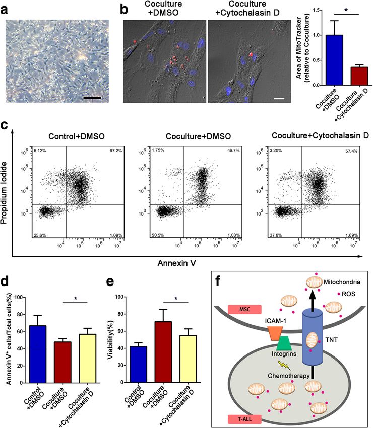
Wang et al. Journal of Hematology & Oncology (2018) 11:11                                                       Page 2 of 13

Background                                                     can move between cells, through tunneling nanotubes
Medical advances have improved the survival of adult           (TNTs), microvesicles, or gap junctions, leading to protec-
patients with acute lymphoblastic leukemia (ALL) over the      tion against tissue injury or resistance to therapeutic
past few decades. T cell acute lymphoblastic leukemia          agents [21, 28–37]. However, there are few studies on the
(T-ALL) is one of the most aggressive hematologic malig-       mechanisms of mitochondria transfer between MSCs and
nancies, accounting for up to 10–15% of pediatric ALL          T-ALL cells. As mitochondrial ROS plays a major role in
and 25% of adult ALL cases [1], and arises from the malig-     the intracellular redox balance [38], it is important to
nant transformation of T cell progenitors [2, 3]. Although     determine whether mitochondrial transfer can be used to
high-dose multi-agent chemotherapy is clinically effective     modify ROS levels.
in most cases, primary drug resistance and relapse are           In this study, we examined bidirectional mitochondria
frequently observed [4], preventing T-ALL from being           transfer between MSCs and T-ALL cells and found that
cured [5, 6].                                                  T-ALL cells exposed to chemotherapeutic drugs trans-
  Recent studies have shown that the bone marrow               ferred many mitochondria to MSCs but received few
milieu, especially mesenchymal stem cells (MSCs), has          from MSCs. This process facilitates the proliferation and
pro-survival effects on leukemia cells and protects            survival of leukemia cells by reducing ROS levels.
leukemic cells from chemotherapy [7–11]. On one hand,          Furthermore, by impeding cell adhesion, the mitochon-
soluble factor-mediated drug resistance has been proposed      dria transfer was disturbed, therefore decreasing the
to contribute to MSC-induced chemoresistance. Iwamoto          survival rate under chemotherapy. These results suggest
and colleagues found that MSCs secreted asparagine that        that mitochondria transfer may be a candidate target for
was taken up by ALL cells and thus protected ALL cells         T-ALL treatment.
from asparaginase treatment [12]. On the other hand, cell
adhesion-mediated drug resistance is also an important         Methods
mechanism of MSC-induced chemoresistance. For                  Cell culture
example, Mudry et al. reported that MSCs interacted with       Human T-ALL cell line Jurkat was purchased from the
leukemia cells by increasing the expression of vascular cell   Cell Bank of the Chinese Academy of Sciences (Shanghai,
adhesion molecule-1 (VCAM-1), protecting leukemia cells        China). The culture medium was RPMI 1640 (Hyclone,
from cytarabine and etoposide cytotoxicity [13]. However,      Logan, UT, USA) supplemented with fetal bovine serum
the role of MSCs in T-ALL cell drug resistance remains         (FBS; Gibco, Grand Island, NY, USA), penicillin, and
unclear, and thus, intensive studies on the mechanisms by      streptomycin (Sigma, St. Louis, MO, USA).
which MSCs protect T-ALL cells are needed to develop              For collection of human primary T-ALL cells, 10 enrolled
T-ALL treatments.                                              T-ALL patients were previously untreated and newly
  Excessive intracellular reactive oxygen species (ROS)        diagnosed at the Department of Haematology, Nanfang
can induce the apoptosis of cells [14–16]. An important        Hospital, Southern Medical University (Guangzhou, China),
mechanism for chemotherapeutic agents is to induce             and Department of Pediatrics, Sun Yat-Sen Memorial
cancer cell apoptosis by enhancing intracellular ROS           Hospital, Sun Yat-Sen University (Guangzhou, China).
levels. Such agents include paclitaxel, anthracyclines,        Consent was provided according to the Declaration of
ara-C, and methotrexate (MTX), among others [17].              Helsinki. Informed consent was obtained following institu-
Mitochondria are the most important source of cellular         tional guidelines, and approval was obtained from the insti-
ROS [18–22]. Therefore, upregulating mitochondrial             tutional review board of Sun Yat-Sen University. All human
ROS levels is a potential strategy for killing cancer cells    bone marrow or peripheral blood samples were obtained
[23, 24], including T-ALL cells [25]. Jitschin et al.          with written informed consent. Primary CD3+ T-ALL cells
reported that induction of mitochondrial ROS in chronic        were isolated through density gradient centrifugation on
lymphocytic leukemia (CLL) cells with PK11195, a drug          standard Ficoll-HyPaque and subjected to fluorescence-
that can generate mitochondrial superoxide, resulted in        activated cell sorting (FACS; BD Bioscience Influx, Franklin
cell apoptosis [26]. Our previous research showed that         Lakes, NJ, USA).
MSCs reduced mitochondrial ROS levels in T-ALL cells              MSCs were collected from bone marrow aspirates of
through the ERK pathway and thus protected T-ALL cells         healthy volunteers with informed consent. Isolation and
from chemotherapeutics ara-C or MTX. Accordingly,              characterization of MSCs were performed as we previously
inhibition of the ERK activation with the ERK inhibitor        described [39, 40]. Briefly, the bone marrow aspirates were
PD325901 increased mitochondrial ROS levels and the            diluted, have undergone the density gradient centrifugation,
cell death rate of T-ALL cells [27]. These results indicated   and were counted and planted before purification [41, 42].
that MSCs protect T-ALL cells by decreasing mitochon-          The culture medium was low-glucose DMEM (Hyclone,
drial ROS levels in T-ALL cells. Interestingly, in the past    Logan, UT, USA) supplemented with 10% FBS and 100 IU/
few years, several studies have reported that mitochondria     ml penicillin and streptomycin. To generate GFP-labeled
Wang et al. Journal of Hematology & Oncology (2018) 11:11                                                        Page 3 of 13

MSCs, MSCs were transfected with lentivirus containing         Staining of F-actin in MSCs
lentiviral expression vector pLV/puro-EF1a-GFP [43] using      To visualize TNTs which consist of F-actin, MSCs were
the X-treme GENE HP reagent (Roche) according to the           immersion-fixed in 4% paraformaldehyde. After a brief
manufacturer’s instructions. Three days after transfection,    permeabilization with 0.1% Triton X-100, cell coverslips
GFP-labeled MSCs were purified by FACS (Influx, Becton         were incubated in AlexaFluor 647-conjugated phalloidin
Dickinson).                                                    for 20 min at room temperature.
  Several culture models were used in this article. (1)
Monoculture: Jurkat cells/human primary T-ALL cells            Mitochondrial ROS assessment
(5 × 10⁵ /ml) or MSCs (5 × 10⁴ /ml) were respectively          The levels of mitochondrial ROS were detected using
seeded in 24-well plates. (2) Coculture: MSCs (5 × 10⁴ /ml)    the fluorescent probes MitoSOX™ Red (Molecular
and Jurkat cells/human primary T-ALL cells (5 × 10⁵ /ml)       Probes, Life Technologies, Carlsbad, CA, USA), and
were suspended in RPMI 1640 and seeded in 24-well plates.      fluorescent intensity was measured by flow cytometry
(3) Transwell: Jurkat cells/human primary T-ALL cells          (FACScan; Becton Dickinson, San Diego, CA, USA).
(5 × 10⁵ /ml) were seeded in the Transwell inserts
(Millipore), which were inserted into the 24-well plates       Annexin V/PI flow cytometry analysis
with preseeded MSCs (5 × 10⁴ /ml). The relative measure-       Jurkat cells from monoculture or coculture system were
ments were performed after coculture for 1 to 3 days.          treated with ara-C or MTX for 2 days and harvested by
                                                               centrifugation. Jurkat cells were then stained with annexin
Reagents and antibodies                                        V/propidium iodide (PI) assay kit (BIOSCI BIOTECH,
Ara-C and MTX were purchased from Pharmacia Pty Ltd.           Shanghai, China) according to the manufacturer’s instruc-
(NSW, Australia) and Calbiochem (San Diego, CA, USA),          tion. The apoptotic population was immediately evaluated
respectively. 300nM ara-C or 100 nM MTX were used to           by flow cytometry. The percentages of early apoptotic cells
cause cytotoxicity in Jurkat cells. 18-α-GA, dynasore, and     (annexin V+/PI−) and late apoptotic cells (annexin V+/PI+)
cytochalasin D were purchased from Sigma-Aldrich and           were analyzed and graphed.
were used in a concentration of 50, 50, and 1 μM, respect-
ively. Neutralizing anti-ICAM-1 antibody (MS305PABX)           RNA isolation and qRT-PCR analysis
was purchased from Invitrogen (Carlsbad, CA, USA). Alexa       Total mRNA from MSCs was extracted using an RNeasy
Fluor™ 647 phalloidin was purchased from Thermo Fisher         Mini Kit (Qiagen), and complementary DNA (cDNA) was
Scientific.                                                    synthesized using a QuantiTect Reverse Transcription Kit
                                                               (Qiagen) according to the manufacturers’ protocols. qRT-
                                                               PCRs were carried out using SYBR Green qPCR SuperMix
Cell viability assay                                           (Roche, Indianapolis, IN, USA) and a LightCycler 480
Cell viability was determined using a CCK-8 assay kit          Detection System (Roche) as described by the manufacturer.
(Dojindo Laboratories, Kumamoto, Japan) according to           Target mRNA levels were normalized with respect to those
the manufacturer’s instructions. The principle of this assay   of β-actin. The primer sequences used for qRT-PCR are
is that some components of the CCK-8 assay kit will be         listed in Additional file 1: Table S1.
reduced by mitochondria to produce formazan so as to be
detectable. Briefly, 5 × 10⁴ Jurkat cells in 100 μl culture    Statistical analyses
media were plated to a 96-well plate in suspension. Then,      All experiments were performed at least three separate
the samples were incubated with CCK-8 solution (10 μl)         times. All data are expressed as the mean ± S.E.M. Com-
for 4 h at 37 °C; the absorbance in each well was quanti-      parisons among groups were performed using one-way
fied at 450 nm using an automated enzyme-linked                analysis of variance (ANOVA) or Student’s t test. Statistical
immunosorbent assay reader (Tecan, Salzburg, Austria).         differences were determined by GraphPad Prism 5.0
Cell viability was calculated according to the manufac-        software (GraphPad Software Inc., CA, USA). A two-sided
turer’s instructions.                                          P value < 0.05 was considered to be statistically significant.
                                                                 For the other experimental procedures, please see
Fluorescence staining of mitochondria                          Additional file 1.
MitoTracker Red (Molecular Probes) was used to label
mitochondria. Jurkat cells or MSCs were incubated with         Results
200 nM MitoTracker Red in culture media for 10 min at          Jurkat cells transfer mitochondria to MSCs when exposed
37 °C. Excess of the dye was washed out with PBS. Then,        to chemotherapeutic drugs
4 days later, stained cells were then seeded for monocul-      We previously found that MSCs could protect T-ALL
ture and coculture. We verified the feasibility of mito-       cells from chemotherapeutic cell death in indirect
chondria dye method in Additional file 1: Figure S1.           (Transwell) and direct coculture system. Furthermore,
Wang et al. Journal of Hematology & Oncology (2018) 11:11                                                        Page 4 of 13

we showed that exposure of T-ALL cells to MSCs                  TNT containing mitochondria in it (Fig. 2a). Then, 18-α-
decreased mitochondrial ROS levels via the ERK/Drp1             GA (a blocker of gap junctions), the dynamin inhibitor
pathway under both culture conditions, However, when            dynasore (a blocker of microvesicle endocytosis), and the
exposed to chemotherapeutic drugs, Jurkat cells in direct       potent actin polymerization inhibitor cytochalasin D (a
contact with MSCs exhibited significantly lower mito-           blocker of TNT formation) were added to the coculture
chondrial ROS levels than cells in the Transwell system         system with ara-C or MTX. Mitochondria transfer was then
[27]. We thus wondered whether there were other                 analyzed with flow cytometry. Prior to coculture, Jurkat
mechanisms by which MSCs decrease ROS levels in                 cells were stained with MitoTracker Red to label mitochon-
Jurkat cells in a cytotoxic environment. As mitochondria        dria. We observed that, compared with DMSO, cytochala-
are the key source of intracellular ROS, alterations in         sin D significantly decreased the number of Red+ MSCs,
mitochondrial number and function could influence the           whereas 18-α-GA or dynasore treatment had no significant
intracellular ROS levels. We thus explored whether              effect on the number of Red+ MSCs (Fig. 2b, c). Therefore,
mitochondria transfer occurred between MSCs and                 these data show that TNTs are the key mechanism by
Jurkat cells and participated in MSC-induced leukemia           which mitochondria transfer from Jurkat cells to MSCs.
cell chemoresistance. First, MSCs were labeled with green
fluorescent protein (GFP) by lentiviral transduction to dis-    Inhibition of mitochondria transfer decreases
tinguish them from Jurkat cells in the coculture system.        MSC-induced chemoresistance in Jurkat cells
These cells were then purified via fluorescence-activated       We next examined whether mitochondria transfer could
cell sorting (FACS). Prior to coculture experiments, we         lead to decreased ROS levels and drug resistance in
also labeled MSCs and Jurkat cells with the mitochondria-       Jurkat cells. First, we analyzed the mitochondrial ROS
specific dye MitoTracker Red to observe mitochondria            levels in Jurkat cells in the coculture system, which
transfer between MSCs and Jurkat cells. Twelve hours            showed reduced ROS comparing with non-coculture
later, 300 nM ara-C or 100 nM MTX was added to the              control group, and treatment with cytochalasin D
coculture system. After 2 days of coculture, we quantified      increased the mitochondrial ROS levels in Jurkat cells
mitochondria transfer by flow cytometry. The results            upon exposure to ara-C or MTX (Fig. 3a, b). And the
showed that 32.20 ± 5.21% (ara-C-treated group) or 30.00        mitochondrial DNA damage caused by chemotherapeu-
± 4.31% (MTX-treated group) of GFP-labeled MSCs were            tics also decreased after coculture (Additional file 1:
Red+, indicating that approximately 30% of the MSCs             Figure S2). We further performed annexin V/PI flow
received mitochondria from Jurkat cells (Fig. 1a). We also      cytometry analysis and a cell viability assay and found that
stained GFP-labeled MSCs with MitoTracker Red before            Jurkat cells treated with cytochalasin D had an increased
coculture with Jurkat cells. However, only 0.59 ± 0.14%         apoptosis rate (Fig. 3c, d) and decreased cell viability
(ara-C-treated group) or 0.62 ± 0.15% (MTX-treated              (Fig. 3e), indicating that blocking mitochondria transfer
group) of the Jurkat cells were Red+ after 2 days of cocul-     decreased the capacity of MSCs to protect Jurkat cells from
ture, indicating that few Jurkat cells received mitochondria    drug cytotoxicity. Although there are slight differences in
from MSCs (Fig. 1b). Taken together, these results showed       numerical values between CCK-8 and annexin V/PI results,
that Jurkat cells could transfer mitochondria to MSCs           which may be due to different processing methods, the
when treated with chemotherapeutic drugs. We further            tendencies consist with each other and both support the
performed confocal microscopy to directly observe mito-         conclusions. Taken together, these results demonstrate that
chondria transfer. We first labeled Jurkat cells with           mitochondria transfer contributes to the MSC-induced
MitoTracker Red before coculture with GFP-labeled               chemoresistance of Jurkat cells.
MSCs. After 3 days of coculture, specific fields of view as
well as side views of confocal imaging showed that mito-        ICAM-1-mediated Jurkat cell/MSC adhesion contributes to
chondrial Red fluorescence was internalized in GFP-             MSC-induced chemoresistance
labeled MSCs (Fig. 1c). In addition, the areas of red foci in   Intriguingly, we found that most Jurkat cells adhered to
GFP-labeled MSCs increased in a time-dependent manner           MSCs in the direct coculture system. Furthermore, we
from day 1 to day 3 (Fig. 1d, e), indicating that mitochon-     performed confocal stacking with z-spacing and analyzed
dria transfer from Jurkat cells to MSCs was dynamic.            side views and found that Jurkat cells adhered to MSCs
                                                                closely (Fig. 4a). To determine which molecules were
Jurkat cells transfer mitochondria to MSCs via                  involved in the adhesion, we analyzed the major adhesion
intercellular tunneling nanotubes (TNTs)                        molecules for T cells including cadherins (N-cadherin,
Mitochondria transfer between cells has been reported to        E-cadherin, and P-cadherin), selectins (E-selectin, P-
be mediated by tunneling nanotubes (TNTs), microvesi-           selectin, and L-selectin), and the Ig family (ICAM-1,
cles and gap junction [36]. In our study, mitochondria          ICAM-2, VCAM-1, and PECAM-1) [44–46]. As shown in
transfer through TNTs was confirmed by the presence of          Fig. 4b, the mRNA level of ICAM-1was strikingly induced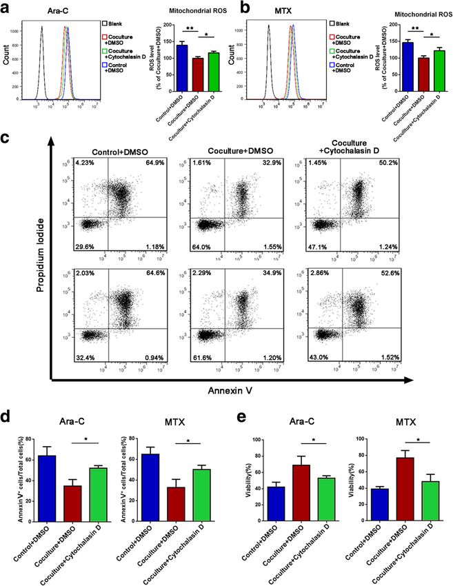
Wang et al. Journal of Hematology & Oncology (2018) 11:11                                                                                    Page 5 of 13

 Fig. 1 Jurkat cells transfer mitochondria to MSCs when exposed to ara-C or MTX. a Flow cytometry analysis of MitoTracker Red uptake by MSCs (GFP+ gated)
 cocultured with MitoTracker Red-labeled Jurkat cells after 300 nM ara-C or 100 nM MTX was added for 48 h. b Flow cytometry analysis of
 MitoTracker Red uptake by Jurkat cells (GFP− gated) cocultured with MitoTracker Red-labeled GFP+ MSCs after 300 nM ara-C or 100 nM MTX was
 added for 48 h. c Representative confocal microscopy images show that Jurkat cell-derived mitochondria (Red+) were internalized in MSCs(GFP+).
 Scale bar, 10 μm. d Representative confocal images show that MSCs received Jurkat cell-derived mitochondria at different time points (1, 2, and 3 days
 after coculture). Scale bar, 10 μm. e The areas of red foci per field were calculated by ImageJ software. The data are presented as the mean ± S.E.M. of
 three independent experiments (*P < 0.05; **P < 0.01; t test)

in MSCs cocultured with Jurkat cells, whereas the other ad-                    Jurkat cells (Fig. 4c, d), indicating that ICAM-1 was crucial
hesion molecules did not show significant changes. We fur-                     for cell adhesion between Jurkat cells and MSCs. We further
ther tested the role of ICAM-1 in Jurkat cell/MSC adhesion                     explored whether Jurkat cell/MSC adhesion contributed to
using a blocking antibody against ICAM-1(anti-ICAM-1).                         MSC-induced chemoresistance. In the coculture system
The results demonstrated that treatment with 20 mg/ml                          with ara-C or MTX, we also tested whether MSC-induced
anti-ICAM-1 significantly decreased the number of adhering                     chemoresistance could be influenced by anti-ICAM-1. We
Wang et al. Journal of Hematology & Oncology (2018) 11:11 Page 6 of 13
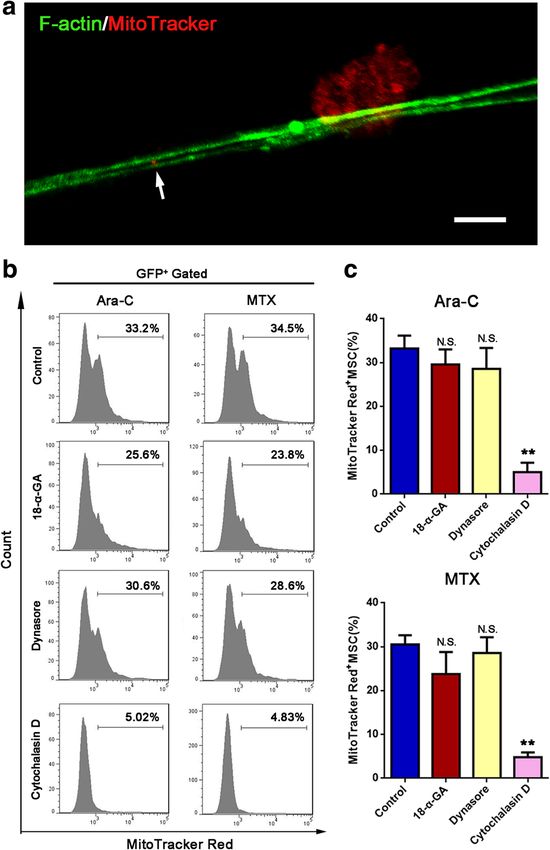
Fig. 2 Mitochondria transfer from Jurkat cells to MSCs can be blocked by cytochalasin D. a Representative confocal microscopy images show the presence
of TNTs containing mitochondria (arrow). Scale bar, 5 μm. b 18-α-GA (50 μM), dynasore (50 μM), or cytochalasin D (1 μM) was added to the coculture system
with ara-C or MTX for 48 h. Flow cytometry analysis of Jurkat cell-derived mitochondria uptake by MSCs (GFP+ gated). c The percentage of Red+ MSCs in
each group was analyzed and graphed. The results are expressed as the mean ± S.E.M. of three independent experiments (*P < 0.05; **P < 0.01; t test)
observed that Jurkat cells exposed to anti-ICAM-1 showed contributed to chemoresistance through mitochondria
an increased cell death rate (Fig. 4e) and decreased cell via- transfer. Jurkat cell/MSC adhesion was blocked by anti-
bility (Fig. 4f). Taken together, these results indicate that ICAM-1, and then, mitochondria transfer was analyzed with
ICAM-1-mediated Jurkat cell/MSC adhesion contributes to confocal microscopy and flow cytometry. Before coculture,
MSC-induced chemoresistance. Jurkat cells were stained with MitoTracker Red to label
mitochondria. Mitochondria transfer from Jurkat cells to
Mitochondria transfer from Jurkat cells to MSCs is MSCs was obviously inhibited by anti-ICAM-1 treatment
mediated by Jurkat cell/MSC adhesion (Fig. 5a, b), which was further confirmed by flow cytometry
Based on our finding that ICAM-1-mediated Jurkat cell/ analysis (Fig. 5c, d). On the other hand, we measured the
MSC adhesion contributed to MSC-induced chemoresis- ROS levels in MSCs after coculture with Jurkat cells. With
tance in Jurkat cells, we explored whether this adhesion the transfer of mitochondria from Jurkat cells to MSCs, theWang et al. Journal of Hematology & Oncology (2018) 11:11 Page 7 of 13
Fig. 3 Inhibition of mitochondria transfer decreases the effects of MSC-induced chemoresistance. a, b Cytochalasin D (1 μM) was added to the coculture
system with ara-C or MTX for 48 h. The levels of mitochondrial ROS in Jurkat cells were examined by MitoSOX staining. c The apoptosis rate was
determined using annexin V/PI staining and FACS. d The percentages of annexin V-positive cells were calculated. e A CCK-8 assay was used to assess
Jurkat cell viability. The data above are presented as the mean ± S.E.M. of three independent experiments (*P < 0.05; **P < 0.01; t test)
ROS levels in MSCs increased, so that the metabolic activity Red before coculture with MSCs. After 2 days of coculture
of MSCs also increased (Additional file 1: Figure S3). Taken with ara-C or MTX, some primary human T-ALL cells
together, these results indicate that cell adhesion between had adhered to MSCs (Fig. 6a); confocal imaging showed
Jurkat cells and MSCs facilitates the process of mitochondria that mitochondrial Red fluorescence was internalized in
transfer in the presence of chemotherapeutic drugs. MSCs. The areas of red foci in MSCs significantly de-
creased when cytochalasin D was added to the coculture
MSC-induced chemoresistance of primary T-ALL cells is system (Fig. 6b), indicating that mitochondria transfer was
ameliorated by inhibiting TNT formation impaired when TNT formation was blocked. Similarly, the
Finally, we asked whether our findings in Jurkat cells areas of red foci in MSCs also decreased when anti-
could be recapitulated in human primary T-ALL cells. We ICAM-1 was added (Additional file 1: Figure S4). To
labeled human primary T-ALL cells with MitoTracker further examine the exact effect of cytochalasin D on theWang et al. Journal of Hematology & Oncology (2018) 11:11 Page 8 of 13 Fig. 4 Increased expression of ICAM-1 in MSCs facilitates cell adhesion and protects Jurkat cells from chemotherapeutic drugs. a Confocal top view and side view of Jurkat cells (prestained with MitoTracker Red) and GFP-labeled MSCs in a coculture system. Scale bar, 10 μm. b mRNA expression of adhesion molecules in MSCs cocultured with Jurkat cells. c Representative photos of Jurkat cells and MSCs in the coculture system after removal of the nonadhesive Jurkat cells. Scale bar, 50 μm. d The relative adhesion ratio was calculated as the ratio of the number of Jurkat cells adhered to MSCs in the anti-ICAM-1 treated group to that in the DMSO-treated group. e The death rate of Jurkat cells was examined by FACS. f A CCK-8 assay was used to assess Jurkat cell viability. The data above are presented as the mean ± S.E.M. of three independent experiments (*P < 0.05; **P < 0.01; t test) survival of human primary T-ALL cells, we performed Discussion annexin V/PI flow cytometry analysis and a cell viability As one of the most aggressive hematologic malignancies, assay, and the results were consistent with our cell line T-ALL is usually treated with multiple chemotherapeutic experiments. Human primary T-ALL cells cocultured with drugs clinically, but observations of primary drug resist- MSCs and treated with cytochalasin D had a higher ance during treatment are quite frequent. Although it is apoptosis rate (Fig. 6c, d) and lower cell viability (Fig. 6e) widely accepted that MSCs are involved in the pro- than those cocultured with MSCs only. These results survival effects, the exact role of MSCs under the indicate that blocking mitochondria transfer by inhibiting chemotherapy remains unclear. Here, we demonstrated TNT formation decreases the capacity of MSCs to protect that upon the induction of oxidative stress by chemo- T-ALL cells from drug cytotoxicity. therapeutic drugs, T-ALL cells were able to transfer
Wang et al. Journal of Hematology & Oncology (2018) 11:11                                                                            Page 9 of 13

 Fig. 5 Cell-cell adhesion mediates mitochondria transfer from Jurkat cells to MSCs. Anti-ICAM-1 (20 mg/ml) was added to the coculture system
 with MTX for 48 h. a Confocal images showed that mitochondria transfer was inhibited by anti-ICAM-1. Scale bar, 10 μm. b The areas of red foci
 per field were calculated by ImageJ software. c Flow cytometry analysis of MitoTracker Red uptake by MSCs (GFP+ population) cocultured with
 MitoTracker Red-labeled Jurkat cells for 48 h. d The percentage of Red+ MSCs in each group was analyzed and graphed. The data above are
 presented as the mean ± S.E.M. of three independent experiments (*P < 0.05; **P < 0.01; t test)

mitochondria to MSCs. This process was mediated by                         affect cellular fate. For example, mitochondria trans-
TNTs and ICAM-1 contributing to the cell adhesion-                         ferred from MSCs to tumor cells could increase the
mediated drug resistance. A graphical abstract is shown                    oxidative phosphorylation and ATP production [47].
to describe this mechanism briefly (Fig. 6f ).                             Some cancer cells could also induce stromal cells to pro-
  MSCs can trigger the drug resistance of tumor cells via                  duce oncometabolites to fuel their metabolism through
two main strategies, soluble factor-mediated drug                          mitochondria transfer [50]. Meanwhile, it is reported
resistance and cell adhesion-mediated drug resistance                      that mitochondrial loss in MSCs could decrease ATP
[47]. For the former, drug resistance can be triggered by                  concentrations in these cells, thereby decreasing their
MSCs secreting cytokines, chemokines, growth factors                       secretory capacity and interfering the cytokine secretion
[27], and exosomes [48], and for the latter, MSCs can in-                  which played an important role in maintaining the
duce drug resistance by adhering to cancer cells, including                microenvironment [28]. On the other hand, MSCs might
melanoma cells [40] and leukemia cells [49]. In this study,                eliminate the transferred mitochondria to stabilize the
we found that mitochondria transfer between MSCs and                       intracellular homeostasis. Phinney et al. firstly figured
T-ALL cells is also a mechanism that induces chemoresis-                   out that MSCs eliminated their damaged mitochondria
tance in tumor cells. According to the literature, intercel-               by exporting them to neighboring macrophages for
lular mitochondria transfer can be mediated by TNTs,                       recycling [51], so as to decrease the oxidative pressure in
microvesicles, or gap junctions. We confirmed that TNTs                    the microenvironment, associated with better survival
played a major role in the mitochondria transfer between                   and increased regrowth potential. In our study, mito-
MSCs and T-ALL cells, and inhibition of TNTs led to                        chondria transfer helps reduce the ROS level in Jurkat
decreased MSC-induced chemoresistance. This finding                        cells, so as to induce chemoresistance. Taken together,
may thus provide a novel strategy for T-ALL treatment.                     investigating the fate of transferred mitochondria helps
  Many studies have demonstrated that the intercellular                    to understand the crosstalk in leukemic microenviron-
transferred mitochondria were still functional and can                     ment and offers probable therapy strategies.
Wang et al. Journal of Hematology & Oncology (2018) 11:11                                                                                  Page 10 of 13

 Fig. 6 Inhibition of TNT formation ameliorates MSC-induced chemoresistance on primary T-ALL cells. Cytochalasin D (1 μM) was added to the
 coculture system with ara-C or MTX for 48 h. a Representative photos of human primary T-ALL cells and MSCs in the coculture system after the
 removal of nonadhesive human primary T-ALL cells. Scale bar, 100 μm. b Representative confocal microscopy images show that human primary
 T-ALL cell-derived mitochondria (Red+) were internalized in MSCs. Scale bar, 20 μm. The areas of red foci per field were calculated by ImageJ software.
 c The apoptosis rate was determined using annexin V/PI staining and FACS. d The percentages of annexin V-positive cells were calculated. e A CCK-8
 assay was used to assess human primary T-ALL cell viability. f Graphic abstract: T-ALL cell/MSC adhesion-mediated mitochondria transfer contributes to
 MSC-induced chemoresistance. The data above are presented as the mean ± S.E.M. of three independent experiments (*P < 0.05; **P < 0.01; t test)

   In contrast to the myriad reports demonstrating that                       relationships. On the other hand, in cancers such as
MSCs could transfer mitochondria to various kinds of                          acute myeloid leukemia (AML), MSCs are impaired in
cells, including cortical neurons [52], cardiomyocytes                        their growth properties and osteogenic differentiation
[53], renal tubular cells [30], lung epithelium cells [28],                   potential [56]. Interestingly, here, we demonstrated that
lung adenocarcinoma cells [32], osteosarcoma cells [54],                      MSCs could receive mitochondria from T-ALL cells, and
macrophage [51], and acute myeloid leukemia cells [55],                       it is likely that this transfer would lead to MSC damage in
mitochondria transfer from other cells to MSCs has                            T-ALL patients. Since MSCs play an important role in
rarely been demonstrated. In our study, we found that                         tissue repair [57, 58], they are worthy of further investiga-
T-ALL cells could transfer mitochondria to adhering                           tion. Moreover, it is reported that AML cells can import
MSCs when treated with ara-C or MTX. This newly                               mitochondria from MSCs in order to better withstand
identified process complements the existing knowledge                         chemotherapy [55, 59]. Thus, we verified our conclusion
of mitochondria transfer and provides a novel perspec-                        by comparing ALL cells with AML cells and found they
tive regarding mitochondria transfer in intercellular                         have different adhesive capacity and mitochondria transfer
Wang et al. Journal of Hematology & Oncology (2018) 11:11                                                                     Page 11 of 13

direction (Additional file 1: Figure S5). The difference in    mitochondria transfer to MSCs which is mediated by
transfer direction may also be due to their different meta-    TNTs. Adhesion between MSCs and T-ALL cells is
bolic state. T-ALL cells prefer glycolysis after coculture,    frequently observed in coculture systems. ICAM-1 is the
while AML cells have more oxidative phosphorylation            major adhesion molecule. Our results demonstrate that
[60]. ALL cells export mitochondria to reduce intracellular    this mechanism should be considered in future clinical
ROS, while AML cells import mitochondria for the               investigations. Targeting mitochondria transfer may be a
demand of oxidative phosphorylation.                           potential strategy for the chemoresistance of T-ALL.
  Disrupted oxidative stress metabolism is a common            Hopefully, this study will serve as a precedent for applying
feature of cancer cells [61, 62], and this phenomenon          similar therapeutic strategies in the chemoresistance of
has also been observed in T-ALL cells [63]. As a result,       other leukemias.
ROS levels are higher in T-ALL cells than in non-
leukemic cells. Since excess ROS can lead to leukemia          Additional file
cell deaths, the induction of intracellular oxidative stress
has been shown to be an important anti-cancer mechan-           Additional file 1: Table S1. Primer used to amplify the human
ism of leukemia chemotherapy [64]. Thus, the promo-             transcripts during real-time quantitative PCR. Figure S1. Verification of the
                                                                feasibility of mitochondria dye method. Figure S2. Mitochondrial DNA
tion of mitochondrial ROS production can be observed            damage caused by drug-induced ROS in Jurkat cells. Figure S3. ROS levels in
in T-ALL cells treated with paclitaxel, anthracyclines,         MSCs increase after coculture with Jurkat cells. Figure S4. Anti-ICAM-1
ara-C, and MTX, among others. In our previous study,            decreases mitochondria transfer between human primary T-ALL cells and
                                                                MSCs. Figure S5. MSCs export mitochondria to AML cells (HL-60 cells) but not
we found that MSC-mediated chemoresistance of T-ALL             ALL cells (Jurkat cells) under chemotherapy. (DOCX 3488 kb)
cells was dependent on decreased mitochondrial ROS in
T-ALL cells, and the ERK/Drp1 signaling pathway was
                                                               Abbreviations
involved in the downregulation of ROS levels. However,         ALL: Acute lymphoblastic leukemia; AML: Acute myeloid leukemia;
mitochondrial ROS in T-ALL cells decreased to a larger         ANOVA: Analysis of variance; CCK-8: Cell counting kit-8;
extent in a coculture system than in a Transwell system,       cDNA: Complementary deoxyribonucleic acid; CLL: Chronic lymphocytic
                                                               leukemia; FACS: Fluorescence-activated cell sorting; FBS: Fetal bovine serum;
indicating that an unknown mechanism mediated the              GFP: Green fluorescent protein; ICAM-1: Intercellular adhesion molecule -1;
MSC-induced chemoresistance. Here, we found that cell          MSCs: Mesenchymal stem cells; PECAM-1: Platelet endothelial cell adhesion
adhesion-mediated mitochondria transfer from T-ALL             molecule-1; PI: Propidium iodide; qRT-PCR: Quantitative real-time polymerase
                                                               chain reaction; ROS: Reactive oxygen species; T-ALL: T cell acute
cells to MSCs can reduce oxidative stress by decreasing        lymphoblastic leukemia; TNTs: Tunneling nanotubes; VCAM-1: Vascular cell
mitochondrial ROS. This finding solved the question            adhesion molecule-1
raised by our previous study. Additionally, Ishikawa et
al. observed the direct transfer of intercellular ROS me-      Acknowledgements
                                                               We thank the Department of Haematology in Nanfang Hospital and
diated by connexin-43 from hematopoietic stem cells to         Department of Pediatrics in Sun Yat-Sen Memorial Hospital for sample
bone marrow stromal cells [65]. This finding suggested         collection.
that the direct transfer of mitochondrial ROS via TNTs
might also be another mechanism for decreasing ROS in          Funding
                                                               This work was supported by the National Key Research and Development
T-ALL cells. Unfortunately, due to the limitations of the      Program of China (2017YFA0105501), the National Natural Science
existing experimental techniques, the two mechanisms           Foundation of China (81425016), the Natural Science Foundation of
have yet to be discriminated.                                  Guangdong Province (S2013030013305, 2015A030312013, and
                                                               2017A030310237), the Key Scientific and Technological Projects of
  Additionally, we also found that treatment with anti-        Guangdong Province (2014B020226002, 2015B020226004, and
ICAM-1 significantly blocked mitochondria transfer, indi-      2014B020228003), the Key Scientific and Technological Program of
cating that mitochondria transfer was mediated by T-ALL        Guangzhou City (201400000003-3, 201508020262) the Guangdong Province
                                                               Universities and Colleges Pearl River Scholar Funded Scheme (GDUPS, 2013),
cell/MSC adhesion. Combined with our finding that block-       the Key Scientific and Technological Projects of Guangdong Province
ing mitochondria transfer with cytochalasin D abolished        (2014B020225007, 2017B020231001), the Key Scientific and Technological
the capacity of MSCs to protect T-ALL cells, we concluded      Projects of Guangdong Province (2014B020212009), the Key Scientific and
                                                               Technological Program of Guangzhou City (201508020254), and the China
that T-ALL cell/MSC adhesion-mediated mitochondria             Postdoctoral Science Foundation (2016M602583, 2017T100657).
transfer contributed to MSC-induced chemoresistance.
Thus, inhibition of T-ALL cell/MSC adhesion-mediated           Availability of data and materials
mitochondria transfer may be a novel strategy for T-ALL        All data generated in the study are included in the present article (and its
                                                               supplementary information files).
treatment.
                                                               Authors’ contributions
Conclusions                                                    JW, XL, and YQ contribute equally to the work. JW and APX designed the
Our results elucidate the role of MSCs in the chemoresis-      research. JW, XL, YQ, YS, JC, BW, XW, QK, XS, YW, YH, HL, and TW performed
                                                               the research. RL and QL provided the sample collection. JW, XL, and YQ
tance of T-ALL cells. T-ALL cells manage chemotherapy-         analyzed the data. JW, XL, YQ, BW, and XW wrote the paper. All authors read
induced intracellular oxidative stress by targeting            and approved the final manuscript.
Wang et al. Journal of Hematology & Oncology (2018) 11:11 Page 12 of 13
Ethics approval and consent to participate 13. Mudry RE, Fortney JE, York T, Hall BM, Gibson LF. Stromal cells regulate survival
Patients gave informed consent for additional aliquot of the marrow aspirate of B-lineage leukemic cells during chemotherapy. Blood. 2000;96(5):1926–32.
to be used for research purposes in accordance with the ethical guidelines 14. Redza-Dutordoir M, Averill-Bates DA. Activation of apoptosis signalling
of Sun Yat-Sen Memorial Hospital, Sun Yat-Sen University, and Nanfang Hos- pathways by reactive oxygen species. Biochim Biophys Acta. 2016;1863(12):
pital, Southern Medical University. 2977–92.
15. Kashyap MK, Amaya-Chanaga CI, Kumar D, Simmons B, Huser N, Gu Y, Hallin
Consent for publication M, Lindquist K, Yafawi R, Choi MY, et al. Targeting the CXCR4 pathway using
Not applicable. a novel anti-CXCR4 IgG1 antibody (PF-06747143) in chronic lymphocytic
leukemia. J Hematol Oncol. 2017;10(1):112.
Competing interests 16. Ling S, Xie H, Yang F, Shan Q, Dai H, Zhuo J, Wei X, Song P, Zhou L, Xu X,
The authors declare that they have no competing interests. et al. Metformin potentiates the effect of arsenic trioxide suppressing
intrahepatic cholangiocarcinoma: roles of p38 MAPK, ERK3, and mTORC1. J
Hematol Oncol. 2017;10(1):59.
Publisher’s Note 17. Conklin KA. Cancer chemotherapy and antioxidants. J Nutr. 2004;134(11):
Springer Nature remains neutral with regard to jurisdictional claims in 3201s–4s.
published maps and institutional affiliations. 18. Liesa M, Palacin M, Zorzano A. Mitochondrial dynamics in mammalian
health and disease. Physiol Rev. 2009;89(3):799–845.
Author details 19. Youle RJ, van der Bliek AM. Mitochondrial fission, fusion, and stress. Science.
1
Program of Stem Cells and Regenerative Medicine, Affiliated Guangzhou 2012;337(6098):1062–5.
Women and Children’s Hospital, Zhongshan School of Medicine, Sun Yat-Sen 20. Tan AS, Baty JW, Dong LF, Bezawork-Geleta A, Endaya B, Goodwin J,
University, Guangzhou 510080, China. 2Center for Stem Cell Biology and Bajzikova M, Kovarova J, Peterka M, Yan B, et al. Mitochondrial genome
Tissue Engineering, Key Laboratory for Stem Cells and Tissue Engineering, acquisition restores respiratory function and tumorigenic potential of cancer
Ministry of Education, Sun Yat-Sen University, 74# Zhongshan 2nd Road, cells without mitochondrial DNA. Cell Metab. 2015;21(1):81–94.
Guangzhou, Guangdong, China. 3Biotherapy Center, the Third Affiliated 21. Osswald M, Jung E, Sahm F, Solecki G, Venkataramani V, Blaes J, Weil S,
Hospital, Sun Yat-Sen University, Guangzhou 510080, China. 4The First Horstmann H, Wiestler B, Syed M, et al. Brain tumour cells interconnect to a
Affiliated Hospital of Xi’an Jiaotong University Medical College, Xi’an, Shaanxi functional and resistant network. Nature. 2015;528(7580):93.
710061, China. 5Department of Hematology, Nanfang Hospital, Southern 22. CHO D, Kim KY, Bushong EA, Mills EA, Boassa D, Shih T, Kinebuchi M, Phan
Medical University, Guangzhou 510515, China. 6Key Laboratory of Protein S, Zhou Y, Bihlmeyer NA, et al. Transcellular degradation of axonal
Modification and Degradation, School of Basic Medical Sciences, Affiliated mitochondria. Proc Natl Acad Sci U S A. 2014;111(26):9633–8.
Cancer Hospital and Institute of Guangzhou Medical University, Guangzhou 23. Zhang L, Chen QS, Xu PP, Qian Y, Wang AH, Xiao D, Zhao Y, Sheng Y, Wen
511436, China. 7Department of Biochemistry, Zhongshan School of Medicine, XQ, Zhao WL. Catechins induced acute promyelocytic leukemia cell
Sun Yat-Sen University, Guangzhou 510080, China. apoptosis and triggered PML-RARalpha oncoprotein degradation. J Hematol
Oncol. 2014;7:75.
Received: 17 November 2017 Accepted: 12 January 2018 24. Banjerdpongchai R, Kongtawelert P, Khantamat O, Srisomsap C,
Chokchaichamnankit D, Subhasitanont P, Svasti J. Mitochondrial and
endoplasmic reticulum stress pathways cooperate in zearalenone-induced
References apoptosis of human leukemic cells. J Hematol Oncol. 2010;3:50.
1. Lonetti A, Cappellini A, Bertaina A, Locatelli F, Pession A, Buontempo F, 25. Li XY, Fang P, Mai JT, Choi ET, Wang H, Yang XF. Targeting mitochondrial
Evangelisti C, Evangelisti C, Orsini E, Zambonin L, et al. Improving nelarabine reactive oxygen species as novel therapy for inflammatory diseases and
efficacy in T cell acute lymphoblastic leukemia by targeting aberrant PI3K/ cancers. J Hematol Oncol. 2013;6:19.
AKT/mTOR signaling pathway. J Hematol Oncol. 2016;9(1):114. 26. Jitschin R, Hofmann AD, Bruns H, Giessl A, Bricks J, Berger J, Saul D, Eckart
2. Pui CHEW. Acute lymphoblastic leukaemia. N Engl J Med. 1998;399:605–15. MJ, Mackensen A, Mougiakakos D. Mitochondrial metabolism contributes to
3. Wallaert A, Durinck K, Taghon T, Van Vlierberghe P, Speleman F. T-ALL and oxidative stress and reveals therapeutic targets in chronic lymphocytic
thymocytes: a message of noncoding RNAs. J Hematol Oncol. 2017;10(1):66. leukemia. Blood. 2014;123(17):2663–72.
4. Pui CHRL, Look AT. Acute lymphoblastic leukaemia. Lancet. 2008;371:1030–43. 27. Cai JY, Wang JC, Huang YN, Wu HX, Xia T, Xiao JQ, Chen XY, Li HY, Qiu Y,
5. Bassan R, Hoelzer D. Modern therapy of acute lymphoblastic leukemia. J Wang YN, et al. ERK/Drp1-dependent mitochondrial fission is involved in
Clin Oncol. 2011;29(5):532–43. the MSC-induced drug resistance of T-cell acute lymphoblastic leukemia
6. Szczepanski TOA, van der Velden VH, San MJ, van Dongen JJ. Minimal cells. Cell Death Dis. 2016;7(11):e2459.
residual disease in leukaemia patients. Lancet Oncol. 2001;2:409–17. 28. Islam MN, Das SR, Emin MT, Wei M, Sun L, Westphalen K, Rowlands DJ,
7. Zhang B, Li M, McDonald T, Holyoake TL, Moon RT, Campana D, Shultz L, Quadri SK, Bhattacharya S, Bhattacharya J. Mitochondrial transfer from bone-
Bhatia R. Microenvironmental protection of CML stem and progenitor cells marrow-derived stromal cells to pulmonary alveoli protects against acute
from tyrosine kinase inhibitors through N-cadherin and Wnt-beta-catenin lung injury. Nat Med. 2012;18(5):759–U153.
signaling. Blood. 2013;121(10):1824–38. 29. Pasquier J, Guerrouahen BS, Al Thawadi H, Ghiabi P, Maleki M, Abu-Kaoud N,
8. Wang L, O'Leary H, Fortney J, Gibson LF. Ph+/VE-cadherin+ identifies a stem Jacob A, Mirshahi M, Galas L, Rafii S, et al. Preferential transfer of
cell like population of acute lymphoblastic leukemia sustained by bone mitochondria from endothelial to cancer cells through tunneling nanotubes
marrow niche cells. Blood. 2007;110(9):3334–44. modulates chemoresistance. J Transl Med. 2013;11:94.
9. Yang Y, Mallampati S, Sun BH, Zhang J, Kim SB, Lee JS, Gong Y, Cai Z, Sun 30. Plotnikov EY, Khryapenkova TG, Galkina SI, Sukhikh GT, Zorov DB. Cytoplasm
XP. Wnt pathway contributes to the protection by bone marrow stromal and organelle transfer between mesenchymal multipotent stromal cells and
cells of acute lymphoblastic leukemia cells and is a potential therapeutic renal tubular cells in co-culture. Exp Cell Res. 2010;316(15):2447–55.
target. Cancer Lett. 2013;333(1):9–17. 31. Spees JL, Olson SD, Whitney MJ, Prockop DJ. Mitochondrial transfer
10. Schmidt T, Masouleh BK, Loges S, Cauwenberghs S, Fraisl P, Maes C, Jonckx between cells can rescue aerobic respiration. Proc Natl Acad Sci U S A.
B, De Keersmaecker K, Kleppe M, Tjwa M, et al. Loss or inhibition of stromal- 2006;103(5):1283–8.
derived PIGF prolongs survival of mice with imatinib-resistant Bcr-Abl1(+) 32. Liu KM, Ji KQ, Guo L, Wu W, Lu HX, Shan PY, Yan CZ. Mesenchymal stem
leukemia. Cancer Cell. 2011;19(6):740–53. cells rescue injured endothelial cells in an in vitro ischemia-reperfusion
11. Tabe Y, Jin L, Iwabuchi K, Wang RY, Ichikawa N, Miida T, Cortes J, Andreeff model via tunneling nanotube like structure-mediated mitochondrial
M, Konopleva M. Role of stromal microenvironment in nonpharmacological transfer. Microvasc Res. 2014;92:10–8.
resistance of CML to imatinib through Lyn/CXCR4 interactions in lipid rafts. 33. Bukoreshtliev NV, Wang X, Hodneland E, Gurke S, Barroso JFV, Gerdes HH.
Leukemia. 2012;26(5):883–92. Selective block of tunneling nanotube (TNT) formation inhibits intercellular
12. Iwamoto S, Mihara K, Downing JR, Pui CH, Campana D. Mesenchymal cells organelle transfer between PC12 cells. FEBS Lett. 2009;583(9):1481–8.
regulate the response of acute lymphoblastic leukemia cells to 34. Gurke S, Barroso JFV, Hodneland E, Bukoreshtliev NV, Schlicker O, Gerdes
asparaginase. J Clin Invest. 2007;117(4):1049–57. HH. Tunneling nanotube (TNT)-like structures facilitate a constitutive,Wang et al. Journal of Hematology & Oncology (2018) 11:11 Page 13 of 13
actomyosin-dependent exchange of endocytic organelles between normal 54. Cho YM, Kim JH, Kim M, Park SJ, Koh SH, Ahn HS, Kang GH, Lee JB, Park KS,
rat kidney cells. Exp Cell Res. 2008;314(20):3669–83. Lee HK. Mesenchymal stem cells transfer mitochondria to the cells with
35. Vallabhaneni KC, Haller H, Dumler I. Vascular smooth muscle cells initiate virtually no mitochondrial function but not with pathogenic mtDNA
proliferation of mesenchymal stem cells by mitochondrial transfer via mutations. PLoS One. 2012;7(3):e32778.
tunneling nanotubes. Stem Cells Dev. 2012;21(17):3104–13. 55. Moschoi R, Imbert V, Nebout M, Chiche J, Mary D, Prebet T, Saland E,
36. Hsu YC, Wu YT, Yu TH, Wei YH. Mitochondria in mesenchymal stem cell Castellano R, Pouyet L, Collette Y, et al. Protective mitochondrial transfer
biology and cell therapy: from cellular differentiation to mitochondrial from bone marrow stromal cells to acute myeloid leukemic cells during
transfer. Semin Cell Dev Biol. 2016;52:119–31. chemotherapy. Blood. 2016;128(2):253–64.
37. Dong LF, Kovarova J, Bajzikova M, Bezawork-Geleta A, Svec D, Endaya B, 56. Geyh S, Rodriguez-Paredes M, Jager P, Khandanpour C, Cadeddu RP,
Sachaphibulkij K, Coelho AR, Sebkova N, Ruzickova A, et al. Horizontal Gutekunst J, Wilk CM, Fenk R, Zilkens C, Hermsen D, et al. Functional
transfer of whole mitochondria restores tumorigenic potential in inhibition of mesenchymal stromal cells in acute myeloid leukemia.
mitochondrial DNA-deficient cancer cells. Elife. 2017;6. Leukemia. 2016;30(3):683–91.
38. Nakahira K, Cloonan SM, Mizumura K, Choi AMK, Ryter SW. Autophagy: a 57. Zhao K, Liu Q. The clinical application of mesenchymal stromal cells in
crucial moderator of redox balance, inflammation, and apoptosis in lung hematopoietic stem cell transplantation. J Hematol Oncol. 2016;9(1):46.
disease. Antioxid Redox Sign. 2014;20(3):474–94. 58. Kim N, Cho SG. Clinical applications of mesenchymal stem cells. Korean J
39. Peng Y, Chen X, Liu Q, Zhang X, Huang K, Liu L, Li H, Zhou M, Huang F, Fan Intern Med. 2013;28(4):387–402.
Z, et al. Mesenchymal stromal cells infusions improve refractory chronic 59. Marlein CR, Zaitseva L, Piddock RE, Robinson SD, Edwards DR, Shafat MS,
graft versus host disease through an increase of CD5+regulatory B cells Zhou Z, Lawes M, Bowles KM, Rushworth SA. NADPH oxidase-2 derived
producing interleukin 10. Leukemia. 2015;29(3):636–46. superoxide drives mitochondrial transfer from bone marrow stromal cells to
40. Wang JC, Wang YN, Wang SC, Cai JY, Shi JQ, Sui X, Cao Y, Huang WJ, Chen leukemic blasts. Blood. 2017;130(14):1649–60.
XY, Cai ZJ, et al. Bone marrow-derived mesenchymal stem cell-secreted IL-8 60. Suganuma K, Miwa H, Imai N, Shikami M, Gotou M, Goto M, Mizuno S,
promotes the angiogenesis and growth of colorectal cancer. Oncotarget. Takahashi M, Yamamoto H, Hiramatsu A, et al. Energy metabolism of
2015;6(40):42825–37. leukemia cells: glycolysis versus oxidative phosphorylation. Leuk Lymphoma.
41. Vereb Z, Poliska S, Albert R, Olstad OK, Boratko A, Csortos C, Moe MC, Facsko A, 2010;51(11):2112–9.
Petrovski G. Role of human corneal stroma-derived mesenchymal-like stem 61. Pelicano H, Carney D, Huang P. ROS stress in cancer cells and therapeutic
cells in corneal immunity and wound healing. Sci Rep. 2016;6:26227. implications. Drug Resist Update. 2004;7(2):97–110.
42. Frank O, Heim M, Jakob M, Barbero A, Schafer D, Bendik I, Dick W, Heberer 62. Renschler MF. The emerging role of reactive oxygen species in cancer
M, Martin I. Real-time quantitative RT-PCR analysis of human bone marrow therapy. Eur J Cancer. 2004;40(13):1934–40.
stromal cells during osteogenic differentiation in vitro. J Cell Biochem. 2002; 63. Silva A, Girio A, Cebola I, Santos CI, Antunes F, Barata JT. Intracellular
85(4):737–46. reactive oxygen species are essential for PI3K/Akt/mTOR-dependent IL-7-
43. Zhang XR, Huang WJ, Chen XY, Lian YF, Wang JC, Cai C, Huang L, Wang T, mediated viability of T-cell acute lymphoblastic leukemia cells. Leukemia.
Ren J, Xiang AP. CXCR5-overexpressing mesenchymal stromal cells exhibit 2011;25(6):960–7.
enhanced homing and can decrease contact hypersensitivity. Mol Ther. 64. Tong LY, Chuang CC, Wu SY, Zuo L. Reactive oxygen species in redox
2017;25(6):1434–47. cancer therapy. Cancer Lett. 2015;367(1):18–25.
44. Pribila JT, Quale AC, Mueller KL, Shimizu Y. Integrins and T cell-mediated 65. Ishikawa ET, Gonzalez-Nieto D, Ghiaur G, Dunn SK, Ficker AM, Murali B,
immunity. Annu Rev Immunol. 2004;22:157–80. Madhu M, Gutstein DE, Fishman GI, Barrio LC, et al. Connexin-43 prevents
45. Bromley SK, Burack WR, Johnson KG, Somersalo K, Sims TN, Sumen C, Davis hematopoietic stem cell senescence through transfer of reactive oxygen
MM, Shaw AS, Allen PM, Dustin ML. The immunological synapse. Annu Rev species to bone marrow stromal cells. Proc Natl Acad Sci U S A. 2012;
Immunol. 2001;19:375–96. 109(23):9071–6.
46. Ren GW, Zhao X, Zhang LY, Zhang JM, L'Huillier A, Ling WF, Roberts AI, Le
AD, Shi ST, Shao CS, et al. Inflammatory cytokine-induced intercellular
adhesion molecule-1 and vascular cell adhesion molecule-1 in
mesenchymal stem cells are critical for immunosuppression. J Immunol.
2010;184(5):2321–8.
47. Meads MB, Gatenby RA, Dalton WS. Environment-mediated drug resistance:
a major contributor to minimal residual disease. Nat Rev Cancer. 2009;9(9):
665–A674.
48. Ji RB, Zhang B, Zhang X, Xue JG, Yuan X, Yan YM, Wang M, Zhu W, Qian H,
Xu WR. Exosomes derived from human mesenchymal stem cells confer
drug resistance in gastric cancer. Cell Cycle. 2015;14(15):2473–83.
49. Jacamo R, Chen Y, Wang ZQ, Ma WC, Zhang M, Spaeth EL, Wang Y, Battula
VL, Mak PY, Schallmoser K, et al. Reciprocal leukemia-stroma VCAM-1/VLA-4-
dependent activation of NF-kappa B mediates chemoresistance. Blood.
2014;123(17):2691–702.
50. Martinez-Outschoorn UE, Pavlides S, Howell A, Pestell RG, Tanowitz HB,
Sotgia F, Lisanti MP. Stromal-epithelial metabolic coupling in cancer:
integrating autophagy and metabolism in the tumor microenvironment. Int
J Biochem Cell Biol. 2011;43(7):1045–51. Submit your next manuscript to BioMed Central
51. Phinney DG, Di Giuseppe M, Njah J, Sala E, Shiva S, St Croix CM, Stolz DB, and we will help you at every step:
Watkins SC, Di YP, Leikauf GD, et al. Mesenchymal stem cells use
extracellular vesicles to outsource mitophagy and shuttle microRNAs. Nat • We accept pre-submission inquiries
Commun. 2015;6:8472. • Our selector tool helps you to find the most relevant journal
52. Babenko VA, Silachev DN, Zorova LD, Pevzner IB, Khutornenko AA,
• We provide round the clock customer support
Plotnikov EY, Sukhikh GT, Zorov DB. Improving the post-stroke
therapeutic potency of mesenchymal multipotent stromal cells by • Convenient online submission
cocultivation with cortical neurons: the role of crosstalk between cells. • Thorough peer review
Stem Cell Transl Med. 2015;4(9):1011–20.
• Inclusion in PubMed and all major indexing services
53. Plotnikov EY, Khryapenkova TG, Vasileva AK, Marey MV, Galkina SI, Isaev NK,
Sheval EV, Polyakov VY, Sukhikh GT, Zorov DB. Cell-to-cell cross-talk • Maximum visibility for your research
between mesenchymal stem cells and cardiomyocytes in co-culture. J Cell
Mol Med. 2008;12(5a):1622–31. Submit your manuscript at
www.biomedcentral.com/submitYou can also read