Activation or exhaustion of CD8+ T cells in patients with - COVID-19 - Nature
←
→
Page content transcription
If your browser does not render page correctly, please read the page content below
www.nature.com/cmi

REVIEW ARTICLE                          OPEN

Activation or exhaustion of CD8+ T cells in patients with
COVID-19
                                                  1,3 ✉
Min-Seok Rha1,2 and Eui-Cheol Shin

© The Author(s) 2021

    In addition to CD4+ T cells and neutralizing antibodies, CD8+ T cells contribute to protective immune responses against SARS-CoV-2
    in patients with coronavirus disease 2019 (COVID-19), an ongoing pandemic disease. In patients with COVID-19, CD8+ T cells
    exhibiting activated phenotypes are commonly observed, although the absolute number of CD8+ T cells is decreased. In addition,
    several studies have reported an upregulation of inhibitory immune checkpoint receptors, such as PD-1, and the expression of
    exhaustion-associated gene signatures in CD8+ T cells from patients with COVID-19. However, whether CD8+ T cells are truly
    exhausted during COVID-19 has been a controversial issue. In the present review, we summarize the current understanding of CD8+
    T-cell exhaustion and describe the available knowledge on the phenotypes and functions of CD8+ T cells in the context of
    activation and exhaustion. We also summarize recent reports regarding phenotypical and functional analyses of SARS-CoV-2-
    specific CD8+ T cells and discuss long-term SARS-CoV-2-specific CD8+ T-cell memory.

    Keywords: CD8+ T cell; Activation; T-cell exhaustion; SARS-CoV-2; COVID-19

    Cellular & Molecular Immunology (2021) 18:2325–2333; https://doi.org/10.1038/s41423-021-00750-4

INTRODUCTION                                                                                 In COVID-19 patients, the CD8+ T-cell population undergoes
Since the initial reports of pneumonia cases of unknown origin in                         quantitative and qualitative changes. Decreased cell number and
Wuhan, China, in late December 2019 [1], novel severe acute                               activation phenotypes are frequently observed, particularly in
respiratory syndrome coronavirus 2 (SARS-CoV-2) has been rapidly                          severe disease [16, 20–24]. Previous studies have also reported
spreading worldwide. Coronavirus disease 2019 (COVID-19),                                 exhaustion phenotypes of CD8+ T cells in patients with severe
caused by SARS-CoV-2 infection, manifests with a broad spectrum                           COVID-19 based on the upregulation of inhibitory receptors (IRs)
of clinical symptoms, from asymptomatic infection to critical                             [20, 25–30], which may impair host defenses and result in poor
disease [2]. COVID-19 has threatened public health and had a                              disease outcomes. In contrast, no significant evidence of CD8+
devastating economic impact. Global efforts are underway to                               T-cell exhaustion has been observed in several single-cell RNA
control the COVID-19 pandemic. Prophylactic COVID-19 vaccines                             sequencing (scRNA-seq) analyses [31, 32]. However, all of these
using various platforms have been approved since December                                 studies have the limitation of their conclusions relying on the
2020, and their administration has started in populations                                 expression of IRs or transcripts related to T-cell exhaustion without
throughout the world [3–7].                                                               information on the antigen specificity of CD8+ T cells and their
   A better understanding of host immune responses to SARS-CoV-2                          effector functions. Our previous study using major histocompat-
is crucial to the development of effective vaccines and therapeutics                      ibility complex class I (MHC-I) multimers demonstrated that PD-1+
and ending the current pandemic. SARS-CoV-2 infection elicits the                         SARS-CoV-2-specific CD8+ T cells are functionally active in terms of
activation of both innate and adaptive immunity [8–11]. In adaptive                       interferon (IFN)-γ production, implying that these cells are not
immunity, CD8+ T cells play an essential role in controlling viral                        truly exhausted [33].
infection by killing virus-infected cells and producing effector                             Several reviews have already summarized and discussed
cytokines. Since the emergence of COVID-19, remarkable progress                           different aspects of CD8+ T-cell responses to SARS-CoV-2 in terms
has been made in understanding CD8+ T-cell responses against                              of cross-reactivity, kinetics, and protective roles [34–39]. In the
SARS-CoV-2. It is now clear that SARS-CoV-2-specific CD8+ T-cell                           current review, we focus on the activation and exhaustion of CD8+
responses are detected in the acute and convalescent phases of                            T cells in patients with COVID-19. We summarize the current
COVID-19 [12–17]. In addition, recent studies using animal models                         understanding of CD8+ T-cell exhaustion and discuss available
have reported that CD8+ T cells contribute to protection from the                         knowledge regarding the activation and exhaustion of CD8+
development of severe COVID-19 [18, 19].                                                  T cells in the context of COVID-19.

1
 Laboratory of Immunology and Infectious Diseases, Graduate School of Medical Science and Engineering, Korea Advanced Institute of Science and Technology (KAIST), Daejeon,
Republic of Korea. 2Department of Otorhinolaryngology, Yonsei University College of Medicine, Seoul, Republic of Korea. 3The Center for Epidemic Preparedness, KAIST, Daejeon,
Republic of Korea. ✉email: ecshin@kaist.ac.kr

Received: 30 April 2021 Accepted: 20 July 2021
Published online: 19 August 2021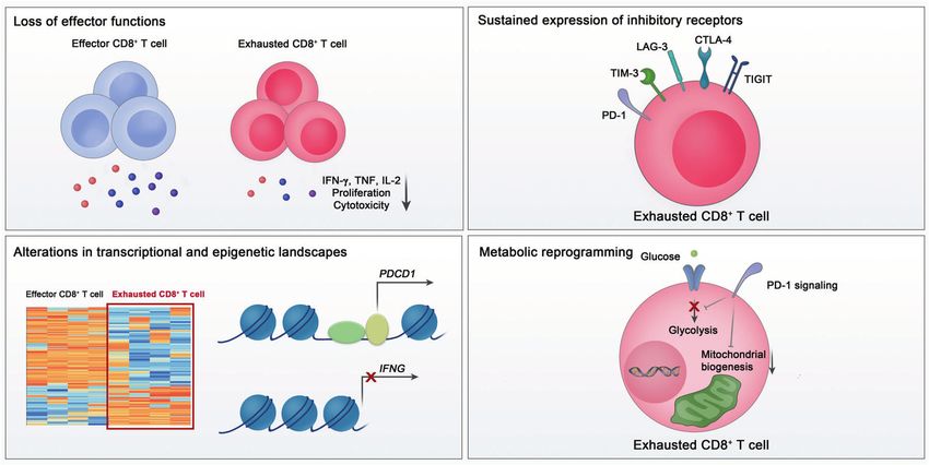
M.-S. Rha and E.-C. Shin
                  2326

                         Fig. 1 Key features of exhausted CD8+ T cells. Exhausted CD8+ T cells are characterized by a loss of effector functions, sustained expression of
                         inhibitory receptors, altered transcriptional and epigenetic landscape, and metabolic reprogramming
1234567890();,:

                         THE CHARACTERISTICS OF EXHAUSTED CD8+ T CELLS                              between viral load and the severity of exhaustion in chronic viral
                         An overview of CD8+ T-cell exhaustion                                      infection can be explained by functional impairment of exhausted
                         During acute viral infection, naive CD8+ T cells that recognize            CD8+ T cells. Furthermore, exhausted CD8+ T cells respond poorly
                         antigens presented on MHC-I by their T-cell receptors (TCRs) are           to homeostatic cytokines, including IL-7 and IL-15 [56], in relation
                         activated and undergo clonal expansion and differentiation into            to their low expression of CD127 and CD122 [57].
                         effector CD8+ T cells [40, 41]. Effector CD8+ T cells produce                 The functions of exhausted CD8+ T cells may vary across
                         cytokines, including IFN-γ and tumor necrosis factor (TNF), and            diseases, possibly related to antigens and the immune micro-
                         directly kill target cells [42]. In the subsequent contraction phase       environment. The absence of CD4+ cells has been shown to
                         following antigen clearance, a small proportion of effector CD8+           contribute to CD8+ T-cell exhaustion [58, 59]. In addition, a recent
                         T cells differentiate into memory CD8+ T cells [40, 41]. Memory            study reported that hypoxia, which is frequently observed in
                         CD8+ T cells rapidly exert effector functions upon antigen re-             cancer, promotes functional impairment of T cells in the presence
                         encounter, playing a crucial role in host protection during                of continuous TCR stimulation [60].
                         reinfection [41].
                            On the other hand, when antigens persist in chronic viral               Sustained expression of inhibitory receptors
                         infection or cancer, the development of memory CD8+ T cells fails,         Another key feature of exhausted CD8+ T cells is sustained
                         and the effector functions of CD8+ T cells become impaired [43, 44].       expression of IRs [43, 44]. IRs counteract T-cell activation to avoid
                         This state of CD8+ T cells is called “exhaustion.” CD8+ T-cell             exaggerated immune activation. In particular, in antigen-persisting
                         exhaustion was first reported in a previous study using a mouse             conditions, IRs mediate T-cell exhaustion by negatively regulating
                         model of chronic lymphocytic choriomeningitis virus (LCMV)                 the activation of antigen-specific T cells.
                         infection [45]. LCMV-specific CD8+ T cells that are continuously               Among the various IRs, PD-1 is a key molecule responsible for
                         stimulated by antigens exhibit impaired effector functions and             T-cell exhaustion [43, 44]. PD-1 is a transmembrane glycoprotein
                         limited proliferation compared to conventional memory CD8+                 receptor belonging to the CD28 family [61]. An immunoreceptor
                         T cells [46]. These findings have also been observed in human               tyrosine-based inhibitory motif and an immunoreceptor tyrosine-
                         patients with chronic viral infection or cancer [47, 48]. T-cell           based switch motif are located in the intracellular region of PD-1
                         exhaustion is evidently the main mechanism underlying immune               [44]. PD-1 has two ligands: PD-L1 (CD274 or B7-H1) and PD-L2 (B7-
                         dysfunction during chronic viral infection and cancer [43, 44, 49],        DC) [62]. PD-L1 is expressed not only by immune cells but also by
                         and virus antigen-specific and tumor antigen-specific CD8+ T cells           nonimmune cells, including tumor cells, whereas PD-L2 is mainly
                         exhibit features of T-cell exhaustion and dysfunction [47, 48, 50–53].     expressed by antigen-presenting cells [63]. In the case of T cells,
                         CD8+ T cell exhaustion is now considered a distinct differentiation        PD-1 expression is mainly induced and sustained by TCR-mediated
                         state of CD8+ T cells, with several key features (Fig. 1).                 stimulation, but PD-1 expression can also be induced by cytokines
                                                                                                    and other stimuli [62]. PD-1/PD-L1 engagement inhibits T-cell
                         The loss of effector function                                              activation via the recruitment of SHP-2 and subsequent depho-
                         CD8+ T-cell exhaustion is characterized by progressive and                 sphorylation of signaling molecules [43, 64, 65]. PD-1 blockade has
                         hierarchical impairment of effector functions. Generally, IL-2             been demonstrated to reinvigorate exhausted CD8+ T cells and
                         production and proliferative capacity become compromised early,            reduce viral load during chronic LCMV infection [66, 67]. In tumor
                         followed by defects in TNF production and cytotoxicity [54]. The           models, the blockade of PD-1 signaling also enhances the
                         loss of IFN-γ production occurs in more severely exhausted CD8+            functions of CD8+ T cells, with robust antitumor effects [68, 69].
                         T cells [55]. When antigen stimulation is excessive, clonal deletion       On the basis of these results, cancer immunotherapy targeting PD-
                         or apoptosis of antigen-specific CD8+ T cells occurs, which is              1 has been developed and shown to have clinical benefits in
                         considered the end stage of CD8+ T-cell exhaustion [54].                   multiple types of cancer [70–74].
                         Functional loss of exhausted CD8+ T cells eventually results in a             In addition to PD-1, exhausted T cells express a battery of IRs,
                         failure to eliminate the virus or tumor cells. Therefore, a correlation    including TIM-3, LAG-3, TIGIT, and CTLA-4 [75, 76]. Although

                                                                                                                    Cellular & Molecular Immunology (2021) 18:2325 – 2333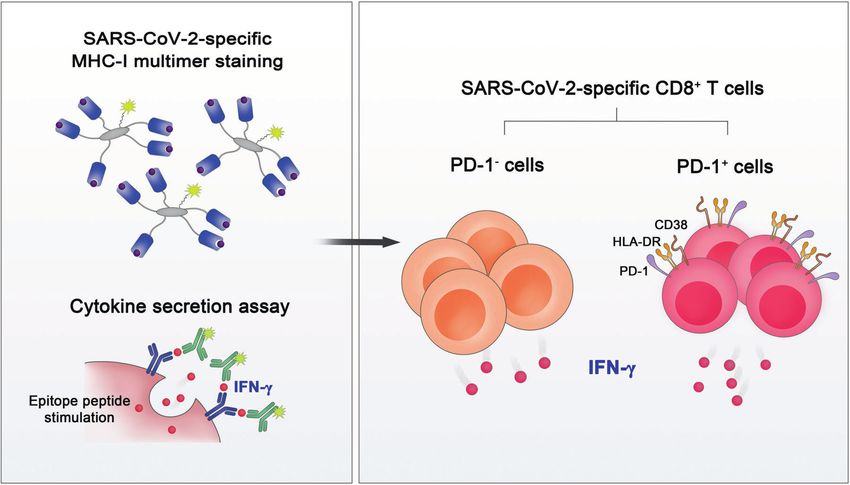
M.-S. Rha and E.-C. Shin
                                                                                                                                               2327
individual expression of PD-1 or other IRs is not sufficient to          characteristics of CD8+ T-cell exhaustion are individually insuffi-
indicate CD8+ T-cell exhaustion, the coexpression of multiple IRs is    cient to identify exhausted CD8+ T cells. In particular, because the
considered a main characteristic of exhaustion. In exhausted CD8+       majority of IRs are also transiently expressed in effector CD8+
T cells, several IRs are coexpressed with PD-1 and provide a            T cells during activation, IR expression is not a unique feature of
synergistic inhibitory effect [53, 77, 78]. Exhausted CD8+ T cells      exhausted CD8+ T cells [44, 101]. A previous study also showed no
with a higher number of coexpressed IRs have more severe                impairment of cytokine production in CD8+ T cells expressing a
exhaustion [77]. Simultaneous blockade of multiple IRs leads to         diverse array of IRs, indicating that IR expression may not be
robust reinvigoration of exhausted T cells in cancers and chronic       directly linked to dysfunction [102]. In transcriptomic analyses of
viral infections [70, 76].                                              CD8+ tumor-infiltrating lymphocytes from tumor-bearing mice,
                                                                        many IRs are present in the activation/dysfunction gene module
Changes in the epigenetic and transcriptional landscape                 but not in the dysfunctional gene module [103]. Furthermore,
In exhausted virus-specific CD8+ T cells from chronically LCMV-          genes related to the cell cycle pathway, migration, cytotoxic
infected mice, the expression of multiple genes is altered,             molecules, and costimulatory receptors are commonly upregu-
including genes related to TCR and cytokine signaling pathways,         lated in both exhausted and activated CD8+ T cells [79].
costimulatory pathways, and energy metabolism, as well as genes            Therefore, simultaneous consideration of diverse features,
encoding IRs and transcription factors [79]. Several studies using a    including dysfunction, sustained IR expression, transcriptional
mouse model of chronic LCMV infection have also shown that              and epigenetic alterations, and metabolic derangement, is needed
various transcription factors, including T-bet, Eomes, Blimp1, NFAT,    to identify bona fide exhausted CD8+ T cells and uncouple them
TCF1, IRF4, and TOX, are involved in CD8+ T-cell exhaustion [80–        from activated CD8+ T cells.
86]. In addition, BATF, which is commonly upregulated in both
HIV-specific CD8+ T cells from HIV progressors and Jurkat cells
following PD-1 ligation, mediates PD-1-induced suppression of           AN OVERVIEW OF CD8+ T-CELL RESPONSES AGAINST SARS-
T cells in vivo [87]. Although a master transcription factor specific    COV-2 IN PATIENTS WITH COVID-19
to exhaustion has not yet been identified, multiple transcription        Since the outbreak of COVID-19, we have gained much informa-
factors are associated with exhaustion-specific gene expression          tion about CD8+ T-cell responses to SARS-CoV-2. Early studies
and function [80, 81, 87, 88].                                          reported that SARS-CoV-2-specific CD8+ T-cell responses are
   Epigenetic regulation at the chromatin level also plays an           successfully elicited by SARS-CoV-2 infection [12, 13, 17]. SARS-
important role in controlling the differentiation and fate of CD8+      CoV-2-specific CD8+ T-cell responses have been identified in
T cells. Recent technological advances in epigenetics have              ~70% of convalescent individuals after recovery from COVID-19
enabled us to investigate the epigenetic characteristics of             [12]. These responses are specific to a wide range of SARS-CoV-2
exhausted CD8+ T cells. Previous studies using an assay for             antigens, including spike, nucleocapsid, and membranous pro-
transposase-accessible chromatin with high-throughput sequen-           teins, as well as other nonstructural proteins [12, 13, 17].
cing (ATAC-seq) have shown that the epigenetic landscape of                A series of studies suggest a critical role of CD8+ T cells in
exhausted CD8+ T cells is distinct from that of effector and            protecting against the development of severe COVID-19. SARS-
memory CD8+ T cells [89, 90]. Remarkable differences in the             CoV-2-specific CD8+ T-cell responses correlate with low disease
accessible chromatin regions were observed between exhausted            severity during the acute phase [104]. Memory T-cell responses
CD8+ T cells and effector/memory CD8+ T cells [89, 90]. For             have been detected in COVID-19 convalescent individuals even in
example, several open chromatin regions in the Ifng locus are           the absence of SARS-CoV-2-specific antibodies [105]. In addition,
present in effector and memory CD8+ T cells but not in exhausted        CD8+ T cells from the bronchoalveolar lavage fluid of patients
CD8+ T cells [90]. In contrast, open chromatin regions related to       with severe/critical COVID-19 exhibit a lack of dominant clones
IRs, such as PD-1, are specific to exhausted CD8+ T cells [89, 90].      compared to those from the bronchoalveolar lavage fluid of
                                                                        patients with mild disease [106].
Metabolic reprogramming                                                    Recently, studies using animal models revealed the importance
The activation and clonal expansion of CD8+ T cells are                 of CD8+ T cells in controlling SARS-CoV-2 infection. Limited viral
accompanied by alterations in cellular metabolism. During acute         clearance in the respiratory tract was observed in CD8+-depleted
infection, a transition from mitochondrial oxidative phosphoryla-       convalescent rhesus macaques upon SARS-CoV-2 rechallenge,
tion to glycolysis is required for differentiation into effector CD8+   implying that memory CD8+ T cells are required for the clearance
T cells [91–93]. Memory precursor T cells alter their cellular          of SARS-CoV-2 [18]. Furthermore, T-cell vaccination that does not
metabolism to oxidative phosphorylation and fatty acid oxidation        elicit neutralizing antibodies partially protects SARS-CoV-2-
[94]. In transcriptomic analysis, substantial alterations have been     infected mice from severe disease [19].
observed in genes involved in metabolism and bioenergetic
pathways in exhausted CD8+ T cells, suggesting that CD8+ T-cell
exhaustion is accompanied by metabolic alterations [79].                THE CD8+ T-CELL POPULATION IN PATIENTS WITH COVID-19
Exhausted CD8+ T cells are known to undergo metabolic                   The upregulation of activation markers and inhibitory
reprogramming, including decreased glycolysis and dysregulated          receptors
mitochondrial energetics [95]. Moreover, PD-1 signaling sup-            There is a growing body of evidence that circulating CD8+ T cells
presses glycolysis and promotes fatty acid oxidation in CD8+            from patients with severe COVID-19 exhibit an activated
T cells by inhibiting PI3K/Akt and MEK/ERK signaling [96].              phenotype characterized by increased expression of CD38, HLA-
Furthermore, PD-1 blockade restores glycolysis in exhausted             DR, and Ki-67 [16, 20–22, 107]. In addition, a recent study
CD8+ T cells [97].                                                      analyzing airway immune cells revealed that CD8+ T cells from the
                                                                        airways of patients with COVID-19 were predominantly tissue-
                                                                        resident memory T cells and that these cells have an elevated
UNCOUPLING T-CELL EXHAUSTION FROM ACTIVATION                            proportion of activated cells [108].
Considering that CD8+ T-cell exhaustion results from persistent            An exhausted CD8+ T-cell phenotype with an upregulation of
stimulation of T cells, it is challenging to distinguish T-cell         IRs, such as PD-1, TIM-3, LAG-3, CTLA-4, NKG2A, and CD39, has
exhaustion from activation. The surface markers and transcrip-          been described in patients with COVID-19, particularly in those
tional signatures of exhausted CD8+ T cells closely overlap with        with severe disease [20, 25–29]. In addition, an scRNA-seq analysis
those of activated CD8+ T cells [88, 98–100]. In addition, most         of peripheral blood mononuclear cells (PBMCs) showed that the

Cellular & Molecular Immunology (2021) 18:2325 – 2333
M.-S. Rha and E.-C. Shin
2328
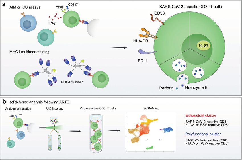
Fig. 2 Phenotype of SARS-CoV-2-specific CD8+ T cells. The phenotype of SARS-CoV-2-specific CD8+ T cells was examined using a ex vivo
stimulation-based functional assays, MHC-I multimer staining, and b single-cell RNA sequencing (scRNA-seq) following antigen-reactive T-cell
enrichment (ARTE). In the acute phase, SARS-CoV-2-specific CD8+ T cells express activation markers (CD38 and HLA-DR), PD-1, Ki-67, and
cytotoxic proteins (perforin and granzyme B). In scRNA-seq analysis of virus-reactive CD8+ T cells, the proportion of the “exhaustion” cluster,
characterized by increased expression of exhaustion-associated genes, was higher in SARS-CoV-2-reactive CD8+ T cells than in influenza A
virus (IAV)- or respiratory syncytial virus (RSV)-reactive CD8+ T cells. In addition, the proportion of the “polyfunctional” cluster expressing high
levels of genes encoding cytokines was lower in SARS-CoV-2-reactive CD8+ T cells than in IAV- or RSV-reactive CD8+ T cells. AIM activation-
induced marker, ICS intracellular cytokine staining
exhaustion score in the CD8+ effector cluster was significantly exert higher effector functions, including the production of IL-2
higher in patients with severe COVID-19 than in healthy donors and IL-17A and the expression of the degranulation marker
and patients with moderate disease [109]. Moreover, increased CD107a, upon anti-CD3/CD28 stimulation compared to cells from
PD-L1 expression has been reported in basophils and eosinophils healthy donors [25]. However, these studies examined the
from patients with severe COVID-19 [110]. functions of the CD8+ T-cell population following ex vivo
In contrast, a number of studies have reported no evidence of stimulation with pan-T cell stimulants, not SARS-CoV-2 antigens;
CD8+ T-cell exhaustion in patients with COVID-19, even in those thus, they lack information on the antigen specificity of CD8+
with severe cases. An early study performing scRNA-seq analysis of T cells.
PBMCs found that the T-cell exhaustion module score was not
significantly changed in CD8+ T cells from patients with COVID-19,
even in patients with severe cases with acute respiratory distress SARS-COV-2-SPECIFIC CD8+ T CELLS IN PATIENTS WITH
syndrome, compared to healthy donors [31]. In addition, a recent COVID-19
study using single-cell cellular indexing of transcriptomes and The phenotype of SARS-CoV-2-specific CD8+ T cells
epitopes by sequencing (CITE-seq) and TCR sequencing described Considering that only a proportion of the CD8+ T-cell population
that a cluster of exhausted CD8+ T cells was not associated with is specific to the infecting virus, it is important to examine the
COVID-19 [32]. In that study, the exhaustion of clonally expanded phenotype and functions of viral-antigen-specific CD8+ T cells, not
CD8+ T cells, as evaluated by IR expression, was not associated the total CD8+ T cell population, during viral infection. SARS-CoV-
with disease severity [32]. 2-specific CD8+ T cells from COVID-19 patients have been
Discrepancies in the results may be derived from several factors. investigated by many researchers (Fig. 2). Early studies examined
First, there were differences in the criteria for disease severity SARS-CoV-2-specific CD8+ T-cell responses using ex vivo
among studies. Second, the exhaustion gene sets used in the stimulation-based functional assays, such as intracellular cytokine
analysis or the detailed method of analysis for the transcriptomic staining and activation-induced marker assays [12, 13, 15, 17]. In
data were different. Third, the demographics of the study cohorts addition, scRNA-seq analysis following antigen-reactive T-cell
need to be considered. enrichment (ARTE) allowed us to investigate SARS-CoV-2-reactive
CD8+ T cells at the transcriptome level [111, 112]. However, all of
The functions of CD8+ T cells in patients with COVID-19 these assays have inherent limitations in that ex vivo stimulation
Several studies have reported that CD8+ T cells from patients with may change the phenotype of CD8+ T cells. Moreover,
COVID-19 exhibit a decreased cytokine-producing capacity upon stimulation-based functional assays detect functioning T cells,
stimulation with PMA/ionomycin [23, 27]. In contrast, another not virus-specific nonfunctioning cells. In contrast, MHC multimer
study reported that CD8+ T cells from patients with COVID-19 techniques, which directly detect antigen-specific T cells, do not
Cellular & Molecular Immunology (2021) 18:2325 – 2333M.-S. Rha and E.-C. Shin
2329
Fig. 3 Functional analysis of SARS-CoV-2-specific MHC-I multimer+CD8+ T cells. MHC-I multimer staining in combination with cytokine
secretion assays revealed that both PD-1+ and PD-1– cells among SARS-CoV-2-specific CD8+ T cells are functional in terms of IFN-γ production
have these caveats [113]. Several studies using MHC-I multimers SARS-CoV-2-reactive CD8+ T cells from COVID-19 patients than in
have examined the phenotypes of SARS-CoV-2-specific CD8+ IAV- and RSV-reactive CD8+ T cells from healthy donors [111],
T cells [16, 17, 33, 114, 115]. suggesting that SARS-CoV-2-reactive CD8+ T cells have a reduced
In the acute phase of COVID-19, SARS-CoV-2-specific MHC-I capacity to secrete effector cytokines.
multimer+CD8+ T cells express activation markers (CD38 and HLA-
DR), Ki-67, IRs (PD-1, TIM-3, and LAG-3), and cytotoxic proteins The functions of PD-1-expressing SARS-CoV-2-specific CD8+
(perforin and granzyme B), indicating that these cells are activated T cells
and proliferate with a high cytotoxic capacity [16]. This finding is To investigate the effector functions of SARS-CoV-2-specific CD8+
in line with the result that SARS-CoV-2-reactive CD8+ T cells T cells, our group performed MHC-I multimer staining, followed by
detected by stimulation-based assays express CD38, HLA-DR, Ki- proliferation assays and cytokine secretion assays (Fig. 3) [33].
67, and PD-1 [16]. Similar results were observed in our analysis SARS-CoV-2-specific MHC-I multimer+ T cells from individuals who
with MHC-I multimer staining. In a longitudinal analysis, we found recovered from COVID-19 showed robust proliferation upon
that the expression of PD-1 and CD38 in MHC-I multimer+ cells ex vivo antigen challenge. In addition, despite the lower frequency
decreases during the course of COVID-19 [33]. We also observed of IFN-γ-producing cells in SARS-CoV-2-specific CD8+ T cells than
an inverse correlation between the expression of PD-1 and CD38 IAV-specific CD8+ T cells, IFN-γ was produced by SARS-CoV-2-
in MHC-I multimer+ cells and the number of days since symptom specific CD8+ T cells regardless of their PD-1 expression. The same
onset. These kinetics suggest that PD-1 expression in SARS-CoV-2- results were observed when we analyzed SARS-CoV-2-specific
specific CD8+ T cells is transient, not persistent, in patients with MHC-I multimer+ cells from acute COVID-19 patients. These
COVID-19. Thus far, relatively few studies have examined the findings indicate that PD-1+ cells among SARS-CoV-2-specific
expression of IRs other than PD-1 in SARS-CoV-2-specific CD8+ MHC-I multimer+ cells are not exhausted but functionally active in
T cells. In the acute phase of severe COVID-19, a considerable the acute and early convalescent phases of COVID-19 and that PD-
proportion of SARS-CoV-2-specific CD8+ T cells express TIM-3, 1 needs to be considered an activation marker rather than an
LAG-3, TIGIT, and CTLA-4 [16]. The expression of TIM-3 and TIGIT in exhaustion marker in patients with COVID-19. In addition, there
SARS-CoV-2-specific CD8+ T cells tended to be lower among was no significant difference between patients with severe and
patients who recovered from mild COVID-19 than among patients nonsevere COVID-19 in regard to IFN-γ production by SARS-CoV-2-
with acute severe COVID-19 [16]. specific MHC-I multimer+CD8+ T cells. However, our study relied
A recent scRNA-seq analysis of virus-reactive CD8+ T cells on MHC-I multimers specific to HLA-A*02-restricted epitopes from
obtained using ARTE demonstrated that the proportion of the the spike protein. CD8+ T cells specific to other SARS-CoV-2
“exhaustion” CD8+ T-cell cluster characterized by increased epitopes restricted by other HLA-I allotypes may differ in
expression of exhaustion-associated genes, including HAVCR2 phenotype and function. In addition, given that the impairment
(TIM-3) and LAG3, was higher in SARS-CoV-2-reactive CD8+ T cells of IFN-γ production occurs in the later stage of T-cell exhaustion
from COVID-19 patients than in influenza A virus (IAV)- and [55], the production capacities of other cytokines, such as IL-2 and
respiratory syncytial virus (RSV)-reactive CD8+ T cells from healthy TNF, and cytotoxicity need to be examined further in SARS-CoV-2-
donors [111]. Intriguingly, the exhaustion cluster showed sig- specific MHC-I multimer+CD8+ T cells.
nificant enrichment of cytotoxicity-related genes, such as GZMB, Thus far, the phenotype and functions of SARS-CoV-2-specific
GZMA, GZMH, PRF1, and TBX21, indicating that this cluster is not CD8+ T cells have been analyzed primarily in peripheral blood
associated with dysfunction. On the other hand, the proportion of [16, 17, 33, 114, 115]. However, previous studies in animal models
the cluster expressing high levels of genes encoding cytokines, of respiratory viral infections have shown that tissue-resident
including IFNG, TNF, CCL3, CCL4, XCL1, and XCL2, was lower in memory T cells in the respiratory tract critically contribute to
Cellular & Molecular Immunology (2021) 18:2325 – 2333M.-S. Rha and E.-C. Shin
2330
protection from viral infection [116, 117]. In patients with COVID- alteration occurs more rapidly than T-cell exhaustion [132].
19, the expression of tissue-residency markers (CD69 and CD103) Furthermore, whether the differentiation state and transcriptional
and activation markers (PD-1 and HLA-DR) is higher in airway profiles of functionally impaired CD8+ T cells in respiratory viral
CD8+ T cells than in their peripheral blood counterparts [108], infections are similar to those of exhausted T cells is not clear.
indicating that tissue-resident CD8+ T cells with an activated Before the COVID-19 pandemic, little was known about the
phenotype are enriched in the airways. Therefore, additional functional impairment or exhaustion of CD8+ T cells during
studies are needed to investigate whether SARS-CoV-2-specific respiratory viral infections in humans. Further investigations with
CD8+ T cells in the respiratory tract are exhausted or functional in functional, transcriptomic, epigenetic, and metabolic profiling are
patients with COVID-19. needed to clarify T-cell exhaustion in acute respiratory viral
Moreover, comprehensive investigations on the transcriptional infections.
and epigenetic dynamics of SARS-CoV-2-specific CD8+ T cells
would provide new insights into the differentiation trajectories of
CD8+ T cells and clarify whether CD8+ T cells are truly exhausted CONCLUDING REMARKS AND PERSPECTIVES
during the course of COVID-19. Since the emergence of COVID-19, global efforts have rapidly
increased our knowledge of the immune responses to SARS-CoV-
The development of SARS-CoV-2-specific T-cell memory 2, including CD8+ T-cell responses. However, information regard-
Accumulating evidence suggests that SARS-CoV-2-specific T-cell ing the role of SARS-CoV-2-specific CD8+ T cells in protective
responses are maintained in convalescent individuals up to immunity is still limited. In addition, the differentiation dynamics
10 months post infection, indicating that SARS-CoV-2-specific of CD8+ T cells during the course of COVID-19, particularly
T-cell memory develops successfully and is long lasting [118–124]. whether SARS-CoV-2-specific CD8+ T cells become exhausted,
As CD8+ T cells that fail to become functional memory T cells remain enigmatic. Further comprehensive studies on the func-
differentiate into exhausted T cells, these findings suggest that tional, transcriptional, epigenetic, and metabolic landscapes of
CD8+ T-cell exhaustion may be limited in the majority of patients SARS-CoV-2-specific CD8+ T cells would help answer this question.
with COVID-19. Moreover, considering that virus-specific effector T cells are
Among subsets of memory T cells, stem cell-like memory T recruited to the site of inflammation, SARS-CoV-2-specific CD8+
(TSCM) cells are characterized by a high self-renewal capacity and a T cells in the respiratory tract should be investigated. Deeper
multipotent ability to generate diverse memory subsets [125, 126]. investigation of CD8+ T cells will help not only control the
Stem-like CD8+ memory T-cell progenitors have been described ongoing COVID-19 pandemic but also prepare for any upcoming
as being composed of two distinct subsets based on PD-1 and pandemics.
TIGIT expression [127]. Our group recently showed that the
majority of SARS-CoV-2-specific TSCM cells from convalescent
COVID-19 patients are PD-1–TIGIT– cells, suggesting that these REFERENCES
cells are not exhausted-like progenitors [124]. These findings also 1. Zhou P, Yang XL, Wang XG, Hu B, Zhang L, Zhang W, et al. A pneumonia
support SARS-CoV-2-specific CD8+ T cells being rarely exhausted outbreak associated with a new coronavirus of probable bat origin. Nature.
in patients with COVID-19. Limited exhaustion of SARS-CoV-2- 2020;579:270–3.
specific CD8+ T cells and successful development of TSCM cells 2. Huang C, Wang Y, Li X, Ren L, Zhao J, Hu Y, et al. Clinical features of patients
infected with 2019 novel coronavirus in Wuhan, China. Lancet.
lead to host protection upon re-exposure to SARS-CoV-2 among 2020;395:497–506.
COVID-19 convalescent individuals. 3. Baden LR, El Sahly HM, Essink B, Kotloff K, Frey S, Novak R, et al. Efficacy and
safety of the mRNA-1273 SARS-CoV-2 vaccine. N Engl J Med. 2021;384:403–16.
CD8+ T-cell exhaustion and vaccine-induced memory T-cell 4. Polack FP, Thomas SJ, Kitchin N, Absalon J, Gurtman A, Lockhart S, et al. Safety
responses and efficacy of the BNT162b2 mRNA Covid-19 vaccine. N Engl J Med.
Currently available vaccines using diverse platforms have been 2020;383:2603–15.
shown to elicit protective T-cell immunity [4, 7, 128–130]. 5. Ramasamy MN, Minassian AM, Ewer KJ, Flaxman AL, Folegatti PM, Owens DR,
Currently, COVID-19 vaccines are administered not only to et al. Safety and immunogenicity of ChAdOx1 nCoV-19 vaccine administered in
a prime-boost regimen in young and old adults (COV002): a single-blind, ran-
unexposed individuals but also to COVID-19 convalescent
domised, controlled, phase 2/3 trial. Lancet. 2021;396:1979–93.
individuals. Given that exhausted CD8+ T cells lose their potential 6. Stephenson KE, Le Gars M, Sadoff J, de Groot AM, Heerwegh D, Truyers C, et al.
to differentiate into memory T cells, the potential CD8+ T-cell Immunogenicity of the Ad26.COV2.S vaccine for COVID-19. JAMA.
exhaustion in individuals who have had COVID-19 can impede 2021;325:1535–44.
vaccine-induced development of T-cell memory. However, 7. Keech C, Albert G, Cho I, Robertson A, Reed P, Neal S, et al. Phase 1-2 trial of a
because CD8+ T-cell exhaustion is not evident in patients with SARS-CoV-2 recombinant spike protein nanoparticle vaccine. N Engl J Med.
COVID-19, it is assumed that COVID-19-experienced individuals 2020;383:2320–32.
successfully develop functional CD8+ T-cell memory following 8. Merad M, Martin JC. Pathological inflammation in patients with COVID-19: a key
vaccination. Recent studies have reported that a single dose of role for monocytes and macrophages. Nat Rev Immunol. 2020;20:355–62.
9. Lee JS, Park S, Jeong HW, Ahn JY, Choi SJ, Lee H, et al. Immunophenotyping of
mRNA vaccine robustly induces spike-specific T-cell responses in
COVID-19 and influenza highlights the role of type I interferons in development
COVID-19 convalescent individuals [131]. of severe COVID-19. Sci Immunol. 2020;5:eabd1554.
10. Giamarellos-Bourboulis EJ, Netea MG, Rovina N, Akinosoglou K, Antoniadou A,
Antonakos N, et al. Complex immune dysregulation in COVID-19 patients with
THE EXHAUSTED-LIKE PHENOTYPES OF CD8+ T CELLS IN severe respiratory failure. Cell Host Microbe. 2020;27:992–1000.e1003.
RESPIRATORY VIRAL INFECTIONS 11. Sette A, Crotty S. Adaptive immunity to SARS-CoV-2 and COVID-19. Cell.
An exhausted-like phenotype of CD8+ T cells has been reported in 2021;184:861–80.
several studies of respiratory viral infections using mouse models. 12. Grifoni A, Weiskopf D, Ramirez SI, Mateus J, Dan JM, Moderbacher CR, et al.
PD-1 upregulation on virus-specific CD8+ T cells and an Targets of T cell responses to SARS-CoV-2 coronavirus in humans with COVID-19
disease and unexposed individuals. Cell. 2020;181:1489–501.e15.
impairment of their effector functions have been observed during
13. Le Bert N, Tan AT, Kunasegaran K, Tham C, Hafezi M, Chia A, et al. SARS-CoV-2-
infection with respiratory viruses, such as human metapneumo- specific T cell immunity in cases of COVID-19 and SARS, and uninfected controls.
virus or influenza virus [132–134]. Similar to T-cell exhaustion Nature. 2020;584:457–62.
during chronic viral infections, the PD-1 pathway primarily 14. Nelde A, Bilich T, Heitmann JS, Maringer Y, Salih HR, Roerden M, et al. SARS-CoV-
mediates functional impairment of CD8+ T cells in acute 2-derived peptides define heterologous and COVID-19-induced T cell recogni-
respiratory virus infection [132, 134]. However, this functional tion. Nat Immunol. 2021;22:74–85.
Cellular & Molecular Immunology (2021) 18:2325 – 2333M.-S. Rha and E.-C. Shin
2331
15. Weiskopf D, Schmitz KS, Raadsen MP, Grifoni A, Okba N, Endeman H, et al. 42. Kaech SM, Wherry EJ. Heterogeneity and cell-fate decisions in effector and
Phenotype and kinetics of SARS-CoV-2-specific T cells in COVID-19 patients with memory CD8(+) T cell differentiation during viral infection. Immunity.
acute respiratory distress syndrome. Sci Immunol. 2020;5:eabd2071. 2007;27:393–405.
16. Sekine T, Perez-Potti A, Rivera-Ballesteros O, Strålin K, Gorin JB, Olsson A, et al. 43. Hashimoto M, Kamphorst AO, Im SJ, Kissick HT, Pillai RN, Ramalingam SS, et al.
Robust T cell immunity in convalescent individuals with asymptomatic or mild CD8 T cell exhaustion in chronic infection and cancer: opportunities for inter-
COVID-19. Cell. 2020;183:158–68.e14. ventions. Annu Rev Med. 2018;69:301–18.
17. Peng Y, Mentzer AJ, Liu G, Yao X, Yin Z, Dong D, et al. Broad and strong memory 44. McLane LM, Abdel-Hakeem MS, Wherry EJ. CD8 T cell exhaustion during chronic
CD4(+) and CD8(+) T cells induced by SARS-CoV-2 in UK convalescent indivi- viral infection and cancer. Annu Rev Immunol. 2019;37:457–95.
duals following COVID-19. Nat Immunol. 2020;21:1336–45. 45. Moskophidis D, Lechner F, Pircher H, Zinkernagel RM. Virus persistence in
18. McMahan K, Yu J, Mercado NB, Loos C, Tostanoski LH, Chandrashekar A, et al. acutely infected immunocompetent mice by exhaustion of antiviral cytotoxic
Correlates of protection against SARS-CoV-2 in rhesus macaques. Nature. effector T cells. Nature. 1993;362:758–61.
2021;590:630–4. 46. Gallimore A, Glithero A, Godkin A, Tissot AC, Plückthun A, Elliott T, et al.
19. Zhuang Z, Lai X, Sun J, Chen Z, Zhang Z, Dai J, et al. Mapping and role of T cell Induction and exhaustion of lymphocytic choriomeningitis virus-specific cyto-
response in SARS-CoV-2-infected mice. J Exp Med. 2021;218:e20202187. toxic T lymphocytes visualized using soluble tetrameric major histocompatibility
20. Song JW, Zhang C, Fan X, Meng FP, Xu Z, Xia P, et al. Immunological and complex class I-peptide complexes. J Exp Med. 1998;187:1383–93.
inflammatory profiles in mild and severe cases of COVID-19. Nat Commun. 47. Day CL, Kaufmann DE, Kiepiela P, Brown JA, Moodley ES, Reddy S, et al. PD-1
2020;11:3410. expression on HIV-specific T cells is associated with T-cell exhaustion and dis-
21. Mathew D, Giles JR, Baxter AE, Oldridge DA, Greenplate AR, Wu JE, et al. Deep ease progression. Nature. 2006;443:350–4.
immune profiling of COVID-19 patients reveals distinct immunotypes with 48. Baitsch L, Baumgaertner P, Devêvre E, Raghav SK, Legat A, Barba L, et al.
therapeutic implications. Science. 2020;369:eabc8511. Exhaustion of tumor-specific CD8(+) T cells in metastases from melanoma
22. Kuri-Cervantes L, Pampena MB, Meng W, Rosenfeld AM, Ittner C, Weisman AR, patients. J Clin Invest. 2011;121:2350–60.
et al. Comprehensive mapping of immune perturbations associated with severe 49. Pauken KE, Wherry EJ. Overcoming T cell exhaustion in infection and cancer.
COVID-19. Sci Immunol. 2020;5:eabd7114. Trends Immunol. 2015;36:265–76.
23. Mazzoni A, Salvati L, Maggi L, Capone M, Vanni A, Spinicci M, et al. Impaired 50. Fourcade J, Sun Z, Benallaoua M, Guillaume P, Luescher IF, Sander C, et al.
immune cell cytotoxicity in severe COVID-19 is IL-6 dependent. J Clin Invest. Upregulation of Tim-3 and PD-1 expression is associated with tumor antigen-
2020;130:4694–703. specific CD8+ T cell dysfunction in melanoma patients. J Exp Med.
24. Varchetta S, Mele D, Oliviero B, Mantovani S, Ludovisi S, Cerino A, et al. Unique 2010;207:2175–86.
immunological profile in patients with COVID-19. Cell Mol Immunol. 51. Matsuzaki J, Gnjatic S, Mhawech-Fauceglia P, Beck A, Miller A, Tsuji T, et al.
2021;18:604–12. Tumor-infiltrating NY-ESO-1-specific CD8+ T cells are negatively regulated by
25. De Biasi S, Meschiari M, Gibellini L, Bellinazzi C, Borella R, Fidanza L, et al. Marked LAG-3 and PD-1 in human ovarian cancer. Proc Natl Acad Sci USA.
T cell activation, senescence, exhaustion and skewing towards TH17 in patients 2010;107:7875–80.
with COVID-19 pneumonia. Nat Commun. 2020;11:3434. 52. Riches JC, Davies JK, McClanahan F, Fatah R, Iqbal S, Agrawal S, et al. T cells from
26. Zheng HY, Zhang M, Yang CX, Zhang N, Wang XC, Yang XP, et al. Elevated CLL patients exhibit features of T-cell exhaustion but retain capacity for cytokine
exhaustion levels and reduced functional diversity of T cells in peripheral blood production. Blood. 2013;121:1612–21.
may predict severe progression in COVID-19 patients. Cell Mol Immunol. 53. Jin HT, Anderson AC, Tan WG, West EE, Ha SJ, Araki K, et al. Cooperation of Tim-3
2020;17:541–3. and PD-1 in CD8 T-cell exhaustion during chronic viral infection. Proc Natl Acad
27. Zheng M, Gao Y, Wang G, Song G, Liu S, Sun D, et al. Functional Sci USA. 2010;107:14733–8.
exhaustion of antiviral lymphocytes in COVID-19 patients. Cell Mol Immunol. 54. Wherry EJ, Blattman JN, Murali-Krishna K, van der Most R, Ahmed R. Viral per-
2020;17:533–5. sistence alters CD8 T-cell immunodominance and tissue distribution and results
28. Diao B, Wang C, Tan Y, Chen X, Liu Y, Ning L, et al. Reduction and functional in distinct stages of functional impairment. J Virol. 2003;77:4911–27.
exhaustion of T cells in patients with coronavirus disease 2019 (COVID-19). Front 55. Mackerness KJ, Cox MA, Lilly LM, Weaver CT, Harrington LE, Zajac AJ. Pro-
Immunol. 2020;11:827. nounced virus-dependent activation drives exhaustion but sustains IFN-gamma
29. Laing AG, Lorenc A, Del Molino Del Barrio I, Das A, Fish M, Monin L, et al. A transcript levels. J Immunol. 2010;185:3643–51.
dynamic COVID-19 immune signature includes associations with poor prog- 56. Wherry EJ, Barber DL, Kaech SM, Blattman JN, Ahmed R. Antigen-independent
nosis. Nat Med. 2020;26:1623–35. memory CD8 T cells do not develop during chronic viral infection. Proc Natl
30. Schultheiß C, Paschold L, Simnica D, Mohme M, Willscher E, von Wenserski L, Acad Sci USA. 2004;101:16004–9.
et al. Next-generation sequencing of T and B cell receptor repertoires from 57. Shin H, Blackburn SD, Blattman JN, Wherry EJ. Viral antigen and extensive
COVID-19 patients showed signatures associated with severity of disease. division maintain virus-specific CD8 T cells during chronic infection. J Exp Med.
Immunity. 2020;53:442–455.e4. 2007;204:941–9.
31. Wilk AJ, Rustagi A, Zhao NQ, Roque J, Martínez-Colón GJ, McKechnie JL, et al. A 58. Matloubian M, Concepcion RJ, Ahmed R. CD4+ T cells are required to sustain
single-cell atlas of the peripheral immune response in patients with severe CD8+ cytotoxic T-cell responses during chronic viral infection. J Virol.
COVID-19. Nat Med. 2020;26:1070–6. 1994;68:8056–63.
32. Liu C, Martins AJ, Lau WW, Rachmaninoff N, Chen J, Imberti L, et al. Time- 59. Aubert RD, Kamphorst AO, Sarkar S, Vezys V, Ha SJ, Barber DL, et al. Antigen-
resolved systems immunology reveals a late juncture linked to fatal COVID-19. specific CD4 T-cell help rescues exhausted CD8 T cells during chronic viral
Cell. 2021;184:1836–57.e22. infection. Proc Natl Acad Sci USA. 2011;108:21182–7.
33. Rha MS, Jeong HW, Ko JH, Choi SJ, Seo IH, Lee JS, et al. PD-1-expressing SARS- 60. Scharping NE, Rivadeneira DB, Menk AV, Vignali P, Ford BR, Rittenhouse NL, et al.
CoV-2-specific CD8(+) T cells are not exhausted, but functional in patients with Mitochondrial stress induced by continuous stimulation under hypoxia rapidly
COVID-19. Immunity. 2021;54:44–52.e3. drives T cell exhaustion. Nat Immunol. 2021;22:205–15.
34. Rha MS, Kim AR, Shin EC. SARS-CoV-2-specific T cell responses in patients with 61. Yokosuka T, Takamatsu M, Kobayashi-Imanishi W, Hashimoto-Tane A, Azuma M,
COVID-19 and unexposed individuals. Immune Netw. 2021;21:e2. Saito T. Programmed cell death 1 forms negative costimulatory microclusters
35. Lipsitch M, Grad YH, Sette A, Crotty S. Cross-reactive memory T cells and herd that directly inhibit T cell receptor signaling by recruiting phosphatase SHP2. J
immunity to SARS-CoV-2. Nat Rev Immunol. 2020;20:709–13. Exp Med. 2012;209:1201–17.
36. Chen Z, John Wherry E. T cell responses in patients with COVID-19. Nat Rev 62. Schildberg FA, Klein SR, Freeman GJ, Sharpe AH. Coinhibitory pathways in the
Immunol. 2020;20:529–36. B7-CD28 ligand-receptor family. Immunity. 2016;44:955–72.
37. Altmann DM, Boyton RJ. SARS-CoV-2 T cell immunity: specificity, function, 63. Keir ME, Butte MJ, Freeman GJ, Sharpe AH. PD-1 and its ligands in tolerance and
durability, and role in protection. Sci Immunol. 2020;5:eabd6160. immunity. Annu Rev Immunol. 2008;26:677–704.
38. Karlsson AC, Humbert M, Buggert M. The known unknowns of T cell immunity to 64. Hui E, Cheung J, Zhu J, Su X, Taylor MJ, Wallweber HA, et al. T cell costimulatory
COVID-19. Sci Immunol. 2020;5:eabe8063. receptor CD28 is a primary target for PD-1-mediated inhibition. Science.
39. de Candia P, Prattichizzo F, Garavelli S, Matarese G. T Cells: warriors of SARS- 2017;355:1428–33.
CoV-2 Infection. Trends Immunol. 2021;42:18–30. 65. Freeman GJ, Long AJ, Iwai Y, Bourque K, Chernova T, Nishimura H, et al.
40. Cui WG, Kaech SM. Generation of effector CD8+ T cells and their conversion to Engagement of the PD-1 immunoinhibitory receptor by a novel B7 family
memory T cells. Immunol Rev. 2010;236:151–66. member leads to negative regulation of lymphocyte activation. J Exp Med.
41. Kaech SM, Cui W. Transcriptional control of effector and memory CD8+ T cell 2000;192:1027–34.
differentiation. Nat Rev Immunol. 2012;12:749–61.
Cellular & Molecular Immunology (2021) 18:2325 – 2333M.-S. Rha and E.-C. Shin
2332
66. Barber DL, Wherry EJ, Masopust D, Zhu B, Allison JP, Sharpe AH, et al. Restoring 92. O’Sullivan D, Pearce EL. Targeting T cell metabolism for therapy. Trends
function in exhausted CD8 T cells during chronic viral infection. Nature. Immunol. 2015;36:71–80.
2006;439:682–7. 93. Chang CH, Pearce EL. Emerging concepts of T cell metabolism as a target of
67. Shin EC, Rehermann B. Taking the brake off T cells in chronic viral infection. Nat immunotherapy. Nat Immunol. 2016;17:364–8.
Med. 2006;12:276–7. 94. van der Windt GJ, Pearce EL. Metabolic switching and fuel choice during T-cell
68. Hirano F, Kaneko K, Tamura H, Dong H, Wang S, Ichikawa M, et al. Blockade of differentiation and memory development. Immunol Rev. 2012;249:27–42.
B7-H1 and PD-1 by monoclonal antibodies potentiates cancer therapeutic 95. Bengsch B, Johnson AL, Kurachi M, Odorizzi PM, Pauken KE, Attanasio J, et al.
immunity. Cancer Res. 2005;65:1089–96. Bioenergetic insufficiencies due to metabolic alterations regulated by the
69. Curiel TJ, Wei S, Dong H, Alvarez X, Cheng P, Mottram P, et al. Blockade of B7-H1 inhibitory receptor PD-1 are an early driver of CD8(+) T cell exhaustion.
improves myeloid dendritic cell-mediated antitumor immunity. Nat Med. Immunity. 2016;45:358–73.
2003;9:562–7. 96. Patsoukis N, Bardhan K, Chatterjee P, Sari D, Liu B, Bell LN, et al. PD-1 alters T-cell
70. Ribas A, Wolchok JD. Cancer immunotherapy using checkpoint blockade. Sci- metabolic reprogramming by inhibiting glycolysis and promoting lipolysis and
ence. 2018;359:1350–5. fatty acid oxidation. Nat Commun. 2015;6:6692.
71. Garon EB, Rizvi NA, Hui R, Leighl N, Balmanoukian AS, Eder JP, et al. Pem- 97. Chang CH, Qiu J, O'Sullivan D, Buck MD, Noguchi T, Curtis JD, et al. Metabolic
brolizumab for the treatment of non-small-cell lung cancer. N Engl J Med. competition in the tumor microenvironment is a driver of cancer progression.
2015;372:2018–28. Cell. 2015;162:1229–41.
72. Ribas A, Hamid O, Daud A, Hodi FS, Wolchok JD, Kefford R, et al. Association of 98. Marraco SAF, Neubert NJ, Verdeil G, Speiser DE. Inhibitory receptors beyond T
pembrolizumab with tumor response and survival among patients with cell exhaustion. Front Immunol. 2015;6:1–14.
advanced melanoma. JAMA. 2016;315:1600–9. 99. Singer M, Wang C, Cong L, Marjanovic ND, Kowalczyk MS, Zhang H, et al. A
73. Le DT, Durham JN, Smith KN, Wang H, Bartlett BR, Aulakh LK, et al. Mismatch distinct gene module for dysfunction uncoupled from activation in tumor-
repair deficiency predicts response of solid tumors to PD-1 blockade. Science. infiltrating T cells (vol 166, pg 1500, 2016). Cell. 2017;171:1221–3.
2017;357:409–13. 100. Fuertes Marraco SA, Neubert NJ, Verdeil G, Speiser DE. Inhibitory receptors
74. Ansell SM, Lesokhin AM, Borrello I, Halwani A, Scott EC, Gutierrez M, et al. PD-1 beyond T cell exhaustion. Front Immunol. 2015;6:310.
blockade with nivolumab in relapsed or refractory Hodgkin’s lymphoma. N Engl 101. Wherry EJ, Kurachi M. Molecular and cellular insights into T cell exhaustion. Nat
J Med. 2015;372:311–9. Rev Immunol. 2015;15:486–99.
75. Anderson AC, Joller N, Kuchroo VK. Lag-3, Tim-3, and TIGIT: co-inhibitory 102. Legat A, Speiser DE, Pircher H, Zehn D, Fuertes Marraco SA. Inhibitory receptor
receptors with specialized functions in immune regulation. Immunity. expression depends more dominantly on differentiation and activation than
2016;44:989–1004. “exhaustion” of human CD8 T cells. Front Immunol. 2013;4:455.
76. Attanasio J, Wherry EJ. Costimulatory and coinhibitory receptor pathways in 103. Singer M, Wang C, Cong L, Marjanovic ND, Kowalczyk MS, Zhang H, et al. A
infectious disease. Immunity. 2016;44:1052–68. distinct gene module for dysfunction uncoupled from activation in tumor-
77. Blackburn SD, Shin H, Haining WN, Zou T, Workman CJ, Polley A, et al. Cor- infiltrating T cells. Cell. 2016;166:1500–11.e9.
egulation of CD8+ T cell exhaustion by multiple inhibitory receptors during 104. Rydyznski Moderbacher C, Ramirez SI, Dan JM, Grifoni A, Hastie KM, Weiskopf D,
chronic viral infection. Nat Immunol. 2009;10:29–37. et al. Antigen-specific adaptive immunity to SARS-CoV-2 in acute COVID-19 and
78. Gros A, Robbins PF, Yao X, Li YF, Turcotte S, Tran E, et al. PD-1 identifies the associations with age and disease severity. Cell. 2020;183:996–1012.e19.
patient-specific CD8(+) tumor-reactive repertoire infiltrating human tumors. J 105. Schwarzkopf S, Krawczyk A, Knop D, Klump H, Heinold A, Heinemann FM, et al.
Clin Invest. 2014;124:2246–59. Cellular immunity in COVID-19 convalescents with PCR-confirmed infection but
79. Wherry EJ, Ha SJ, Kaech SM, Haining WN, Sarkar S, Kalia V, et al. Molecular with undetectable SARS-CoV-2-specific IgG. Emerg Infect Dis. 2021;27:122–9.
signature of CD8+ T cell exhaustion during chronic viral infection. Immunity. 106. Liao M, Liu Y, Yuan J, Wen Y, Xu G, Zhao J, et al. Single-cell landscape of
2007;27:670–84. bronchoalveolar immune cells in patients with COVID-19. Nat Med.
80. Kao C, Oestreich KJ, Paley MA, Crawford A, Angelosanto JM, Ali MA, et al. 2020;26:842–4.
Transcription factor T-bet represses expression of the inhibitory receptor PD-1 107. Adamo S, Chevrier S, Cervia C, Zurbuchen Y, Raeber ME, Yang L, et al. Profound
and sustains virus-specific CD8+ T cell responses during chronic infection. Nat dysregulation of T cell homeostasis and function in patients with severe COVID-
Immunol. 2011;12:663–71. 19. Allergy. 2021. https://doi.org/10.1111/all.14866.
81. Paley MA, Kroy DC, Odorizzi PM, Johnnidis JB, Dolfi DV, Barnett BE, et al. Pro- 108. Szabo PA, Dogra P, Gray JI, Wells SB, Connors TJ, Weisberg SP, et al. Longitudinal
genitor and terminal subsets of CD8+ T cells cooperate to contain chronic viral profiling of respiratory and systemic immune responses reveals myeloid cell-
infection. Science. 2012;338:1220–5. driven lung inflammation in severe COVID-19. Immunity. 2021;54:797–814.e6.
82. Man K, Gabriel SS, Liao Y, Gloury R, Preston S, Henstridge DC, et al. Transcription 109. Zhang JY, Wang XM, Xing X, Xu Z, Zhang C, Song JW, et al. Single-cell landscape
factor IRF4 promotes CD8(+) T cell exhaustion and limits the development of of immunological responses in patients with COVID-19. Nat Immunol.
memory-like T cells during chronic infection. Immunity. 2017;47:1129–41.e5. 2020;21:1107–18.
83. Khan O, Giles JR, McDonald S, Manne S, Ngiow SF, Patel KP, et al. TOX tran- 110. Vitte J, Diallo AB, Boumaza A, Lopez A, Michel M, Allardet-Servent J, et al. A
scriptionally and epigenetically programs CD8(+) T cell exhaustion. Nature. granulocytic signature identifies COVID-19 and its severity. J Infect Dis.
2019;571:211–8. 2020;222:1985–96.
84. Beltra JC, Manne S, Abdel-Hakeem MS, Kurachi M, Giles JR, Chen Z, et al. 111. Kusnadi A, Ramírez-Suástegui C, Fajardo V, Chee SJ, Meckiff BJ, Simon H, et al.
Developmental relationships of four exhausted CD8(+) T cell subsets reveals Severely ill COVID-19 patients display impaired exhaustion features in SARS-
underlying transcriptional and epigenetic landscape control mechanisms. CoV-2-reactive CD8(+) T cells. Sci Immunol. 2021;6:eabe4782.
Immunity. 2020;52:825–41.e8. 112. Koh JY, Shin EC. Landscapes of SARS-CoV-2-reactive CD8(+) T cells: hetero-
85. Martinez GJ, Pereira RM, Äijö T, Kim EY, Marangoni F, Pipkin ME, et al. The geneity of host immune responses against SARS-CoV-2. Signal Transduct Target
transcription factor NFAT promotes exhaustion of activated CD8(+) T cells. Ther. 2021;6:146.
Immunity. 2015;42:265–78. 113. Altman JD, Moss PA, Goulder PJ, Barouch DH, McHeyzer-Williams MG, Bell JI, et al.
86. Shin H, Blackburn SD, Intlekofer AM, Kao C, Angelosanto JM, Reiner SL, et al. A Phenotypic analysis of antigen-specific T lymphocytes. Science. 1996;274:94–96.
role for the transcriptional repressor Blimp-1 in CD8(+) T cell exhaustion during 114. Schulien I, Kemming J, Oberhardt V, Wild K, Seidel LM, Killmer S, et al. Char-
chronic viral infection. Immunity. 2009;31:309–20. acterization of pre-existing and induced SARS-CoV-2-specific CD8(+) T cells. Nat
87. Quigley M, Pereyra F, Nilsson B, Porichis F, Fonseca C, Eichbaum Q, et al. Med. 2021;27:78–85.
Transcriptional analysis of HIV-specific CD8+ T cells shows that PD-1 inhibits T 115. Habel JR, Nguyen T, van de Sandt CE, Juno JA, Chaurasia P, Wragg K, et al.
cell function by upregulating BATF. Nat Med. 2010;16:1147–51. Suboptimal SARS-CoV-2-specific CD8(+) T cell response associated with the
88. Doering TA, Crawford A, Angelosanto JM, Paley MA, Ziegler CG, Wherry EJ. prominent HLA-A*02:01 phenotype. Proc Natl Acad Sci USA. 2020;117:24384–91.
Network analysis reveals centrally connected genes and pathways involved in 116. McMaster SR, Wilson JJ, Wang H, Kohlmeier JE. Airway-resident memory CD8
CD8+ T cell exhaustion versus memory. Immunity. 2012;37:1130–44. T cells provide antigen-specific protection against respiratory virus challenge
89. Sen DR, Kaminski J, Barnitz RA, Kurachi M, Gerdemann U, Yates KB, et al. The through rapid IFN-gamma production. J Immunol. 2015;195:203–9.
epigenetic landscape of T cell exhaustion. Science. 2016;354:1165–9. 117. Pizzolla A, Nguyen T, Smith JM, Brooks AG, Kedzieska K, Heath WR, et al. Resi-
90. Pauken KE, Sammons MA, Odorizzi PM, Manne S, Godec J, Khan O, et al. Epi- dent memory CD8(+) T cells in the upper respiratory tract prevent pulmonary
genetic stability of exhausted T cells limits durability of reinvigoration by PD-1 influenza virus infection. Sci Immunol. 2017;2:eaam6970.
blockade. Science. 2016;354:1160–5. 118. Bilich T, Nelde A, Heitmann JS, Maringer Y, Roerden M, Bauer J, et al. T cell and
91. Buck MD, O’Sullivan D, Pearce EL. T cell metabolism drives immunity. J Exp Med. antibody kinetics delineate SARS-CoV-2 peptides mediating long-term immune
2015;212:1345–60. responses in COVID-19 convalescent individuals. Sci Transl Med. 2021;13:eabf7517.
Cellular & Molecular Immunology (2021) 18:2325 – 2333M.-S. Rha and E.-C. Shin
2333
119. Dan JM, Mateus J, Kato Y, Hastie KM, Yu ED, Faliti CE, et al. Immunological 134. Erickson JJ, Lu P, Wen S, Hastings AK, Gilchuk P, Joyce S, et al. Acute viral
memory to SARS-CoV-2 assessed for up to 8 months after infection. Science. respiratory infection rapidly induces a CD8+ T cell exhaustion-like phenotype. J
2021;371:eabf4063. Immunol. 2015;195:4319–30.
120. Zuo J, Dowell AC, Pearce H, Verma K, Long HM, Begum J, et al. Robust SARS-
CoV-2-specific T cell immunity is maintained at 6 months following primary
infection. Nat Immunol. 2021;22:620–6. ACKNOWLEDGEMENTS
121. Jiang XL, Wang GL, Zhao XN, Yan FH, Yao L, Kou ZQ, et al. Lasting antibody and This research was supported by the 2020 Joint Research Project of the Institutes of
T cell responses to SARS-CoV-2 in COVID-19 patients three months after infec- Science and Technology.
tion. Nat Commun. 2021;12:897.
122. Breton G, Mendoza P, Hägglöf T, Oliveira TY, Schaefer-Babajew D, Gaebler C,
et al. Persistent cellular immunity to SARS-CoV-2 infection. J Exp Med. 2021;218:
e20202515. AUTHOR CONTRIBUTIONS
123. Sherina N, et al. Persistence of SARS-CoV-2-specific B and T cell responses in M-SR and E-CS conceived and designed the work; collected and analyzed the
convalescent COVID-19 patients 6-8 months after the infection. Med (N Y). relevant reports; and wrote the manuscript.
2021;2:281–95.e4.
124. Jung JH, Rha MS, Sa M, Choi HK, Jeon JH, Seok H, et al. SARS-CoV-2-specific T cell
memory is sustained in COVID-19 convalescent patients for 10 months with COMPETING INTERESTS
successful development of stem cell-like memory T cells. Nat Commun. The authors declare no competing interests.
2021;12:4043.
125. Gattinoni L, Lugli E, Ji Y, Pos Z, Paulos CM, Quigley MF, et al. A human memory T
cell subset with stem cell-like properties. Nat Med. 2011;17:1290–7. ADDITIONAL INFORMATION
126. Gattinoni L, Speiser DE, Lichterfeld M, Bonini C. T memory stem cells in health Correspondence and requests for materials should be addressed to E.-C.S.
and disease. Nat Med. 2017;23:18–27.
127. Galletti G, De Simone G, Mazza E, Puccio S, Mezzanotte C, Bi TM, et al. Two Reprints and permission information is available at http://www.nature.com/
subsets of stem-like CD8(+) memory T cell progenitors with distinct fate reprints
commitments in humans. Nat Immunol. 2020;21:1552–62.
128. Anderson EJ, Rouphael NG, Widge AT, Jackson LA, Roberts PC, Makhene M, et al.
Safety and immunogenicity of SARS-CoV-2 mRNA-1273 vaccine in older adults.
N Engl J Med. 2020;383:2427–38.
129. Ewer KJ, Barrett JR, Belij-Rammerstorfer S, Sharpe H, Makinson R, Morter R, et al.
T cell and antibody responses induced by a single dose of ChAdOx1 nCoV-19 Open Access This article is licensed under a Creative Commons
(AZD1222) vaccine in a phase 1/2 clinical trial. Nat Med. 2021;27:270–8. Attribution 4.0 International License, which permits use, sharing,
130. Sadoff J, Le Gars M, Shukarev G, Heerwegh D, Truyers C, de Groot AM, et al. adaptation, distribution and reproduction in any medium or format, as long as you give
Interim results of a phase 1-2a trial of Ad26.COV2.S Covid-19 vaccine. N Engl J appropriate credit to the original author(s) and the source, provide a link to the Creative
Med. 2021;384:1824–35. Commons license, and indicate if changes were made. The images or other third party
131. Reynolds CJ, Pade C, Gibbons JM, Butler DK, Otter AD, Menacho K, et al. Prior material in this article are included in the article’s Creative Commons license, unless
SARS-CoV-2 infection rescues B and T cell responses to variants after first vac- indicated otherwise in a credit line to the material. If material is not included in the
cine dose. Science. 2021. https://doi.org/10.1126/science.abh1282. article’s Creative Commons license and your intended use is not permitted by statutory
132. Erickson JJ, Gilchuk P, Hastings AK, Tollefson SJ, Johnson M, Downing MB, et al. regulation or exceeds the permitted use, you will need to obtain permission directly
Viral acute lower respiratory infections impair CD8+ T cells through PD-1. J Clin from the copyright holder. To view a copy of this license, visit http://creativecommons.
Invest. 2012;122:2967–82. org/licenses/by/4.0/.
133. Rutigliano JA, Sharma S, Morris MY, Oguin TH, McClaren JL, Doherty PC, et al.
Highly pathological influenza A virus infection is associated with augmented
expression of PD-1 by functionally compromised virus-specific CD8+ T cells. J © The Author(s) 2021
Virol. 2014;88:1636–51.
Cellular & Molecular Immunology (2021) 18:2325 – 2333You can also read