IN SILICO ANALYSIS SUGGESTS THE RNAI ENHANCING ANTIBIOTIC ENOXACIN AS A POTENTIAL INHIBITOR OF SARS COV 2 INFECTION
←
→
Page content transcription
If your browser does not render page correctly, please read the page content below
www.nature.com/scientificreports
OPEN In silico analysis suggests
the RNAi‑enhancing antibiotic
enoxacin as a potential inhibitor
of SARS‑CoV‑2 infection
Amirhossein Ahmadi1 & Sharif Moradi 2*
COVID-19 has currently become the biggest challenge in the world. There is still no specific medicine
for COVID-19, which leaves a critical gap for the identification of new drug candidates for the
disease. Recent studies have reported that the small-molecule enoxacin exerts an antiviral activity
by enhancing the RNAi pathway. The aim of this study is to analyze if enoxacin can exert anti-SARS-
CoV-2 effects. We exploit multiple computational tools and databases to examine (i) whether the
RNAi mechanism, as the target pathway of enoxacin, could act on the SARS-CoV-2 genome, and
(ii) microRNAs induced by enoxacin might directly silence viral components as well as the host cell
proteins mediating the viral entry and replication. We find that the RNA genome of SARS-CoV-2 might
be a suitable substrate for DICER activity. We also highlight several enoxacin-enhanced microRNAs
which could target SARS-CoV-2 components, pro-inflammatory cytokines, host cell components
facilitating viral replication, and transcription factors enriched in lung stem cells, thereby promoting
their differentiation and lung regeneration. Finally, our analyses identify several enoxacin-targeted
regulatory modules that were critically associated with exacerbation of the SARS-CoV-2 infection.
Overall, our analysis suggests that enoxacin could be a promising candidate for COVID-19 treatment
through enhancing the RNAi pathway.
Since the emergence of SARS-CoV-2 virus in November 2019, COVID-19 has become the biggest challenge in
the world1. Although several efforts are currently underway to develop COVID-19 vaccines, there is an urgent
need to find new, effective treatments in order to decrease the mortality rate especially in regions and countries
with the highest number of COVID-19 cases and d eaths2,3. Various strategies including blocking viral entry into
the host c ells4–6, inhibiting viral r eplication7,8 and reducing cytokine s torms9,10 have been proposed to relieve
patients from COVID-19 symptoms. Based on these strategies, hundreds of clinical trials have been conducted
and only a few drugs have been shown to slightly shorten the time to recovery or weakly reduce the rate of death
among hospitalized p atients8,11. However, there is still no specific efficacious medicine for COVID-19 that clearly
reduces the death rate, making it critically inevitable to look for, and evaluate, new drug candidates for the effec-
tive treatment of COVID-19. As a potent antiviral strategy, the innate immune system could be exploited to fight
the deadly infection caused by SARS-CoV-2.
The innate immune system, which functions as the first line of defense against viruses in the majority of
mammalian cells, consists of the interferon (IFN) and the RNA interference (RNAi) pathways as major immune
mechanisms against various v iruses12. The IFN pathway is activated by viral components, thereby transcription-
ally activating a large number of the so-called IFN-stimulated genes (ISGs)13. The activation of ISGs induces the
production and secretion of various cytokines and chemokines which in turn recruit a large number of immune
cells to the site of infection14. This pathway appears to be more active in mature cells (i.e. less active in stem and
progenitor cells which are frequently infected by many viruses)15. Moreover, it can lead to a potentially life-
threatening immune reaction called the cytokine release syndrome or cytokine storm which is resulted from an
exaggerated immune response (i.e. a hyperactive IFN-mediated response) to the viral infection16. This immune
overreaction is injurious to the host cells and might be induced by the SARS-CoV-2 i nfection17. In contrast, the
RNAi pathway is an IFN-independent process of fighting viruses (therefore, does not induce a cytokine storm)
1
Department of Biological Science and Technology, Faculty of Nano and Bio Science and Technology,
Persian Gulf University, Bushehr 75169,, Iran. 2Department of Stem Cells and Developmental Biology,
Cell Science Research Center, Royan Institute for Stem Cell Biology and Technology, ACECR, Tehran, Iran.
*
email: sh.moradi@royan-rc.ac.ir
Scientific Reports | (2021) 11:10271 | https://doi.org/10.1038/s41598-021-89605-6 1
Vol.:(0123456789)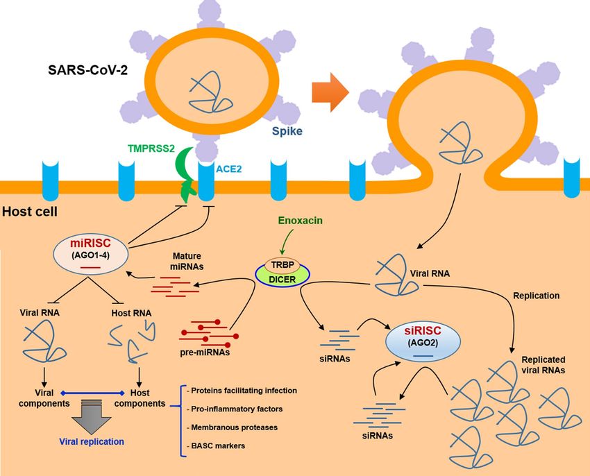
www.nature.com/scientificreports/
and is typically more active in embryonic and non-mature (e.g. stem and progenitor) cells18. It involves the
efficient degradation of the large viral RNAs which form secondary double-stranded structures thereby serving
as substrates of the RNAi p athway18.
Fluoroquinolones such as enoxacin are broad-spectrum synthetic antibiotics used in different clinical condi-
tions like urinary tract-, respiratory-, and systemic i nfections19,20. Although quinolones are known to typically
inhibit DNA replication by targeting bacterial DNA g yrases21, a growing body of evidence has revealed that
some members of this family of antibiotics could also inhibit viral helicases, attenuate cytokine production and
pro-inflammatory reactions22, and more importantly enhance the RNAi process as an inflammation-free innate
immune defense against viral i nfections23.
RNAi acts as a sequence-specific gene silencing process in which double-stranded RNAs (dsRNAs) such as
short hairpin RNAs (shRNAs), viral RNAs, and microRNA (miRNA) precursors are cleaved by the RNase enzyme
DICER to yield small interfering RNA (siRNA) duplexes. One strand of these duplexes is then preferentially
incorporated into the so-called RNA‐induced silencing complex (RISC) to target complementary transcripts
through Watson–Crick base‐pairing interactions24. The initiating dsRNAs can be either exogenous (e.g. viral
RNAs or shRNAs) or endogenous (e.g. pre-miRNA transcripts) which are processed to generate siRNAs and
miRNAs25,26. miRNAs are short non-coding RNAs processed by DICER which regulate gene expression at the
post-transcriptional level, thereby modulating virtually all biological p athways27–29. Importantly, the RNAi path-
way appears to be a particularly potent antiviral process, as many viruses have evolved several RNAi-suppressing
strategies including encoding the viral suppressor of RNAi (VSR) proteins to minimize the RNAi p athway30,31.
Therefore, compounds such as enoxacin which could serve as RNAi enhancers might be ideal candidates for
antiviral therapy of COVID-19.
Studies have shown that enoxacin could enhance RNAi activity through its binding to, and stimulating,
TAR RNA binding protein (TRBP), as the main cofactor of DICER, thereby facilitating the binding of DICER
to target RNAs32,33. This enhancement in the RNAi pathway subsequently leads to a more potent RNAi (i.e.
siRNA and miRNA) effect on the target RNAs, either mRNAs (targets of miRNAs) or viral RNAs (targets of the
virally-derived siRNAs)23,34,35. Interestingly, enoxacin has recently been shown to exert a potent antiviral activity
against several types of viruses such as Zika virus, Dengue virus, human immunodeficiency virus (HIV/AIDS),
and langat virus in in vitro, organoid, and animal models by enhancing the RNAi p athway36–39, suggesting that
the RNAi-enhancing activity of enoxacin could serve as a general antiviral strategy including against the novel
coronavirus. Here, we exploited several in silico analyses to predict if enoxacin could exert anti-SARS-CoV-2
effects. Our results indicated that the RNA genome of SARS-CoV-2 might be processed by the endonuclealytic
activity of DICER. Moreover, we determined a set of putative miRNA targets of enoxacin and found that a frac-
tion of these miRNAs could restrict the entry of SARS-CoV-2 into the host cells by targeting key cell surface
proteins. Another fraction of enoxacin-responsive miRNAs showed the potential to repress both host transcripts
mediating the replication of the virus and viral transcripts encoding important viral proteins. Finally, other
enoxacin-induced miRNAs appeared to potentially silence the cytokine storm driven by the SARS-CoV-2 infec-
tion as well as promote bronchiolar stem cell differentiation, thereby empowering the regeneration of the lung
parenchyma. Overall, our findings strongly suggest that enoxacin and possibly other RNAi-enhancing members
of the fluoroquinolones might serve as antiviral drugs able to be repositioned for effective COVID-19 therapy.
Results
The RNA genome of SARS‑CoV‑2 might be a suitable substrate for DICER. Since enoxacin
has been reported to exert its anti-viral activity by means of enhancing the RNAi pathway through binding
and stimulating the activity of TRBP, the physical partner of DICER23,33, we first investigated if the single-
stranded RNA genome of SARS-CoV-2 might be processed by the RNAi machinery. As DICER acts on hairpin
RNA structures, we used three methods to predict these precursor structures in the viral genome. The SM-
based method, which predicts pre-miRNAs from given sequences using sequence-structure motif strategies40,
predicted 145 hairpin structures and miRNA precursors potentially derived from the SARS-CoV-2 genome
(Table S1), suggesting that DICER could act on the SARS-CoV-2 genome and gradually degrade it down to
siRNAs. Moreover, we utilized two other hairpin-predicting tools, i.e. the Ab-initio and the BLASTN method
(a miRBase feature), to examine if other different methods of hairpin prediction similarly identify potential
DICER-acting genomic regions. Of note, in the Ab-initio method an approximation of miRNA hairpin structure
is first searched for, before reconstituting the pre-miRNA structure41, while the BLASTN approach searches
for a human miRNA homolog in the viral genome. Using these two approaches, we could predict 518 and 69
hairpin/miRNA precursors, respectively (Table S1), which could serve as binding sites for recruiting DICER and
stimulating its RNase activity. Notably, comparing the results obtained with the three hairpin-predicting tools
revealed that 12 hairpin structures were commonly predicted by all these approaches in nearly the same regions
of the SARS-CoV-2 genome (Table 1). These results strongly suggest that the SARS-CoV-2 genome intrinsically
harbors multiple hairpin structures which could be processed by DICER/TRBP complex, allowing for the effi-
cient degradation of the coronavirus RNA genome through the RNAi pathway.
Enoxacin might target the proteins mediating SARS‑CoV‑2 entry into host cells. To predict
if enoxacin-stimulated miRNAs could target SARS-CoV-2 entry receptors, we first sought to determine the
miRNAs that are reported in several published research to be upregulated by enoxacin (Table S2). Analysis of
enoxacin-induced miRNAs across multiple types of cell lines (i.e. prostate cancer cells, HEK cells, and melanoma
cells) revealed that enoxacin could upregulate a large set of mature miRNAs. Indeed, 268 miRNAs were found
to be upregulated under enoxacin treatment (Table S2). Comparing these 268 miRNAs with the miRNA expres-
sion profile of the lung tissue (see the list of miRNAs in Table S3) showed that 137 of enoxacin-upregulated
Scientific Reports | (2021) 11:10271 | https://doi.org/10.1038/s41598-021-89605-6 2
Vol:.(1234567890)
www.nature.com/scientificreports/
Ab-initio-based method SM-based method BLASTN method
1 Sequences Location Sequences Location Sequences Location
5′GCCUUUGGAGGCUGUGUGUUC
5′CUUUGGAGGCUGUGUGUUCUC
UCUUAUGUUGGUUGCCAUAAC
UUAUGUUGGUUGCCAUAACAA 5′UCUUAUGUUGGUUGCCAUAAC
AAGUGUGCCUAUUGGGUUCCA
2 1498–1621 GUGUGCCUAUUGGGUUCCACG 1479–1582 AAGUGUGCCUAUUGGGUUCCA 1499–1554
CGUGCUAGCGCUAACAUAGGU
UGCUAGCGCUAACAUAGGUUG CGUGCUAGCGCUAA 3′
UGUAACCAUACAGGUGUUGUU
UAACCAUACAGGUGUUGUU 3′
GGAGAAGGUUCCGAAGGU 3′
5′UGAACUUGAUGAAAGGAUUGA
UAAAGUACUUAAUGAGAAGUG 5′AAGUACUUAAUGAGAAGUGCU
CUCUGCCUAUACAGUUGAACU CUGCCUAUACAGUUGAACUCG
5′CACCACUGGGCAUUGAUUUAG
3 CGGUACAGAAGUAAAUGAGUU 2832–2972 GUACAGAAGUAAAUGAGUUCG 2816–2917 2884–2919
AUGAGUGGAGUAUGG 3′
CGCCUGUGUUGUGGCAGAUGC CCUGUGUUGUGGCAGAUGCUG
UGUCAUAAAAACUUUGCAACC UCAUAAAAACUUUGCAA 3′
AGUAUCUGAAUUA 3′
5′GUGAUACAUUCUGUGCUGGUA
GUACAUUUAUUAGUGAUGAAG 5′CUGACCAGUCUUCUUACAUCG
UUGCGAGAGACUUGUCACUAC UUGAUAGUGUUACAGUGAAGA 5′UUUGAUAAAGCUGGUCAAAAG
4 AGUUUAAAAGACCAAUAAAUC 7736–7876 AUGGUUCCAUCCAUCUUUACU 7715–7718 ACUUAUGAAAGACAUUCUCUC 7778–7826
CUACUGACCAGUCUUCUUACA UUGAUAAAGCUGGUCAAAAGA UCUCAUU 3′
UCGUUGAUAGUGUUACAGUGA CUUAUGAAAGACAUUCUCU 3′
AGAAUGGUUCCAU 3′
5′UAAUAACACUAAAGGUUCAUU
5′AGUCUUCUUACAUCGUUGAUA
GCCUAUUAAUGUUAUAGUUUU 5′AAAGGUUCAUUGCCUAUUAAU
GUGUUACAGUGAAGAAUGGUU
5 7831–7911 UGAUGGUAAAUCAAAAUGUGA 7851–7949 GUUAUAGUUUUUGAUGGUAAA 7862–7908
CCAUCCAUCUUUACUUUGAUA
AGAAUCAUCUGCAAAAUCAGC UCAAA 3′
AAGCUGGUCAAAAGACU 3′
GUCUGUUUACUACA 3′
5′AACCACCACAAACCUCUAUCA
5′UAUUUUAGUGGAGCAAUGGAU
CCUCAGCUGUUUUGCAGAGUG 5′CUGGUAAAGUUGAGGGUUGUA
ACAACUAGCUACAGAGAAGCU
6 10051–10133 GUUUUAGAAAAAUGGCAUUCC 10016–10119 UGGUACAAGUAACUUGUGGUA 10083–10129
GCUUGUUGUCAUCUCGCAAAG
CAUCUGGUAAAGUUGAGGGUU CAACU 3′
GCUCUCAAUGACUUCAGUA 3′
GUAUGGUACAAGUAACUUG 3′
5′CAGCUGAUGCACAAUCGUUUU
UAAACGGGUUUGCGGUGUAAG 5′AGGACGAAGAUGACAAUUUAA
UGCAGCCCGUCUUACACCGUG UUGAUUCUUACUUUGUAGUUA 5′AGACACACUUUCUCUAACUAC
7 CGGCACAGGCACUAGUACUGA 13660–13710 AGAGACACACUUUCUCUAACU 13615–13719 CAACAUGAAGAAACAAUUUAU 13660–13710
UGUCGUAUACAGGGCUUUUGA ACCAACAUGAAGAAACAAUUU AAUUUACUU 3′
CAUCUACAAUGAUAAAGUAGC AUAAUUUACUUAAGGAUUGU 3′
UG 3′
5′CAGCUGAUGCACAAUCGUUUU
UAAACGGGUUUGCGGUGUAAG 5′AGGACGAAGAUGACAAUUUAA
UGCAGCCCGUCUUACACCGUG UUGAUUCUUACUUUGUAGUUA
5′ACUUUCUCUAACUACCAACAU
8 CGGCACAGGCACUAGUACUGA 13660–13710 AGAGACACACUUUCUCUAACU 13615–13719 13366–13692
GAAGAA 3′
UGUCGUAUACAGGGCUUUUGA ACCAACAUGAAGAAACAAUUU
CAUCUACAAUGAUAAAGUAGC AUAAUUUACUUAAGGAUUGU 3′
UG 3′
5′UGGCUUAUACCCAACACUCAA
5′GUAGUAGAAUUAUACCUGCAC
UAUCUCAGAUGAGUUUUCUAG
GUGCUCGUGUAGAGUGUUUUG 5′GUUUUGAUAAAUUCAAAGUGA
CAAUGUUGCAAAUUAUCAAAA
9 17227–17348 AUAAAUUCAAAGUGAAUUCAA 17224–17318 AUUCAACAUUAGAACAGUAUG 17261–17316
GGUUGGUAUGCAAAAGUAUUC
CAUUAGAACAGUAUGUCUUUU UCUUUUGUACUGUA 3′
UACACUCCAGGGACCACCUGG
GUACUGUAAA3′
UACUGGUAAGAGUCA3′
5′GAGGGUUUUUUCACUUACAUU
UGUGGGUUUAUACAACAAAAG 5′CUUAAAUUAAGGGGUACUGCU
CUAGCUCUUGGAGGUUCCGUG GUUAUGUCUUUAAAAGAAGGU 5′GUGACUAUUGACUAUACAGAA
10 GCUAUAAAGAUAACAGAACAU 21397–21539 CAAAUCAAUGAUAUGAUUUUA 21411–21512 AUUUCAUUUAUGCUUUGGUGU 21433–21471
UCUUGGAAUGCUGAUCUUUAU UCUCUUCUUAGUAAAGGUAGA AAAGAUG 3′
AAGCUCAUGGGACACUUCGCA CUUAUAAUUAGAGAAAA3′
UGGUGGACAGCCUUU3′
5′GUACUUGGACAAUCAAAAAGA
5′AGAAUGUUCUCUAUGAGAACC
GUUGAUUUUUGUGGAAAGGGC 5′CAAAAAGAGUUGAUUUUUGUG
AAAAAUUGAUUGCCAACCAAU
11 24646–24726 UAUCAUCUUAUGUCCUUCCCU 24658–24751 GAAAGGGCUAUCAUCUUAUGU 24672–24718
UUAAUAGUGCUAUUGGCAAAA
CAGUCAGCACCUCAUGGUGUA CCUUC 3′
UUCAAGACUCACUUUCU3′
GUCUUCUUG3′
5′UACAUUUGGCUAGGUUUUAUA
5′CCUCAAAAAGAGAUGGCAACU
GCUGGCUUGAUUGCCAUAGUA
AGCACUCUCCAAGGGUGUUCA
AUGGUGACAAUUAUGCUUUGC 5′UGUUGUUGUUUGUAACAGUUU
CUUUGUUUGCAACUUGCUGUU
12 UGUAUGACCAGUUGCUGUAGU 25564–25691 25583–25702 ACUCACACCUUUUGCUCGUUG 25643–25689
GUUGUUUGUAACAGUUUACUC
UGUCUCAAGGGCUGUUGUUCU CUGCU 3′
ACACCUUUUGCUCGUUGCUGC
UGUGGAUCCUGCUGCAAAUUU
UGGCCUUGAAGCCC3′
G3′
Table 1. Stem-loop structures co-predicted by three different methods in nearly the same genomic regions of
the SARS-CoV-2.
miRNAs were also expressed in the lung tissue. To minimize the false positives, we used these 137 miRNAs (that
were both enoxacin-induced and lung tissue-expressed) for our subsequent analyses. Next, we attempted to
Scientific Reports | (2021) 11:10271 | https://doi.org/10.1038/s41598-021-89605-6 3
Vol.:(0123456789)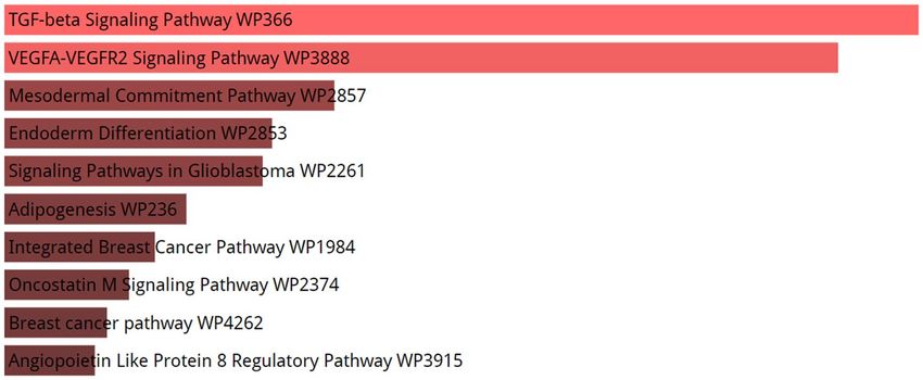
www.nature.com/scientificreports/

                                            Figure 1.  Venn diagram analysis of enoxacin-induced miRNAs and miRNAs targeting cell entry receptors
                                            necessary for SARS-CoV-2 infection. Entry receptors including ACE2 and ACE could be targeted by two
                                            enoxacin-induced miRNAs.

                                             Target transcripts   Targeting miRNAs
                                                                  hsa-miR-582-5p, hsa-miR-452-5p, hsa-miR-214-3p, hsa-miR-1208, hsa-miR-181b-5p, hsa-miR-181c-5p, hsa-miR-
                                             TMPRSS2
                                                                  98-5p
                                             TMPRSS11D            hsa-miR-574-3p, hsa-miR-186-5p, hsa-miR-23a-3p
                                             CTSL                 hsa-miR-501-5p, hsa-miR-518a-5p
                                             FURIN                hsa-miR-20a-5p, hsa-miR-483-3p, hsa-miR-17-5p, hsa-miR-4286, hsa-miR-140-3p, hsa-miR-497-5p, hsa-miR-107

                                            Table 2.  Enoxacin-induced miRNAs that can target membranous proteases facilitating SARS-CoV-2 entry.

                                            define miRNAs that could potentially target ACE2 as the main entry receptor of SARS-CoV-2 on cell surface as
                                            well as ACE as an alternative entry receptor of the virus (Table S4). To this end, we considered only those miR-
                                            NAs which were co-predicted by three different miRNA target prediction tools (i.e. miRanda, TargetScan, and
                                            miRDB) in addition to the miRNAs on miRTarBase that have previously been experimentally verified to regu-
                                            late these two cell-surface receptors. This stringent approach could considerably increase the reliability of our
                                            miRNA target analyses. Using this strategy, 25 and 14 miRNAs were found to potentially repress ACE2 and ACE
                                            genes, respectively. Importantly, two of the miRNAs reported to be induced by enoxacin, i.e. hsa-miR-362-5p
                                            and hsa-miR-582-5p, were among these miRNAs that can target ACE2 (Fig. 1A) and ACE (Fig. 1B), respectively,
                                            suggesting that enoxacin might be able to reduce the efficiency with which SARS-CoV-2 enters the host cells
                                            through cell surface receptors.
                                                SARS-CoV-2 cell entry is reported to also depend on the activity of certain cell proteases particularly the
                                            transmembrane serine proteinase 2 (TMPRSS2) and 11D (TMPRSS11D), cathepsin L (CTSL), and FURIN.
                                            Judging from the analysis of miRNAs co-predicted or validated to target these proteases, we found they can be
                                            direct targets of a fraction of the enoxacin-induced miRNAs. Indeed, seven, two, three, and seven of the enoxa-
                                            cin-induced miRNAs were found to putatively target TMPRSS2, CTSL, TMPRSS11D, and FURIN, respectively
                                            (Table 2). These collective results highlight enoxacin’s potential to restrict the entry of the novel coronavirus
                                            into the cells.

                                            Enoxacin might target intracellular proteins interacting with SARS‑CoV‑2 components. To
                                            perform enrichment analysis on the SARS-CoV-2-interacting host genes that might be affected by enoxacin,
                                            we used the union of co-predicted (from miRanda, PITA, TargetScan, and miRDB) and validated targets (from
                                            miRTarBase) of the miRNAs upregulated by enoxacin. Next, to find out if these targets are also expressed in
                                            the lung tissue (to enhance the reliability of our in silico analysis), the intersection of these target genes and
                                            the genes expressed in the lung tissue was determined (see the list of mRNA transcripts in Table S3). Then, the
                                            PPI network of these targets was extracted using STRING database. Totally, 3893 genes were predicted to be
                                            targeted by enoxacin-upregulated miRNAs in the lung (Table S5). As shown in Fig. 2, the pathway enrichment
                                            analysis of these genes revealed that the TGF-β signaling pathway was the most significant pathway targeted by
                                            enoxacin (p-value = 5.782e−20). In addition, we determined the top four protein modules (M1 to M4) in the
                                            PPI network of the targeted genes (Fig. 3). The pathway enrichment analysis of these modules (Table 3) high-
                                            lighted that M1 was related to MHC class I-mediated antigen processing and TGF-β signaling pathway; M2 was
                                            mainly associated with M phase pathway and PI3K-Akt signaling; M3 was primarily consisted of endocytosis

          Scientific Reports |   (2021) 11:10271 |                 https://doi.org/10.1038/s41598-021-89605-6                                                                4

Vol:.(1234567890)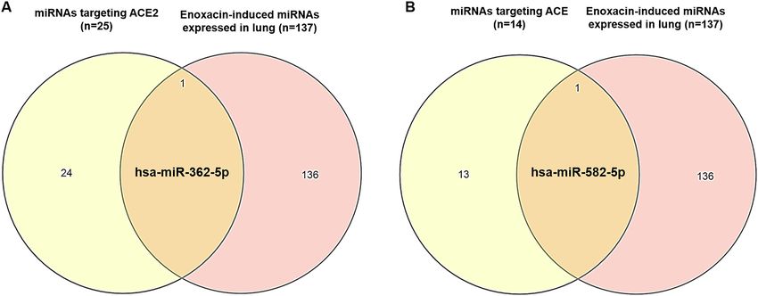
www.nature.com/scientificreports/
Figure 2. Enrichment analysis of genes which are predicted to be targeted by enoxacin-induced miRNAs. The
Panther enrichment analysis using Enrichr showed that genes putatively targeted by enoxacin-induced miRNAs
were mostly involved in TGF-β signaling (p-value = 5.782e−20). The lighter the red color is, the more significant
the p-value.
and EGF/EGFR signaling; and the M4 module highlighted the involvement of VEGF/VEGFR2 and PI3K-Akt
pathways (Table 3). Furthermore, as shown in Fig. 4, the enrichment analysis of the top 25 hub genes iden-
tified by Cytohubba showed that hub genes mostly belonged to MHC class I-mediated antigen presentation
(p-value = 7.684e−25), suggesting that enoxacin might modulate the immune system. It is also noteworthy that
we depicted the PPI network for 386 genes which were observed to be simultaneously co-targeted by 10 or more
enoxacin-induced miRNAs and repeated all the above analyses. Our data revealed a highly similar set of results
(data not shown), suggesting that the overall impact of enoxacin might be mostly mediated by a fraction of its
target miRNAs.
On the other hand, we used the BioGRID database to obtain human proteins that interact with SARS-CoV-2
proteins (Table S6) for which the PPI network was depicted (Fig. S1A). The results of KEGG pathway enrichment
analysis (Fig. S2A) revealed that these 321 proteins were involved in protein processing in endoplasmic reticulum
and RNA transport (p-value = 0.0001441), which highlights potential intracellular machineries facilitating SARS-
CoV-2 infection. In addition, the top four modules of the interaction network of these human proteins (Fig. S1B)
were observed to be mostly involved in RNA transport and AMPK signaling pathway (enrichment analysis by
KEGG, p-value = 2.651e−5 and 1.956e−4 respectively). (Fig. S2B). Furthermore, analysis of the nine hub proteins
from host cells interacting with SARS-CoV-2 proteins (Fig. S3) demonstrated that they were associated with
insulin signaling and cell junction (enrichment analysis by KEGG, p-value = 0.001625 and 0.002486, respectively).
The proteins and cellular pathways described above appear to interact with SARS-CoV-2 viruses, thereby
facilitating its infection. We, therefore, hypothesized that enoxacin might down-regulate some of these host
proteins to perturb the viral life cycle. Our analysis revealed that enoxacin-upregulated miRNAs could target
103 of these 321 proteins (Table S7). Taken together, the induction of certain miRNAs by enoxacin could lead
to the repression of host proteins mediating the infection of the novel coronavirus.
A set of miRNAs targeting SARS‑CoV‑2 components are upregulated by enoxacin. A fraction
of enoxacin-upregulated miRNAs might directly inhibit the viral RNA genome. The miRDB search tool pre-
dicted 900 miRNAs that might target the SARS-CoV-2 RNA genome (Table S8). Venn diagram analysis showed
that 26 out of these miRNAs (with target score > 70) can be upregulated by enoxacin. These 26 miRNAs were pre-
dicted to directly bind and target various regions of the SARS-CoV-2 RNA genome which encode key viral
components (Table S9); notably, a fraction of these miRNAs appear to target multiple viral components simul-
taneously (Table 4). Furthermore, we compared the enoxacin-induced miRNAs with the miRNAs previously
reported to target SARS-CoV-2 genome via different prediction methods42–44. Venn diagram analysis revealed
that hsa-miR-455-5p, hsa-miR-623, hsa-miR-193a-5p, hsa-miR-602, hsa-miR-222, hsa-miR-378a-3p, hsa-miR-
34a-5p, and hsa-miR-98-5p which were reported in these studies to target SARS-CoV-2 genome, were among
miRNAs upregulated by enoxacin (Fig. S4). We, therefore, conclude that the RNA genome of the SARS-CoV-2
might be regulated by certain human miRNAs that are enoxacin-inducible.
Enoxacin‑enhanced miRNAs may exert immunomodulatory effects against the
SARS‑CoV‑2‑induced cytokine storm. An exaggerated immune response in the respiratory system to
the SARS-CoV-2 infection has been suggested to contribute to the high mortality rate seen in patients with
COVID-199,45. To predict if enoxacin could attenuate such cytokine storms in patients with COVID-19, we
extracted the anti-inflammatory miRNAs through literature review and found 25 miRNAs frequently reported
to exert immunomodulatory effects46,47. Interestingly, our data showed that nine out of these 25 miRNAs includ-
ing hsa-miR-21, hsa-miR-17, hsa-miR-146a, hsa-miR-155, hsa-miR-181b, hsa-miR-181c, hsa-miR-31, hsa-miR-
92a, and hsa-miR-223 could be upregulated by enoxacin (Fig. S5). Of note, we noticed that some of the enoxa-
cin-upregulated miRNAs targeted PIK3CA and GSK3B as major components of PI3K signaling which plays a
Scientific Reports | (2021) 11:10271 | https://doi.org/10.1038/s41598-021-89605-6 5
Vol.:(0123456789)www.nature.com/scientificreports/
Figure 3. The PPI network (top four modules) of genes potentially targeted by enoxacin-induced miRNAs.
The PPI network of genes predicted to be targeted by enoxacin-induced miRNAs were depicted by Cytoscape
(only interactions with the confidence of a combined score > 0.400 were included) and protein modules were
identified by MCODE (cutoff criteria were ‘degree cutoff = 2’, ‘k-core = 2’, ‘node score cutoff = 0.2’, and ‘maximum
depth = 100). M: Module.
Modules Classification system Pathways p-value
Bioplanet 2019 Antigen presentation: folding, assembly, and peptide loading of class I MHC proteins 1.698e−86
1 WikiPathways 2019 TGF-β signaling pathway 3.646e−6
KEGG Ubiquitin-mediated proteolysis 1.510e−60
Bioplanet 2019 M phase pathway 1.226e−39
2 WikiPathways 2019 PI3K-Akt signaling pathway 1.170e−17
KEGG Splicesome 2.026e−23
Bioplanet 2019 Endocytosis 2.710e−14
3 WikiPathways 2019 EGF/EGFR signaling pathway 3.439e−7
KEGG Endocytosis 2.291e−13
Bioplanet 2019 Pathways in cancer 4.469e−17
4 WikiPathways 2019 VEGF/VEGFR2 signaling pathway 4.704e−18
KEGG PI3K-Akt signaling pathway 2.333e−19
Table 3. GO and pathway enrichment analysis of top four modules in the PPI network of genes predicted to
be targeted by enoxacin-induced miRNAs.
key role in driving inflammatory responses (data not shown). In conclusion, enoxacin might prove beneficial in
fighting the SARS-CoV-2 infection via promoting immunomodulatory effects.
Scientific Reports | (2021) 11:10271 | https://doi.org/10.1038/s41598-021-89605-6 6
Vol:.(1234567890)www.nature.com/scientificreports/
Figure 4. Hub genes in the PPI network could be targeted by enoxacin-induced miRNAs. (A) Twenty-five hub
genes were identified by Cytohubba and MCC method. (B) The KEGG pathway enrichment analysis showed
that these genes were mostly associated with MHC class I-mediated antigen processing (p-value = 1.698e−86).
The lighter the red color is, the more significant the p-value.
SARS-CoV-2 components Targeting miRNAs
NSP1 hsa-miR-125b-2-3p, hsa-miR-382-5p
hsa-miR-513a-3p, hsa-miR-376a-3p, hsa-miR-583, hsa-miR-186-5p, hsa-miR-495-3p, hsa-miR-3065-5p,
NSP2
hsa-miR-125-2-3p
hsa-miR-485-3p, hsa-miR-20a-3p, hsa-miR-23a-3p, hsa-miR-520a-3p, hsa-miR-376a-3p, hsa-miR-452-5p,
hsa-miR-382-5p, hsa-miR-576-5p, hsa-miR-583, hsa-miR-181c-5p, hsa-miR-181b-5p, hsa-miR-497-5p,
NSP3
hsa-miR-186-5p, hsa-miR-545-3p, hsa-miR-30b-5p, hsa-miR-505-3p, hsa-miR-518a-5p, hsa-miR-29c-3p, hsa-
miR-29b-3p, hsa-miR-495-3p, hsa-miR-29a-3p, hsa-miR-194-5p, hsa-miR-3065-5p, hsa-miR-125b-2-3p
hsa-miR-3065-5p, hsa-miR-125b-2-3p, hsa-miR-107, hsa-miR-513a-3p, hsa-miR-382-5p, hsa-miR-583,
NSP4 hsa-miR-181c-5p, hsa-miR-181b-5p, hsa-miR-186-5p, hsa-miR-30b-5p, hsa-miR-518a-5p, hsa-miR-29a-3p,
hsa-miR-29b-3p, hsa-miR-495-3p, hsa-miR-29a-3p, hsa-miR-194-5p
hsa-miR-485-3p, hsa-miR-583, hsa-miR-181c-5p, hsa-miR-181b-5p, hsa-miR-30b-5p, hsa-miR-518a-5p, hsa-
NSP6
miR-29c-3p, hsa-miR-29b-3p, hsa-miR-495-3p, hsa-miR-29a-3p, hsa-miR-194-5p
NSP7 hsa-miR-518a-5p
NSP8 hsa-miR-20a-3p, hsa-miR-382-5p, hsa-miR-576-5p, hsa-miR-181c-5p, hsa-miR-181b-5p
NSP9 hsa-miR-495-3p, hsa-miR-194-5p
ORF3a hsa-miR-3065-5p, hsa-miR-497-5p, hsa-miR-545-3p, hsa-miR-518a-5p
ORF5 hsa-miR-107
ORF6 hsa-miR-513a-3p
ORF7a hsa-miR-452-5p, hsa-miR-186-5p, hsa-miR-125b-2-3p
ORF8 hsa-miR-181c-5p, hsa-miR-181b-5p, hsa-miR-30b-5p, hsa-miR-513a-3p, hsa-miR-376a-3p
hsa-miR-497-5p, hsa-miR-545-3p, hsa-miR-29c-3p, hsa-miR-29b-3p, hsa-miR-29a-3p, hsa-miR-107, hsa-
Nucleocapsid
miR-513a-3p, hsa-miR-20a-3p, hsa-miR-382-5p
hsa-miR-186-5p, hsa-miR-545-3p, hsa-miR-505-3p, hsa-miR-495-3p, hsa-miR-3065-5p, hsa-miR-125b-2-3p,
RdRp hsa-miR-107, hsa-miR-23a-3p, hsa-miR-520a-3p, hsa-miR-376a-3p hsa-miR-452-5p, hsa-miR-382-5p, hsa-
miR-497-5p
hsa-miR-518a-5p, hsa-miR-29c-3p, hsa-miR-29b-3p, hsa-miR-495-3p, hsa-miR-29a-3p, hsa-miR-3065-5p,
Spike hsa-miR-125b-2-3p, hsa-miR-107, hsa-miR-513a-3p, hsa-miR-485-3p, hsa-miR-20a-3p, hsa-miR-23a-3p, hsa-
miR-376a-3p, hsa-miR-382-5p, hsa-miR-576-5p, hsa-miR-497-5p, hsa-miR-186-5p, hsa-miR-545-3p
hsa-miR-29c-3p, hsa-miR-29b-3p, hsa-miR-29a-3p, hsa-miR-194-5p, hsa-miR-3065-5p, hsa-miR-513a-3p,
Helicase
hsa-miR-452-5p, hsa-miR-382-5p, hsa-miR-576-5p, hsa-miR-30b-5p, hsa-miR-505-3p, hsa-miR-518a-5p
hsa-miR-29c-3p, hsa-miR-29b-3p, hsa-miR-495-3p, hsa-miR-29a-3p, hsa-miR-194-5p, hsa-miR-513a-3p, hsa-
2OMT
miR-20a-3p, hsa-miR-576-5p, hsa-miR-181c-5p, hsa-miR-181b-5p, hsa-miR-186-5p
hsa-miR-29c-3p, hsa-miR-29b-3p, hsa-miR-495-3p, hsa-miR-29a-3p, hsa-miR-376a-3p, hsa-miR-181c-5p,
3′-5′ exonuclease
hsa-miR-181b-5p, hsa-miR-30b-5p, hsa-miR-505-3p
hsa-miR-495-3p, hsa-miR-194-5p, hsa-miR-3065-5p, hsa-miR-125b-2-3p, hsa-miR-485-3p, hsa-miR-376a-3p,
3C-like proteinase
hsa-miR-576-5p, hsa-miR-583, hsa-miR-30b-5p, hsa-miR-505-3p
hsa-miR-513a-3p, hsa-miR-376a-3p, hsa-miR-497-5p, hsa-miR-30b-5p, hsa-miR-495-3p, hsa-miR-3065-5p,
endoRNAse
hsa-miR-513a-3p
5′-UTR hsa-miR-505-3p
Table 4. SARS-CoV-2 components can be targeted by enoxacin-induced miRNAs.
Scientific Reports | (2021) 11:10271 | https://doi.org/10.1038/s41598-021-89605-6 7
Vol.:(0123456789)www.nature.com/scientificreports/
Enoxacin might promote bronchiolar stem cell differentiation, reversing viral negative effects
on lung parenchyma. As with SARS-CoV infection (which can promote the severe acute respiratory
syndrome, SARS), the SARS-CoV-2 infection triggers and promotes lung injury as a typical symptom of hos-
pitalized COVID-19 p atients48. Cumulative evidence suggests that the bronchio-alveolar stem cells (BASCs),
which are characterized by “Sca-1+ CD34+ CD45− Pecam−” markers, participate in tissue regeneration after lung
injury49. Importantly, these cells appear to be a prime target of SARS-CoV infection50. To predict if miRNAs
upregulated by enoxacin might play a role in lung regeneration, we extracted 95 previously reported miRNAs
targeting the developmental stage-specific transcription factors and key marker genes of B ASCs50. Venn diagram
analysis showed that 27 out of these miRNAs could be upregulated by enoxacin treatment (Table S10). Particu-
larly, hsa-let-7d, hsa-let-7g, and hsa-let-7c (predicted to target CD3450) were among the miRNAs upregulated by
enoxacin. Thus, enoxacin might induce BASC differentiation (necessary for lung tissue repair upon injury) by
upregulating certain miRNAs.
Discussion
The RNAi enhancer enoxacin has been proposed as a repurposed drug candidate for targeting multiple
cancers51,52 and several viral d iseases36,37,53,54. For example, Yan-Peng et al. reported that enoxacin augmented
virus-derived siRNA levels in Zika virus-infected human neural progenitor cells and brain organoids, highlight-
ing that enoxacin can promote viral RNA-genome degradation36. We hypothesized that the RNAi-enhancing
activity of enoxacin might similarly interfere with SARS-CoV-2 infection. In line with this, Bartoszewski et al.
suggested that SARS-CoV-2 may act through the depletion of specific host m iRNAs55, explaining, at least partly,
why enhancing the host-cell miRNA activity might be a viable therapeutic option against SARS-CoV-2 replica-
tion. In addition, Chow et al. found that most of the differentially expressed miRNAs in Calu3 cells infected with
SARS-CoV-2 were d ownregulated56. Interestingly, we found that some of these down-regulated miRNAs such as
hsa-miR-194-5p, hsa-miR-21-5p, and hsa-miR-940 can be upregulated by enoxacin (see Table S2).
Moreover, we found hundreds of stem-loop structures in the RNA genome of SARS-CoV-2 which could
be binding sites for the DICER/TRBP complex as the direct target of enoxacin’s stimulatory effect. Several
small RNAs have experimentally been found to be derived from our predicted stem-loop regions of the
SARS-CoV-2 genome. Merino et al. reported eight SARS-CoV-2-derived small RNA molecules experimen-
tally confirmed by small-RNA sequencing in the SARS-CoV-2-infected human Calu-3 cells57. Interestingly,
six of these genuine SARS-CoV-2 small RNAs were predicted using the SM-based or Ab-initio methods (see
Table S1) to be derived from the SARS-CoV-2 genomic regions which included the regions 396–496, 555–634,
26742–26873,27002–27104 (predicted by the SM-based approach), 26903–27016, and 29541–29625 (predicted
by the Ab-initio approach). The identification of these putative DICER binding sites in the SARS-CoV-2 genome
is important for the targeting of viral RNA genome, as intramolecular stem-loop structures in RNA molecules
are known to be typical substrates for Dicer enzymes58,59. Finally, the fact that the SARS-CoV-2 genome encodes
VSRs (i.e. nucleocapsid and SARS-CoV-2-7a proteins) further supports the importance of the RNAi pathway as
a crucial antiviral defense mechanism in mammalian c ells60,61.
In addition to the direct action of DICER/TRBP complex on the SARS-CoV-2 genome, our results suggested
that 26 enoxacin-induced miRNAs could target different regions of the SARS-CoV-2 genome (see Table S9).
Experimentally, eight out of these 26 miRNAs have previously been reported to target SARS-CoV-2 genome42–44,
providing further support for our in silico findings. Altogether, these findings suggest the RNAi pathway as an
effective antiviral mechanism against various viruses including coronaviruses, and support the application of
the RNAi enhancer enoxacin in potential inhibition of the SARS-CoV-2 infection.
Targeting viral entry receptors and cell-membrane-associated proteases necessary for SARS-CoV-2 infection
has been proposed as a rational strategy to treat COVID-1962. Our results showed that some of enoxacin-induced
miRNAs could potentially target the cell-surface receptors and cell-membrane-associated proteases necessary
for SARS-CoV-2 infection. In this regard, hsa-miR-214 and has-miR-98, which we found to potentially target
TMPRSS2 using several miRNA target prediction and validation databases, have been experimentally verified
to target TMPRSS2 in Caco-2, HMVEC-L, and HUVEC c ells63,64. Finally, microarray a nalyses65 support our
data regarding the high likelihood of hsa-miR-107 to repress FURIN, which codes for another cell-membrane-
associated protease involved in SARS-CoV-2 p athogenesis66.
Following the entry of SARS-CoV-2 into the lung epithelial cells, these cells start secreting inflammatory
factors to recruit various leukocytes to the lung tissue, helping to suppress the infection67,68. However, the over-
production of inflammatory mediators might lead to the so-called acute respiratory distress syndrome (ARDS)
which promotes the destruction of the lung parenchyma in patients with severe COVID-1969. Among these
inflammatory mediators, IL-6 is strikingly upregulated in the blood samples from non-survivor individuals69,
explaining why the IL-6 inhibitor Tocilizumab is currently being used in patients with A RDS9.
Notably, we found that nine of the enoxacin-induced miRNAs had previously been found to exert immu-
nomodulatory effects46. The probable immunomodulatory effect of enoxacin is not surprising given that several
reports have independently shown the immunomodulatory activities of fluoroquinolones in diverse c ontexts22.
It is worthwhile to note that the enoxacin-enhanced miRNAs hsa-miR-21, hsa-miR-146a, hsa-miR-92a, hsa-
miR-181b, and hsa-miR-223 have been reported to induce anti-inflammatory effects in part by decreasing IL-6
expression70–74. Moreover, overexpression of hsa-miR-21 in lipopolysaccharide-induced macrophages was
reported to significantly decrease IL-6 and increase anti-inflammatory IL-10 s ecretion70. Further, hsa-miR-
146a overexpression in human retinal endothelial cells or in lipopolysaccharide-induced macrophages reduced
the IL-6 s ecretion75,76. Furthermore, hsa-miR-146s can negatively regulate TNFα-induced inflammatory path-
way in m acrophages77 and reduce IL-6 secretion in primary human small airway epithelial c ells78. Another
enoxacin-induced miRNA, hsa-miR-92a, was found to directly target mitogen-activated protein kinase kinase
Scientific Reports | (2021) 11:10271 | https://doi.org/10.1038/s41598-021-89605-6 8
Vol:.(1234567890)www.nature.com/scientificreports/
4, decreasing TNFα and IL-6 production in m acrophages79. Interestingly, hsa-miR-181b was found to decrease
IL-6 expression in lipopolysaccharide-induced macrophages80. Finally, hsa-miR-223, a crucial regulator of the
innate immune responses, was found to directly target poly (adenosine diphosphatase-ribose) polymerase-1
(PARP-1)81 and NLRP82, thereby suppressing inflammation. Taken together, these collective evidence strongly
suggest that the miRNAs induced by enoxacin are functionally involved in dampening inflammation in various
biological contexts, and that the potential enoxacin-driven immunomodulation might play an important part
in the effective treatment of COVID-19 patients.
We also found that a fraction of the enoxacin-induced miRNAs particularly hsa-let-7c, hsa-let-7g, and hsa-
let-7d could contribute to BASC differentiation possibly by targeting the stem cell marker CD3450. In accordance
with this result, over-expression of hsa-let-7 family members has frequently been reported to inhibit various stem
cell states and promote multi-lineage differentiation27,83,84. BASCs were found to be activated upon different lung
injuries, and differentiate into multiple cell lineages for lung regeneration85. Since alveolar damage and pulmonary
fibrosis are the main pathological findings in patients with severe COVID-1986, triggering lung stem cell pools
to differentiate may enhance lung repair and regeneration85. In addition, Mallick et al. reported that 15 miRNAs
were downregulated in SARS-CoV-infected B ASCs50, of which the expression of hsa-let-7c, hsa-let-7d, hsa-let-
7g, hsa-miR-186, hsa-miR-98, and hsa-miR-223 can be restored by enoxacin-induced miRNAs (see Table S2).
The PPI network the target genes of enoxacin-induced, lung-expressed miRNAs revealed that these genes
were mainly associated with MHC class I-mediated antigen processing, TGF-β signaling pathway, PI3K-AKT
signaling, and endocytosis. In this regard, Xia et al.87 demonstrated in mice that MHC class I exacerbated the
vesicular stomatitis virus (VSV)-induced infection in the lung by disrupting the type I IFN signaling, and that
the viral load in MHC class I-deficient macrophages was decreased. Moreover, the possible inhibitory effect of
enoxacin on TGF-β signaling is interesting because TGF-β signaling (i) drives the chronic adaptive immune
responses in patients with A RDS88, resulting in rapid and massive edema and fibrosis in these patients (for
this reason, its blockade has been suggested as a potential treatment of COVID-19)89 and (ii) increases FURIN
expression in well-differentiated primary human bronchial epithelial cells90 (therefore, its blockade may attenu-
ate the SARS-CoV-2 entry into the cells). In addition, the inhibitors of PI3K-AKT signaling, which we suggest
to be targeted by enoxacin, have been proposed as drug candidates in COVID-19 treatments91. This argument is
supported by the point that PI3K-AKT signaling is required for establishing persistent SARS-CoV infection in
Vero E6 cells92. The possible effect of enoxacin on endocytosis pathway is also notable as some proteins of this
pathway are interactors of SARS-CoV-2 p roteins93. Overall, it seems that enoxacin upregulates a specific set of
miRNAs in lung cells which can restrict the entry, replication, and infection of SARS-CoV-2 through suppress-
ing several intracellular pathways.
It is also noteworthy that our study has a number of limitations. First, enoxacin-induced miRNA profiles
were extracted from studies on cancer cells which may not completely reflect the actual effects of enoxacin on
SARS-CoV-2-infected cells, although the molecular interaction of enoxacin with its target, TRBP, does not
appear to be mechanistically different in different types of cell. To minimize this limitation, we considered only
those enoxacin-induced miRNAs that were also expressed by lung cells. Second, our in silico results need to be
investigated experimentally in both in vitro and animal models of SARS-CoV-2 infection, although we provided
a large body of experimental evidence from previously published research for our in silico findings. Altogether,
our analysis strongly suggests that enoxacin could be a promising drug candidate for COVID-19 treatment.
Conclusion
Enoxacin belongs to the fluoroquinolone family of synthetic antibiotics which was recently found to enhance
the maturation of TRBP/DICER-dependent small RNAs, leading to a global increase in the concentration of
these regulatory RNAs. The enhancement of the RNAi pathway by enoxacin has been frequently found to exert
detrimental effects on the replication of several types of viruses, suggesting that enoxacin might similarly inhibit
the infection caused by the novel coronavirus. Using several in silico analyses, we observed that enoxacin could
promote the DICER/TRBP-mediated degradation of the SARS-CoV-2 RNA genome, as our data suggested that
the viral genomic RNA can be a suitable substrate for the DICER activity. We could also find several enoxacin-
upregulated miRNAs that are predicted to directly target the viral genome. Importantly, several enoxacin-induced
miRNAs were suggested to inhibit not only the viral components mediating viral entry into host cells and its
intracellular replication but could also target certain host proteins interacting with the viral components. There-
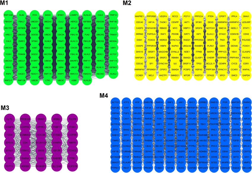
fore, enoxacin might be able to exert antiviral effects against the SARS-CoV-2 infection. Figure 5 illustrates a
schematic model of how the SARS-CoV-2 infection might be suppressed by the application of enoxacin. The
potential antiviral effects of enoxacin could be probably further increased when enoxacin treatment is accom-
panied by the delivery of a shRNA sequence directly targeting the SARS-CoV-2 genome. In this way, not only
enoxacin could restrict the viral replication per se as suggested in this study but also can enhance the processing
of the delivered shRNA in order to provide a more potent inhibitory effect on the novel coronavirus. Finally,
since there are other fluoroquinolones which similarly exhibit RNAi enhancing effects, it might be possible to
also use those fluoroquinolone members for targeting the novel coronavirus infection. Further investigations are
needed to examine how enoxacin or other family members might modulate the infection caused by SARS-CoV-2.
Materials and Methods
Prediction of miRNA‑ and shRNA/siRNA precursors in the SARS‑CoV‑2 genome. The shRNA/
miRNA precursor structures in the SARS-CoV-2 genome were predicted by three web server tools. First, the
ab initio method was used to predict miRNA hairpin structures in the entire 29,903-nucleotide genome of
SARS-CoV-2 (NC_045512.2) using miRNAFold with default p arameters41,94. The entire SARS-CoV-2 genome
was also analyzed by the sequence-structure motif-based (SM-based) miRNA prediction method using the Reg-
Scientific Reports | (2021) 11:10271 | https://doi.org/10.1038/s41598-021-89605-6 9
Vol.:(0123456789)www.nature.com/scientificreports/
Figure 5. Modeling of the potential SARS-CoV-2 inhibition by enoxacin. Enoxacin enhances the RNAi
pathway through binding to TRBP, the physical partner of DICER. This interaction enhances the dicing of viral
RNA genome directly by DICER as well as upregulates certain mature miRNAs which could target SARS-CoV-2
RNA genome and viral transcripts including VSRs through RISC complexes. Enoxacin-induced miRNAs might
also target entry receptors and membranous proteases in host cells, pro-inflammatory genes in the immune
cells, and stem cell markers in BASCs. It might also suppress the interactions between certain viral and host
RNA molecules which mediate and facilitate viral replication and infection. ( ) designates mutual
interaction. miRISC: miRNA-induced silencing complex; siRISC: siRNA-induced silencing complex.
ulatory RNA web tool with default parameters (http://www.regulatoryrna.org/webserver/SSMB/pre-miRNA/
home.html). The BLASTN method was also run to predict precursor structures using the miRBase search tool95.
Obtaining the list of enoxacin‑induced miRNAs and prediction of their target genes. The lists
of enoxacin-induced miRNAs were obtained from previous experiments analyzing the effects of enoxacin on
human embryonic kidney cells (HEK293)33, human prostate cancer cell lines (LNCaP and DU145)34, and the
human melanoma cell line A37535. All miRNAs induced by enoxacin across different cell lines were extracted
using Venn diagram. The expression profiles of miRNAs and mRNAs of the human lung tissue were obtained
from the IMOTA database, an interactive multi-omics-tissue a tlas96. The target genes of miRNAs were co-pre-
dicted by four databases ( miRDB97, PITA98, miRanda99, and T
argetScan100) provided by m
iRWalk101. To further
increase the likelihood of obtaining true-positive miRNA targets, only transcripts with at least two binding
sites for any given miRNA were extracted from the miRWalk 2.0 atlas101. Validated target genes (FDR < 0.05) of
enoxacin-induced miRNAs were obtained from the miRTarBase d atabase102 via the MIENTURNET web t ool103.
Gene ontology (GO) and biological pathway analyses. Gene set enrichment analysis of the predicted
target genes for up-regulated miRNAs was performed using Enrichr as an enrichment analysis web applica-
tion which provides access to 35 gene-set libraries104. Several features of the Enrichr database including KEGG,
Scientific Reports | (2021) 11:10271 | https://doi.org/10.1038/s41598-021-89605-6 10
Vol:.(1234567890)www.nature.com/scientificreports/
Wikipathways, and BioPlanet were used for GO analysis. P < 0.05 was considered to indicate statistical signifi-
cance and the results were ranked by P-value.
Protein–protein interaction (PPI) network analysis, module selection, and identification of
hub genes. The PPI networks were constructed to infer interaction among proteins using the online STRING
database (http://string-db.org/). Interactions with the confidence of a combined score > 0.400 were imported
into Cytoscape to construct the PPI network. We used MCODE to identify the modules in the PPI network105.
The cutoff criteria were ‘degree cutoff = 2’, ‘k-core = 2’, ‘node score cutoff = 0.2’, and ‘maximum depth = 100’. Hub
genes were identified using the Cytoscape plugin cytoHubba by MCC method, as described p reviously106.
Determining the putative miRNAs which target the viral and host components. Prediction of
the human miRNAs that could target the viral RNA genome was performed using the miRDB custom search web
tool107. This database allows for submission of the entire SARS-CoV-2 genome and provides the list of human
miRNAs potentially targeting different regions of the viral genome. Prediction of the human miRNAs that target
host genes interacting with the virus was performed using miRDB, miRanda, and TargetScan databases. Vali-
dated miRNA targets were obtained from miRTarBase and considered in combination with co-predicted gene
targets for gene ontology and biological pathway analyses.
Received: 8 October 2020; Accepted: 29 April 2021
References
1. Yang, P. & Wang, X. COVID-19: A new challenge for human beings. Cell. Mol. Immunol. 17, 555–557. https://doi.org/10.1038/
s41423-020-0407-x (2020).
2. Khuroo, M. S., Khuroo, M., Khuroo, M. S., Sofi, A. A. & Khuroo, N. S. COVID-19 vaccines: A race against time in the middle
of death and devastation!. J. Clin. Exp. Hepatol. 10, 610–621. https://doi.org/10.1016/j.jceh.2020.06.003 (2020).
3. Abd El-Aziz, T. M. & Stockand, J. D. Recent progress and challenges in drug development against COVID-19 coronavirus
(SARS-CoV-2)-an update on the status. Infect. Genet. Evol. 83, 104327. https://doi.org/10.1016/j.meegid.2020.104327 (2020).
4. Datta, P. K., Liu, F., Fischer, T., Rappaport, J. & Qin, X. SARS-CoV-2 pandemic and research gaps: Understanding SARS-CoV-2
interaction with the ACE2 receptor and implications for therapy. Theranostics 10, 7448–7464. https://doi.org/10.7150/thno.
48076 (2020).
5. Wu, Y. et al. A noncompeting pair of human neutralizing antibodies block COVID-19 virus binding to its receptor ACE2. Science
368, 1274–1278. https://doi.org/10.1126/science.abc2241 (2020).
6. Hoffmann, M. et al. SARS-CoV-2 cell entry depends on ACE2 and TMPRSS2 and is blocked by a clinically proven protease
inhibitor. Cell 181, 271-280.e8. https://doi.org/10.1016/j.cell.2020.02.052 (2020).
7. Caly, L., Druce, J. D., Catton, M. G., Jans, D. A. & Wagstaff, K. M. The FDA-approved drug ivermectin inhibits the replication
of SARS-CoV-2 in vitro. Antiviral. Res. 178, 104787. https://doi.org/10.1016/j.antiviral.2020.104787 (2020).
8. Wang, Y. et al. Remdesivir in adults with severe COVID-19: A randomised, double-blind, placebo-controlled, multicentre trial.
Lancet 395, 1569–1578. https://doi.org/10.1016/S0140-6736(20)31022-9 (2020).
9. Guaraldi, G. et al. Tocilizumab in patients with severe COVID-19: A retrospective cohort study. Lancet. Rheumatol. 2, e474–e484.
https://doi.org/10.1016/S2665-9913(20)30173-9 (2020).
10. Roudsari, P. P. et al. Auxiliary role of mesenchymal stem cells as regenerative medicine soldiers to attenuate inflammatory
processes of severe acute respiratory infections caused by COVID-19. Cell. Tissue. Bank. 21, 405–425. https://doi.org/10.1007/
s10561-020-09842-3 (2020).
11. Tobaiqy, M. et al. Therapeutic management of COVID-19 patients: A systematic review. Infect. Prev. Pract. 2, 100061. https://
doi.org/10.1016/j.infpip.2020.100061 (2020).
12. Berkhout, B. RNAi-mediated antiviral immunity in mammals. Curr. Opin. Virol. 32, 9–14. https://d oi.o rg/1 0.1 016/j.c oviro.2 018.
07.008 (2018).
13. Schoggins, J. W. Interferon-stimulated genes: Roles in viral pathogenesis. Curr. Opin. Virol. 6, 40–46. https://doi.org/10.1016/j.
coviro.2014.03.006 (2014).
14. Echebli, N. et al. Stage-specific IFN-induced and IFN gene expression reveal convergence of type I and type II IFN and highlight
their role in both acute and chronic stage of pathogenic SIV infection. PLoS ONE 13, e0190334. https://doi.org/10.1371/journ
al.pone.0190334 (2018).
15. Schuster, S., Miesen, P. & van Rij, R. P. Antiviral RNAi in insects and mammals: Parallels and differences. Viruses 11, 448. https://
doi.org/10.3390/v11050448 (2019).
16. Shimabukuro-Vornhagen, A. et al. Cytokine release syndrome. J. Immunother. Cancer 6, 56. https://doi.org/10.1186/s40425-
018-0343-9 (2018).
17. Ye, Q., Wang, B. & Mao, J. The pathogenesis and treatment of theCytokine Storm’in COVID-19. J. Infect. 80, 607–613. https://
doi.org/10.1016/j.jinf.2020.03.037 (2020).
18. Maillard, P. V., Van der Veen, A. G., Poirier, E. Z. & Reis e Sousa, C. Slicing and dicing viruses: Antiviral RNA interference in
mammals. EMBO. J. 38, e100941. https://doi.org/10.15252/embj.2018100941 (2019).
19. Zhanel, G., Ennis, K. & Vercaigne, L. A critical review of the fluoroquinolones: Focus on respiratory tract infections. Drugs 62,
13–59. https://doi.org/10.2165/00003495-200262010-00002 (2002).
20. Zhanel, G. G. et al. The new fluoroquinolones: A critical review. Can. J. Infect. Dis. 10, 207–238. https://doi.org/10.1155/1999/
378394 (1999).
21. Siddiqui, S. et al. Simian virus 40 large T antigen as a model to test the efficacy of flouroquinolones against viral helicases.
Bioinformation 14, 75–79. https://doi.org/10.6026/97320630014075 (2018).
22. Dalhoff, A. Immunomodulatory activities of fluoroquinolones. Infection 33, 55–70. https://doi.org/10.1007/s15010-005-8209-8
(2005).
23. Zhang, Q., Zhang, C. & Xi, Z. Enhancement of RNAi by a small molecule antibiotic enoxacin. Cell. Res. 18, 1077–1079. https://
doi.org/10.1038/cr.2008.287 (2008).
24. Hammond, S. M., Caudy, A. A. & Hannon, G. J. Post-transcriptional gene silencing by double-stranded RNA. Nat. Rev. Genet.
2, 110–119. https://doi.org/10.1038/35052556 (2001).
Scientific Reports | (2021) 11:10271 | https://doi.org/10.1038/s41598-021-89605-6 11
Vol.:(0123456789)www.nature.com/scientificreports/
25. Carthew, R. W. & Sontheimer, E. J. Origins and mechanisms of miRNAs and siRNAs. Cell 136, 642–655. https://doi.org/10.
1016/j.cell.2009.01.035 (2009).
26. Moradi, S. et al. 10th Royan Institute’s International Summer School on “Molecular Biomedicine: From diagnostics to thera-
peutics”. BioEssays 42, e2000042. https://doi.org/10.1002/bies.202000042 (2020).
27. Shahriari, F. et al. MicroRNA profiling reveals important functions of miR-125b and let-7a during human retinal pigment
epithelial cell differentiation. Exp. Eye. Res. 190, 107883. https://doi.org/10.1016/j.exer.2019.107883 (2019).
28. Moradi, S. et al. Small RNA sequencing reveals Dlk1-Dio3 locus-embedded microRNAs as major drivers of ground-state pluri-
potency. Stem. Cell Rep. 9, 2081–2096. https://doi.org/10.1016/j.stemcr.2017.10.009 (2017).
29. Gross, N., Kropp, J. & Khatib, H. MicroRNA signaling in embryo development. Biology 6, 34. https://d oi.o
rg/1 0.3 390/b
iolog y603
0034 (2017).
30. Cui, L. et al. The nucleocapsid protein of coronaviruses acts as a viral suppressor of RNA silencing in mammalian cells. J. Virol.
89, 9029–9043 (2015).
31. Csorba, T., Kontra, L. & Burgyán, J. Viral silencing suppressors: Tools forged to fine-tune host-pathogen coexistence. Virology
479, 85–103. https://doi.org/10.1016/j.virol.2015.02.028 (2015).
32. Fareh, M. et al. TRBP ensures efficient Dicer processing of precursor microRNA in RNA-crowded environments. Nat. Commun.
7, 1–11. https://doi.org/10.1038/ncomms13694 (2016).
33. Shan, G. et al. A small molecule enhances RNA interference and promotes microRNA processing. Nat. Biotechnol. 26, 933–940.
https://doi.org/10.1038/nbt.1481 (2008).
34. Sousa, E. J. et al. Enoxacin inhibits growth of prostate cancer cells and effectively restores microRNA processing. Epigenetics 8,
548–558. https://doi.org/10.4161/epi.24519 (2013).
35. Valianatos, G. et al. A small molecule drug promoting miRNA processing induces alternative splicing of MdmX transcript and
rescues p53 activity in human cancer cells overexpressing MdmX protein. PLoS ONE 12, e0185801. https://doi.org/10.1371/
journal.pone.0185801 (2017).
36. Xu, Y.-P. et al. Zika virus infection induces RNAi-mediated antiviral immunity in human neural progenitors and brain organoids.
Cell. Res. 29, 265–273. https://doi.org/10.1038/s41422-019-0152-9 (2019).
37. Scroggs, S. L. et al. Old drugs with new tricks: Efficacy of fluoroquinolones to suppress replication of flaviviruses. Viruses 12,
1022. https://doi.org/10.3390/v12091022 (2020).
38. Scroggs, S. L. et al. Evolution of resistance to fluoroquinolones by dengue virus serotype 4 provides insight into mechanism of
action and consequences for viral fitness. Virolgy 552, 94–106. https://doi.org/10.1016/j.virol.2020.09.004 (2020).
39. Shah, P. S., Pham, N. P. & Schaffer, D. V. HIV develops indirect cross-resistance to combinatorial RNAi targeting two distinct
and spatially distant sites. Mol. Ther. 20, 840–848. https://doi.org/10.1038/mt.2012.3 (2012).
40. Liu, X., He, S., Skogerbø, G., Gong, F. & Chen, R. Integrated sequence-structure motifs suffice to identify microRNA precursors.
PLoS ONE 7, e32797. https://doi.org/10.1371/journal.pone.0032797 (2012).
41. Tav, C., Tempel, S., Poligny, L. & Tahi, F. miRNAFold: A web server for fast miRNA precursor prediction in genomes. Nucleic.
Acids. Res. 44, W181–W184. https://doi.org/10.1093/nar/gkw459 (2016).
42. Demirci, M. D. S. & Adan, A. Computational analysis of microRNA-mediated interactions in SARS-CoV-2 infection. PeerJ 8,
e9369. https://doi.org/10.7717/peerj.9369 (2020).
43. Liu, Z. et al. Implications of the virus-encoded miRNA and host miRNA in the pathogenicity of SARS-CoV-2. Preprint at https://
arxiv.org/abs/2004.04874 (2020).
44. Sardar, R., Satish, D., Birla, S. & Gupta, D. Integrative analyses of SARS-CoV-2 genomes from different geographical locations
reveal unique features potentially consequential to host-virus interaction, pathogenesis and clues for novel therapies. Heliyon
6, e04658. https://doi.org/10.1016/j.heliyon.2020.e04658 (2020).
45. Tang, Y. et al. Cytokine storm in COVID-19: The current evidence and treatment strategies. Front. Immunol. 11, 1708. https://
doi.org/10.3389/fimmu.2020.01708 (2020).
46. Tahamtan, A., Teymoori-Rad, M., Nakstad, B. & Salimi, V. Anti-inflammatory microRNAs and their potential for inflammatory
diseases treatment. Front. Immunol. 9, 1377. https://doi.org/10.3389/fimmu.2020.01708 (2018).
47. Botta, C. et al. Immunomodulatory activity of microRNAs: Potential implications for multiple myeloma treatment. Curr. Cancer.
Drug. Targets. 17, 819–838. https://doi.org/10.2174/1568009617666170330154756 (2017).
48. Wang, Y. et al. Temporal changes of CT findings in 90 patients with COVID-19 pneumonia: A longitudinal study. Radiology
296, E55–E64. https://doi.org/10.1148/radiol.2020200843 (2020).
49. Salwig, I. et al. Bronchioalveolar stem cells are a main source for regeneration of distal lung epithelia in vivo. EMBO. J. 38,
e102099. https://doi.org/10.15252/embj.2019102099 (2019).
50. Mallick, B., Ghosh, Z. & Chakrabarti, J. MicroRNome analysis unravels the molecular basis of SARS infection in bronchoalveolar
stem cells. PLoS ONE 4, e7837. https://doi.org/10.1371/journal.pone.0007837 (2009).
51. Yadav, V. & Talwar, P. J. B. Repositioning of fluoroquinolones from antibiotic to anti-cancer agents: An underestimated truth.
Biomed. Pharmacother. 111, 934–946. https://doi.org/10.1016/j.biopha.2018.12.119 (2019).
52. Melo, S. et al. Small molecule enoxacin is a cancer-specific growth inhibitor that acts by enhancing TAR RNA-binding protein
2-mediated microRNA processing. Proc. Natl. Acad. Sci. USA 108, 4394–4399. https://d oi.o
rg/1 0.1 073/p nas.1 01472 0108 (2011).
53. Richter, S., Parolin, C., Palumbo, M. & Palù, G. Antiviral properties of quinolone-based drugs. Curr. Drug. Targets. Infect. Disord.
4, 111–116. https://doi.org/10.2174/1568005043340920 (2004).
54. Mottola, C. et al. In vitro antiviral activity of fluoroquinolones against African swine fever virus. Vet. Microbiol. 165, 86–94.
https://doi.org/10.1016/j.vetmic.2013.01.018 (2013).
55. Bartoszewski, R. et al. SARS-CoV-2 may regulate cellular responses through depletion of specific host miRNAs. Am. J. Physiol.
Lung. Cell. Mol. Physiol. 319, L444–L455. https://doi.org/10.1152/ajplung.00252.2020 (2020).
56. Chow, J.T.-S. & Salmena, L. J. G. Prediction and analysis of SARS-CoV-2-targeting MicroRNA in human lung epithelium. Genes
11, 1002. https://doi.org/10.3390/genes11091002 (2020).
57. Merino, G. A. et al. Novel SARS-CoV-2 encoded small RNAs in the passage to humans. Bioinformatics https://doi.org/10.1093/
bioinformatics/btaa1002 (2020).
58. Lee, K.-M., Gong, Y.-N. & Shih, S.-R. Methods for detection and study of virus-derived small RNAs produced from the intra-
molecular base-pairing region of the picornavirus genome. Methods 183, 4–12. https://doi.org/10.1016/j.ymeth.2019.08.011
(2019).
59. Sabin, L. R. et al. Dicer-2 processes diverse viral RNA species. PLoS ONE 8, e55458. https://doi.org/10.1371/journal.pone.00554
58 (2013).
60. Mu, J. et al. SARS-CoV-2-encoded nucleocapsid protein acts as a viral suppressor of RNA interference in cells. Sci. China. Life.
Sci. 63, 1413–1416. https://doi.org/10.1007/s11427-020-1692-1 (2020).
61. Karjee, S. & Mukherjee, S. K. RNAi suppressor: The hidden weapon of SARS-CoV. J. Biosci. 45, 1–6. https://doi.org/10.1007/
s12038-020-00071-0 (2020).
62. Baughn, L. B. et al. Targeting TMPRSS2 in SARS-CoV-2 Infection. Mayo. Clin. Proc. 95, 1989–1999. https://doi.org/10.1016/j.
mayocp.2020.06.018 (2020).
63. Kaur, T. et al. Tmprss2 specific miRNAs as promising regulators for SARS-CoV-2 entry checkpoint. Virus. Res. 294, 198275.
https://doi.org/10.1016/j.virusres.2020.198275 (2021).
Scientific Reports | (2021) 11:10271 | https://doi.org/10.1038/s41598-021-89605-6 12
Vol:.(1234567890)You can also read