The role of senescence in the development of non-alcoholic fatty liver disease and progression to non-alcoholic steatohepatitis
←
→
Page content transcription
If your browser does not render page correctly, please read the page content below
Article type   : Review
Accepted Article
             The role of senescence in the development of non-alcoholic fatty liver
             disease and progression to non-alcoholic steatohepatitis

             Alkistis-Maria Papatheodoridi1,*, Lampros Chrysavgis1,*, Michael Koutsilieris1,# and
             Antonios Chatzigeorgiou1,2,#

             1
              Department of Physiology, Medical School, National and Kapodistrian University of
             Athens, 75 Mikras Asias Str., 11527, Athens, Greece
              2
                Institute for Clinical Chemistry and Laboratory Medicine, University Hospital and Faculty
             of Medicine Carl Gustav Carus of TU Dresden, Fetscherstrasse 74, 01307 Dresden, Germany

             * contributed equally as first authors
             # contributed equally as senior authors

             E-mail list:
             Alkistis-Maria Papatheodoridi: alkistispapath@gmail.com,
             Lampros Chrysavgis: lchrisaugis@gmail.com
             Michael Koutsilieris: mkoutsil@med.uoa.gr
             Antonios Chatzigeorgiou: achatzig@med.uoa.gr

             Keywords: cell cycle arrest, fibrosis, senescence associated secretory phenotype (SASP),
             senolytics

             Corresponding author
             Assist. Prof. Antonios Chatzigeorgiou (MD, PhD)
             Department of Physiology, Medical School, National and Kapodistrian University of Athens,
             75 Mikras Asias Str., 11527, Athens, Greece
             Email: achatzig@med.uoa.gr
             Tel: +30-2107462623

             This article has been accepted for publication and undergone full peer review but has not
             been through the copyediting, typesetting, pagination and proofreading process, which may
             lead to differences between this version and the Version of Record. Please cite this article as
             doi: 10.1002/hep.30834
             This article is protected by copyright. All rights reserved.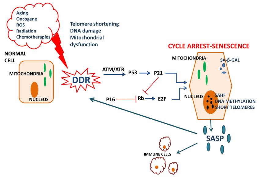
Abbreviations
                   1.
Accepted Article
                       NASH: Non Alcoholic Steatohepatitis
                   2.  NAFLD: Non Alcoholic Fatty Liver Disease
                   3.  ROS: Reactive oxygen species
                   4.  ATM: Ataxia-telangiectasia mutated
                   5.  SA-β-GAL: senescence-associated β-galactosidase
                   6.  SASP: senescence associated secretory phenotype
                   7.  IL: interleukins
                   8. AT: Adipose tissue
                   9. HFD: high fat diet
                   10. HCC: hepatocellular carcinoma
                   11. HSC: hepatic stellate cells

             Financial Support
             Supported by grants from the Deutsche Forschungsgemeinschaft (CH 1862/2-1 and CH
             1862/3-1 to AC) and the Hellenic Association for the Study of the Liver (HASL)

             ABSTRACT

             In recent years cellular senescence has generated a lot of interest among researchers due to its

             involvement in normal aging process and also in common human diseases. During

             senescence, cells undergo alterations that include telomere shortening, nuclear area

             enlargement, genomic and mitochondrial DNA damage, leading to irreversible cell cycle

             arrest, and secretion of proinflammatory cytokines. Evidence suggests that the complex

             process of senescence is involved in the development of a plethora of chronic diseases

             including metabolic and inflammatory disorders and tumorigenesis. Recently, several human

             and animal studies have emphasized the involvement of senescence in the pathogenesis and

             development of liver steatosis including the progression to Non-Alcoholic Steatohepatitis

             This article is protected by copyright. All rights reserved.
(NASH) as characterized by the additional emergence of inflammation, hepatocyte
ballooning and liver fibrosis. The development of Non-Alcoholic Fatty Liver Disease
Accepted Article
(NAFLD) and its progression to NASH are commonly accompanied by several
pathophysiological events including metabolic dysregulation and inflammatory phenomena
occurring within the liver which may contribute to or derive from cellular senescence,
implying that the latter may be both a stimulus and a consequence of the disease. In this
review we summarize the current literature on the impact of cellular senescence in
NAFLD/NASH, and discuss the effectiveness and safety of novel senolytic drugs and
therapeutic options available to delay or treat the disease. Finally we identify the open
questions and issues to be addressed in the near future.
1.Introduction: Molecular characteristics of senescence
The term “senescence” derives from the Latin word “senex” meaning a man of old age.
Cellular senescence describes a decline in cell division capacity whereby normal diploid
differentiated cells enter a state of cell cycle arrest and lose their ability to proliferate (1,2). It
is triggered by DNA damage in chromosomes and telomeres, provoked by internal or external
stimuli such as aging, oncogene expression and reactive oxygen species(ROS) accumulation
or radiation and chemotherapies respectively (3,4). More specifically, there are two major
mechanisms of cellular senescence; one is replicative senescence which depends on telomere
shortening or erosion, predominantly upon aging, and the other is stress-induced premature
senescence which is mostly telomere-independent and refers to intracellular or environmental
stress factors leading to DNA damage (1,2). Both mechanisms induce a complex multigenic
pathway known as DNA damage response(DDR) which can either activate a repair
mechanism or lead to the inhibition of cell cycle(5). In the latter case, DDR triggers ataxia-
This article is protected by copyright. All rights reserved.telangiectasia mutated(ATM) and Radd3-related protein kinases leading to p53
phosphorylation and subsequent activation of p21 resulting to cell cycle arrest(6,7).
Accepted Article
Concurrently p21 and p16 inhibit the phosphorylation of the retinoblastoma factor(Rb)
allowing it to bind to E2F transcription factor and stop the progression of the cell cycle(8).
Cells undergoing senescence appear enlarged and flattened with enlarged nuclei under light
microscope observation, while biochemical assessment of senescent cells shows the presence
of the senescence biomarker senescence-associated β-galactosidase(SA-β-GAL)(9).
Senescence-associated heterochromatin foci (SAHF) constitute another senescence
biomarker. SAHFs consist of heterochromatin and a group of proteins that contribute to
senescence by repressing the expression of proliferation-promoting genes (10). Importantly,
the senescence-associated secretory phenotype(SASP), a complex mixture of molecular
mediators secreted by senescent cells, serves as an important molecular signature for
senescence(3). SASP includes proinflammatory cytokines, such as interleukin-1b(IL-1b), IL-
6, IL-8, chemokines such as Monocyte Chemoattractant Protein-1(MCP-1), growth factors
such as Human Growth Factor (HGF) and Fibroblast Growth Factor(FGF), proteases such as
matrix Metalloproteinases (MMPs), fibronectin, ROS and nitric oxide(3,11). These factors
alter the tissue microenvironment as they induce inflammation, attract immune cells to
remove senescent cells and induce senescence in neighboring cells in a paracrine manner
(12,13)(Figure 1).
This review aims at providing a synopsis on the current literature dealing with cellular and
molecular aspects influencing cellular senescence during non-alcoholic fatty liver disease
(NAFLD), as well as during its progression to non-alcoholic steatohepatitis(NASH).
This article is protected by copyright. All rights reserved.2.Senescence in health and disease
Accepted Article
2.1:General aspects
Cellular senescence is a complex process that has a dual function, both beneficial and
detrimental in human health. Under physiological conditions, senescence eliminates damaged
cells and is involved in tissue restoration upon acute stress or injury. The secretion of
chemokines and cytokines such as IL-1b, IL8 and MCP-1 through SASP attracts immune
cells leading to immunological clearance of the senescent cells (14,15). Consistently, aging
and chronic stress induce telomere attrition and excessive SASP, leading to accumulation of
senescent cells and insufficient tissue regeneration (14,16). Concurrently, hematopoietic stem
cells decrease with age leading in fewer immune cells and a decline in the immune response,
provoking a vicious cycle of defective clearance of senescent cells(17). The combination of
ineffective regeneration, excessive SASP and inefficient clearance may explain the
accumulation of senescent cells in aged organisms, thus increasing the risk for chronic age-
related diseases such as dementia, osteoarthritis, metabolic dysregulation and
carcinogenesis(16,18).
2.2: Senescence and metabolic dysregulation
Metabolic dysregulation refers to a complex wide range of alterations in glucose and lipids’
metabolism, taking place mainly during diabetes and the metabolic syndrome, which can lead
to several secondary complications such as NAFLD and cardiometabolic disease(19).
Importantly, the expansion of the adipose tissue(AT) and in particular the increase of
adipocyte size during obesity leads to upregulation of leptin and downregulation of
adiponectin production by the AT(20,21). Besides, the increased adipocyte size leads to
hypoxia-induced oversecretion of cytokines and chemokines such as Tumor Necrosis
Factor(TNF), IL-6 and MCP-1 by the adipocytes as well as by the inflammatory immune
This article is protected by copyright. All rights reserved.cells that accumulate in the obese AT(19,22). When reaching the liver, the aforementioned
mediators together with the increased levels of free fatty acids and apolipoproteins, observed
Accepted Article
during metabolic dysregulation, lead to liver injury and development of
NAFLD/NASH(20,21).
Metabolic dysregulation is thought to favor cellular senescence in metabolic tissues, such as
the AT and the pancreas, further perpetuating a status of metabolic dyshomeostasis of these
tissues. For instance, obesity induces excessive ROS production, increased production of
cytokines and high expression of SA-β-GAL as well as p53, p16 and p21 in the AT of both
mice and humans (23,24). Along this line, p53 ablation in mice ameliorated insulin resistance
under obese conditions (24). Consistently, high expression of p14 has been reported in
subcutaneous AT from diabetic individuals, having also a positive correlation with p21 in the
same tissue(25). Consistently, SA-β-GAL was more abundant in preadipocytes and
endothelial cells isolated from obese rats and humans as compared to that of lean ones(26).
Senescence-related phenomena are also implicated in pancreatic islet dysfunction during
obesity. Deletion of p27 in Irs2-deficient or db/db diabetic mice improved hyperglycaemia by
inducing compensatory insulin production due to improved maintenance of their islet mass
(27). Besides, senescent endothelial cells express p16INK4a and SA-β-GAL at atherosclerotic
plaques and gene polymorphisms in p21 affect the risk of development coronary artery
disease and myocardial infraction (28,29).
3.The role of senescence in NAFLD/NASH
NAFLD is one of the most common chronic liver diseases affecting approximately 25% of
the population worldwide and its prevalence increases along with aging, obesity and diabetes
(30). Its diagnosis depends on clinical and histological criteria which include triglyceride
accumulation in hepatocytes, defined as steatosis, in individuals that do not consume
This article is protected by copyright. All rights reserved.excessive amounts of alcohol. NAFLD frequently progresses to NASH which is characterized
by the presence of steatosis, inflammation, necrosis and fibrosis (30,31). Advanced fibrosis
Accepted Article
observed in some NASH patients may lead to cirrhosis in 10-15% of these patients or even to
hepatocellular carcinoma(HCC) (30). Currently there is increasing interest in the association
between NAFLD/NASH and senescence.
3.1:Evidence from animal studies
Animal studies have shown a relation between senescence and steatosis. Zhang et.al.
investigated whether the p21 and p16 senescence-associated pathways were involved in
NAFLD pathogenesis, by studying rats on a high-fat diet(HFD). Upon categorizing the rats
into obesity-prone and obesity-resistant ones, they observed that the former, which developed
more severe steatosis, displayed increased hepatic mRNA levels of p16 and p21 and
decreased p53 and phosphorylated-Rb as compared to the latter(10). The acetylation levels of
histones H3 and H4 were increased, the trimethylation of H3-Lys-27 was reduced at the p21-
promoter and the dimethylation levels of the H3-Lys-4 of the p21-coding region was higher
in livers of obesity-prone rats compared to obesity-resistant rats(10). Likewise, Ogrodnik et
al., in order to establish a relationship between hepatic fat deposition and hepatocyte
senescence tested three different dietary regimens in C57BL/6 mice whereby one group was
fed ad-libetum(AL), the other received restricted-diet(RD) and the third group, which
consisted of half AL and half RD animals, was subjected to a dietary crossover at nine
months, when the regimen switched for the following three months prior to sacrifice(32). As
expected, long-term AL-feeding induced fatty liver development and presented several
markers of senescence in the hepatocytes, including increased senescence-associated damage
foci, as determined by the presence of γH2AX, increased senescence-associated distention of
satellites and larger nuclear areas(32). Aiming to identify the relation between senescence
and NAFLD, two different experimental strategies were implemented; p16-expressing
This article is protected by copyright. All rights reserved.senescent cells were genetically eliminated by utilizing the INK-ATTAC mice, while a
senolytic drug cocktail was administered into db/db mice. In both cases a reduction in the
Accepted Article
number of senescent cells was accompanied by ameliorated hepatic lipid accumulation(32).
Finally, analysis of human liver biopsies from NAFLD patients, further confirmed that the
senescence markers telomere-associated damage foci and p21 are related to the severity of
the disease(32).
Several senescence-related proteins have been shown to be associated with the development
of NAFLD and its progression to NASH. For instance, Daugherity et al. showed that ATM
increases in HFD-fed mice and obese ATM-deficient mice were characterized by the
presence of fewer apoptotic hepatocytes and finally less hepatic fibrosis than the wild
type(WT) mice(33). Nevertheless, no difference was observed in liver steatosis between WT
and ATM-deficient mice(33). In contrast to the aforementioned improved liver phenotype
observed during ATM-deficiency, in another study, HFD-fed ATM-deficient mice were more
insulin resistant and were presented with lipodystrophy, predominantly in the subcutaneous
AT. This was attributed to impaired adipocyte differentiation under ATM-deficiency due to
defective induction of transcriptional factors such as C/EBPa and Peroxisome proliferator-
activated receptor gamma(PPAR-γ)(34). Likewise, Schneider et.al. observed glucose
dyshomeostasis and acceleration of atherosclerosis in ATM+/- mice compared to ATM+/+
mice, both on a ApoE-/- background, accompanied by deteriorated hepatic insulin
signaling(35).
The senescence marker protein 30(SMP30), an antioxidant protein that protects against
apoptosis and decreases with aging, is also related to the development of NAFLD(36,37).
Both SMP-30-knockout mice on a Leprdb/db background as well as SMP30/Superoxide
dismutase-1 double-knockout mice developed hepatic steatosis and presented higher levels of
hepatic oxidative stress and superoxide anion radicals compared to WT mice. These findings
This article is protected by copyright. All rights reserved.were attributed to decreased levels of hepatic Apolipoprotein B, and transcription factors
levels involved in lipid metabolism (36,37). Similarly, NASH induction by methionine and
Accepted Article
choline-deficient diet into p53-deficient male mice resulted in slower disease progression
accompanied by lower ROS accumulation and lipid peroxidation as well as fewer apoptotic
cells compared to the WT mice(38). Interestingly, experiments performed with primary
cultured hepatocytes showed that Transforming Growth Factor-beta(TGF-β) induces ROS
production resulting in apoptosis and steatosis, while p53-deficiency decreases its
levels(38,39). Likewise, examination of liver biopsies from NAFLD patients showed
increased p53 levels compared to controls(38). These results indicate that proteins involved
in senescence are overexpressed in NAFLD.
Several lines of evidence suggest that senescence is associated with important changes in
DNA methylation affecting also genes related to lipid metabolism(40,41). To investigate this
hypothesis, Tryndyak et al. induced NAFLD via a choline- and folate-deficient diet into two
different mouse strains, namely the A/J and WSB/EiJ mice, which are known to have
different susceptibility to the development of the disease. Indeed, the more severe steatosis,
observed in the WSB/EiJ mice, as compared to the A/J mice, was accompanied by different
DNA and histone methylation profiles of NAFLD-related genes and genes related to hepatic
lipid accumulation and subsequent alteration in their expression (42). These changes suggest
that alterations in the epigenetic profile of hepatocytes could determine the severity of
NAFLD.
Aging itself, induces cellular senescence leading to development and disease progression of
NAFLD through several pathways(1,4,43). Age-related mitochondrial dysfunction and
elevated oxidative stress trigger fatty liver disease in aged mice on a HFD(44). Upregulation
of the cyclin-dependent kinase-4(cdk4) with aging induces phosphorylation of CCAAT-
This article is protected by copyright. All rights reserved.enhancer-binding protein(C/EBPα) and formation of C/EBPa-p300 complexes leading to
steatosis, while pharmacological inhibition of cdk4 reduces hepatic lipid accumulation
Accepted Article
(45,46).
Although the abovementioned studies imply that senescence is variously involved in NAFLD
pathogenesis and progression, it is of interest whether data from animal studies can be
translated into the human system, in order to design therapeutic strategies in the future.
3.2: The involvement of senescence in human NAFLD/NASH
Data from human clinical studies further support the hypothesis that senescence is associated
with NAFLD. Telomeres were found to be shorter and nuclear area was lower in liver
biopsies from NAFLD patients, as compared to controls, although the latter increased with
age in patients with NAFLD (47). DNA damage, indicated by γH2AX expression, was higher
in patients with NAFLD accompanied by increased p21 expression, indicating a cell cycle
arrest at the G1/S phase(47). Interestingly, both p21 and nuclear area were correlated to the
fibrosis stage(47). Another study between NAFLD patients and healthy individuals,
demonstrated that the relative nuclear size of hepatocytes in NAFLD patients was
significantly larger than the predicted normal value of healthy population, while no difference
in telomere length was observed between the two populations(48). Nevertheless, the telomere
length negatively correlated to nuclear size both in NAFLD patients and healthy controls,
while the increment of nuclear size correlated with age only in healthy individuals,
suggesting that in NAFLD the nuclear enlargement proceeds independently of age(48).
Consistently, Ping et.al. investigated the validity of telomere length as predictive factor of
NAFLD development in T2DM patients, who participated in a 6-year cohort study. Patients
who developed NAFLD within this period had shorter telomeres in peripheral blood
leukocytes at the end of the follow-up period compared to the patients who did not develop
steatosis(49). Similarly, Laish et al. showed that NAFLD patients had shorter telomeres in
This article is protected by copyright. All rights reserved.their peripheral lymphocytes and higher expression of telomerase reverse transcriptase
mRNA as compared to healthy controls(50). These findings support the role of telomere
Accepted Article
dysfunction and senescence in general in NAFLD.
Accordingly, another study observed that SNP-related variants of the CDKN1A gene,
encoding the p21 protein, may be related to the progress of NAFLD. Specifically, genotyping
of lymphocyte DNA, collected in parallel to a hepatic biopsy of NAFLD patients, showed
that the SNP rs762623 seems to influence the development of fibrosis in NAFLD, but does
not affect the progression once fibrosis has started (51). This suggests that the initiation and
progression of fibrosis in NAFLD may have different underlying pathophysiology and
highlights how genetic variations of senescence-related genes might play a role in the
development and progression of the disease. Additionaly, Park etal. demonstrated a
significant reduction of hepatic SMP30-protein levels in a stage-dependent manner in
NAFLD patients, as evaluated by their NAFLD-activity score, while the levels of SMP30
were inversely correlated to the extent of fibrosis(52).
In addition, genomic instability, featured by the cellular presence of micronuclei(MN),
nucleoplasmic bridges(NPBs) and nuclear buds(NBUDs), is considered a form of DNA
damage and thus is indicative for cellular senescence. All these three indexes were found to
be upregulated in NAFLD patients as compared to control individuals(53).
DNA methylation is another senescence marker which seems to be associated with NAFLD
including its progression to NASH(54-57). By performing an epigenome-wide association
study of DNA methylation in whole blood for γ-glutamyltransferase, alanine transaminase
and aspartate transaminase levels, Nano et.al. showed that methylation of SLC7A11 was
associated with reduced risk for hepatic steatosis(56). By determining SLC7A11 gene
expression in nine hepatoma cell lines they found that in the HepaRG cell line, SLC7A11 had
the highest expression and this was associated to high expression of lipid-related genes (56).
This article is protected by copyright. All rights reserved.Another gene the methylation of which is considered to be associated with the progression of
NAFLD into NASH is that of PPAR-γ as its promoter was found to be hyper-methylated in
Accepted Article
both liver tissue and circulating DNA of NAFLD patients, while its altered methylation
profile in circulating DNA could predict the emergence of fibrosis and thus the severity of the
disease (54).
Likewise, Murphy et al. showed that different methylation patterns distinguished patients
with mild from advanced NAFLD, with the latter featured by more hypo-methylated genes in
their biopsies(55). Further analysis revealed that the tissue repair genes were overexpressed,
consistent with methylation and expression being inversely related, while the metabolism
associated-genes were hyper-methylated and as a consequence down-regulated in advanced
NAFLD(55). Another study identified two different methylated region networks related to
NAFLD progression and aging. The first was associated to downregulation of genes related
to transcriptional regulation and cell proliferation, while the second to upregulation of genes
associated to lipid metabolism(58). A study performing methylation analysis of six tumor
suppressor genes and immunohistochemical analysis of oxidative DNA damage in biopsies
from NAFLD patients, found that the levels of oxidative DNA damage were closely related
to disease progression and to the DNA methylation of tumor suppressor genes (57). The latter
likely implies that oxidative DNA damage is an instigator for carcinogenesis-related
alterations in DNA methylation during NAFLD/NASH.
Of note, several studies have suggested that DNA methylation signatures can be used as a
marker of biological age(40,59). Loomba et.al. recently studied the peripheral blood DNA
methylation signatures in biopsy-proven NASH patients and used this marker to assess age
acceleration in these patients and compare it to that of healthy controls. They showed that
patients with stage 3 fibrosis, scored according to the NASH CRN-classification, had
increased age acceleration, which further correlated to the hepatic collagen content(41). A
This article is protected by copyright. All rights reserved.genome-wide methylation analysis highlighted differently methylated CpG islands which
were related to alteration in the expression of a plethora of developmental pathways(41).
Accepted Article
These data indicate that steatohepatitis may induce altered methylation profiles in a plethora
of cells, apart from the hepatocytes, even in peripheral blood cells.
3.3: Senescence and NASH to HCC progression
Hepatocyte senescence is thought to act as a protective mechanism against the development
of hepatocellular carcinoma(HCC). Indeed, Xue et.al. showed that restoration of p53
expression in p53-deficient murine liver tumors resulted in cellular senescence and tumor
regression into undetectable levels(60). Importantly, the secretion of chemokines and
cytokines by senescent cells led to immune cell recruitment and subsequent immunological
clearance, wherein several cellular players of the innate immunity, including macrophages
and neutrophils, were shown to participate(60). Similarly, transduction of transposable
elements expressing the Nras-oncogene into pre-malignant murine hepatocytes induced their
senescence and triggered their clearance by monocytes/macrophages in a CD4+ T-cell-
dependent manner(61). Of note, the presence of DNA in the cytoplasm, especially under
tumorigenic conditions, may act as a senescence-initiating signal and the process depends on
a cytoplasmic DNA sensing pathway, composed by the nucleotidyl transferase cGAMP
synthase(cGAS) and the adaptor protein ‘Stimulator of Interferon Genes’(STING) that is
finally essential for the induction of senescence and SASP(62). More precisely, interaction of
cGAS with cytoplasmic DNA triggers the formation of cyclic GMP-AMP(cGAMP) from
GTP and ATP that in turn activates STING and thereby senescence(63,64). Indeed, under
Ras-oncogene-mediated tumorigenic conditions, STING-deficient mice displayed reduced
immune cell accumulation and clearance of senescent cells in the liver, leading to increased
tumor growth(63).
This article is protected by copyright. All rights reserved.Intriguingly, although NAFLD/NASH is characterized by increased senescence, the disease
occasionally evolves to HCC. Unlike other chronic liver diseases, like viral or alcoholic
Accepted Article
hepatitis, this may happen even in the absence of cirrhosis(65,66). Indeed, Ertle et.al.
demonstrated that nearly half of the NAFLD/NASH-induced HCC patients had no evidence
of cirrhosis(65), implying that other mechanisms taking place under NAFLD/NASH may
play a role in this process. For instance, obesity was previously linked with hepatic stellate
cells’(HSCs) senescence and SASP which promote carcinogenesis(67). Depletion of
senescent HSCs or ablation of a SASP inducer in mice, namely IL-1β, prevented HCC
development(67). Yoshimoto et.al. related these findings to an increase in gut microbiota
population which produces the DNA damaging metabolite, deoxycholic acid(DCA),
promoting SASP in HSCs(67). Moreover, in human NASH-related HCC, cancer-associated
fibroblasts and non-tumoral HSCs demonstrated increased expression of senescence- and
SASP-markers compared to those deriving from conventional HCC(68). Further mechanistic
studies are needed to elucidate the role of senescence in NASH-associated HCC.
3.4:NASH resolution and the role of senescence
So far, little knowledge exists about the mechanisms involved in NASH resolution and the
requisite tissue restoration, while the majority of studies are focusing on the resolution of
NASH-related intratissular inflammation(69). Indeed, during resolution of chronic liver
damage, independently of etiology, the removal of the toxic factor causing the injury,
provokes a shift of the previously inflammatory hepatic microenvironment into more
restorative(69). Dendritic cells, NK cells as well as CD11bhiF4/80intLy6Clo restorative
macrophages are of cardinal importance in this process by secreting fibrolytic MMPs and by
provoking senescence or apoptosis of activated HSCs in a paracrine-dependent manner,
leading thus to degradation and recession of the excessive extracellular matrix(69,70).
This article is protected by copyright. All rights reserved.Importantly, senescence of HSCs is thought to be indispensable for the attenuation of hepatic
fibrosis and thus NASH resolution. Notably, p53- and p53/INK4a/ARF-deficient mice, as
Accepted Article
well as mice with HSC-specific p53-ablation treated with CCl4 developed more severe
fibrosis than the WT animals, attributed to the presence of activated rather than senescent
HSCs(71). Likewise, senescent HSCs are characterized by downregulation of the expression
of genes associated with cell cycle progression and extracellular matrix production and
upregulation of those encoding SASP components, including fibrolytic MMPs and cytokines,
favoring their immune clearance and thus fibrosis resolution(71). Moreover, overexpression
or treatment with the SASP component IL-22 was shown to drive HSCs to senescence in vivo
and in vitro leading to reduced hepatic fibrosis(72). Similarly, Nishizawa etal. showed that
administration of human recombinant Insulin Growth Factor-I(IGF-I) attenuates steatosis,
inflammation and fibrosis in mice treated to develop NASH or cirrhosis and drives HSCs to
senescence. Importantly, these beneficial effects of IGF-I administration were not observed in
p53-deficient mice(73).
Additionally, senescence may influence NASH resolution by affecting the liver regeneration
process that is needful for the reformation of the tissue. Importantly, the age- or/and steatosis-
associated accumulation of senescent hepatocytes is linked to impaired regenerative capacity
of the liver(74,75). Of interest, a spatiotemporal interrelation between SASP and liver
regeneration may be of decisive importance during NASH resolution. Indeed, a recent study
by Ritschka et al. showed that transient exposure of mouse keratinocytes to SASP led to
increased regenerative capacity of those cells, while induction of senescence in single cells in
mouse livers resulted to increased stem cell markers in the surrounding tissue. Oppositely,
prolonged exposure of keratinocytes to SASP provoked maintenance of the cells to a
senescent status, implying overall that SASP acts as a double-edged sword for tissue
regeneration in a time-dependent manner(76). In addition, chronic activation of HSCs due to
This article is protected by copyright. All rights reserved.ageing or NASH and their subsequent chemokine and ROS production leads to decreased
activation and proliferation of Liver Progenitor Cells, which are required for liver
Accepted Article
regeneration following hepatic injury(77). Overall, further studies are essential to reveal the
role of senescence in liver regeneration during NASH resolution.
4. Conclusion and future perspectives
Steatotic hepatocytes often display severe DNA damage and express markers of cell cycle
arrest, indicating that they have entered a senescent state and implying that senescence is
involved in NAFLD/NASH pathogenesis(figure 2)(32,47). Taken that hepatic senescence is
causally induced by HFD and aging, both well-established risk factors for NAFLD
development, it could be considered as a secondary phenomenon during NAFLD emergence
and progression. Nevertheless, depletion of senescent cells in vivo attenuated steatosis, while
senescence induction in vitro and in vivo promoted hepatocyte lipid deposition, suggesting
that senescence plays indeed an important role in NAFLD pathogenesis(32). Notably, a
vicious cycle of cytokine-induced senescence during NAFLD cannot be excluded(78).
However, intriguingly, NAFLD-associated senescence may also have beneficial impacts on
NAFLD to NASH progression. For instance, senescent HSCs produce less extracellular
matrix components and more MMPs, thereby alleviating fibrosis advancement(71).
Concurrently, hepatocyte senescence is required for damaged, pre-cancerous cells’ detection
and clearance, preventing thus liver carcinogenesis(60).
As until now, the golden standard for NAFLD diagnosis and monitoring is the invasive liver
biopsy. Thus, current research efforts are focusing on whether senescence markers measured
in serum or plasma, defined as ‘liquid biopsy’, may reflect disease severity and progression.
These include altered expression or methylation status of senescence-related genes,
metabolomic analyses, markers of oxidative stress or other molecular indicators of
This article is protected by copyright. All rights reserved.senescence, such as telomere length(41,51,79). Further studies should be conducted in order
to prove their potential role as easily accessible biomarkers.
Accepted Article
Concurrently, no approved drug exists so far for the treatment of NAFLD/NASH and the
current management of the disease predominantly focuses on adequate control of the
metabolic profile of the patient putting exercise and diet in the first line for disease
prevention and therapy. In NAFLD patients, clinical trials show that long-term diet and
exercise reduce hepatic steatosis(80), while in aged obese patients even short-term
intervention attenuates hepatic lipid accumulation and improves overall patients’ metabolic
status(81). A recent retrospective cohort study revealed that in non-obese middle aged
individuals, loss of skeletal mass and increase of fat mass with aging is associated with
NAFLD development(82). Exercise may be of therapeutic value for these patients as well.
Meanwhile, novel senolytic drugs have already been developed and animal studies currently
under way may shed more light on their efficacy. The kinase inhibitors dasatinib and
quercetin induce apoptosis in senescent cells in vitro and in vivo(32,83). Administration of
these agents in mice with fatty liver eliminated hepatic senescent cells and decreased lipid
accumulation (32). SASP may serve as a potential target for senolytic agents. The targeting of
signaling pathways such as the Janus kinase–signal transducer and activator of transcription
(JAK-STAT) pathway may limit SASP effect (84). Likewise, IL-22 administration in mice
limited liver fibrosis and led mouse HSCs to senescence(72). In parallel, restoring telomerase
activity by inserting the telomerase gene in senescent murine hepatocytes improved liver
function(85). This may constitute a form of gene therapy for humans in the future and a topic
for future research.
Whereas, at first glance, senolytic agents may seem as a propitious therapeutic opportunity
for several chronic senescence-related diseases, we should bear in mind that cellular
senescence is a normal and sometimes beneficial process. Potential side effects such as
This article is protected by copyright. All rights reserved.rampant cell proliferation and tumorigenesis may prohibit their applicability to human
patients. Thus, the therapeutic potential of senescence remains elusive and although
Accepted Article
senescence has a vast clinical significance, more studies should be conducted in order to
clarify its role not only in NAFLD but also in most clinical medical specialties.
REFERENCES
1.Aravinthan A.Cellular senescence: a hitchhiker's guide.Hum Cell 2015;28:51-64.
2.Campisi J, d'Adda di Fagagna F.Cellular senescence: when bad things happen to good cells.Nat Rev
Mol Cell Biol 2007;8:729-740.
3.Coppé J-P,Desprez P-Y,Krtolica A, Campisi J.The senescence-associated secretory phenotype: the
dark side of tumor suppression.Annual review of pathology 2010;5:99-118.
4.Regulski MJ.Cellular Senescence: What, Why, and How.Wounds 2017;29:168-174.
5.Jackson SP, Bartek J.The DNA-damage response in human biology and disease.Nature
2009;461:1071-1078.
6.Bakkenist CJ, Kastan MB.DNA damage activates ATM through intermolecular autophosphorylation
and dimer dissociation.Nature 2003;421:499-506.
7.Georgakilas AG,Martin OA, Bonner WM.p21: A Two-Faced Genome Guardian.Trends in Molecular
Medicine 2017;23:310-319.
8.Rubin SM,Gall AL,Zheng N, Pavletich NP.Structure of the Rb C-terminal domain bound to E2F1-DP1:
a mechanism for phosphorylation-induced E2F release.Cell 2005;123:1093-1106.
9.Itahana K,Campisi J, Dimri GP.Methods to detect biomarkers of cellular senescence: the
senescence-associated beta-galactosidase assay.Methods Mol Biol 2007;371:21-31.
10.Zhang X,Zhou D,Strakovsky R,Zhang Y, Pan YX.Hepatic cellular senescence pathway genes are
induced through histone modifications in a diet-induced obese rat model.Am J Physiol Gastrointest
Liver Physiol 2012;302:G558-564.
11.Tchkonia T,Zhu Y,van Deursen J,Campisi J, Kirkland JL.Cellular senescence and the senescent
secretory phenotype: therapeutic opportunities.J Clin Invest 2013;123:966-972.
12.Acosta JC,O'Loghlen A,Banito A,Guijarro MV,Augert A,Raguz S,Fumagalli M,et al.Chemokine
signaling via the CXCR2 receptor reinforces senescence.Cell 2008;133:1006-1018.
13.Acosta JC,Banito A,Wuestefeld T,Georgilis A,Janich P,Morton JP,Athineos D,et al.A complex
secretory program orchestrated by the inflammasome controls paracrine senescence.Nat Cell Biol
2013;15:978-990.
14.Hoenicke L, Zender L.Immune surveillance of senescent cells--biological significance in cancer-
and non-cancer pathologies.Carcinogenesis 2012;33:1123-1126.
15.Ovadya Y,Landsberger T,Leins H,Vadai E,Gal H,Biran A,Yosef R,et al.Impaired immune surveillance
accelerates accumulation of senescent cells and aging.Nat Commun 2018;9:5435.
16.Wei W, Ji S.Cellular senescence: Molecular mechanisms and pathogenicity.J Cell Physiol
2018;233:9121-9135.
17.Rossi DJ,Bryder D,Seita J,Nussenzweig A,Hoeijmakers J, Weissman IL.Deficiencies in DNA damage
repair limit the function of haematopoietic stem cells with age.Nature 2007;447:725-729.
18.He S, Sharpless NE.Senescence in Health and Disease.Cell 2017;169:1000-1011.
19.Chatzigeorgiou A, Chavakis T.Immune Cells and Metabolism.Handb Exp Pharmacol 2016;233:221-
249.
This article is protected by copyright. All rights reserved.20.Wree A,Schlattjan M,Bechmann LP,Claudel T,Sowa JP,Stojakovic T,Scharnagl H,et al.Adipocyte cell
size, free fatty acids and apolipoproteins are associated with non-alcoholic liver injury progression in
severely obese patients.Metabolism 2014;63:1542-1552.
21.Adolph TE,Grander C,Grabherr F, Tilg H.Adipokines and Non-Alcoholic Fatty Liver Disease:
Accepted Article
Multiple Interactions.Int J Mol Sci 2017;18.
22.Skurk T,Alberti-Huber C,Herder C, Hauner H.Relationship between adipocyte size and adipokine
expression and secretion.J Clin Endocrinol Metab 2007;92:1023-1033.
23.Schafer MJ,White TA,Evans G,Tonne JM,Verzosa GC,Stout MB,Mazula DL,et al.Exercise Prevents
Diet-Induced Cellular Senescence in Adipose Tissue.Diabetes 2016;65:1606-1615.
24.Minamino T,Orimo M,Shimizu I,Kunieda T,Yokoyama M,Ito T,Nojima A,et al.A crucial role for
adipose tissue p53 in the regulation of insulin resistance.Nat Med 2009;15:1082-1087.
25.Markowski DN,Thies HW,Gottlieb A,Wenk H,Wischnewsky M, Bullerdiek J.HMGA2 expression in
white adipose tissue linking cellular senescence with diabetes.Genes Nutr 2013;8:449-456.
26.Tchkonia T,Morbeck DE,Von Zglinicki T,Van Deursen J,Lustgarten J,Scrable H,Khosla S,et al.Fat
tissue, aging, and cellular senescence.Aging Cell 2010;9:667-684.
27.Uchida T,Nakamura T,Hashimoto N,Matsuda T,Kotani K,Sakaue H,Kido Y,et al.Deletion of Cdkn1b
ameliorates hyperglycemia by maintaining compensatory hyperinsulinemia in diabetic mice.Nat Med
2005;11:175-182.
28.Helgadottir A,Thorleifsson G,Manolescu A,Gretarsdottir S,Blondal T,Jonasdottir A,Jonasdottir A,et
al.A common variant on chromosome 9p21 affects the risk of myocardial infarction.Science
2007;316:1491-1493.
29.Minamino T,Yoshida T,Tateno K,Miyauchi H,Zou Y,Toko H, Komuro I.Ras induces vascular smooth
muscle cell senescence and inflammation in human atherosclerosis.Circulation 2003;108:2264-2269.
30.Younossi Z,Anstee QM,Marietti M,Hardy T,Henry L,Eslam M,George J,et al.Global burden of
NAFLD and NASH: trends, predictions, risk factors and prevention.Nat Rev Gastroenterol Hepatol
2018;15:11-20.
31.Chalasani N,Younossi Z,Lavine JE,Charlton M,Cusi K,Rinella M,Harrison SA,et al.The diagnosis and
management of nonalcoholic fatty liver disease: Practice guidance from the American Association for
the Study of Liver Diseases.Hepatology 2018;67:328-357.
32.Ogrodnik M,Miwa S,Tchkonia T,Tiniakos D,Wilson CL,Lahat A,Day CP,et al.Cellular senescence
drives age-dependent hepatic steatosis.Nature Communications 2017;8:15691.
33.Daugherity EK,Balmus G,Al Saei A,Moore ES,Abi Abdallah D,Rogers AB,Weiss RS,et al.The DNA
damage checkpoint protein ATM promotes hepatocellular apoptosis and fibrosis in a mouse model
of non-alcoholic fatty liver disease.Cell Cycle 2012;11:1918-1928.
34.Takagi M,Uno H,Nishi R,Sugimoto M,Hasegawa S,Piao J,Ihara N,et al.ATM Regulates Adipocyte
Differentiation and Contributes to Glucose Homeostasis.Cell Rep 2015;10:957-967.
35.Schneider JG,Finck BN,Ren J,Standley KN,Takagi M,Maclean KH,Bernal-Mizrachi C,et al.ATM-
dependent suppression of stress signaling reduces vascular disease in metabolic syndrome.Cell
Metab 2006;4:377-389.
36.Kondo Y,Hasegawa G,Okada H,Senmaru T,Fukui M,Nakamura N,Sawada M,et al.Lepr(db/db) Mice
with senescence marker protein-30 knockout (Lepr(db/db)Smp30(Y/-)) exhibit increases in small
dense-LDL and severe fatty liver despite being fed a standard diet.PLoS One 2013;8:e65698.
37.Kondo Y,Masutomi H,Noda Y,Ozawa Y,Takahashi K,Handa S,Maruyama N,et al.Senescence marker
protein-30/superoxide dismutase 1 double knockout mice exhibit increased oxidative stress and
hepatic steatosis.FEBS Open Bio 2014;4:522-532.
38.Tomita K,Teratani T,Suzuki T,Oshikawa T,Yokoyama H,Shimamura K,Nishiyama K,et
al.p53/p66Shc-mediated signaling contributes to the progression of non-alcoholic steatohepatitis in
humans and mice.J Hepatol 2012;57:837-843.
39.Elston R, Inman GJ.Crosstalk between p53 and TGF-beta Signalling.J Signal Transduct
2012;2012:294097.
40.Horvath S.DNA methylation age of human tissues and cell types.Genome Biology 2013;14:3156.
This article is protected by copyright. All rights reserved.41.Loomba R,Gindin Y,Jiang Z,Lawitz E,Caldwell S,Djedjos CS,Xu R,et al.DNA methylation signatures
reflect aging in patients with nonalcoholic steatohepatitis.JCI Insight 2018;3.
42.Tryndyak VP,Han T,Fuscoe JC,Ross SA,Beland FA, Pogribny IP.Status of hepatic DNA methylome
predetermines and modulates the severity of non-alcoholic fatty liver injury in mice.BMC Genomics
Accepted Article
2016;17:298.
43.Fontana L,Zhao E,Amir M,Dong H,Tanaka K, Czaja MJ.Aging promotes the development of diet-
induced murine steatohepatitis but not steatosis.Hepatology (Baltimore, Md.) 2013;57:995-1004.
44.Lohr K,Pachl F,Moghaddas Gholami A,Geillinger KE,Daniel H,Kuster B, Klingenspor M.Reduced
mitochondrial mass and function add to age-related susceptibility toward diet-induced fatty liver in
C57BL/6J mice.Physiol Rep 2016;4.
45.Nguyen P,Valanejad L,Cast A,Wright M,Garcia JM,El-Serag HB,Karns R,et al.Elimination of Age-
Associated Hepatic Steatosis and Correction of Aging Phenotype by Inhibition of cdk4-C/EBPalpha-
p300 Axis.Cell Rep 2018;24:1597-1609.
46.Jin J,Valanejad L,Nguyen TP,Lewis K,Wright M,Cast A,Stock L,et al.Activation of CDK4 Triggers
Development of Non-alcoholic Fatty Liver Disease.Cell Rep 2016;16:744-756.
47.Aravinthan A,Scarpini C,Tachtatzis P,Verma S,Penrhyn-Lowe S,Harvey R,Davies SE,et
al.Hepatocyte senescence predicts progression in non-alcohol-related fatty liver disease.J Hepatol
2013;58:549-556.
48.Nakajima T,Nakashima T,Okada Y,Jo M,Nishikawa T,Mitsumoto Y,Katagishi T,et al.Nuclear size
measurement is a simple method for the assessment of hepatocellular aging in non-alcoholic fatty
liver disease: Comparison with telomere-specific quantitative FISH and p21
immunohistochemistry.Pathol Int 2010;60:175-183.
49.Ping F,Li ZY,Lv K,Zhou MC,Dong YX,Sun Q, Li YX.Deoxyribonucleic acid telomere length shortening
can predict the incidence of non-alcoholic fatty liver disease in patients with type 2 diabetes
mellitus.J Diabetes Investig 2017;8:174-180.
50.Laish I,Mannasse-Green B,Hadary R,Biron-Shental T,Konikoff FM,Amiel A, Kitay-Cohen Y.Telomere
Dysfunction in Nonalcoholic Fatty Liver Disease and Cryptogenic Cirrhosis.Cytogenet Genome Res
2016;150:93-99.
51.Aravinthan A,Mells G,Allison M,Leathart J,Kotronen A,Yki-Jarvinen H,Daly AK,et al.Gene
polymorphisms of cellular senescence marker p21 and disease progression in non-alcohol-related
fatty liver disease.Cell Cycle 2014;13:1489-1494.
52.Park H,Ishigami A,Shima T,Mizuno M,Maruyama N,Yamaguchi K,Mitsuyoshi H,et al.Hepatic
senescence marker protein-30 is involved in the progression of nonalcoholic fatty liver disease.J
Gastroenterol 2010;45:426-434.
53.Karaman H,Karaman A,Donmez-Altuntas H,Bitgen N,Hamurcu Z,Oguz A, Karakukcu
C.Investigation of genome instability in patients with non-alcoholic steatohepatitis.World J
Gastroenterol 2013;19:5295-5301.
54.Hardy T,Zeybel M,Day CP,Dipper C,Masson S,McPherson S,Henderson E,et al.Plasma DNA
methylation: a potential biomarker for stratification of liver fibrosis in non-alcoholic fatty liver
disease.Gut 2017;66:1321-1328.
55.Murphy SK,Yang H,Moylan CA,Pang H,Dellinger A,Abdelmalek MF,Garrett ME,et al.Relationship
between methylome and transcriptome in patients with nonalcoholic fatty liver
disease.Gastroenterology 2013;145:1076-1087.
56.Nano J,Ghanbari M,Wang W,de Vries PS,Dhana K,Muka T,Uitterlinden AG,et al.Epigenome-Wide
Association Study Identifies Methylation Sites Associated With Liver Enzymes and Hepatic
Steatosis.Gastroenterology 2017;153:1096-1106 e1092.
57.Nishida N,Yada N,Hagiwara S,Sakurai T,Kitano M, Kudo M.Unique features associated with
hepatic oxidative DNA damage and DNA methylation in non-alcoholic fatty liver disease.J
Gastroenterol Hepatol 2016;31:1646-1653.
This article is protected by copyright. All rights reserved.58.Hotta K,Kitamoto A,Kitamoto T,Ogawa Y,Honda Y,Kessoku T,Yoneda M,et al.Identification of
differentially methylated region (DMR) networks associated with progression of nonalcoholic fatty
liver disease.Sci Rep 2018;8:13567.
59.Sakaki M,Ebihara Y,Okamura K,Nakabayashi K,Igarashi A,Matsumoto K,Hata K,et al.Potential roles
Accepted Article
of DNA methylation in the initiation and establishment of replicative senescence revealed by array-
based methylome and transcriptome analyses.PLoS One 2017;12:e0171431.
60.Xue W,Zender L,Miething C,Dickins RA,Hernando E,Krizhanovsky V,Cordon-Cardo C,et
al.Senescence and tumour clearance is triggered by p53 restoration in murine liver
carcinomas.Nature 2007;445:656.
61.Kang TW,Yevsa T,Woller N,Hoenicke L,Wuestefeld T,Dauch D,Hohmeyer A,et al.Senescence
surveillance of pre-malignant hepatocytes limits liver cancer development.Nature 2011;479:547-
551.
62.Yang H,Wang H,Ren J,Chen Q, Chen ZJ.cGAS is essential for cellular senescence.Proc Natl Acad Sci
U S A 2017;114:E4612-E4620.
63.Dou Z,Ghosh K,Vizioli MG,Zhu J,Sen P,Wangensteen KJ,Simithy J,et al.Cytoplasmic chromatin
triggers inflammation in senescence and cancer.Nature 2017;550:402-406.
64.Ablasser A,Goldeck M,Cavlar T,Deimling T,Witte G,Rohl I,Hopfner KP,et al.cGAS produces a 2'-5'-
linked cyclic dinucleotide second messenger that activates STING.Nature 2013;498:380-384.
65.Ertle J,Dechene A,Sowa JP,Penndorf V,Herzer K,Kaiser G,Schlaak JF,et al.Non-alcoholic fatty liver
disease progresses to hepatocellular carcinoma in the absence of apparent cirrhosis.Int J Cancer
2011;128:2436-2443.
66.Mittal S,El-Serag HB,Sada YH,Kanwal F,Duan Z,Temple S,May SB,et al.Hepatocellular Carcinoma in
the Absence of Cirrhosis in United States Veterans is Associated With Nonalcoholic Fatty Liver
Disease.Clin Gastroenterol Hepatol 2016;14:124-131 e121.
67.Yoshimoto S,Loo TM,Atarashi K,Kanda H,Sato S,Oyadomari S,Iwakura Y,et al.Obesity-induced gut
microbial metabolite promotes liver cancer through senescence secretome.Nature 2013;499:97-101.
68.Lee JS,Yoo JE,Kim H,Rhee H,Koh MJ,Nahm JH,Choi JS,et al.Tumor stroma with senescence-
associated secretory phenotype in steatohepatitic hepatocellular carcinoma.PLoS One
2017;12:e0171922.
69.Cordero-Espinoza L, Huch M.The balancing act of the liver: tissue regeneration versus fibrosis.J
Clin Invest 2018;128:85-96.
70.Ramachandran P,Pellicoro A,Vernon MA,Boulter L,Aucott RL,Ali A,Hartland SN,et al.Differential
Ly-6C expression identifies the recruited macrophage phenotype, which orchestrates the regression
of murine liver fibrosis.Proc Natl Acad Sci U S A 2012;109:E3186-3195.
71.Krizhanovsky V,Yon M,Dickins RA,Hearn S,Simon J,Miething C,Yee H,et al.Senescence of activated
stellate cells limits liver fibrosis.Cell 2008;134:657-667.
72.Kong X,Feng D,Wang H,Hong F,Bertola A,Wang FS, Gao B.Interleukin-22 induces hepatic stellate
cell senescence and restricts liver fibrosis in mice.Hepatology 2012;56:1150-1159.
73.Nishizawa H,Iguchi G,Fukuoka H,Takahashi M,Suda K,Bando H,Matsumoto R,et al.IGF-I induces
senescence of hepatic stellate cells and limits fibrosis in a p53-dependent manner.Sci Rep
2016;6:34605.
74.Wang MJ,Chen F,Li JX,Liu CC,Zhang HB,Xia Y,Yu B,et al.Reversal of hepatocyte senescence after
continuous in vivo cell proliferation.Hepatology 2014;60:349-361.
75.Shirai M,Yamauchi H,Nakayama H,Doi K, Uetsuka K.Expression of epidermal growth factor
receptor protein in the liver of db/db mice after partial hepatectomy.Exp Toxicol Pathol
2007;59:157-162.
76.Ritschka B,Storer M,Mas A,Heinzmann F,Ortells MC,Morton JP,Sansom OJ,et al.The senescence-
associated secretory phenotype induces cellular plasticity and tissue regeneration.Genes Dev
2017;31:172-183.
77.Pibiri M.Liver regeneration in aged mice: new insights.Aging (Albany NY) 2018;10:1801-1824.
This article is protected by copyright. All rights reserved.78.Kandhaya-Pillai R,Miro-Mur F,Alijotas-Reig J,Tchkonia T,Kirkland JL, Schwartz S.TNFalpha-
senescence initiates a STAT-dependent positive feedback loop, leading to a sustained interferon
signature, DNA damage, and cytokine secretion.Aging (Albany NY) 2017;9:2411-2435.
79.Sookoian S, Pirola CJ.Cell-free DNA methylation as liquid biopsy for the assessment of fibrosis in
Accepted Article
patients with nonalcoholic steatohepatitis: a gap between innovation and
implementation.Hepatobiliary Surg Nutr 2017;6:117-121.
80.Dong F,Zhang Y,Huang Y,Wang Y,Zhang G,Hu X,Wang J,et al.Long-term lifestyle interventions in
middle-aged and elderly men with nonalcoholic fatty liver disease: a randomized controlled trial.Sci
Rep 2016;6:36783.
81.Shah K,Stufflebam A,Hilton TN,Sinacore DR,Klein S, Villareal DT.Diet and exercise interventions
reduce intrahepatic fat content and improve insulin sensitivity in obese older adults.Obesity (Silver
Spring) 2009;17:2162-2168.
82.Lee MJ,Kim EH,Bae SJ,Kim GA,Park SW,Choe J,Jung CH,et al.Age-Related Decrease in Skeletal
Muscle Mass Is an Independent Risk Factor for Incident Nonalcoholic Fatty Liver Disease: A 10-Year
Retrospective Cohort Study.Gut Liver 2019;13:67-76.
83.Zhu Y,Tchkonia T,Pirtskhalava T,Gower AC,Ding H,Giorgadze N,Palmer AK,et al.The Achilles' heel
of senescent cells: from transcriptome to senolytic drugs.Aging Cell 2015;14:644-658.
84.Xu M,Tchkonia T,Ding H,Ogrodnik M,Lubbers ER,Pirtskhalava T,White TA,et al.JAK inhibition
alleviates the cellular senescence-associated secretory phenotype and frailty in old age.Proc Natl
Acad Sci U S A 2015;112:E6301-6310.
85.Rudolph KL,Chang S,Millard M,Schreiber-Agus N, DePinho RA.Inhibition of experimental liver
cirrhosis in mice by telomerase gene delivery.Science 2000;287:1253-1258.
Figure Legends
Figure 1: Molecular aspects of cellular senescence
Aging, oncogene expression, reactive oxygen species accumulation (ROS), radiation and
chemotherapy cause DNA damage, telomere shortening and mitochondrial dysfunction,
inducing thus the DNA Damage Response (DDR). DDR activates ataxia-telangiectasia
mutated (ATM) and Radd3-related (ATR) protein kinases, leading to p53 phosphorylation.
p53 activates p21, leading to cell cycle arrest. p21 and p16 inhibit the phosphorylation of the
retinoblastoma factor (Rb), allowing it to bind to the E2F transcription factor and stop the
progression of cell cycle. Senescent cells appear larger and more flattened; they express the
senescence biomarker senescence-associated β galactosidase (SA-β-GAL) and are
characterized by the presence of senescence-associated heterochromatin foci (SAHF) and
altered DNA methylation signatures. The senescence associated secretory phenotype (SASP)
is a complex mixture of pro-inflammatory cytokines, chemokines, growth factors, proteases,
fibronectin, ROS and ions such as nitric oxide, secreted by senescent cells. Altogether these
factors induce inflammation, attract immune cells to remove senescent cells and at the same
time induce senescence in the neighboring cells in a paracrine manner.
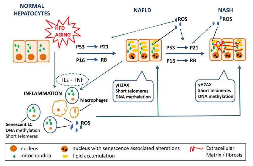
This article is protected by copyright. All rights reserved.Figure 2: Senescence in NAFLD/NASH
Aging and high fat diet (HFD) induce steatosis and senescence in normal hepatocytes via the
Accepted Article
p53-p21 and the p16-Rb pathway. Hepatocytes of NAFLD patients display senescence
markers such as increased expression of γH2AX, short telomeres and altered DNA
methylation patterns. These senescent cells secrete interleukin-1b (IL-1b), IL-6, chemokines,
and ROS [senescence associated secretory phenotype (SASP) components] leading both to
reinforcement of the senescent pathway in the neighboring cells and to disease progression to
NASH. NASH hepatocytes also express senescent markers. At the same time, aging and HFD
as well as senescent hepatocytes induce inflammation by interleukins’ and TNF secretion,
macrophage activation and senescence of the lymphocytes. Overall, these factors induce
steatosis to normal hepatocytes, while favoring further progression of NAFLD to NASH.
This article is protected by copyright. All rights reserved.Accepted Article
This article is protected by copyright. All rights reserved.Accepted Article
This article is protected by copyright. All rights reserved.You can also read