Calcium channel ITPR2 and mitochondria-ER contacts promote cellular senescence and aging - Nature
←
→
Page content transcription
If your browser does not render page correctly, please read the page content below
ARTICLE
                  https://doi.org/10.1038/s41467-021-20993-z                 OPEN

                  Calcium channel ITPR2 and mitochondria–ER
                  contacts promote cellular senescence and aging
                  Dorian V. Ziegler1, David Vindrieux1, Delphine Goehrig1, Sara Jaber1, Guillaume Collin1, Audrey Griveau1,
                  Clotilde Wiel1, Nadia Bendridi2, Sophia Djebali 3, Valerio Farfariello4, Natacha Prevarskaya 4, Léa Payen1,
                  Jacqueline Marvel 3, Sébastien Aubert5, Jean-Michel Flaman1, Jennifer Rieusset2, Nadine Martin1 &
                  David Bernard 1 ✉
1234567890():,;

                  Cellular senescence is induced by stresses and results in a stable proliferation arrest
                  accompanied by a pro-inflammatory secretome. Senescent cells accumulate during aging,
                  promoting various age-related pathologies and limiting lifespan. The endoplasmic reticulum
                  (ER) inositol 1,4,5-trisphosphate receptor, type 2 (ITPR2) calcium-release channel and cal-
                  cium fluxes from the ER to the mitochondria are drivers of senescence in human cells. Here
                  we show that Itpr2 knockout (KO) mice display improved aging such as increased lifespan, a
                  better response to metabolic stress, less immunosenescence, as well as less liver steatosis
                  and fibrosis. Cellular senescence, which is known to promote these alterations, is decreased
                  in Itpr2 KO mice and Itpr2 KO embryo-derived cells. Interestingly, ablation of ITPR2 in vivo
                  and in vitro decreases the number of contacts between the mitochondria and the ER and their
                  forced contacts induce premature senescence. These findings shed light on the role of
                  contacts and facilitated exchanges between the ER and the mitochondria through ITPR2 in
                  regulating senescence and aging.

                  1 Centre de Recherche en Cancérologie de Lyon, Inserm U1052, CNRS UMR 5286, Centre Léon Bérard, Université de Lyon, Lyon, France. 2 CarMeN

                  Laboratory, INSERM UMR-1060, Lyon 1 University, INRA U1397, F-69921 Oullins, France. 3 Centre International de Recherche en Infectiologie, Inserm U1111,
                  CNRS UMR5308, École Normale Supérieure de Lyon, Université de Lyon, Université Claude Bernard Lyon 1, Lyon, France. 4 INSERM U1003, Laboratoire
                  d’Excellence, Canaux Ioniques d’Intérêt Thérapeutique, Équipe Labellisée Par la Ligue Nationale Contre le Cancer, SIRIC ONCOLille, Université des Sciences et
                  Technologies de Lille, Villeneuve d’Ascq, France. 5 Institut de Pathologie, Centre de Biologie Pathologie, CHRU de Lille, Faculté de Médecine, Université de
                  Lille, Lille Cedex, France. ✉email: david.bernard@lyon.unicancer.fr

                  NATURE COMMUNICATIONS | (2021)12:720 | https://doi.org/10.1038/s41467-021-20993-z | www.nature.com/naturecommunications                                    1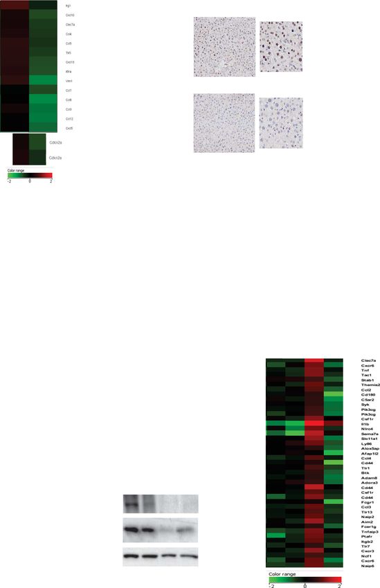
ARTICLE                                                                 NATURE COMMUNICATIONS | https://doi.org/10.1038/s41467-021-20993-z

C
         ellular senescence is an important process regulating dif-          compared to control littermates (Fig. 1b). This decrease was
         ferent pathophysiological processes from embryonic                  associated with an increased proportion of both naive CD4+
         development to aging. In particular, it promotes various            and CD8+ T cells, and an overall increase in naive/memory
age-related diseases and shortens the lifespan1–3. Cellular senes-           T cells (Supplementary Fig. 1b), strongly supporting that ITPR2
cence is characterized by a stable cell cycle arrest and a pro-              promotes immunosenescence in aged mice.
inflammatory senescent-associated secretory program (SASP),                      Since Itpr2 is highly expressed in the liver25, we examined the
both involved in the pathophysiological effects of senescent                 structure and function of the liver in aged mice. Compared to WT
cells4,5. Although downstream factors, such as p16, directly                 mice, the livers of old Itpr2 KO mice displayed no marks of
blocking cell cycle or activating the SASP are largely described,            macroscopic steatosis (Fig. 1c) and fewer lipid droplets
the upstream molecular and sub-cellular mechanisms controlling               (Supplementary Fig. 1c). Accumulation of lipid is a key feature
these factors are largely unknown. The network and activity of               of liver steatosis26, mainly through increased triacylglycerol
mitochondria are dysregulated during cellular senescence, though             (TAG) synthesis. Whole-genome transcriptome analysis revealed
the characterization and cause of these alterations are largely              that loss of Itpr2 reduced the cellular lipid metabolic Gene
unknown6.                                                                    Ontology signature and the fatty acid metabolism Gene set in the
   Calcium critically regulates many cellular and molecular pro-             liver of old mice (Supplementary Fig. 1d, e). More specifically,
cesses including but not limited to secretion, autophagy, migra-             mRNA levels of the fatty acid biosynthesis enzymes Elovl1
tion, proliferation and cell death7. More recently calcium has               (elongation of very long-chain fatty acids protein 1) and Fabp7
been shown to be regulated during cellular senescence and to                 (free fatty acid-binding protein 7) were significantly reduced in
impact its outcome8,9. We and others have recently shown that                the liver of old Itpr2 KO female mice (Fig. 1d). Moreover, Itpr2
transfer of calcium from the endoplasmic reticulum (ER) through              KO mice displayed decreased liver fibrosis (Fig. 1e) and presented
inositol 1,4,5-trisphosphate receptor (ITPR or IP3R) ER channels             a decreased blood aspartate aminotransferase (AST) level (Fig. 1f),
to the mitochondria and its subsequent mitochondrial accumu-                 a marker of damaged liver27.
lation leads to cellular senescence in normal human cells9–12.                  We next investigated the ability of old WT and Itpr2 KO mice
Contact sites between mitochondria and ER, also called                       to respond to a metabolic challenge, since during aging, liver
mitochondria-ER contacts (MERCs) or mitochondria-associated                  dysfunction contributes to an altered response28. Although no
ER membranes (MAMs), have emerged as hotspots for calcium                    difference between the two genotypes was observed in the
transfer and signaling13–16. ITPR2 can be part of MERC sites,                regulation of glycaemia during a glucose tolerance test in 20-
coordinating among others calcium transfer16. Potential role of              month-old mice, 26-month-old WT male mice had lost their
these MERC sites in cellular senescence is currently unknown.                ability to regulate their glycaemia, whereas KO littermates
   Here, we show that ITPR2 promotes some age-associated                     responded normally to glucose injection (Fig. 1g), suggesting
hallmarks and aging in mice. This is associated with increased               that Itpr2 KO mice were protected from age-induced glucose
cellular senescence and MERCs in mice. Accordingly, Itpr2 KO                 intolerance. Furthermore, Itpr2 KO resulted in the abolition of
mouse embryonic fibroblasts (MEFs) display less senescence,                   age-related increased basal blood glycaemia (Supplementary
reduced MERCs and limited mitochondrial calcium. Further-                    Fig. 1f). This parameter could not be assessed in females as most
more, forcing MERCs by using a synthetic linker induces pre-                 of the females were already dead at this age (Fig. 1a). Besides, no
mature senescence. Taken together, these results support that                significant differences were observed between old WT and Itpr2
ITPR2 and increased MERCs are important regulators of cellular               KO mice for the following parameters: bone mineral density and
senescence and aging.                                                        content (Supplementary Fig. 1g), weight (Supplementary Fig. 1h),
                                                                             and tumor lesions (Supplementary Fig. 1i).
                                                                                Together, these results support that ITPR2 promotes some
Results                                                                      alterations associated with physiological aging.
Itpr2 knockout increases lifespan and limits age-related phe-
notypes in mice. In order to investigate the importance of ITPR2
in physiological aging, we studied and monitored cohorts of Itpr2            Loss of Itpr2 reduces cellular senescence in mice and their
knockout mice (KO)17 up to their death. Remarkably, Itpr2 KO                 derived cells. As increased cellular senescence is known to pro-
enhanced the median and maximum lifespan of female mice by                   mote all of the age-related alterations described above4,5 and as
23% and 39%, respectively (Fig. 1a). Loss of Itpr2 did not affect            the ITPR2 calcium channel is a positive regulator of cellular
the survival of male mice (Supplementary Fig. 1a). The differ-               senescence in human cells10, we wondered whether loss of Itpr2
ences in lifespan between males and females are well described.              decreased cellular senescence in mice and in mice-derived cells.
Indeed, many studies report striking gender-associated lifespan              Transcriptomic analyses revealed that liver from 23-month-old
differences following a modification in regimen or gene expres-               Itpr2 KO mice presented fewer markers of cellular senescence, as
sion, though the mechanisms underlying such differences remain               evidenced by a decrease in the inflammatory response signature, a
unclear18–21. According to our results, WT males lived longer                signature that can be related to pro-inflammatory SASP29 and
than WT females as already reported22, suggesting that Itpr2                 includes ccl3, ccl4, ccl12, cxcl5 and cxcl10, and p16ink4a mRNA
could contribute to the intrinsic lifespan difference between male           levels (Fig. 2a and Supplementary Fig. 2a). These results were
and female mice.                                                             confirmed in a larger group of mice by RT-qPCR on ccl3 and
   Aging is a complex systemic process involving numerous vital              p16ink4a (Fig. 2b) and by the immunohistochemical analysis of
organs. Several aging-related features were monitored: immuno-               the p16INK4A protein (Fig. 2c).
senescence designated as immune aging23, liver alterations,                     Reminiscent of Itpr2 loss extending the lifespan of female
response to a metabolic challenge, bone density and tumor                    animals only, ccl3 and p16ink4a mRNA levels were markedly
lesions. Immunosenescence, essentially characterized by the                  reduced in the liver of old Itpr2 KO females compared to livers of
exhaustion of naive T cells and accumulation of memory                       old WT females. A similar but weaker trend towards a reduction
T cells, was examined by quantifying the differences between                 in ccl3 and p16ink4a expression was observed in the liver of old
these two populations24 in the spleens of 23-month old WT and                Itrp2 KO males compared to the liver of old WT males
Itpr2 KO mice. Itpr2 KO mice displayed a decreased number of                 (Supplementary Fig. 2b). In addition, p16ink4a mRNA levels were
both memory CD4+ T cells and effector memory CD8+ T cells                    two-fold higher in the liver of old WT females compared to WT

2                NATURE COMMUNICATIONS | (2021)12:720 | https://doi.org/10.1038/s41467-021-20993-z | www.nature.com/naturecommunications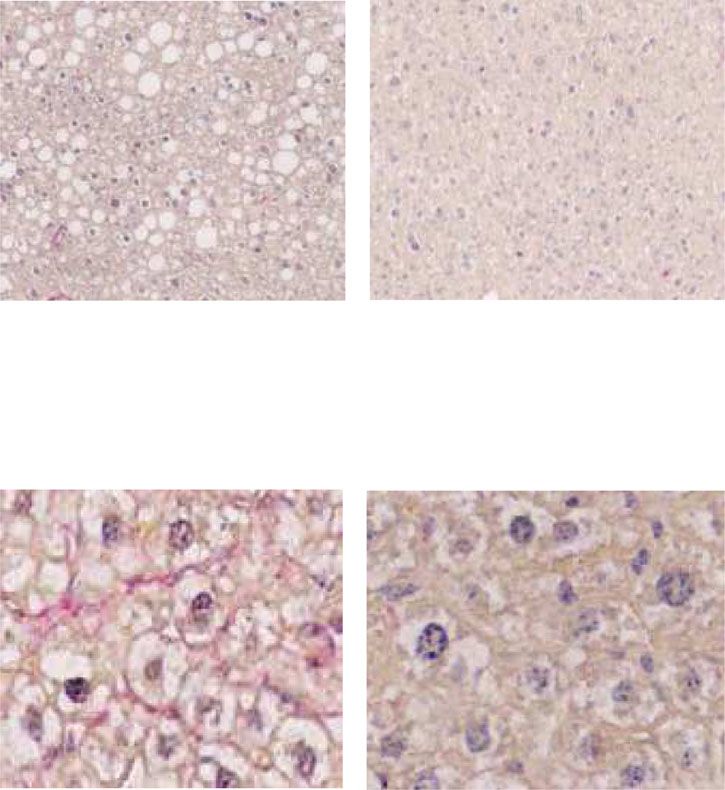
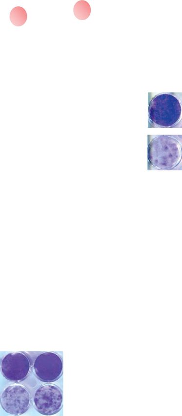
NATURE COMMUNICATIONS | https://doi.org/10.1038/s41467-021-20993-z                                                                ARTICLE

males, consistent with the difference in lifespan observed above                   To further confirm a link between the loss of Itpr2 and
between the sexes (Fig. 1a and Supplementary Fig. 1a). In line                  decreased levels of senescence, beyond the liver, we explored the
with a role for cellular senescence in promoting liver aging and                replicative potential of Itpr2 KO- and WT-derived MEFs. Late
dysfunction30–32, intrahepatic fibrosis level as well as blood                   passage Itpr2 KO MEFs showed a decreased level of cellular
AST levels and lipid droplets quantity were positively correlated               senescence, as illustrated by a decrease in the level of several
with the p16ink4a senescence marker in the liver of old mice                    cellular senescence markers, namely the senescence associated-β-
(Fig. 2d–f).                                                                    galactosidase activity (SA-β-Gal) (Fig. 2g and Supplementary

NATURE COMMUNICATIONS | (2021)12:720 | https://doi.org/10.1038/s41467-021-20993-z | www.nature.com/naturecommunications                        3
ARTICLE                                                                   NATURE COMMUNICATIONS | https://doi.org/10.1038/s41467-021-20993-z

Fig. 1 Itpr2 knockout increases lifespan and limits age-related phenotypes in mice. a Survival curves of Itpr2 WT (n = 9) or KO (n = 11) C57BL/6 female
mice. Log-rank test. b Immunophenotyping of the spleens of 23-month-old mice displaying a relative number of memory (CD44high) CD4+ and CD8+ T-
cells in Itpr2 WT (n = 13) and KO (n = 10) female and male mice. Mean ± SEM. Unpaired two-tailed Student t-test. c Micrographs of liver sections stained
with Sirius Red of 23-month-old male and female Itpr2 WT (n = 12) and KO (n = 10) mice. Scale bar: 25 µm. Quantification of the percentage of mice
presenting macroscopic steatosis. d mRNA level extracted from microarray data in the livers of 23-month-old WT (n = 4) and KO (n = 4) female mice.
Mean ± SEM. Unpaired two-tailed Student t-test. e Micrographs of liver sections stained with Sirius Red. Scale bar: 10 µm. Quantification of relative
intrahepatic collagen fibers according to Sirius Red staining in 23-month-old male and female Itpr2 WT (n = 12) and KO (n = 10) mice. Mean ± SEM. Two-
tailed Mann–Whitney U Test. f Quantification of relative blood AST level of 23-month-old Itpr2 WT (n = 14) and KO (n = 13) female and male mice. Mean
± SEM. Unpaired two-tailed Student t-test. g Intraperitoneal glucose-tolerance test of adult (20-month-old) and old (26-month-old) Itpr2 WT (n = 6) and
KO (n = 8) male mice. Mean ± SEM. Unpaired two-tailed Mann–Whitney U Test. *p < 0.05; **p < 0.01; ***p < 0.001.

Fig. 2c), cell proliferation (Supplementary Fig. 2d), p16ink4a                 were not decreased in old Itpr2 KO MEFs (Supplementary
mRNA (Fig. 2h) and protein (Fig. 2i) levels and finally a                       Fig. 3j), excluding this bias in the analysis of MERCs. As observed
reversion of the enhanced inflammatory response as evidenced by                 in vivo, the number of MERCs was also positively correlated with
transcriptomic data (Fig. 2j and Supplementary Fig. 2e).                       the senescence marker SA-β-Gal in MEFs (Fig. 3f), substantiating
   Because ITPR2 can mediate calcium efflux from the ER to the                  once again a link between MERCs and cellular senescence.
mitochondria and mitochondrial calcium can mediate                                Hence, these results support that fewer and more relaxed
senescence10,12, we determined calcium levels in the late passage              MERCs in Itrp2 KO mice and their derived MEFs are correlated
Itpr2 KO and WT MEFs, using ratiometric genetic reporters.                     with lower levels of cellular senescence.
Itpr2 KO MEFs displayed higher ER calcium levels and lower
mitochondrial calcium levels when compared to the WT late                      Increasing MERCs induces premature senescence. Loss of Itpr2
passage MEFs (Supplementary Fig. 2f, g), as well as decreased                  decreases cellular senescence and this effect is correlated with a
mitochondrial depolarization and mitochondrial ROS production                  lower number of MERCs raising the possibility that MERCs
(Supplementary Fig. 2h, i). These results strongly suggested that              directly promote cellular senescence. In order to test this
old Itpr2 KO MEFs, which undergo less senescence, have                         hypothesis, we used a synthetic linker containing a domain
moderated calcium fluxes from the ER to mitochondria and                        anchored to the ER membrane and another one anchored to the
attenuated mitochondrial dysfunction.                                          mitochondrial outer membrane, as previously described36, to
   In conclusion, these results support that loss of Itpr2 reduces             force the interaction between mitochondria and the ER (Fig. 4a).
the level of cellular senescence, limits ER-mitochondrial calcium              TEM analysis showed that constitutive expression of MERC lin-
fluxes, and is correlated with an improvement in the structure                  kers increased the number of MERCs (Fig. 4b and Supplementary
and function of the liver of old mice.                                         Fig. 4a), increased the total length of the ER-mitochondria
                                                                               interface, and brought mitochondria and ER membranes closer
Loss of Itpr2 diminishes MERCs. ITPR2 can be part of and                       according to distances measured between ER and mitochondria
promotes the MERCs16,33, which are hotspots for calcium                        in MERCs, probably also tightening some pre-existing MERCs
exchange between the ER and the mitochondria15,34. Moreover,                   (Fig. 4c, d and Supplementary Fig. 4b) without increasing the
ITPR2 promotes cellular senescence in human cells by favoring                  perimeter and the number of mitochondria (Supplementary
calcium transfer to the mitochondria10, and we observed here that              Fig. 4c). Remarkably, forcing MERCs also reduced resting cal-
late passage senescence-resistant Itpr2 KO MEFs showed                         cium in the ER while promoting in the meantime its accumula-
decreased levels of mitochondrial calcium (Supplementary                       tion into the mitochondria (Fig. 4e and Supplementary Fig. 4d).
Fig. 2g). We thus evaluated whether cellular senescence regulated              Importantly, increasing MERCs via the constitutive expression of
by ITPR2 could involve MERCs.                                                  linkers led to premature senescence in cells as shown by their
   Assessment of the number of MERCs was performed by                          decreased ability to proliferate and incorporate EdU (Fig. 4f, g),
examining close proximity between ER, using ITPR1, and                         an increased proportion of SA-β-Gal-positive cells (Fig. 4h) and
mitochondria, using VDAC1, both proteins being components                      an increased expression of p16INK4A and various SASP compo-
of MERCs, as previously described and validated in the liver of                nents, including CCL3, IL8 and IL1-β (Fig. 4i).
mice using proximity ligation assay (PLA)35. The number of                        Mechanistically, the linker-induced mitochondrial depolariza-
MERCs was 2-fold lower in the liver of old Itpr2 KO mice                       tion and mitochondrial ROS accumulation (Fig. 4j, k), which can
compared to WT littermates (Fig. 3a and Supplementary Fig. 3a)                 be induced by increased mitochondrial calcium and also are well-
and these changes were not due to changes in ITPR1 and VDAC1                   known senescence inducers10,12,37. Accordingly, N-Acetyl
levels (Supplementary Fig. 3b, c). The number of MERCs was also                Cysteine (NAC) antioxidant treatment (Fig. 4l–n) as well as
correlated with the level of the senescence marker p16INK4A                    knockdown of p53 (Supplementary Fig. 4e–g), a downstream
(Fig. 3b and Supplementary Fig. 3d). Remarkably the number of                  effector of ROS38,39, prevented linker-induced senescence.
MERCs in mice was positively correlated with the relative level of             Activation of the pro-inflammatory arm of the SASP can be
intrahepatic fibrosis (Supplementary Fig. 3e). Further supporting               NF-κB-dependent38,40,41. Knockdown of RelA, a member of the
a link between ITPR2 levels, senescence and the number of                      NF-κB family of the transcription factor, did not revert
MERCs, late passage Itpr2 KO MEFs displayed a decrease in the                  proliferation arrest (Supplementary Fig. 4h, i), yet reverted the
number of MERCs compared to old and senescent WT MEFs,                         induction of IL-8 and IL1-β but not of CCL3 in linker-expressing
according to both PLAs between ITPR1 and VDAC1 (Fig. 3c),                      cells (Supplementary Fig. 4i), indicating that the pro-
without any change in ITPR1 and VDAC1 levels (Supplementary                    inflammatory SASP induced by the linker is partly regulated by
Fig. 3f, g), and transmission electronic microscopy (TEM) (Fig. 3d             NF-κB activation.
and Supplementary Fig. 3h). Beyond the number of MERCs, the                       In conclusion, forcing contacts between ER and mitochondrial
distance between the ER and the mitochondria in MERCs                          membranes is sufficient to trigger premature cellular senescence,
increased in late passage Itpr2 KO MEFs (Fig. 3e and                           involving a mitochondrial ROS/p53 pathway and a partial NF-
Supplementary Fig. 3i). Perimeter and number of mitochondria                   κB-dependent SASP induction.

4                  NATURE COMMUNICATIONS | (2021)12:720 | https://doi.org/10.1038/s41467-021-20993-z | www.nature.com/naturecommunications
NATURE COMMUNICATIONS | https://doi.org/10.1038/s41467-021-20993-z ARTICLE Discussion that loss of Itpr2 decreases the level of senescence in vitro and In this study, we unraveled a role for the calcium channel ITPR2 in vivo and improves aging, suggesting that ITPR2 regulates aging in the regulation of lifespan, physiological aging and cellular by impacting cellular senescence. senescence. During these last few years, the functional link We mainly studied the impact of Itpr2 in old mice by focusing on between cellular senescence, age-related alterations and lifespan the liver for several reasons. ITPR2 belongs to a family of proteins has been demonstrated by eliminating the senescent cells using comprising two other members, ITPR1 and ITPR3, which are genetic or pharmacological tools3,42. In our study, we observed known to exert some redundant activities on cancer sensitivity43 or NATURE COMMUNICATIONS | (2021)12:720 | https://doi.org/10.1038/s41467-021-20993-z | www.nature.com/naturecommunications 5
ARTICLE                                                                   NATURE COMMUNICATIONS | https://doi.org/10.1038/s41467-021-20993-z

Fig. 2 Loss of Itpr2 in mice reduces key features of cellular senescence. a Inflammatory response gene ontology obtained from the genes downregulated
in the livers of 23-month-old Itpr2 WT (n = 4) and KO (n = 4) female mice, according to transcriptomic microarray analyses, corrected p-value = 0.019.
b Relative ccl3 and p16ink4a mRNA levels in liver of 23-month-old Itpr2 WT (n = 14) and KO (n = 15) female and male mice. Mean ± SEM. Unpaired two-
tailed Mann–Whitney U Test. c Micrographs of liver sections and quantification of p16INK4A-positive cells, stained by immunohistochemistry (IHC) in the
livers of 23-month-old mice Itpr2 WT (n = 4) and KO (n = 4) female mice. Mean ± SEM. Unpaired two-tailed Student t-test (*p = 0.0331). d–e Linear
regression analyses between intrahepatic fibrosis level or blood AST level and p16ink4a mRNA levels in the livers of 23-month-old female and male mice
(n = 14). Two-tailed Spearman Rank Correlation test. f Linear regression analysis between the number of lipid droplets and p16ink4a mRNA levels in the
liver of 23-month-old female mice (n = 7). Two-tailed Spearman Rank Correlation test. g Quantification of SA-ß-galactosidase-positive cells in Itpr2 WT
(n = 8) and KO (n = 7) MEFs at early and late passage. Mean ± SEM. Early vs. Late: Paired two-tailed Student t-test (ns: non-significant). Late WT vs. Late
KO: Unpaired two-tailed Welch’s t-test. h p16ink4a mRNA level in WT (n = 5) and KO (n = 5) MEFs at late passage. Mean ± SEM. Unpaired two-tailed
Welch’s t-test. i p16INK4A, Itpr2 and tubulin protein levels in Itpr2 WT and KO MEFs at late passage. j Inflammatory response gene ontology extracted from
microarray of Itpr2 WT (n = 3) and KO (n = 3) MEFs at early and late passage, corrected p-value = 2.292−12. *p < 0.05; **p < 0.01; ***p < 0.001; ns non-
significant.

on the regulation of senescence10. In the bone, where the loss of              demonstrating also a link between MERCs integrity, cellular
Itpr2 does not have any significant beneficial impact, its mRNA                  senescence and aging. ITPR2 could exert this new structural
level is less abundant than Itpr1 (Supplementary Fig. 5). Of note, in          activity by being a tethering species or/and by promoting tether
the liver, Itpr2 is known to be highly expressed compared to the two           formation by other factors. For instance, as GRP75 links ER and
other Itprs (Supplementary Fig. 5)25. Moreover, cellular senescence            mitochondria by simultaneously binding to ITPRs and
is involved in age-related liver alterations30,31. Consequently, the           VDACs13,55, ITPR2 loss might destabilize this ER-mitochondrial
liver appears to be a perfect candidate organ to examine the impact            tethering system. Taken together, our results and the recent data
of Itpr2 deletion on cellular senescence and organ aging and its               from literature support that ITPR2 regulates senescence by its
associated alterations. In line with this hypothesis, Itpr2 KO delays          canonical calcium channel function and by its non-canonical
age-related features of the liver, including both steatosis and fibrosis.       structural function promoting MERC.
Moreover, decreased circulating AST levels as well as a better                    Interestingly, ITPR1, which can regulate senescence similarly
response to a metabolic challenge observed in Itpr2 KO are                     to ITPR210, and the linker, which induces MERCs, have also been
expected to depend, at least in part, on a better liver function during        shown to promote steatosis and to alter glucose homeostasis in
aging44. Altogether, our work unravels that liver ITPR2, while it is           obese mice56, reinforcing our current observations in Itpr2 KO
the most abundant ITPR in this organ, participates actively to its             mice during aging. Obesity is known to promote cellular senes-
chronological decline.                                                         cence, which mediates organismal dysfunctions during obesity
   Cellular senescence induced by short-term injury stimulates                 including steatosis and glucose homeostasis alterations31,32,57.
wound healing45, protects from fibrosis especially in the liver46–48            Even though cellular senescence was not investigated in the study
and enhances stemness and regeneration49, whereas long-term                    by Arruda56, we can speculate that the effects of ITPR1 and
accumulation of senescent cells promotes fibrosis and degenera-                 MERCs are mediated, at least in part, by cellular senescence.
tion in various contexts50. Accordingly, liver regeneration of                 ITPR1 and ITPR2 may thus promote senescence and subsequent
young Itpr2 KO mice after hepatectomy is reduced compared to                   key drivers of age-related defects in the liver, including steatosis
their control littermates51. In this latter study, senescence was not          and alterations of glucose homeostasis.
examined but we can speculate that it might contribute to liver                   Structural changes at the interface between the ER and the
regeneration. Interestingly, another in vivo study in the brain                mitochondria induced by ITPR2 likely mediate part of the effect
reported the beneficial effect of Itpr2 loss in persistent damage-              of ITPR2 on senescence. Indeed, we have shown that inducing
induced phenotype52. Once again, senescence has not been                       MERCs by constitutively expressing a synthetic linker leads to
evaluated in this photothrombosis-induced cerebral ischemia, but               premature senescence. MERCs are known to be involved in
ischemia injuries are known to induce senescence in other                      multiple cellular signaling processes, including calcium transfer to
models53,54. Overall, ITPR2, as a regulator of cellular senescence,            the mitochondria13,14,34,58. Mitochondrial calcium contributes to
could exert both beneficial effects after short-term injury in young            mitochondrial bioenergetics regulation59, and its rise can con-
individuals but detrimental effects after chronic injuries such as             tribute to cellular senescence by inducing ROS production9–12.
during aging.                                                                  We observed that forced contacts between mitochondria and ER
   Aside from the role of ITPR2 in cellular senescence in vivo and             induce mitochondrial calcium accumulation, mitochondrial ROS
age-related physiological declines, our work also shed light on its            accumulation and p53-dependent senescence, in line with our
role as a potent regulator of the formation and/or maintenance of              previous results deciphering that ITPR2 regulates senescence in a
MERCs. Of note, in a previous study, we did not observe any                    ROS-dependent manner9–12. As an association had been reported
changes in the MERCs during oncogene-induced senescence in                     between mitochondria, ROS, NF-κB transcription factors and
immortalized human mammary epithelial cells according to co-                   SASP60,61, we decided to investigate this relationship. We
immunofluorescence experiments10. Discrepancies between these                   demonstrated the involvement of the RelA NF-κB transcription
results might be due to the difference in the senescence system                factor in regulating some SASP factors during linker-induced
used or/and to the fact that our current study used more sensitive             senescence, and observed a decrease in NF-κB activity in old Itpr2
approaches to evaluate MERCs. Itpr2 KO mice liver or MEFs                      KO liver and MEFs according to GSEA analysis (Supplementary
display a decreased number of MERCs. This decrease occurred                    Fig. 6). MERCs also constitute signaling platforms involved in
only in senescent MEFs when compared to non-senescent ones.                    pro-inflammatory responses, notably via both formation and
How ITPR2 regulates MERCs remains unclear but it has recently                  regulation of the inflammasome NLRP362,63. In addition, NLRP3
been identified as required to maintain MERC33. Indeed,                         has been associated with the production of SASP components64.
according to Bartok and colleagues, ITPR2 participates in                      Interestingly, Itpr2 KO liver and MEFs display a dampened
maintaining close contacts33. Our results support and extend                   inflammatory response whereas inducing MERCs enhances the
these observations by using both Itpr2 KO MEFs and mice,                       inflammatory response, as evidenced by Il8 and ccl3 expression.

6                  NATURE COMMUNICATIONS | (2021)12:720 | https://doi.org/10.1038/s41467-021-20993-z | www.nature.com/naturecommunications
NATURE COMMUNICATIONS | https://doi.org/10.1038/s41467-021-20993-z                                                                 ARTICLE

This inflammatory response can promote paracrine senescence                      p53-dependent cell cycle arrest and a p53- and NF-κB-dependent
and mediate part of the pro-aging effects of cellular senescence2,3.            SASP, as suggested by our results, and (ii) by other mechanisms,
We can thus speculate that ITPR2, through higher amounts of                     for instance inflammasome activation which also triggers pro-
functional and closer MERCs, could contribute to cellular                       inflammatory SASP production known to mediate autocrine
senescence and aging by two complementary and synergistic                       senescence. Beyond MERCs and calcium transfer from the ER to
mechanisms, namely (i) the increased mitochondrial calcium                      the mitochondria, cytosolic calcium and its signaling could also
level and the subsequent ROS production which can induce a                      contribute to the senescence phenotype9, in line with the decrease

NATURE COMMUNICATIONS | (2021)12:720 | https://doi.org/10.1038/s41467-021-20993-z | www.nature.com/naturecommunications                         7
ARTICLE                                                                            NATURE COMMUNICATIONS | https://doi.org/10.1038/s41467-021-20993-z

Fig. 3 Loss of Itpr2 diminishes contacts between mitochondria and the ER. a Proximity ligation assay (PLA) using VDAC1 (outer mitochondrial
membrane) and ITPR1 (ER membrane) antibodies in the livers of 23-month-old Itpr2 WT (n = 4) and KO (n = 4) female mice. Dots are formed when the
distance between ITPR1 and VDAC1 is below 50 nm. Representative pictures (scale bar: 5 µm) and quantification shown as mean ± SEM. Unpaired two-
tailed Student t-test. b Linear regression analysis between the number of MERCs and the percentage of p16INK4Apositive cells in the liver of 23-month-old
Itpr2 WT (n = 4) and KO (n = 4) female mice. Two-tailed Spearman Rank Correlation test. c PLA using Vdac1 and Itpr1 antibodies in early and late passage
Itpr2 WT and KO MEFs. Representative pictures (scale bar: 5 µm) and mean ± SEM of n = 10 fields, examined over n = 4 independent experiments. Two-
way ANOVA. Tukey’s multiple comparisons test. d Quantification of the number of MERCs normalized to mitochondria number in late passage Itpr2 WT
and KO MEFs. MERCs are defined by a distance 520 nm)
                                                                                        least 30 fields per condition was performed using Operetta CLS High-Content
(Calcium-free reporter). Ratio F(470–500 nm)/F(>520 nm) was calculated. For
                                                                                        Analysis System (PerkinElmer). Columbus™ software (Perkinelmer) was used. After
experiments involving the Linker, detection of RFP was monitored for F(580 ±
                                                                                        evaluating the number of cells (using Hoescht staining), dots were counted by the
30 nm), and only RFP positive cells were analysed. In order to proceed to single-cell
                                                                                        software and normalized against this number. Each dot on PLA bar chart repre-
analyses, LSM files were converted to Columbus™ software (Perkinelmer) and
                                                                                        sents a mean of n = 50–70 cells analyzed.
single-cell measurement of fluorescence intensity was performed.
                                                                                            For transmission electron microscopy, 1:1 volume of glutaraldehyde 4% was
                                                                                        added to the culture medium and cells were incubated 15 min at 4 °C.
RNA extraction, reverse transcription, and real-time quantitative PCR. RNA              Glutaraldehyde/medium was discarded and 1:1 volume of glutaraldehyde 4
was extracted with phenol-chloroform using Upzol (Dutscher, Brumath, France).           %/cacodylate 0.2 M pH 7.4 was added. Cells were then fixed in glutaraldehyde 2%,

8                     NATURE COMMUNICATIONS | (2021)12:720 | https://doi.org/10.1038/s41467-021-20993-z | www.nature.com/naturecommunications
NATURE COMMUNICATIONS | https://doi.org/10.1038/s41467-021-20993-z ARTICLE washed three times for 1 h at 4 °C, post-fixed with 2% OsO4 1 h at 4 °C, and macro FiJi software kindly provided by G. Hajnoczky67. Using this macro, the dehydrated with an increasing ethanol gradient. Impregnation was performed with number of mitochondria per cell was calculated and the perimeter of each Epon A (50%) plus Epon B (50%) plus DMP30 (1.7%). Inclusion was obtained by mitochondrion forming a MERC contact and associated with ER membrane in polymerisation at 60 °C for 72 hr. Ultrathin sections (~70 nm thick) were cut on a TEM micrographs was assessed. UCT (Leica) ultramicrotome, mounted on 200 mesh copper grids and contrasted with uranyl acetate and lead citrate. Acquisition of 4–10 fields per cell and 10-20 cells per condition was performed with a Jeol 1400JEM (Tokyo, Japan) Immunoblot and immunohistochemistry. For immunoblot experiments, cells transmission electron microscope equipped with an Orius 600 camera and Digital were lysed in RIPA buffer. After protein quantification, 30 μg of proteins were Micrograph at CIQLE platform (UCBL-Lyon). MERCs were determined as loaded and resolved by SDS-PAGE electrophoresis and transferred to nitrocellulose distance below 50 nm between ER and OMM membranes. Quantification of membranes (Bio-Rad). Membranes were blocked with TBS Tween/Milk 5% for 1 h MERCs number per mitochondria, length of MERCs and mean/minimal distances and incubated at 4 °C with primary antibodies overnight. Primary antibodies and
ARTICLE                                                                           NATURE COMMUNICATIONS | https://doi.org/10.1038/s41467-021-20993-z

Fig. 4 Inducing MERCs promotes premature senescence. a Graphical representation of the artificial genetic linker to induce MERCs formation. OMM
outer mitochondrial membrane; MIS mitochondrial Intermembrane Space. b Using transmission electron microscopy, MERCs number was calculated in
Ctrl/Linker-infected MRC5 cells. Mean ± SEM of n = 10 (Ctrl) and n = 11 (Linker) cells. Unpaired two-tailed Student t-test. c Mean ER-mitochondria
distance in Ctrl/Linker-infected MRC5 cells. Mean ± SEM of n = 53 (Ctrl) and n = 80 (Linker) MERCs. Unpaired two-tailed Student t-test. d Mitochondrial
membrane length associated with ER, depending on the distance (0-50 nm) between mitochondria and ER in Ctrl/Linker-infected MRC5 cells. Mean ± SEM
of n = 53 (Ctrl) and n = 80 (Linker) MERCs, representative of n = 3 independent experiments. Multiple t-tests. e Steady-state mitochondrial calcium levels
in Ctrl/Linker-infected cells. n: number of analyzed RFP-positive cells representative of n = 3 independent experiments. Mean ± SEM. Two-tailed
Mann–Whitney U Test. f Crystal violet staining for Ctrl/Linker-infected MRC5 cells 12 days after infection, representative of n = 3 independent
experiments. g Quantification of EdU-positive cells for Ctrl/Linker-infected MRC5 cells. Mean ± SEM of n = 3 independent experiments. Paired two-tailed
Student t-test. h Quantification of SA-ß-galactosidase-positive cells in Ctrl/Linker-infected MRC5 cells. Mean ± SEM of n = 4 independent experiments.
Paired two-tailed Student t-test. i RT-qPCR representing relative p16INK4A, CCL3, IL8, and IL1-ß mRNA levels in Ctrl/Linker-infected MRC5 cells. Mean ±
SEM of n = 7 independent experiments. Unpaired two-tailed Welch’s t-test. j Single-cell analysis on RFP-positive Ctrl/Linker-infected cells of mitochondrial
membrane depolarisation using JC1 probe. n: number of analyzed cells, representative of n = 3 independent experiments. Box plots represent the first
quartile, median, and third quartile, whiskers corresponding to min/max values. Two-tailed Mann–Whitney U Test. k Single-cell analysis of RFP-positive
Ctrl/Linker-infected cells of mitochondrial ROS fluorescence. n: number of analyzed cells, representative of n = 3 independent experiments. Box plots
represent the first quartile, median, and third quartile, whiskers corresponding to min/max values. Two-tailed Mann–Whitney U Test. l Crystal violet
staining for Ctrl/Linker-infected MRC5 cells treated with vehicle (Veh) or NAC, representative of n = 3 independent experiments. m Quantification of SA-
ß-galactosidase-positive cells in Ctrl/Linker-infected MRC5 cells, treated with Veh or NAC. Mean ± SEM of n = 3 independent experiments. Two-way
ANOVA. Paired Tukey’s multiple comparisons test. n RT-qPCR representing relative p16INK4A, CCL3, IL8, and IL1-ß mRNA levels in Ctrl/Linker-infected
MRC5 cells treated with Veh or NAC. Mean ± SD, representative of n = 3 independent experiments. Two-way ANOVA. Tukey’s multiple comparisons test.
* p < 0.05; **p < 0.01; ***p < 0.001.

incubated with secondary antibody for 1 h at room temperature. Detection was           Blood analysis and phenotyping. Before sacrifice, intracardiac harvesting of blood
performed using ECL kit (Amersham).                                                    was performed and measurement of blood AST level was evaluated using the
    For immunohistochemistry, organs were collected and snap-frozen in liquid          Activated Aspartate Aminotransferase assay on the ARCHITECT c 16000 Sys-
nitrogen for RNA and protein extraction, or fixed in 10% formalin for 24 h and          tems™. After sacrifice spleens from WT or Itpr2 KO mice were collected aseptically
then ethanol 70%, before processing and paraffin embedding. Paraffin-embedded            and single-cell suspensions were prepared in DMEM medium (Invitrogen) con-
murine tissues were serially sectioned at 3-mm thickness. After deparaffinization       taining 2 mM glutamine, 100 mg/ml gentamicin, and 6% FCS. Splenocytes were
and rehydration, the slides were incubated in 3% hydrogen peroxide in distilled        stained for 30 min at 4 °C with the appropriate mixture of mAbs diluted in staining
water to block endogenous peroxidases. For heat-induced antigen retrieval, tissue      buffer (PBS supplemented with 1% FCS [Life Technologies] and 0.09% NaN3
sections were boiled in 10 mmol/L citrate buffer pH 6.0 in a microwave oven for        [Sigma-Aldrich, Saint Quentin-Fallavier, France]). The following Abs (clones) were
15 min. The slides were incubated for 30 min with low-background” antibody             used: anti-Mouse CD3e BV421 Clone 145-2C11 (Ref 562600); Anti-Mouse CD45
diluent (DAKO Real) and overnight at 4 °C with the primary antibody (listed in         Alexa Fluor700 Clone 30-F11 (Ref 560510); Anti-Mouse CD4 BV605 Clone RM4.5
Supplementary Table 2) diluted in the low-background antibody diluent (DAKO            (Ref 563151); Anti-Mouse CD8 APC-Cy7 Clone 53-6.7 (Ref 557654); Anti-Mouse
Real). After rinsing in PBS, the slides were incubated with a biotinylated secondary   CD44 FITC Clone IM7 (Ref 553133); Anti-Mouse CD62L PECy7 Clone MEL-14
antibody bound to a streptavidin peroxidase conjugate (Dako E0468) for 1 h at          (Ref 560516) all BD PharMingen) (Supplementary Table 2). CD4+/CD44high and
room temperature. Slides were treated with Streptavidin HRP (Vector) and then          CD4+/CD44low were considered, respectively, as memory and naive CD4 T cells.
bound antibody was revealed with the DAB peroxidase substrate kit (Vector).            CD8+/CD44high and CD8+/CD44low were considered respectively as effector/
Sections were counterstained with hematoxylin and the slides were finally               memory and naive CD8 T cells. All analyses were performed on a Becton Dick-
dehydrated and mounted. At least 1000 cells taken from five independent fields           inson FACS Fortessa LSRII and analyzed with FlowJo software (TreeStar, Asland,
were quantified.                                                                        OR, USA). An example of the gating strategy is displayed (Supplementary Fig. 8).

                                                                                       Measurements of bone mineral density and bone mineral content. After
Transcriptomic analysis. Transcriptome analysis of liver tissue or MEFs derived        sacrifice, femurs were harvested and stored in EtOH 70%. Bone mineral content
from WT or Itpr2 KO mice were performed using Whole Mouse Genome                       and bone mineral density were determined by dual-energy X-ray absorptiometry
Microarrays 4x44K v2 (Agilent Technologies) and one-color gene expression              (DXA) using a Lunar PIXImus densitometer (Wipro, GE Healthcare).
Agilent workflow. Total RNA was extracted with NucleoSpin® RNA according to
the manufacturer’s recommendations (Macherey-Nagel). cRNAs were synthesized
and labeled with Cy3 dye starting from 100 ng of total RNA using one-color Low         Liver analyses. For Sirius Red staining, livers were collected and fixed in 10%
Input Quick Amp Labeling Kit (Agilent Technologies). After quality control vali-       formalin for 24 h and then ethanol 70%, before processing and paraffin embedding.
dation, 1650 ng of Cy3-labeled cRNAs purified with RNeasy Mini-spin columns             Paraffin-embedded murine tissues were serially sectioned at 3–4-µm thickness, de-
(Qiagen) were hybridized on the 4x44K arrays for 17 h at 65 °C. Microarrays were       waxed and hydrated. Nuclei were stained with Weigert’s haematoxylin for 8 min,
washed and scanned with an Agilent DNA microarray scanner G2565CA (Agilent             and then the slides were washed for 10 min in running tap water. Picro-sirius red
Technologies). Fluorescence signals were extracted and normalized with Feature         (10%) (Cat#365548 and Cat# P6744-1GA, Sigma Aldrich) was used for staining for
Extraction Software Version 10.5.1.1 (Agilent Technologies) and transferred to         1 h. Slides were washed in two baths of acidified water and dehydrated in three
Genespring GX 12.6 software (Agilent Technologies) for data processing and data        baths of 100% ethanol. Slides were cleared in xylene and mounted. Acquisition of at
mining. Expression data were normalized in Genespring using the 75th percentile        least 5 fields per mice has been performed. Quantification of the stained area was
method. Microarray probes were filtered using Agilent flag filter to remove probes        performed using ImageJ software according to its website’s recommendations.
with raw signal below 20 in all the conditions tested. Transcriptomic analysis on          For red oil staining, livers were collected, snap-frozen, frozen-sectioned at 8-µm
livers was performed from four independent female mice for each genotype and           thicknesses. Sections were fixed in formalin, washed with tap water and rinsed with
differentially expressed genes between Itpr2 KO versus WT with fold change             6% isopropanol. Slides were stained with freshly prepared Oil Red O (0.5% of CI
cutoffs > or  or
NATURE COMMUNICATIONS | https://doi.org/10.1038/s41467-021-20993-z ARTICLE
Data representation, reproducibility, and statistical analysis. Demographic 18. Ali, S. S. et al. Gender differences in free radical homeostasis during aging:
data were graphed and processed using Statistica software to compute mean and shorter-lived female C57BL6 mice have increased oxidative stress. Aging Cell
maximum lifespans, and p values (log-rank test) for each cohort. Bar charts 5, 565–574 (2006).
represent mean ± SD or SEM as indicated in the figure legend. Box plots represent 19. Lamming, D. W. et al. Depletion of Rictor, an essential protein component of
the first quartile, median, and third quartile with whiskers corresponding to min mTORC2, decreases male lifespan. Aging Cell 13, 911–917 (2014).
and max values. Each graph related to electron microscopy is representative of n = 20. Austad, S. N. & Fischer, K. E. Sex differences in lifespan. Cell Metab. 23,
3 independent experiments. Statistical analyses for groups were performed as 1022–1033 (2016).
indicated in the figure legend. Depending on the size of sampling, d’Agostini & 21. Kane, A. E., Sinclair, D. A., Mitchell, J. R. & Mitchell, S. J. Sex differences in
Pearson or Shapiro-Wilk normality tests were used before proceeding to any the response to dietary restriction in rodents. Curr. Opin. Physiol. 6, 28–34
analyses. Parametric tests were two-tailed, unpaired or paired: Student’s t test (2018).
(equal variance) or Welch’s t-test (for non-equal variance). Mann–Whitney U Test 22. Kunstyr, I. & Leuenberger, H. G. Gerontological data of C57BL/6J mice. I. Sex
was performed for non-parametric tests. For multiple comparisons (>2), one- or differences in survival curves. J. Gerontol. 30, 157–162 (1975).
two- way ANOVA was performed and subsequent paired or unpaired Tukey’s 23. Pawelec, G. Immunosenenescence: role of cytomegalovirus. Exp. Gerontol. 54,
multiple comparisons test. Two-tailed Spearman Rank Correlation test was used 1–5 (2014).
for correlation analysis. All the statistical analyses were performed using GraphPad 24. Shanley, D. P., Aw, D., Manley, N. R. & Palmer, D. B. An evolutionary
Prism 7 (*P < 0.05; **P < 0.01; ***P < 0.001; ns non-significant).
perspective on the mechanisms of immunosenescence. Trends Immunol. 30,
374–381 (2009).
Reporting summary. Further information on research design is available in the Nature 25. Vervloessem, T., Yule, D. I., Bultynck, G. & Parys, J. B. The type 2 inositol
Research Reporting Summary linked to this article. 1,4,5-trisphosphate receptor, emerging functions for an intriguing
Ca2+-release channel. Biochim. Biophys. Acta 1853, 1992–2005 (2015).
26. Nagle, C. A., Klett, E. L. & Coleman, R. A. Hepatic triacylglycerol
Data availability accumulation and insulin resistance. J. Lipid Res. 50, S74–S79. (2009).
Datasets are available in GEO GSE139982 for MEF analyses and GSE139967 for liver
27. Frith, J., Day, C. P., Henderson, E., Burt, A. D. & Newton, J. L. Non-alcoholic
analyses. Source data are provided with this paper. All remaining data will be available
fatty liver disease in older people. Gerontology 55, 607–613 (2009).
from the corresponding author upon reasonable request.
28. Jackson, R. A. et al. Influence of aging on hepatic and peripheral glucose
metabolism in humans. Diabetes 37, 119–129 (1988).
Received: 26 August 2019; Accepted: 15 December 2020; 29. Salminen, A., Kauppinen, A. & Kaarniranta, K. Emerging role of NF-κB
signaling in the induction of senescence-associated secretory phenotype
(SASP). Cell. Signal. 24, 835–845 (2012).
30. Aravinthan, A. D. & Alexander, G. J. M. Senescence in chronic liver disease: Is
the future in aging? J. Hepatol. 65, 825–834 (2016).
31. Ogrodnik, M. et al. Cellular senescence drives age-dependent hepatic steatosis.
Nat. Commun. 8, 15691 (2017).
References
32. Palmer, A. K. et al. Targeting senescent cells alleviates obesity-induced
1. Baker, D. J. et al. Clearance of p16Ink4a-positive senescent cells delays ageing-
metabolic dysfunction. Aging Cell 0, e12950 (2019).
associated disorders. Nature 479, 232–236 (2011).
33. Bartok, A. et al. IP 3 receptor isoforms differently regulate ER-mitochondrial
2. Baker, D. J. et al. Naturally occurring p16Ink4a-positive cells shorten healthy
contacts and local calcium transfer. Nat. Commun. 10, 1–14. (2019).
lifespan. Nature 530, 184–189 (2016).
34. Marchi, S. et al. Endoplasmic reticulum-mitochondria communication
3. Childs, B. G. et al. Senescent cells: an emerging target for diseases of ageing.
Nat. Rev. Drug Discov. 16, 718–735 (2017). through Ca2+ signaling: the importance of mitochondria-associated
membranes (MAMs). Adv. Exp. Med. Biol. 997, 49–67 (2017).
4. Muñoz-Espín, D. & Serrano, M. Cellular senescence: from physiology to
35. Tubbs, E. et al. Mitochondria-associated endoplasmic reticulum membrane
pathology. Nat. Rev. Mol. Cell Biol. 15, 482–496 (2014).
(MAM) integrity is required for insulin signaling and is implicated in hepatic
5. He, S. & Sharpless, N. E. Senescence in health and disease. Cell 169, 1000–1011
(2017). insulin resistance. Diabetes 63, 3279–3294 (2014).
36. Csordás, G. et al. Structural and functional features and significance of the
6. Ziegler, D. V. & Wiley, C. D. Mitochondrial effectors of cellular senescence:
physical linkage between ER and mitochondria. J. Cell Biol. 174, 915–921
beyond the free radical theory of aging. Aging Cell 14, 1–7 (2015).
(2006).
7. Berridge, M. J., Lipp, P. & Bootman, M. D. The versatility and universality of
calcium signalling. Nat. Rev. Mol. Cell Biol. 1, 11 (2000). 37. Lemasters, J. J., Theruvath, T. P., Zhong, Z. & Nieminen, A.-L. Mitochondrial
calcium and the permeability transition in cell death. Biochim. Biophys. Acta
8. Farfariello, V., Iamshanova, O., Germain, E., Fliniaux, I. & Prevarskaya, N.
1787, 1395–1401 (2009).
Calcium homeostasis in cancer: a focus on senescence. Biochim. Biophys. Acta
38. Acosta, J. C. et al. Chemokine signaling via the CXCR2 receptor reinforces
1853, 1974–1979 (2015).
senescence. Cell 133, 1006–1018 (2008).
9. Martin, N. & Bernard, D. Calcium signaling and cellular senescence. Cell
39. Augert, A. et al. The M-type receptor PLA2R regulates senescence through the
Calcium 70, 16–23 (2018).
p53 pathway. EMBO Rep. 10, 271–277 (2009).
10. Wiel, C. et al. Endoplasmic reticulum calcium release through ITPR2 channels
40. Chien, Y. et al. Control of the senescence-associated secretory phenotype by
leads to mitochondrial calcium accumulation and senescence. Nat. Commun.
NF-κB promotes senescence and enhances chemosensitivity. Genes Dev. 25,
5, 3792 (2014).
11. Borodkina, A. V. et al. Calcium alterations signal either to senescence or to 2125–2136 (2011).
41. Ferrand, M. et al. Screening of a kinase library reveals novel pro-senescence
autophagy induction in stem cells upon oxidative stress. Aging 8, 3400–3418
kinases and their common NF-κB-dependent transcriptional program. Aging
(2016).
7, 986–1003 (2015).
12. Ma, X. et al. The nuclear receptor RXRA controls cellular senescence by
regulating calcium signaling. Aging Cell https://doi.org/10.1111/acel.12831 42. Deursen, J. M.van Senolytic therapies for healthy longevity. Science 364,
636–637 (2019).
(2018).
43. Ouyang, K. et al. Loss of IP3R-dependent Ca2+ signalling in thymocytes leads
13. Brito, O. M. & de, Scorrano, L. An intimate liaison: spatial organization of the
to aberrant development and acute lymphoblastic leukemia. Nat. Commun. 5,
endoplasmic reticulum–mitochondria relationship. EMBO J. 29, 2715–2723
(2010). 4814 (2014).
44. Asrih, M. & Jornayvaz, F. R. Metabolic syndrome and nonalcoholic fatty liver
14. Raturi, A. & Simmen, T. Where the endoplasmic reticulum and the
disease: is insulin resistance the link? Mol. Cell. Endocrinol. 418, 55–65 (2015).
mitochondrion tie the knot: The mitochondria-associated membrane (MAM).
45. Demaria, M. et al. An essential role for senescent cells in optimal wound
Biochim. Biophys. Acta BBA - Mol. Cell Res. 1833, 213–224 (2013).
healing through secretion of PDGF-AA. Dev. Cell 31, 722–733 (2014).
15. van Vliet, A. R., Verfaillie, T. & Agostinis, P. New functions of mitochondria
46. Krizhanovsky, V. et al. Senescence of activated stellate. Cells Limits Liver
associated membranes in cellular signaling. Biochim. Biophys. Acta BBA - Mol.
Fibros. Cell 134, 657–667 (2008).
Cell Res. 1843, 2253–2262 (2014).
47. Kong, X. et al. Interleukin-22 induces hepatic stellate cell senescence and
16. Marchi, S., Patergnani, S. & Pinton, P. The endoplasmic reticulum-
restricts liver fibrosis. Hepatol. Baltim. Md 56, 1150–1159 (2012).
mitochondria connection: one touch, multiple functions. Biochim. Biophys.
Acta 1837, 461–469 (2014). 48. Nishizawa, H. et al. IGF-I induces senescence of hepatic stellate cells and
limits fibrosis in a p53-dependent manner. Sci. Rep. 6, 34605 (2016).
17. Li, X., Zima, A. V., Sheikh, F., Blatter, L. A. & Chen, J. Endothelin-1-induced
49. Milanovic, M., Yu, Y. & Schmitt, C. A. The senescence-stemness alliance - a
arrhythmogenic Ca2+ signaling is abolished in atrial myocytes of inositol-
cancer-hijacked regeneration principle. Trends Cell Biol. 28, 1049–1061
1,4,5-trisphosphate(IP3)-receptor type 2-deficient mice. Circ. Res. 96,
1274–1281 (2005). (2018).
NATURE COMMUNICATIONS | (2021)12:720 | https://doi.org/10.1038/s41467-021-20993-z | www.nature.com/naturecommunications 11ARTICLE NATURE COMMUNICATIONS | https://doi.org/10.1038/s41467-021-20993-z
50. Schafer, M. J. et al. Cellular senescence mediates fibrotic pulmonary disease. (N°2018-144) to D.B. D.V.Z. is supported by the Fondation pour la Recherche Médicale
Nat. Commun. 8, 14532 (2017). FRM (FDT201904008259). We thank Ju Chen for providing Itpr2 KO mice. We thank
51. Khamphaya, T. et al. Nonalcoholic fatty liver disease impairs expression of Julien Ladet and laboratory members for helpful suggestions and collaborations.
the type II inositol 1,4,5-trisphosphate receptor. Hepatology 67, 560–574
(2018).
52. Li, H. et al. Disruption of IP3R2-mediated Ca2+ signaling pathway in Author contributions
astrocytes ameliorates neuronal death and brain damage while reducing D.V., D.G., D.V.Z., G.C., and A.G. managed mice cohort, follow up, tissue processing
behavioral deficits after focal ischemic stroke. Cell Calcium 58, 565–576 and MEFs preparation. D.V.Z., S.J., and D.G. performed tissue staining and analysis.
(2015). D.V.Z. and G.C. performed in vitro senescence assays. C.W. analyzed structures. S.D. and
53. Li, L. U., Zhao, Y. & Zhang, H. P16INK4a upregulation mediated by TBK1 J.M. performed immunophenotyping. F.V. and N.P. provided materials. L.P. analyzed
induces retinal ganglion cell senescence in ischemic injury. Cell Death Dis. 8, blood samples. S.A. processed some murine tissues and performed some analysis. J.M.F.
e2752 (2017). and D.V.Z. performed transcriptomic experiments and analysis. N.B., J.R., and D.V.Z.
54. Maarouf, O. H. et al. Repetitive ischemic injuries to the kidneys result in performed proximity ligation experiments. D.V.Z. performed the other experiments.
lymph node fibrosis and impaired healing. JCI Insight https://doi.org/10.1172/ D.B., N.M., and D.V.Z. designed the study and interpreted the overall results. D.B.
jci.insight.120546 (2018). and N.M. supervised the work. D.V.Z. and D.B. wrote the manuscript with input from
55. Paillusson, S. et al. There’s something wrong with my MAM; the ER- all authors.
mitochondria axis and neurodegenerative diseases. Trends Neurosci. 39,
146–157 (2016).
56. Arruda, A. P. et al. Chronic enrichment of hepatic ER-mitochondria contact Competing interests
sites leads to calcium dependent mitochondrial dysfunction in obesity. Nat. The authors declare no competing interests.
Med. 20, 1427–1435 (2014).
57. Palmer, A. K. et al. Cellular senescence in type 2 diabetes: a therapeutic
opportunity. Diabetes 64, 2289–2298 (2015). Additional information
58. Patergnani, S. et al. Calcium signaling around mitochondria associated The online version contains supplementary material available at https://doi.org/10.1038/
membranes (MAMs). Cell Commun. Signal. 9, 19 (2011). s41467-021-20993-z.
59. Giorgi, C., Marchi, S. & Pinton, P. The machineries, regulation and cellular
functions of mitochondrial calcium. Nat. Rev. Mol. Cell Biol. 19, 713 (2018). Correspondence and requests for materials should be addressed to D.B.
60. Birch, J. & Passos, J. F. Targeting the SASP to combat ageing: mitochondria as
possible intracellular allies? BioEssays News Rev. Mol. Cell. Dev. Biol. https:// Peer review information Nature Communications thanks Gerardo Ferbeyre, Martin
doi.org/10.1002/bies.201600235 (2017). Walsh, and the other, anonymous, reviewer(s) for their contribution to the peer review of
61. Nelson, G., Kucheryavenko, O., Wordsworth, J. & von Zglinicki, T. The this work.
senescent bystander effect is caused by ROS-activated NF-κB signalling. Mech.
Ageing Dev. 170, 30–36 (2018). Reprints and permission information is available at http://www.nature.com/reprints
62. Zhou, R., Yazdi, A. S., Menu, P. & Tschopp, J. A role for mitochondria in
NLRP3 inflammasome activation. Nature 469, 221–225 (2011). Publisher’s note Springer Nature remains neutral with regard to jurisdictional claims in
63. Missiroli, S. et al. Mitochondria-associated membranes (MAMs) and published maps and institutional affiliations.
inflammation. Cell Death Dis. 9, 329 (2018).
64. Acosta, J. C. et al. A complex secretory program orchestrated by the
inflammasome controls paracrine senescence. Nat. Cell Biol. 15, 978–990 Open Access This article is licensed under a Creative Commons
(2013). Attribution 4.0 International License, which permits use, sharing,
65. Suzuki, J. et al. Imaging intraorganellar Ca2+ at subcellular resolution using adaptation, distribution and reproduction in any medium or format, as long as you give
CEPIA. Nat. Commun. 5, 4153 (2014). appropriate credit to the original author(s) and the source, provide a link to the Creative
66. Zhao, Y. et al. An expanded palette of genetically encoded Ca2+ indicators. Commons license, and indicate if changes were made. The images or other third party
Science 333, 1888–1891 (2011). material in this article are included in the article’s Creative Commons license, unless
67. Weaver, D., Bartok, A., Csordas, G. & Hajnoczky, G. A standardized method indicated otherwise in a credit line to the material. If material is not included in the
to quantify ER-mitochondrial interfaces in electron mircographs. Biophys. J. article’s Creative Commons license and your intended use is not permitted by statutory
112, 133a (2017). regulation or exceeds the permitted use, you will need to obtain permission directly from
the copyright holder. To view a copy of this license, visit http://creativecommons.org/
licenses/by/4.0/.
Acknowledgements
We thank the laboratory members for helpful discussions. This work was carried out
with the support of the Fondation ARC pour la recherche sur le cancer and INCA © The Author(s) 2021
12 NATURE COMMUNICATIONS | (2021)12:720 | https://doi.org/10.1038/s41467-021-20993-z | www.nature.com/naturecommunicationsYou can also read