Portable bioluminescent platform for in vivo monitoring of biological processes in non-transgenic animals
←
→
Page content transcription
If your browser does not render page correctly, please read the page content below
ARTICLE
                  https://doi.org/10.1038/s41467-021-22892-9                  OPEN

                  Portable bioluminescent platform for
                  in vivo monitoring of biological processes
                  in non-transgenic animals
                  Aleksey Yevtodiyenko1,2, Arkadiy Bazhin 1, Pavlo Khodakivskyi 1,2, Aurelien Godinat1, Ghyslain Budin1,
                  Tamara Maric 1, Giorgio Pietramaggiori3,4, Sandra S. Scherer3,4, Marina Kunchulia5, George Eppeldauer6,
                  Sergey V. Polyakov6,7, Kevin P. Francis8, Jeffrey N. Bryan 9 & Elena A. Goun1,2 ✉
1234567890():,;

                  Bioluminescent imaging (BLI) is one of the most powerful and widely used preclinical imaging
                  modalities. However, the current technology relies on the use of transgenic luciferase-
                  expressing cells and animals and therefore can only be applied to a limited number of existing
                  animal models of human disease. Here, we report the development of a “portable biolumi-
                  nescent” (PBL) technology that overcomes most of the major limitations of traditional BLI.
                  We demonstrate that the PBL method is capable of noninvasive measuring the activity of
                  both extracellular (e.g., dipeptidyl peptidase 4) and intracellular (e.g., cytochrome P450)
                  enzymes in vivo in non-luciferase-expressing mice. Moreover, we successfully utilize PBL
                  technology in dogs and human cadaver, paving the way for the translation of functional BLI to
                  the noninvasive quantification of biological processes in large animals. The PBL methodology
                  can be easily adapted for the noninvasive monitoring of a plethora of diseases across multiple
                  species.

                  1 Institute of Chemical Sciences and Engineering (ISIC), Swiss Federal Institute of Technology (EPFL), Lausanne, Switzerland. 2 Department of Chemistry,

                  University of Missouri-Columbia, Columbia, MO, USA. 3 Plastic and Reconstructive Surgery, Global Plastic Surgery, Lausanne, Switzerland. 4 Department of
                  Neurosciences, University of Padova, Padova, Italy. 5 Institute of Cognitive Neurosciences, Free University of Tbilisi, Tbilisi, Georgia. 6 National Institute of
                  Standards and Technology (NIST), Gaithersburg, MD, USA. 7 Physics Department, University of Maryland, College Park, MD, USA. 8 Department of
                  Orthopaedic Surgery, David Geffen School of Medicine at UCLA, Santa Monica, CA, USA. 9 Department of Veterinary Medicine and Surgery, University of
                  Missouri-Columbia, Columbia, MO, USA. ✉email: elena.goun@missouri.edu

                  NATURE COMMUNICATIONS | (2021)12:2680 | https://doi.org/10.1038/s41467-021-22892-9 | www.nature.com/naturecommunications                                       1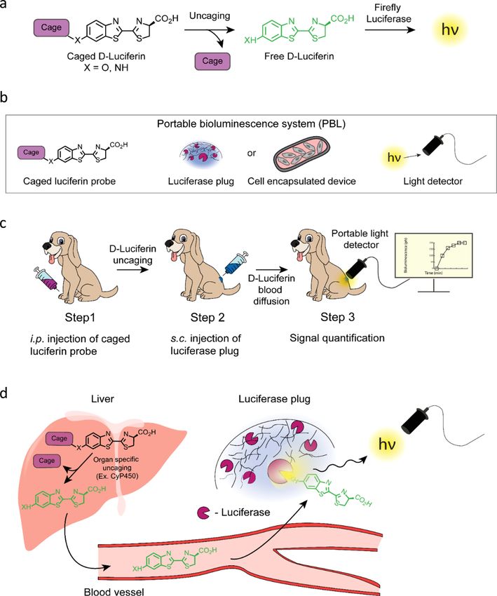
ARTICLE                                                                 NATURE COMMUNICATIONS | https://doi.org/10.1038/s41467-021-22892-9

R
       ecent advances in imaging technologies have revolutionized            using a luciferase-based biodegradable injectable “plug” in
       the fields of biomedical research, especially with respect to          combination with a caged luciferin probe and a highly
       clinical diagnostics and drug discovery. Among the many               sensitive portable light detector. The PBL method can be easily
known preclinical techniques, bioluminescence imaging (BLI)                  applied for the imaging and quantification of a wide variety of
remains one of the most widely used due to its unprecedented                 biological processes for which caged luciferin probes already
sensitivity and ease of use. Current applications of BLI cover a             exist3,4,6,8,9,18–25. We choose to perform the validation studies
wide range of therapeutic areas, in particular cancer and infec-             using enzymatic processes as readout because there is a pressing
tious diseases, but also including neurodegenerative, cardiovas-             demand for more efficient methods for in vivo evaluation of the
cular and metabolic disorders, such as diabetes and obesity1–9.              activities of enzymes such as CYP450 in drug discovery studies. In
   A typical BLI experiment employs a luciferase enzyme as a                 addition to CYP450, which is an important example of an
reporter that generates bioluminescent light upon oxidation of its           intracellular enzyme, we also investigate potential applications of
substrate luciferin. Some of the first in vivo applications of BLI            this technology to therapeutically relevant extracellular enzymes.
relied on the constitutive expression of luciferase enzyme in                Dipeptidyl peptidase 4 (DPP-4) is selected as an example because
cancer cells to monitor tumor growth and metastasis10,11.                    of its essential role in the discovery of drugs for type 2 diabetes
More recently, caged luciferin-based probes were developed,                  and several types of cancer29–32.
opening-up the possibility of extending the application of                      We first demonstrate that the PBL method is capable of non-
BLI to the functional imaging of enzymatic and metabolic pro-                invasive measurements of the activity of DPP-4 and CYP450
cesses, including regulatory proteases; uptake of essential meta-            enzymes in vivo in non-luciferase-expressing mice. Importantly,
bolites (e.g., glucose and fatty acids); fluxes of bioactive small            the sensitivity and accuracy of the method are comparable with
molecules (e.g., H2O2 and H2S); cellular glycosylation; and                  those obtained with standard stationary CCD optical imaging
intracellular delivery and release of therapeutically relevant               instruments (IVIS® Spectrum). Next, we successfully apply PBL
compounds3,6–9,12–26. These functional BLI probes are based on               technology in dogs, paving the way for the translation of func-
the principle that chemically caged luciferin is not a substrate for         tional BLI to the noninvasive quantification of biological pro-
luciferase until it becomes released or uncaged by a specific                 cesses in large animals. Taken together, these results lay an
biological process of interest (e.g., selective enzymatic cleavage,          important foundation for the potential replacement of highly
Fig. 1a). The intensity of the bioluminescent signal quantitatively          invasive and destructive in vivo testing with the PBL methods that
correlates with the amount of free luciferin in the animal, which            would help save millions of animal lives, especially large animals
in turn reflects the level of functional activity of a biological             like dogs. Finally, we also demonstrate that PBL technology has
process of interest. Consequently, the acquired information                  the potential to be used in clinical settings by successfully
reveals the dynamics of a wide range of biological functions that            quantifying bioluminescent signals directly in a human cadaver.
play key roles in physiological and pathological processes, as well          This study demonstrates application of functional BLI technology
as in drug discovery.                                                        for the noninvasive monitoring of biological processes in non-
   Despite all the advantages and widespread applications of BLI             transgenic animals, laying an important foundation for replacing
in the field of preclinical imaging, current BLI technology has a             highly invasive tests with noninvasive readouts and for realizing
number of significant limitations. First, BLI is limited to use               the potential of such methods for clinical translation.
either in luciferase-expressing transgenic mice or in animals
transplanted with luciferase-expressing cells1–4,6–25. While many
relevant luciferase-expressing animal models have been developed             Results
in recent years, they still represent only a small percentage of             General concept of the PBL method. The basic concept of the
existing in vivo models of human disease. Second, current in vivo            developed methodology is illustrated in Fig. 1 and Movie 1. The
imaging instruments usually comprise a small light-tight “black              PBL system depicted on Fig. 1b consists of three main compo-
box” and a cooled charge-coupled device (CCD) camera as a light              nents: a functional bioluminescent probe, which is a caged luci-
detector, making the technology relatively expensive, non-                   ferin compound that can sense a certain biological process of
portable and, most importantly, restricting the use of BLI to                interest (e.g., CYP450 or DPP-4, Fig. 1a), a biocompatible
small animals such as mice and rats. This latter size constraint             luciferase-based bioluminescent light producing reporter “plug”
represents a serious limitation to the use of BLI technology for             or a cell encapsulating device, and a portable light detector. In a
drug development studies because many in vivo tests are per-                 typical experiment, the animal is first injected with a single dose
formed on non-rodent animals such as dogs, cats, rabbits, and                of caged luciferin probe followed by subcutaneous injection of the
nonhuman primates. Third, the present-day instruments also                   luciferase plug a few minutes later. Cell encapsulating device
require keeping animals under prolonged anesthesia during                    transplanted with luciferase-expressing cells is utilized for a long-
imaging, which can impact the animal’s health and be disruptive              term monitoring of biological processes (up to 5 months)33. The
to its metabolism27. As a result of these limitations, many of the           light detector is then immediately affixed on top of the luciferase
widely utilized animal experiments are still performed in a highly           plug and the bioluminescent signal is recorded at specific time
invasive way that leads to the sacrifice of a large number of                 intervals to obtain maximal light output (Fig. 1c). Upon injection
animals. A striking example of such experiments is the toxicology            of a caged probe, free luciferin is released in a target organ (e.g.,
testing of potential therapeutic candidates for the activation of            liver) as a result of uncaging by a specific biological process or
cytochrome p450 (CYP450), a liver enzyme responsible for the                 enzyme such as CYP450 (Fig. 1d). Free luciferin migrates into the
deactivation of the majority of clinically used drugs. This one test         bloodstream and eventually reaches luciferase-based reporter
alone leads to the sacrifice of hundreds of thousands of dogs each            placed under the skin of test animal. The amount of light gen-
year28. Therefore, there is an urgent need for novel technologies            erated by the luciferase plug is proportional to the concentration
that would allow noninvasive monitoring of biological processes              of luciferin in the bloodstream, resulting in a bioluminescent light
in non-transgenic animals, especially large animals such as dogs.            production that directly correlates with the level of functional
   To address the shortcomings of the current BLI technology and             activity of biological process of interest (e.g., enzymatic activity).
to expand its use to non-transgenic and non-rodent animals, we               In all of our experiments we utilized D-luciferin (Fig. 1a, X = O)
develop a “portable bioluminescent” (PBL) system. This system                that is referred as “luciferin” in the rest of the text. However, the
allows noninvasive measurements of biological processes in vivo              same technology could be adapted for the use with other

2                NATURE COMMUNICATIONS | (2021)12:2680 | https://doi.org/10.1038/s41467-021-22892-9 | www.nature.com/naturecommunications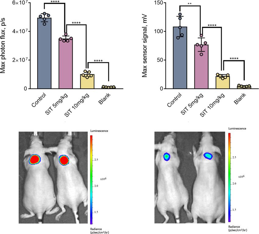
NATURE COMMUNICATIONS | https://doi.org/10.1038/s41467-021-22892-9                                                                            ARTICLE

Fig. 1 General concept of the PBL method for noninvasive in vivo monitoring of biological processes. a Basic principle of functional BLI. A bioluminescent
probe with caged luciferin interacts with the targeted biological process. This reaction leads to uncaging of luciferin, which in turn is able to react with
luciferase to produce bioluminescent light. Importantly, the amount of light generated as the result of this uncaging is proportional to the level of functional
activity of the biological process of interest. b Three main components of the PBL system include (a) a caged luciferin probe that releases free luciferin by a
specific biological process; b luciferase biodegradable plug or a cell encapsulating device with luciferase expressing cells that produces light proportionally
to the amount of released luciferin; c sensitive portable light detector for signal quantification. c A typical PBL experiment employs an administration of a
functional bioluminescent probe based on the caged luciferin scaffold followed by s.c. injection of a biodegradable luciferase plug. The light production is
quantified by the portable light detector that is placed directly on top of the area of the plug. d Upon injection, a caged luciferin probe reaches the target
organ (e.g., liver) where it gets uncaged by a specific biological process or enzyme (e.g., CyP450). Subsequently, free luciferin diffuses into the
bloodstream and eventually reaches the luciferase containing plug. The luciferase enzyme in the plug produces light proportionally to the level of luciferin.
The light is quantified by a sensitive light detector placed on top of the luciferase plug.

luciferases and their corresponding substrates such as Gaussia,                  package with a diameter of 30 mm and height of 40 mm. Inte-
NanoLuc and various red-shifted luciferases, which are all known                 grating the sensor with the amplifier helps to reduce the residual
to provide brighter in vivo signals3,5,8,9,34–40.                                noise. The device resembles a stethoscope and can be easily
                                                                                 applied to small and large animals as well as a human body (Fig. 2
Portable light detector. We developed a compact, portable large-                 and Fig. 5). The “working surface” of the device mechanically
aperture light detector with high sensitivity and low noise spe-                 protects the sensitive electronic components and contains a cir-
cifically for the detection of bioluminescent photon flux, which is                cular optical aperture with 1 cm diameter. We used a commercial
typically relatively low. The design features a large 1 cm2 silicon              Hamamatsu photodiode S1227. The choice of a single-pixel
photodiode in photovoltaic mode with a current-to-voltage                        photodiode over a camera is beneficial due to its low cost, room-
transimpedance amplifier41,42 integrated in a cylindrical                         temperature operation, and, most importantly, portability and

NATURE COMMUNICATIONS | (2021)12:2680 | https://doi.org/10.1038/s41467-021-22892-9 | www.nature.com/naturecommunications                                      3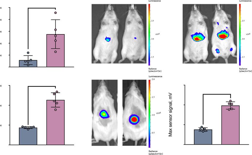
ARTICLE                                                                                         NATURE COMMUNICATIONS | https://doi.org/10.1038/s41467-021-22892-9

             a                                         IVIS Spectum                         b
                                        10 9           ***

                 Max photon flux, p/s                             ****
                                        10 8

                                                                               ****

                                        10 7

                                        10 6
                                                 150         15          1.5      Blank

             c                                     Luciferin dose (mg/kg)                   d                        PBL System

Fig. 2 In vivo measurement of luciferin levels in non-transgenic animals using injectable luciferase plugs. Direct comparison of stationary CCD camera
(IVIS® Spectrum) and PBL readouts. a Maximal photon flux resulting from three groups of nude mice (n = 5) injected s.c. with 100 µL of luciferase plug in
the dorsal area followed by i.p. injection of different doses of luciferin (1.5, 15, and 150 mg/kg) and imaging with the IVIS® Spectrum. The “blank” group of
mice was not injected with luciferin. b Representative images of mice from each luciferin-injected group described in (a). c Experimental setup for the PBL
imaging experiment. The portable light detector is placed directly on the area of the luciferase plug upon administration of luciferin. d Maximum signal
output from the experiment described in (a) except that the measurements were performed using the portable light detector. Data are presented as the
mean ± s. d. (n = 5). Each “n” represents a biologically independent sample. Statistical significance (***P < 0.001; ****P < 0.0001) was calculated using a
two-tailed unpaired t-test. Source data is available as a Source Data file for (a and d).

low noise (low dark current). No spatial optical resolution is                                      previously reported cell encapsulating device should be utilized
required because the detector is placed on top of a luciferase plug                                 for the long-term studies (several months)33. To optimize the
whose position is known. In addition, because the detector is                                       composition of the injectable luciferase plug and to achieve a
placed directly on a light-emitting plug, most of the light emitted                                 bright stable signal in vivo, we tested the effect of different
by the plug could be potentially detected. To detect low light                                      components on the light output using a stationary BLI instrument
levels, we used a low-noise operational amplifier configured as a                                     equipped with a sensitive CCD camera (IVIS® Spectrum, Perkin
current-to-voltage transimpedance amplifier, which is a com-                                         Elmer). The data demonstrated that the luminescence generated
mercial IC chip (Supplementary Fig. 1). The amplification factor                                     by the luciferase plug is directly proportional to the amount of
was set to 1010 V/A with a feedback resistor R = 10 GigaOhm.                                        luciferase enzyme added to the plug and is relatively independent
The detector outputs a voltage reading that is proportional to                                      of the ATP concentration in the range of 1–10 mM (Supple-
optical radiant power absorbed by the diode surface: V = P/r,                                       mentary Fig. 2a, b). As a result of this study, the following
where P is radiant power, V is voltage and r is a proportionality                                   composition of the luciferase plug was chosen for all further
coefficient called responsivity. To find r, we calibrate our detector.                                experiments in mice: 83 μL of Matrigel®, 10 μg of luciferase
We also assess dark noise. Please refer to the Methods section for                                  enzyme, 10 mM ATP, 1 mM Mg2+ and PBS up to a 100 μL total
more detailed description of the detector technology.                                               volume. The Matrigel® matrix was selected for this study because
                                                                                                    it is nontoxic, easy to implant by s.c. injection, and produced
                                                                                                    brighter and more stable signal compared to other matrices
Luciferase-based injectable plug. The other component of the                                        (Supplementary Fig. 2c). Matrigel® based plug was stable for at
PBL method is the luciferase-based injectable plug (hereinafter                                     least 60 min post s.c. injection enabling continuous measurements
referred to as a “luciferase plug”), which contains recombinant                                     of bioluminescent signal and determination of maximal light
luciferase enzyme along with its cofactors and a polymeric matrix                                   output (Supplementary Fig. 3a). These data also demonstrate that
to keep the enzyme and its cofactors intact under the skin of the                                   repetitive measurements taken more than 1 h apart in the same
test animal (Fig. 1b, c, Movie 1). While the luciferase plug is                                     animal will require reinjection of the new plug to achieve better
designed for short-term monitoring of biological processes,                                         consistency of the results.

4                                       NATURE COMMUNICATIONS | (2021)12:2680 | https://doi.org/10.1038/s41467-021-22892-9 | www.nature.com/naturecommunications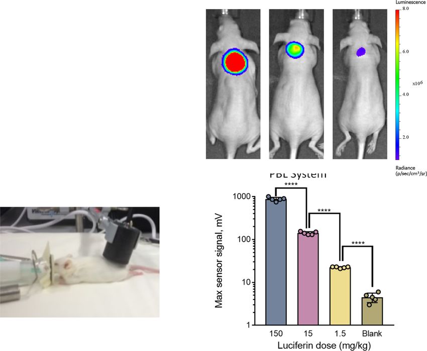
NATURE COMMUNICATIONS | https://doi.org/10.1038/s41467-021-22892-9                                                                  ARTICLE

   Since the surface light intensity is dependent on the depth of               injection of the luciferase plug 10 min post injection of the probe.
the light source, we decided to quantify the dependency of the                  The last group of mice was injected with the plug but not the
bioluminescent light output on the depth of the luciferase plug.                caged luciferin probe and therefore was used as a negative control
We used the cuts of meat from a butcher to quantify signal loss                 (blank). The signal was acquired from all four groups of mice
to a measured tissue thickness (Supplementary Fig. 3b). As                      using either the IVIS® Spectrum or portable light detector. A
expected, the intensity of the detected light was dependent on the              strong bioluminescent signal was observed from the control
depth of the light source. Interestingly, the drop in the signal                group of mice treated with the DPP-4 caged luciferin probe alone
intensity was not as dramatic as we expected with the regular                   with both the IVIS® Spectrum and portable light detector (Fig. 3).
firefly luciferase (about tenfolds per 0.8 cm of meat). We could                  Importantly, the dose dependent reduction in the signal obtained
still see a clear signal even at the depth of 1 cm, suggesting that             from the animals treated with the DPP-4 inhibitor was observed,
this method is suitable for the use with a wide range and                       and these measurements were fully consistent between the IVIS®
concentrations of luminescence imaging probes6,7,9,12–25. Since                 Spectrum and portable light detector readouts (Fig. 3a, b). These
the surface signal is dependent on the depth of the plug, the                   results suggest that the PBL methodology is able to provide an
consistency of the plug injection across study groups is very                   accurate readout of extracellular enzymatic activity in non-
important factor for achieving the best reproducibility of the                  transgenic animals in a noninvasive fashion, and that the
results.                                                                        acquired data is comparable to that obtained with the established
                                                                                stationary “black box” CCD cameras technology.
                                                                                   Next, we examined the feasibility of PBL to measure the
Quantification of blood luciferin levels with luciferase plug. To                activity of an intracellular enzyme, such as CYP450, in vivo. We
investigate whether the amount of light generated by the luci-                  first investigated whether the activity of CYP450 can be detected
ferase plug is proportional to the concentration of luciferin in the            in vivo under conditions previously reported to trigger activation
blood of test animals, we injected different concentrations of                  of the enzyme, namely, upon treatment with a xenobiotic
luciferin solution intraperitoneally (i.p.) into nude mice followed             dexamethasone43. While many different isozymes of CYP450
by s.c. injection of luciferase plugs in the dorsal side of the test            are known, we decided to specifically focus on cytochrome P450
animals. The animals were then immediately anesthetized and                     3A (abbreviated Cyp3a), which is the most common and versatile
placed into the stationary CCD camera (IVIS® Spectrum). The                     isozyme involved in drug metabolism44. The caged luciferin
bioluminescent light output was continuously measured to                        probe specific to this isozyme is fully validated and commercially
determine the maximal optical radiant power (measured in                        available (Luciferin-IPATM, Promega), along with many other
phtons/sec and referred to as “maximum photon flux”). Our data                   cytochrome specific luciferins45. Two cohorts of age-matched
demonstrate that the maximal photon flux resulting from the                      genetically engineered mice that ubiquitously express luciferase
luciferase plug linearly correlates with the amount of injected                 through the beta-actin promoter (FVB-luc+/+ mice) were used
luciferin within a large dynamic range of three orders of mag-                  for this study46. The experimental group of mice was injected
nitude (150, 15 and 1.5 mg/kg doses), suggesting that the plug can              with dexamethasone (abbreviated as “DEX”, 50 mg/kg dose i.p.),
be utilized for accurately quantifying luciferin concentrations in              which was previously shown to specifically cause acute activation
the blood of test animals (Fig. 2a). The representative images of               of Cyp3a47,48, while the control group of mice was treated with
mice injected with the luciferase plug and three different con-                 vehicle alone (vegetable oil). After 24 h, both groups received i.p.
centrations of luciferin are shown on Fig. 2b.                                  injections of the Luciferin-IPATM probe, followed by anesthesia
   The same set of experiments was then performed using the                     of the animals and signal acquisition using the IVIS® Spectrum.
portable light detector, with the procedure for bioluminescence                 As shown in Fig. 4a–c, the bioluminescent signal from the DEX-
measurement similar to that described above using the IVIS®                     treated mice was approximately three times higher than the signal
Spectrum (the experimental setup is shown in Fig. 2c). The results              from the control group, indicating that the probe can successfully
demonstrated in Fig. 2d show a similar linear correlation between               detect Cyp3a activation directly in vivo upon treatment of mice
the luciferin concentration and the maximal optical power                       with DEX.
measured by the portable light detector. Moreover, the error                       Having confirmed the feasibility of measuring Cyp3a using
bars shown in Fig. 2a and Fig. 2d are also comparable. These data               IVIS technology, we repeated the above experiment using non-
indicate that the PBL method is suitable for the sensitive                      transgenic animals and our PBL methodology. Two groups of
quantification of the free luciferin concentration in the blood of               wild-type FVB mice were used, one of which was injected with
non-transgenic animals that do not express the luciferase enzyme.               DEX and the other of which was injected with vehicle (control).
Importantly, the signal linearity correlates with the amount of                 After 24 h, the Luciferin-IPATM probe was injected i.p. into both
injected luciferin over a large dynamic range (three orders of                  groups of mice. The mice were then anesthetized, injected with a
magnitude −3 logs).                                                             luciferase plug and imaged with either the IVIS® Spectrum or
                                                                                portable light detector. As shown in Fig. 4d–f, a significantly
Measurements of enzymatic activities in living mice. To eval-                   higher bioluminescent signal was observed in the animals treated
uate whether this PBL method can be used for accurate mea-                      with DEX than in the vehicle-treated control mice using both the
surements of biological processes using functional bioluminescent               IVIS® Spectrum (Fig. 4d-e) and portable light detector (Fig. 4f).
probes, we first performed a feasibility study with a caged luci-                   In order to compare the imaging results with the conventional
ferin probe previously designed by us for selective sensing of the              measurements of cytochrome activity, we isolated liver samples
activity of DPP-412, a representative example of an extracellular               from wild type and DEX treated FVB-Luc mice and performed
enzyme. Four cohorts of Swiss nude mice (n = 5) were used for                   qPCR analysis of Cyp3a expression. The results of qPCR analysis
this study. Two different doses of 5 and 10 mg/kg of the selective              showed similar folds increase in Cyp3a expression (Supplemen-
DPP-4 inhibitor sitagliptin (abbreviated as “SIT”) were admi-                   tary Fig. 3c). These data suggest that the current invasive and
nistered by oral gavage in PBS buffer to two group of animals,                  time-consuming methods of measuring Cyp3a activity can be
while the third group of mice was treated with PBS only (control).              successfully replaced by the noninvasive bioluminescent detection
Thirty minutes post gavage, three groups of mice received an                    methodologies, especially PBL, which, as shown below, is fully
injection of the DPP-4 caged luciferin probe followed by s.c.                   translatable to large animal studies.

NATURE COMMUNICATIONS | (2021)12:2680 | https://doi.org/10.1038/s41467-021-22892-9 | www.nature.com/naturecommunications                          5
ARTICLE NATURE COMMUNICATIONS | https://doi.org/10.1038/s41467-021-22892-9
a IVIS Spectum b PBL System
c d
Control Sitagliptin
Fig. 3 Noninvasive in vivo measurements of extracellular enzymatic activity (DPP-4) in non-transgenic mice: direct comparison of stationary CCD
camera (IVIS® Spectrum) and PBL readouts. a Maximal photon flux obtained from four groups of nude mice (n = 5) resulting from administration of a
DPP-4-specific caged luciferin probe12 and DPP-4 inhibitor sitagliptin (“SIT”). The “SIT” groups of mice received an oral gavage of different concentrations
of SIT (5 and 10 mg/kg) 30 min prior to injection of the probe, while the control group received a gavage of vehicle only (PBS buffer). All the mice received
s.c. injection of 100 µL of luciferase plug in the dorsal area followed by signal acquisition with the IVIS® Spectrum. The “blank” group of mice received
injection of the plug without caged luciferin probe. b Maximum signal output from the experiment described in (a) except that the measurements were
performed using the portable light detector. c–d Representative images of two mice from the control (c) and 10 mg/kg sitagliptin-treated (d) groups. Data
are presented as the mean ± s. d. (n = 5). Each “n” represents a biologically independent sample. Statistical significance (****P < 0.0001) was calculated
using a two-tailed unpaired t-test. Source data is available as a Source Data file for (a and b).
Application of the PBL method for noninvasive biolumines- routinely utilized to detect drug-induced liver injury in humans
cent signal quantification in large animals and a human post- and experimental animals55,56. While increased ALT serum levels
mortem model. Since the main goal of the PBL method was the generally correlate with hepatocytes damage, induction of serum
translation of functional BLI to large animals, we next decided to ALP reflects the extent of injury to the biliary epithelial cells. Also,
investigate whether this methodology would work in a canine increase in total bilirubin is indicative of hepatic functional
model. While luciferin had been extensively used at relatively high impairment or processing of bilirubin production (hemolysis)55.
concentrations in mice (up to 750 mg/kg)49 with no obvious signs We also measured the levels of GLDH that is a mitochondrial
of adverse effects49–54, no such data were reported for large ani- matrix enzyme responsible for amino acid oxidation and urea
mals like dogs. Thus, we performed a serum chemistry blood production57. In addition, recent clinical studies demonstrated
toxicology analysis upon administration of a clinically relevant high diagnostic potential of this GLDH test in predicting hepatic
amount of luciferin in healthy dog (i.p. injection of 15 mg/kg). We toxicity in patients with various liver pathologies and therefore it
specifically measured the levels of alanine aminotransferase (ALT), has been proposed as more sensitive and specific biomarker of
alkaline phosphatase (ALP), gamma-glutamyltransferase (GGT), liver injury than ALT56. Lastly, we investigated changes in GGT
serum creatine kinase (CK), glutamate dehydrogenase (GLDH) and CK levels that all have been previously used as a diagnostic
and total bilirubin in a commercial laboratory before and 24 h markers of drug-induced liver injury58–60. Our data shown in
after administration of luciferin. These serum biomarkers are Supplementary Table 1 clearly demonstrate that no clinically
6 NATURE COMMUNICATIONS | (2021)12:2680 | https://doi.org/10.1038/s41467-021-22892-9 | www.nature.com/naturecommunicationsNATURE COMMUNICATIONS | https://doi.org/10.1038/s41467-021-22892-9 ARTICLE
a b c
FVB-Luc mice Control DEX
5 10 8 P=0.005
4 10 8
Max photon flux, p/s
3 10 8
2 10 8
1 10 8
0
Control DEX
d e f
W.T. FVB mice Control DEX PBL System
6 10 7 **** 150
****
Max photon flux, p/s
4 10 7 100
2 10 7 50
0 0
Control DEX Control DEX
Fig. 4 Noninvasive in vivo measurements of intracellular enzymatic activity of CYP450: direct comparison of stationary CCD camera (IVIS® Spectrum)
and PBL readouts. a Maximal photon flux resulting from two groups of FVB-luc+/+ mice (n = 5) injected with the Luciferin-IPATM probe, a caged luciferin
reagent specifically designed to measure activation of the Cyp3a isozyme of CYP45045. The “DEX” group of mice was treated with dexamethasone, a
known activator of Cyp3a, 24 h prior to the injection with the Luciferin-IPATM probe, while the control group received a gavage of vehicle only (vegetable
oil). The light output was measured with the IVIS® Spectrum. b–c Representative images of two mice from the control (b) and DEX-treated (c) groups. d
Maximal photon flux obtained from two groups of wild-type non-transgenic FVB mice (n = 5) injected i.p. with the Luciferin-IPATM probe followed by
injection of the luciferase plug (s.c. in the dorsal area) and subsequent imaging by the IVIS® Spectrum. Analogous to the experiment described in (a), the
“DEX” group of mice was treated with DEX 24 h prior to the injection with the Luciferin-IPATM probe while the control group received the gavage of vehicle
only. The light output of the plug was measured with the IVIS® Spectrum. e Representative mouse images from the control and DEX-treated groups in the
experiment described in (d). f Maximum signal output from the experiment described in (d) except that the measurements were performed using the
portable light detector. Data are presented as the mean ± s. d. (n = 5). Each “n” represents a biologically independent sample. Statistical significance (****P
< 0.0001) was calculated using a two-tailed unpaired t-test. Source data is available as a Source Data file for (a, d and f).
relevant elevation was observed in any of these parameters, indi- non-rodent animals with noninvasive measurements using the
cating lack of D-luciferin toxicity in the dog at the concentration PBL method.
studied. Finally, we investigated whether PBL technology has the
Inspired by these results, we investigated whether the amount potential to be translated to human diagnostic applications. For
of light generated by the luciferase plug is proportional to the these experiments, we premixed 100 µL of luciferase plug with
concentration of injected luciferin in dogs. The dogs were three different doses of luciferin (2 µM, 200 nM and 20 nM final
anesthetized, and the luciferase plug was implanted subcuta- concentrations) followed by direct s.c. injection of the mixture
neously in the ventral abdomen. Various concentrations of under the skin of a human cadaver in the area of the upper arm
luciferin (15, 1.5 and 0.15 mg/kg) were then administered via i.p. (Fig. 5c). The portable light detector was then placed directly on
injection followed by the placement of the portable light detector top of the luciferase plug, and the signal was acquired for 15 min.
directly on the area of the luciferase plug (Fig. 5a). As shown in A significant signal was observed even at the lowest luciferin
Fig. 5b, the maximal light output linearly correlated with the concentration, which provided a signal threefold higher than the
amount of injected luciferin within a large dynamic range of three background signal (no plug). Moreover, the signal was propor-
orders of magnitude. Due to strict regulation on animal tional to the luciferin concentrations in the range of three orders
experimentation in large animals, this experiment was performed of magnitude (Fig. 5d). These data demonstrate that the PBL
as a proof of principle with n = 1 per time point. These results technology has full potential to work successfully in humans,
suggest that the PBL method can be utilized for accurate paving the way for the translation of BLI all the way to the clinic.
measurements of the luciferin concentrations in the blood of
large animals such as dogs, and that the acquired data were fully
consistent with the previous data obtained in mice (Fig. 2). Under Discussion
the same experimental conditions, the maximal sensor signal in In comparison to conventional BLI, the PBL method provides a
dogs was ~30 times higher than that in the mouse experiments major technological breakthrough for a number of reasons. It
(15 mg/kg dose; Fig. 2). These data lay an important foundation allows the analysis of a wide range of biological processes by
for the replacement of highly invasive biological tests in large completely removing the need to use luciferase engineered cells or
NATURE COMMUNICATIONS | (2021)12:2680 | https://doi.org/10.1038/s41467-021-22892-9 | www.nature.com/naturecommunications 7ARTICLE NATURE COMMUNICATIONS | https://doi.org/10.1038/s41467-021-22892-9
a b PBL System
c d PBL System
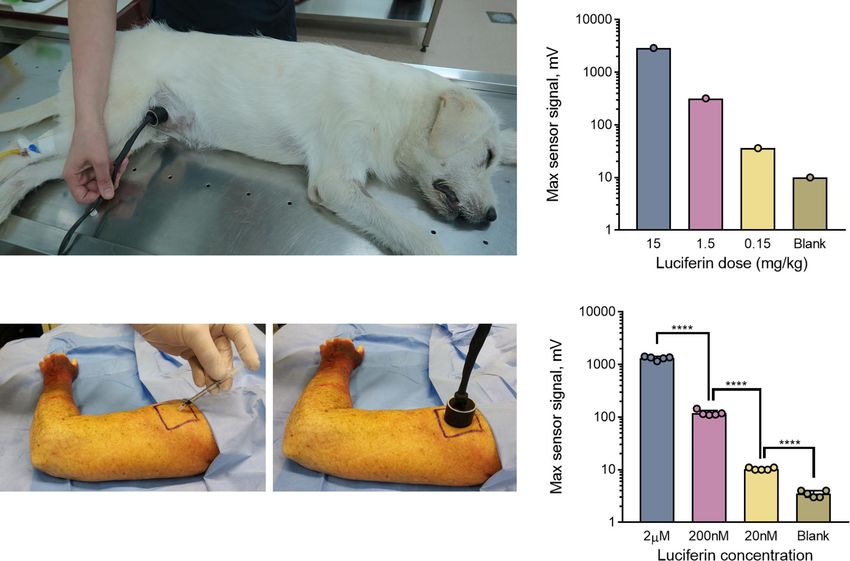
Fig. 5 Noninvasive BL signal quantification in large animals (dogs) and a human post-mortem model using the PBL method. a Experimental setup for
the application of the PBL method in dogs. Three large dogs were anesthetized followed by s.c. injection of 0.5 mL of the luciferase plug in the abdominal
area followed by i.p. injection of 5 mL of luciferin solution at three different concentrations (15 mg/kg, 1.5 mg/kg and 0.15 mg/kg). The portable light
detector is then placed on the area of the luciferase plug, and measurements are obtained for 30 min following the luciferin injection. The experiment was
performed as a proof of principle with n = 1 for each time point with one luciferin injection per dog. b Maximal signal output obtained from dogs injected
with different concentrations of luciferin (15, 1.5 and 0.15 mg/kg in 5 mL of PBS). c Experimental setup for PBL measurements in a human post-mortem
model. The luciferase plug matrix was premixed with three different doses of luciferin (2 µM, 200 nM and 20 nM) followed by direct s.c. injection of the
100 µL of the activated luciferase plug under the skin of a human cadaver. The portable light detector was assembled directly on top of the plug followed by
signal acquisition for 15 min. Three independent injections were performed for each luciferin concentration. d Average maximal signal output obtained by
the portable light detector in the experiment described in (c). Statistical significance (**** P < 0.0001) was calculated using a two-tailed unpaired t-test.
Source data is available as a Source Data file for (b and d).
transgenic animals. This feature dramatically expands the scope isozymes have been previously reported45. Our data in mice
of BLI to many existing animal models of human diseases and clearly demonstrate that bioluminescence readouts can be used
opens-up the opportunity for the use of this powerful modality in for accurate noninvasive quantification of the activity of specific
large animals and potentially humans. Importantly, this work lays isozymes of CYP450 with both PBL technology and standard
the foundation for the replacement of current highly invasive CCD cameras and have full potential to be translated to large
large animals tests performed by utilizing noninvasive readouts, animals such as dogs.
which falls in-line with the requirements of the 3 R principles of In the current study, we utilized a minimally invasive “inject-
animal experimentation. For example, CYP450 is a major able” approach for the development of a luciferase-based biode-
detoxicant of xenobiotics, and measurements of its activity are gradable plug for measuring the luciferin concentration in the
routinely performed by multiple drug development companies as blood of living animals. This approach is ideally suited for the
a part of toxicological studies. The procedure involves the measurements of relatively fast biological processes such as the
administration of high doses of xenobiotics followed by multiple activation of certain enzymes. However, PBL can also be extended
blood withdrawals, after which the animals must be sacrificed. to long-term longitudinal measurements of biological processes
The blood is then analyzed by HPLC-MS methods that usually by repetitive injections of the luciferase plug, since it is composed
provide only a few data points per animal with rather large error of nontoxic and biodegradable material. Moreover, luciferin has
bars due to multiple sample manipulation steps61. In contrast, been used extensively in preclinical settings with repeated injec-
bioluminescent readouts have been shown to provide much tions of rather high doses (up to 750 mg/kg) with no mention of
higher sensitivity and better kinetic parameters than those any harmful side effects49. In addition, it was reported that the
obtained from end-point assays62,63, However, to date such bio- growth rate of luciferase-labeled cell lines in syngenic mouse
luminescent readouts have not been adapted for activity mea- models monitored by repetitive injections of high doses of luci-
surements of CYP450 in live animals despite the fact that a full ferin is identical to the growth rates of wild type cells, suggesting
range of caged luciferin probes for a variety of different CYP450 that luciferin is not toxic to animals and the immune response to
8 NATURE COMMUNICATIONS | (2021)12:2680 | https://doi.org/10.1038/s41467-021-22892-9 | www.nature.com/naturecommunicationsNATURE COMMUNICATIONS | https://doi.org/10.1038/s41467-021-22892-9 ARTICLE
luciferase is extremely weak, if immunogenic at all64,65. We also ability also opens-up the potential for using PBL in veterinary
performed an extensive blood toxicology analysis before and after settings for routine diagnostic testing of companion animals (e.g.,
luciferin injection in dog and did not observe elevation of any noninvasive monitoring for treatment of liver or kidney failure in
clinically relevant parameters indicating lack of luciferin toxicity dogs and cats). The portable and cost-effective features of PBL
at concentration studied. technology, combined with its exquisite sensitivity and quan-
Alternatively, the luciferase plug can be replaced with a cell- tifiability, make this powerful tool a strong candidate for adoption
encapsulated device that remains a stable source of luciferase for in numerous areas of preclinical and clinical research and
up to 5 months33. Indeed, such fully s.c. implanted devices have diagnostics.
already been developed and widely used in humans for passive
immunization against Alzheimer’s disease by providing stable Methods
delivery of recombinant anti-amyloid-β antibodies66 and for Reagents. Matrigel® was purchased from Corning, USA (cat. # 356237); ATP
intrathecal delivery of ciliary neurotrophic factor in amyo- disodium salt was purchased from AppliChem GmbH, Germany (cat. # A1348-
0005); MgSO4 ·7H2O – AlfaAesar, Germany (cat. # A14491-0B); PBS – Thermo-
trophic lateral sclerosis patients67. These cell encapsulating Fisher Scientific, USA (cat.# 10010-015); recombinant firefly luciferase – Sigma-
devices are small in size and require only minimal surgical Aldrich (cat.# SRE0045); D-luciferin – Perkin Elmer, USA (cat.# 122799); DEX –
interventions33,66,67. In addition, novel technologies in the field Sigma-Aldrich (cat.# 31375); QIAGEN RNeasy kit - QIAGEN, Switzerland; Q-Gel
of electrochemical and optical sensors have allowed the creation - QGel SA, Switzerland; and collagen, DEX, sitagliptin – Sigma-Aldrich. DPP-4-
of s.c. implantable devices that can wirelessly send data to specific caged luciferin probe (DAL) and Luciferin-IPATM were chemically syn-
thesized according to the published protocols12,45.
mobile devices and have already been used in clinical practice
for the continuous monitoring of glucose levels in humans68.
Portable light detector. For light detection we used a large-area (1 cm2) silicon
These recent technological advances allow future translation of photodiode, which is a single-pixel device. The photodiode generates electrical
the PBL method to portable and fully implantable “to be worn” current in response to irradiation, and the electrical current is proportional to the
biosensors for in vivo longitudinal measurements of a wide optical power that is absorbed by the diode surface. The current is converted to
range of biological processes. voltage with a low-noise transimpedance amplifier. The output voltage is read by a
stock voltmeter connected to the detector by a BNC cable, see Supplementary Fig. 1
While we utilized a predominantly naturally derived firefly for more details. Our detector design is inspired by the accurate light detection
luciferin-luciferase system in the current study, we also envision method used for standard transfers41,42. To measure responsivity, we calibrate our
the use of the PBL method with other recently developed sub- portable detector with a visible light source at 632 nm using a substitution cali-
strates such as CycLuc134, AkaLumine5,38, naphthyl-luciferins36, bration method. Our portable detector is compared to the trap detector with a
known detection efficiency74. The measured responsivity is 3.32(3) × 109 V/W. To
and several others3,5 that provide much brighter in vivo signals measure dark noise, we block the detectors optical input and obtain a series of
with firefly luciferase due to increased tissue penetration of red- voltage readings with an integration time of 2 s (as it is done in the experiment).
shifted light emission. The method can also be optimized to be We measure the dark noise of 6 × 10⁻¹⁴ W RMS. Therefore, we can resolve an
used with other luciferases such as red-shifted click beetle luci- input optical flux of ~200,000 photons of visible light per second with signal-to-
noise ratio of one. In certain cases, it might be appropriate to integrate for 1 min or
ferase, Gaussia luciferease and NanoLuc3,5,8,9,34–40. Both of the longer, further reducing detection noise to about 1.1 × 10⁻¹⁴ W RMS, which would
latter luciferases should be very effective for use with the PBL yield resolving a flux of ~35,000 photons of visible light per second with signal-to-
method as they are extremely bright and can significantly increase noise ratio of one. To convert optical power expressed in watts to that expressed as
sensitivity of the assay. Indeed, secreted Gaussia luciferase was photon flux, we use Planks formula: F = Pλ0/hc, where F is photon flux, P is power,
previously used to monitor gene expression through a simple h is Planks constant, c is the speed of light, and λ0 is the weighted average
wavelength of the bioluminescence emission spectrum. Unlike an imaging device,
blood sampling in non-transgenic animals to monitor this detector is placed directly on the animal at the injection site of the luciferase
angiogenesis69. In addition, many caged coelenterazines sub- plug, so that it collects nearly all of the bioluminescent light emitted from ~1 cm2
strates have been reported in the literature37. skin surface.
The portable nature of the PBL method also offers additional
advantages over conventional techniques, such as avoiding the Luciferase-based injectable plug. The following optimized composition of the
need to constrain the animals in the light-tight “black box” (e.g., luciferase plug was used for the experiments: 83 μL of Matrigel® matrix, 10 μg of
luciferase enzyme (1 × 10¹¹ units per mg, Sigma-Aldrich SRE0045), 10 mM ATP, 1
IVIS® Spectrum) and the use of anesthesia. Both of these factors mM Mg2+ and PBS up to a 100 μL total volume. ATP, Mg2+, luciferase and PBS
are often associated with additional stress to the animal and have were first premixed in an Eppendorf tube, followed by the addition of ice-cold
been reported to interfere with biological readouts, such as brain Matrigel® matrix. The concentrations of stock solution were as follows: ATP – 100
function, cardiac activity, and general metabolism70–72. Another mM, Mg2+ −0.5 M and luciferase - 10 μg/μL. Typically, 1.2 mL of plug solution
was prepared and stored at −20 °C until use and 100 μL of the plug solution was
important advantage of PBL technology is its extremely low cost used for each mouse injection and experiments in human cadaver. Subcutaneous
(~500–600 USD), which is orders of magnitude lower than that of injection was performed using 1 mL syringe. The size of the plug post injection was
existing stationary “black box” CDD cameras, making the PBL ~7 × 7 mm. 500 μL of the plug solution was used for experiments in dogs.
method an ideal choice for studies that do not require spatial
resolution. For example, the anatomical location of many Optimization of luciferase-based injectable plug. The optimal plug composition
enzymes is organ or tissue specific, such as liver specific CYP450, was developed by testing different concentrations of key components of the plug
various cancer proteases, or gut microbiota enzymes. In addition, such as ATP, matrix, and luciferase enzyme. In order to optimize ATP con-
centration, plugs containing 10 μg of luciferase enzyme, 1 mM Mg2+ and Matrigel®
the location of a biological event can be further controlled by the were supplemented with 1, 5 or 10 mM of ATP. In order to optimize the amount of
route of administration of a functional bioluminescent probe luciferase enzyme, plugs containing 10 mM ATP, 1 mM Mg2+ and Matrigel® were
(e.g., oral gavage for studies on the gastrointestinal absorption of supplemented with 10 or 100 µg of pure luciferase enzyme. Different matrices
metabolites or functions of gut microbiota)18,20,73. including Matrigel®, q-GEL and Collagen 1 were tested in plugs containing 10 μg of
luciferase enzyme, 1 mM Mg2+ and 10 mM ATP. Nude mice were injected with
In conclusion, the PBL method overcomes all the major lim- 150 mg/kg dose of luciferin in PBS followed by s.c. injection of different plug
itations of BLI and provides a major advancement of the powerful formulations at the dorsal side of the mouse. The signals were obtained using IVIS®
BLI modality toward many fundamental and therapeutic appli- Spectrum instrument.
cations. Most importantly, it allows the quantification of multiple
biological processes directly in non-transgenic animals in a Experimental animals. We purchased FVB-luc+/+ mice (full abbreviation: FVB-
noninvasive and simple-to-use fashion. Moreover, our data Tg[CAG-luc, GFP]L2G85Chco/J) from Jackson Laboratory and Swiss nu/nu mice
from Charles River Labs. All animal BLI experiments were reviewed and approved
demonstrate that PBL technology should be translatable to vir- through a license VD2994, VD2849c from the Swiss Cantonal Veterinary Office
tually any large animal for the sensitive quantification of biolo- Committee for Animal Experimentation according to the Swiss National Institu-
gical processes using functional bioluminescent readouts. This tional Guidelines. All in vivo imaging mouse experiments were performed in at
NATURE COMMUNICATIONS | (2021)12:2680 | https://doi.org/10.1038/s41467-021-22892-9 | www.nature.com/naturecommunications 9ARTICLE NATURE COMMUNICATIONS | https://doi.org/10.1038/s41467-021-22892-9
least five animals, and the results were quantified either using Perkin Elmer Living In vivo stability of luciferase based Matrigel® plug. Standard plugs were injected
Image® software (for IVIS® Spectrum images) or a stock voltmeter. The standard s.c. in the dorsal region of nude mice. The mice were injected with 150 mg/kg dose
deviation was calculated using Excel STDEV function. P values were calculated as a of luciferin in PBS at different time points after injection of the plug (5 min, 30 min,
two-tailed t-test using GraphPad Prism v7.03 software. 1 h, 2 h, and 5 h), and immediately imaged with IVIS Spectrum over a period of 15
min.
Measurements of blood luciferin levels in non-transgenic mice using inject-
able luciferase plugs. Different doses of luciferin (150 mg/kg, 15 mg/kg or 1.5 mg/ Dependency of surface light intensity on the depth of the signal source. The
kg in 100 μL of PBS) were administered i.p. to the test animals. Ten minutes post dependency of light detection on the depth of the source signal was investigated
injection of luciferin, 100 μL of the liquid form of the luciferase plug was injected using the standard luciferase plug. The plug was directly mixed with 2 µL of 1 µM
subcutaneously at the dorsal side of the nude mice. The mice were then anesthe- luciferin solution, immediately added to a well of a 96 well plate and imaged using
tized, placed into an IVIS® Spectrum imaging instrument and imaged with auto- IVIS Spectrum (baseline signal). Several slices of about 2 mm retail ham were then
matic settings. A series of sequential images was acquired over a period of 15 min consequently placed over the well, and the plate was imaged after addition of
following the plug injection. Alternatively, the animals were placed in a dark box, each slice.
and the sensor was positioned on top of the luciferase plug. Each experiment was
performed with at least five animals per group. Blood toxicology analysis. Serum samples from a healthy dog before and 24 h
post 15 mg/kg i.p. injection of 5 mL of luciferin solution in sterile PBS were ana-
Noninvasive in vivo measurements of DPP-4 activity. Four cohorts (n = 5) of lyzed using standard analytical techniques. The following parameters were mea-
Swiss nude mice were used for this study. The experimental groups was treated sured: alanine ALT, ALP, GGT, serum CK, GLDH and total bilirubin
with 5 and 10 mg/kg dose of sitagliptin in 100 µL of PBS by oral gavage. The concentrations.
control group was treated with 100 µL of PBS. After 30 min, these groups of mice
were injected with a DPP-4 probe as previously described12. Briefly, mice were Reporting summary. Further information on research design is available in the Nature
injected i.v. with 200 μL of 30 mM GPc peptide in PBS (55 mg/kg). In 15 min the Research Reporting Summary linked to this article.
mice injected i.p. with 100 μL of 10 mM (5.9 mg/kg) CBT in 30% v/v PEG400:70%
water. After another 10 min, the animals were anesthetized, injected s.c. with 100
µL luciferase plug and immediately imaged using either the IVIS® Spectrum (n = 5) Data availability
or portable light detector (n = 5) over a period of 15 min. The last group of mice The data that support the findings of this study are available from the corresponding
was injected with the plug but not caged luciferin probe (blank) and imaged author upon reasonable request. Source data are provided with this paper.
immediately after injection of the plug.
Received: 13 January 2021; Accepted: 29 March 2021;
Noninvasive in vivo measurements of CYP450 activity. Two cohorts of FVB-
luc+/+ mice (n = 5) were used for the first experiment to establish the possibility of
direct imaging of Cyp3a activation in vivo using the Luciferin-IPATM probe. The
experiment layout was based on a previously published procedure45. The experi-
mental cohort of mice was injected i.p. with DEX (50 mg/kg dose in 100 µL of
vegetable oil), and the control cohort was injected with vegetable oil alone (vehicle).
After 24 h, all mice were injected with 0.5 mg of the Luciferin IPATM probe, References
anesthetized and imaged with the IVIS® Spectrum over a period of 15 min. In the 1. Badr, C. E. & Tannous, B. A. Bioluminescence imaging: progress and
next experiment, we used two groups of wild-type FVB mice (n = 5). The applications. Trends Biotechnol. 29, 624–633 (2011).
experiment was conducted using exactly the same procedure as that for the FVB- 2. Li, J., Chen, L., Du, L. & Li, M. Cage the firefly luciferin! - a strategy for
luc+/+ animals, except that the 100 μL of luciferase plug matrix was s.c. injected developing bioluminescent probes. Chem. Soc. Rev. 42, 662–676 (2013).
into the dorsal side of the mice 10 min following the injection of the Luciferin-
3. Mezzanotte, L., van ‘t Root, M., Karatas, H., Goun, E. A. & Lowik, C. In Vivo
IPATM probe followed by imaging with the IVIS® Spectrum over a period of 15
Molecular Bioluminescence Imaging: new Tools and Applications. Trends
min. Next, the same experiment was repeated on the other two groups of wild-type
Biotechnol. 35, 640–652 (2017).
FVB mice (n = 5) except that the portable light detector was used to acquire the
4. Paley, M. A. & Prescher, J. A. Bioluminescence: a versatile technique for
bioluminescent signal.
imaging cellular and molecular features. Medchemcomm 5, 255–267 (2014).
5. Iwano, S. et al. Single-cell bioluminescence imaging of deep tissue in freely
qPCR analysis of Cyp450 expression. Liver tissues were collected from wild type moving animals. Science 359, 935–939 (2018).
and DEX treated mice, mRNA was isolated using QIAGEN RNeasy kit, and gene 6. Prescher, J. A. & Contag, C. H. Guided by the light: visualizing biomolecular
expression was measured by real-time PCR. Two micrograms of RNA were used to processes in living animals with bioluminescence. Curr. Opin. Chem. Biol. 14,
make first strand of cDNA, followed by real-time qPCR using SYBR® Green Master 80–89 (2010).
Mix (Applied Biosystems) in an Applied Biosystems QuantStudio 3 sequence 7. Su, T. A., Bruemmer, K. J. & Chang, C. J. Caged luciferins for bioluminescent
detector system. The results of cycle threshold were plotted into the standard curve activity-based sensing. Curr. Opin. Biotechnol. 60, 198–204 (2019).
separately using Applied Biosystems QuantStudio 3 software and the final value of 8. Xu, T. et al. The Expanding Toolbox of In Vivo Bioluminescent Imaging.
the target gene was normalized to mouse 18 s. The list of qPCR primers is provided Front. Oncol. 6, 150 (2016).
in Supplementary table 2. 9. Yeh, H. W. & Ai, H. W. Development and Applications of Bioluminescent and
Chemiluminescent Reporters and Biosensors. Annu. Rev. Anal. Chem. (Palo
Noninvasive bioluminescent signal quantification in dogs using the PBL Alto Calif.) 12, 129–150 (2019).
method. The study was approved by the Bioethics Committee of the Ivane Ber- 10. Edinger, M. et al. Revealing lymphoma growth and the efficacy of immune
itashvili Center of Experimental Biomedicine (#13/08122017). Larger plugs and cell therapies using in vivo bioluminescence imaging. Blood 101, 640–648
lower luciferin concentrations were used in this study than in the mouse study. (2003).
Three dogs were anesthetized and 0.5 mL of luciferase plug solution was injected s. 11. Sweeney, T. J. et al. Visualizing the kinetics of tumor-cell clearance in living
c. into the hairless abdominal region of the dog as depicted on Fig. 5a followed by i. animals. Proc. Natl Acad. Sci. USA 96, 12044–12049 (1999).
p. injection of different doses of luciferin in 5 mL of sterile PBS with one luciferin 12. Bazhin, A. A. et al. A Universal Assay for Aminopeptidase Activity and Its
injection per dog (15 mg/kg, 1.5 mg/kg and 0.15 mg/kg). The bioluminescent signal Application for Dipeptidyl Peptidase-4 Drug Discovery. Anal. Chem., https://
was then acquired over a period of 30 min using the portable light detector. The doi.org/10.1021/acs.analchem.8b04672 (2018).
animals were kept in a dark room without any additional insulation of the light 13. Cohen, A. S., Dubikovskaya, E. A., Rush, J. S. & Bertozzi, C. R. Real-time
sensor. bioluminescence imaging of glycans on live cells. J. Am. Chem. Soc. 132,
8563–8565 (2010).
Noninvasive bioluminescent signal quantification in a human post-mortem 14. Dragulescu-Andrasi, A., Liang, G. & Rao, J. In vivo bioluminescence
model using the PBL method. The study was approved by the Direction of imaging of furin activity in breast cancer cells using bioluminogenic
Human Morphology Core facility of the Faculty of Biology and Medicine of the substrates. Bioconjug. Chem. 20, 1660–1666, https://doi.org/10.1021/
University of Lausanne (UNIL-CHUV), Switzerland. The body was donated to the bc9002508 (2009).
University of Lausanne, Switzerland for medical research purposes with the con- 15. Gheysens, O. & Mottaghy, F. M. Method of bioluminescence imaging for
sent of the donor. One hundred microliters of luciferase plug solution was mixed molecular imaging of physiological and pathological processes. Methods 48,
with different luciferin concentrations in PBS (2 µM, 200 nM and 20 nM final 139–145 (2009).
concentrations) and injected subcutaneously into the test subject in the area of the 16. Godinat, A. et al. A biocompatible in vivo ligation reaction and its application
upper arm. The portable light detector was then placed directly on top of the for noninvasive bioluminescent imaging of protease activity in living mice.
luciferase plug, and the resulting signal was acquired over a period of 15 min. ACS Chem. Biol. 8, 987–999 (2013).
10 NATURE COMMUNICATIONS | (2021)12:2680 | https://doi.org/10.1038/s41467-021-22892-9 | www.nature.com/naturecommunicationsYou can also read